review article rhein: a review of pharmacological...
TRANSCRIPT

Review ArticleRhein: A Review of Pharmacological Activities
Yan-Xi Zhou,1,2 Wei Xia,3 Wei Yue,1 Cheng Peng,2 Khalid Rahman,4 and Hong Zhang1,5
1Central Laboratory, Shanghai Seventh People’s Hospital, Shanghai 200137, China2Key Laboratory of Standardization of Chinese Herbal Medicines of Ministry of Education, Pharmacy College,Chengdu University of Traditional Chinese Medicine, Chengdu 610075, China3Department of Nuclear Medicine, Shanghai Seventh People’s Hospital, Shanghai 200137, China4School of Pharmacy and Biomolecular Sciences, Faculty of Science, Liverpool John Moores University, Liverpool L3 3AF, UK5Department of Pharmaceutical Botany, School of Pharmacy, Second Military Medical University, Shanghai 200433, China
Correspondence should be addressed to Cheng Peng; [email protected] and Hong Zhang; [email protected]
Received 7 March 2015; Revised 13 May 2015; Accepted 25 May 2015
Academic Editor: Antonella Fioravanti
Copyright © 2015 Yan-Xi Zhou et al. This is an open access article distributed under the Creative Commons Attribution License,which permits unrestricted use, distribution, and reproduction in any medium, provided the original work is properly cited.
Rhein (4, 5-dihydroxyanthraquinone-2-carboxylic acid) is a lipophilic anthraquinone extensively found in medicinal herbs, suchas Rheum palmatum L., Cassia tora L., Polygonum multiflorum Thunb., and Aloe barbadensis Miller, which have been usedmedicinally inChina formore than 1,000 years. Its biological activities related to humanhealth are being explored actively. Emergingevidence suggests that rhein has many pharmacological effects, including hepatoprotective, nephroprotective, anti-inflammatory,antioxidant, anticancer, and antimicrobial activities. The present review provides a comprehensive summary and analysis of thepharmacological properties of rhein, supporting the potential uses of rhein as a medicinal agent.
1. Introduction
Rhein (4,5-dihydroxyanthraquinone-2-carboxylic acid,Figure 1) is a lipophilic anthraquinone extensively found inmedicinal herbs Rheum palmatum L., Cassia tora L., Polygo-num multiflorumThunb. and Aloe barbadensisMiller, and soon, which have been usedmedicinally in China formore than1,000 years. Diarrhea, the most common side effect, is welltolerated in humans. Rhein exhibits linear pharmacokineticsbetween 50 and 200mg [1] and has many pharmacologicaleffects, including hepatoprotective, nephroprotective, anti-inflammatory, antioxidant, anticancer, and antimicrobialactivities (summarized in Table 1). These pharmacologicaleffects lay the foundation for the treatment of hepaticdisease [2], osteoarthritis [3], diabetes [4], atherosclerosis[5], and various cancers, such as nasopharyngeal carcinoma[6], tongue cancer [7], hepatocellular carcinoma [8], andlung cancer [9]. The aim of the present review was to give acomprehensive summary and analysis of the pharmacologicalproperties of rhein, supporting the potential uses of rhein asa medicinal agent.
2. Pharmacology
2.1. Hepatoprotective Activity. Rhein has been shown tomod-ulate cytochrome P450 (CYP) enzymes in rat liver micro-somes. For example, rhein significantly inhibited CYP2E1;inhibition constant (Ki) = 10 𝜇m (mixed); CYP2C9 andCYP3A were also inhibited evidently; Ki = 38 𝜇m (mixed)andKi = 30 𝜇m (mixed), respectively; but rhein revealed onlymild inhibitory effects on CYP1A2 (Ki = 62𝜇m, uncompeti-tive) and CYP2D6 (Ki = 74𝜇m, mixed) [10].
In hepatitis B virus-transgenic mice with nonalcoholicsteatohepatitis induced by a high-fat (HF) diet, rhein wasfound to attenuate the serum levels of total cholesterol,triglyceride, and fasting plasma glucose, ameliorating glucoseand lipid metabolism [11]. Oral administration of rheinsignificantly accelerated energy expenditure and decreasedthe levels of cholesterol and liver triglyceride. It lowered bodyweight, the expression of the lipogenic enzyme sterol regu-latory element-binding protein-1c (SREBP-1c) and its targetgenes in liver, and the transcriptional activity of SREBP-1cthrough its upstream regulator, liver X receptor (LXR). Rhein
Hindawi Publishing CorporationEvidence-Based Complementary and Alternative MedicineVolume 2015, Article ID 578107, 10 pageshttp://dx.doi.org/10.1155/2015/578107

2 Evidence-Based Complementary and Alternative Medicine
OH OH
O
O
COOH
Figure 1: Chemical structure of rhein.
also improved insulin resistance and hepatic steatosis andnormalized alanine aminotransferase (ALT) levels inHFdiet-induced obese mice. Moreover, rhein regulated the T helpersTh1/Th2 responses by inhibition of T-box expressed in T-cells (T-bet) expression and enhancement of GATA-bindingprotein-3 expression through increased signal transducer andactivator of transcription 6 phosphorylation [12].
Fibrosis, characterized by extracellular matrix accumula-tion and disruption of normal tissue structure, is a commoncause of chronic failure of many organs [13]. The recentevidence supports rhein as an antifibrotic agent in hep-atic disorders. In carbon tetrachloride/ethanol-induced liverfibrosis rats, rhein downregulated the levels of serum ALT,hyalauronic acid, procollagen type III, and liver malondi-aldehyde (MDA), upregulated the liver superoxide dismutase(SOD) level, and inhibited the expression of transforminggrowth factor beta 1 (TGF-𝛽1) and alpha-smooth muscleactin (𝛼-SMA), the collagen staining positive area and thegrade of fibrosis in the liver [2]. Furthermore, rheinmarkedlyimproved histological changes of fibrosis and attenuated theexpression of 𝛼-SMA and TGF-𝛽1 in the liver, suggesting itsprotective effect from hepatocyte injury and hepatic fibrosis[14].
2.2. Nephroprotective Activity. Several researches have demo-nstrated the nephroprotective property of rhein both in vivoand in vitro. In Sprague-Dawley rats with immune globulinanephropathy (IgAN), rhein enhanced the expression ofintestinal epithelial tight junction proteins zona occludensprotein-1 and occludin, repaired damaged tight junctions,and protected the intestinal barrier [15]. Oral administrationof rhein (150mg/kg/d) evidently ameliorated renal interstitialfibrotic lesions and attenuated the expression of 𝛼-SMA anddeposition of fibronectin (FN) in mice with renal interstitialfibrosis induced by unilateral ureteral obstruction. Rheinalso suppressed TGF-𝛽1 and its type I receptor expression inobstructed kidneys. In vitro, rhein abolished the 𝛼-SMA andFN expression in rat kidney interstitial fibroblasts cells (NRK-49F) induced by TGF-𝛽1, suggesting that rhein is a potentinhibitor of renal interstitial fibrosis [16].
Rhein markedly ameliorated the glomerular hypertro-phy, mesangial expansion, excessive extracellular matrix,and renal capsule dilation in IgAN rats. Additionally,rhein administration evidently decreased IgA deposition inglomerulus, the volume of urinary red blood cells, 24-hurinary protein excretion, and the expression of upregulatedFN and 𝛼-SMA in renal tissue [17]. In chronic allograft
nephropathy rat models, rhein improved renal functionthrough reductions of renal fibrosis and interstitial inflam-mation and increases of bone morphogenetic protein 7 andhepatic growth factor levels. Furthermore, both FN andcollagen IV were reduced in the extracellular matrix [18].
Rhein was capable of protecting against renal injuryprogression and ameliorating pathological changes by regu-lation of the activities of nuclear factor-kappa B (NF-𝜅B) andcaspase-3 in the early phase of glomerulosclerosis induced byboth unilateral nephrectomy and injection with adriamycininto caudal vein in rats. One of the possible molecularmechanisms by which rhein alleviated renal tissue cell apop-tosis in glomerulosclerosis is that caspase-3 expression inkidney is downregulated [19]. Furthermore, rhein inhibitedthe hypertrophy of renal proximal tubular epithelial cellsinduced by high glucose (30mM) and angiotensin II (10−7M)in rats through significantly decreasing increased cell size,3H-leucine incorporation, and cellular protein content [20].
2.3. Chondroprotective Activity. There has been a largeamount of research on the effects of rhein on osteoarthritis(OA) chondrocytes and tissue separated from human orother animals. Interleukin-1𝛽 (IL-1𝛽) plays a fundamen-tal role in OA pathophysiology and cartilage destruction[3]. Several cells in articular joint tissue produce IL-1𝛽,such as macrophages, synovial cells, and chondrocytes. Thiscytokine contributes to degeneration of articular cartilageby stimulating the cells to produce proteolytic enzymesand by decreasing the anabolism of the chondrocytes [3].Rhein (10−5M) enhanced by 46.5% of aggrecan and 50% ofprostaglandin E
2
, while it reduced by 17–30% of interleukin-6 (IL-6), matrix metalloproteinase (MMP)-3, nitric oxide(NO), and macrophage inflammatory protein-1𝛽 in humanosteoarthritic chondrocytes incubated with 10−10M IL-1𝛽[21]. Rhein markedly decreased IL-1 converting enzymeprotein production [3] and partially increased tissue inhibitorof metalloproteinase-1 (TIMP-1) synthesis and NO produc-tion of IL-1𝛽 [22, 23]. Moreover, rhein slightly decreasedmonocyte chemotactic protein-1 (MCP-1) production, whileit increased the levels of IL-1 receptor antagonist (IL-1RA),cytokine receptors IL-6R, soluble tumor necrosis factor(sTNF) R I and R II, and some chemokines or inter-cellular adhesion molecule (ICAM)-1 in IL-1 (1 ng/mL)-stimulated chondrocytes from osteoarthritic patients [24].Rhein (5–20mg/mL) inhibited 1,25(OH)
2
D3
-induced osteo-calcin release, urokinase plasminogen activator (u-PA) pro-duction, and plasminogen activator inhibitor (PAI)-1 lev-els but increased the levels of insulin-like growth factor-1, prostaglandin E
2,
and cyclooxygenase-2 in human OAprimary subchondral osteoblasts [25].
IL-1 plays an important role in the OA pathogenesis.Rhein (10−7–10−5M) notably blocked IL-1𝛽 production andNO release stimulated by lipopolysaccharide (1 𝜇g/mL) inhuman OA cartilage and synovial tissue cultures. Rhein alsoreversed the inhibitory effect of lipopolysaccharide (LPS)on cartilage 35S uptake and increased IL-1RA content incartilage culture media [26]. Rhein also effectively inhibitedthe synthesis of IL-1𝛽 in human OA synovium, as well as
Evidence-Based Complementary and Alternative Medicine 3
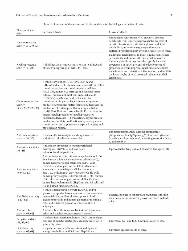
Table 1: Summary of the in vitro and in vivo evidence for the biological activities of rhein.
Pharmacologicaleffect In vitro evidence In vivo evidence
Hepatoprotectiveactivity [2, 7, 10–12]
It modulates cytochrome P450 enzymes, protectshepatocyte from injury and prevents the progress ofhepatic fibrosis in rats, alleviates glucose and lipidmetabolism, increases energy expenditure, andrestrains proinflammatory cytokine expression in mice.
Nephroprotectiveactivity [15–20]
It abolishes the 𝛼-smooth muscle actin (𝛼-SMA) andfibronectin expression of NRK-49F cells.
It alleviates renal fibrosis in mice. It reduces intestinalpermeability and protects the intestinal mucosa inimmune globulin A nephropathy (IgAN), halts theprogression of IgAN, prevents the development ofglomerulosclerosis, improves renal function, reducesrenal fibrosis and interstitial inflammation, and inhibitsthe hypertrophy of renal proximal tubular epithelialcells in rats.
Chondroprotectiveactivity[3, 21, 25, 26, 28–35]
It inhibits cytokines (IL-1𝛽, LPS, TNF-𝛼, andrhIL-1𝛼)-induced effects in human osteoarthritic (OA)chondrocytes, human chondrosarcoma cell lineHEM-C55, human OA cartilage and synovial tissuecultures, human umbilical vein endothelial cells(HUVECs), and bovine and rabbit articularchondrocytes. In particular, it stimulates aggrecanproduction, promotes matrix formation, decreases theproduction of certain proinflammatory mediators(IL-1𝛽, IL-6, IL-8, and prostaglandin E2), corrects thematrix metalloproteinases/metalloproteinasesimbalance, decreases IL-1 converting enzyme proteinproduction, inhibits proliferation of synoviocytes andchondrocytes, and suppresses cathepsin B activity andproteoglycan release.
Anti-inflammatoryactivity [36, 37]
It reduces the transcription and expression ofendothelial cell adhesion molecules.
It inhibits nicotinamide adenine dinucleotidephosphate oxidase (p22phox gp91phox) and cytokines(matrix metalloproteinase-2, activating transcript factor6, and p66Shc).
Antioxidant activity[38–41]
Antioxidant properties in human peripheralneutrophils, HUVECs, and beef heartsubmitochondrial particles.
It prevents the drug-induced oxidative damage in rats.
Anticancer activity[6–9, 42–52]
Anticarcinogenic effects in mouse epidermal cell JB6line, human colon adenocarcinoma cells (Caco-2),human nasopharyngeal carcinoma (NPC) cells,HUVECs, and tongue cancer SCC-4 cells induceapoptosis in human hepatocellular carcinomaBEL-7402 cells, human cervical cancer Ca Ski cells,human promyelocytic leukemia cells (HL-60), humanNPC cells, human tongue cancer cell line (SCC-4),human hepatoblastomaG2 (HepG2) cells, KB cells, andA-549 human lung cancer cells.
Antidiabetic activity[4, 53–62]
It inhibits transforming growth factor 𝛽1
and/orglucose transporter 1 overexpression in human and ratmesangial cells, inhibits glucose uptake in Ehrlichascites tumor cells and human glomerular mesangialcells, and enhances glucose tolerance in 3T3-L1adipocytes.
It decreases glucose concentrations, increases insulinsecretion, and/or improves glucose tolerance in db/dbmice.
Antimicrobialactivity [63, 64]
Antimicrobial effects against bacterium Helicobacterpylori and staphylococcus aureus (S. aureus).
Purgative activity[49, 65, 66]
It induces ion secretion in human CaCo-2 monolayercells and stimulates electrogenic chloride secretion inguinea pig colon.
It increases Na+ and H2O flow in rat colon in-situ.
Lipid-loweringactivity [67, 68]
It regulates cholesterol homeostasis and lipid andenergy metabolism in 3T3-L1 and HepG2 cells. It protects against obesity in mice.
4 Evidence-Based Complementary and Alternative Medicine
the action of this cytokine on the cartilage, by reducing thecontent of chondrocyte IL-1 receptors [27].
Rhein (10−5M) had a weak action on 𝛼4/𝛽1 or 𝛼5/𝛽1receptors in TNF-𝛼 or recombinant human IL-1𝛼- (rhIL-1𝛼-) stimulated chondrocytes (human chondrosarcoma cellline HEM-C55) [28]. Rhein was found to downregulate theproliferation rate of both synoviocytes and chondrocytes,decrease caspase-3/7 activities, and increase the expression ofp21 and/or p27, but not cyclin D1 [29].
After bovine articular chondrocytes were cultured inlow oxygen tension with rhein (10−5M) for 24 h, IL-1𝛽 (10 ng/mL)-activated mitogen activated protein kinase(MAPK) pathway, DNA binding of NF-𝜅B, and activatorprotein-1 (AP-1) were inhibited significantly. NF-𝜅B and AP-1 are two key factors related to the expression of severalproinflammatory genes in chondrocytes. Furthermore, rheincould prevent the procatabolic action of the cytokine byinhibition of the collagenase synthesis and increase thesynthesis of matrix components, such as type II collagenand aggrecan, which might be the mechanism of its disease-modifying effect in OA [30]. Rhein (10−4M) evidentlyprevented increases of MMPs and aggrecanase-1, NF-𝜅B,and AP-1 DNA binding, phosphorylation of extracellularsignal-regulated protein kinase (ERK), and c-Jun NH2-terminal kinase in bovine chondrocytes stimulated by IL-1𝛽(10 ng/mL) in vitro [31]. Rhein dose-dependently inhibitedIL-1𝛽-induced degradation of the inhibitor 𝜅B-𝛼 protein,translocation of the protein p65 (amember of theNF-𝜅B fam-ily) to the nucleus, and NF-𝜅B binding to a specific (gamma-(32)P)-labelled oligonucleotide probe. Rhein also inhibitedinducible NO synthase mRNA and protein synthesis andNO production in a dose-dependent manner [32]. In cul-tured rabbit articular chondrocytes, rhein (0.1–30𝜇M) dose-dependently suppressed the rhIL-1𝛼-induced proteoglycandegradation,MMPs activity, and the expression of proMMPs-1, -3, -9, and -13, while it increased the production of TIMP-1[33, 34]. Rhein at 20𝜇M inhibited the activity of cathepsinB from human liver. In cultured rabbit cartilage challengedwith IL-1𝛽, rhein at 100 𝜇M suppressed cathepsin B activityand proteoglycan release. After treatment with oral diacerein,the prodrug of rhein, at the dose of 25mg/day for 3 months,the progression of OA lesions and osteophyte formation wererestrained in the experimental OA rabbit model [35].
2.4. Anti-Inflammatory Activity. Reducing the expression ofendothelial cell adhesion molecules (ECAMs) is known todecrease inflammation-induced vascular complications. Thetranscription and expression of ECAMs, including ICAM-1, vascular cell adhesion molecule-1 (VCAM-1), and E-SELECTIN, could be reduced by the rhein treatment (10 and20𝜇M) in human umbilical vein endothelial cells (HUVECs).In the presence of LPS stimulation, the transcription andexpression of VCAM-1 were also inhibited by treatment withrhein (10 and 20 𝜇M) [36].
Adjuvant injection elicited the inflammatory edema inrat paw, accompanied by activation of nicotinamide ade-nine dinucleotide phosphate (NADPH) oxidase (p22phoxgp91phox), transcript factor 6 (ATF6) and p66Shc, elevation
of cytokines including MMP-2, and an increase of the p-Akt/Akt ration, which were notably reversed by rhein [37].Rhein (20𝜇M) almost completely inhibited intersegmentalblood vessels formation at both 48 and 72 h after fertilization(hpf) and completely prevented subintestinal vessel plexusformation at 72 hpf in wild type zebra fish embryos. Rheinaffected multiple molecular targets related to angiogenesis,particularly angpt2 and tie2, and also blocked endothelial cellmigration [69].
2.5. Antioxidant Activity. The superabundant productionof reactive oxygen species (ROS) is involved in manypathophysiological processes such as aging, atherosclero-sis, cancer, neurodegenerative disorders, chronic inflam-mation, and degenerative rheumatic disease. Rhein wasobserved to inhibit the ROS production in human periph-eral neutrophils activated by N-formyl-methionyl-leucyl-phenylalanine or phorbol-12-myristate-13-acetate in vitro[38]. Besides, many other mechanisms of the antioxidanteffect of rhein have been revealed. Pretreatment with differ-ent rhein concentrations (2, 4, 8, and 16 𝜇M) significantlydownregulated the mRNA expression of Bid, caspase-3,-8, and -9 and the content of MDA and lactate dehy-drogenase, while it increased NO content and activitiesof NO synthase, SOD, and glutathione peroxidase (GSH-PX) in hydrogen peroxide- (H
2
O2−
) insulted HUVECs,reversing H
2
O2
-induced cell apoptosis [39]. Rhein dramat-ically decreased acetaminophen-induced serum glutamate-pyruvate transaminase, glutamate-oxaloacetic transaminase,creatinine and urea nitrogen levels in the liver, ROS produc-tion, NO andMDA levels, andGSH concentration in the liverand kidney of rats. Rhein also significantly ameliorated thehistopathological damage of the liver and kidney [40]. Rheineliminated the biphasicity of ubiquinone oxidoreductase-(NADH-) induced reaction and caused a substantial stim-ulation of NADH-induced lipid peroxidation in beef heartsubmitochondrial particles. Furthermore, rhein facilitatedboth NADH- and NADPH-induced lipid peroxidation [41].
2.6. Anticancer Activity. Anticarcinogenic effects of rheinon proliferation and metastasis in cells have been inves-tigated in vitro. Rhein inhibited hypertrophic scar fibrob-lasts proliferation in a dose-dependent manner [70]. Rheinalso dose-dependently inhibited 12-O-tetradecanoylphorbol-13-acetate- (TPA-) induced cell transformation and AP-1activation, prevented the phosphorylation of c-Jun proteinand c-Jun NH2-terminal kinase (JNK), did not restrain thephosphorylation of ERK and p38 kinase in mouse epider-mal cell JB6 line [42]. Rhein (0.1 and 1mg/mL) evidentlysuppressed cell proliferation and mitogen-activated protein(MAP) kinase activation in human colon adenocarcinomacells (Caco-2) but significantly lessened H
2
O2
-induced DNAdamage and the elevated MDA and ROS levels induced byH2
O2
/Fe2+ at the concentrations of 0.1–10mg/mL [43].Cancer invasion is believed to be dependent on extra-
cellular matrix remodeling elicited by tumor cells. Rheininhibited invasion and migration in human nasopharyn-geal carcinoma (NPC) cells through downregulation ofthe expression of MMP-9, vascular endothelial growth
Evidence-Based Complementary and Alternative Medicine 5
factor (VEGF), growth factor receptor bound protein 2,son of sevenless-1 and Ras, inhibition of the phosphoryla-tion of ERK, p38 MAPK, and activation of transcriptionfactor NF-𝜅B [6]. Rhein prevented HUVEC tube forma-tion, proliferation, and migration stimulated by vascularendothelial growth factor (VEGF
165
) under normoxic andhypoxic conditions. Moreover, rhein inhibited the activationof phosphatidylinositol 3-kinase (PI3K), phosphorylated-AKT (p-AKT), and phosphorylated ERK, suppressing invitro angiogenesis. Rhein restrained cell cycle and viabilityof hormone-dependent breast cancer cells (MCF-7) andhormone-independent breast cancer cells (MDA-MB-435s)under normoxic or hypoxic conditions. In addition, rheindecreased the expression of hypoxia-inducible factor (HIF)-1𝛼, VEGF
165
, epidermal growth factor (EGF), the phospho-rylation of NF-𝜅B inhibitor, and the activity of heat shockprotein 90𝛼 (Hsp90𝛼) under normoxic or hypoxic conditions[44].
Rhein prevented themRNA expression ofMMP-9, whichplays an important role and is the most associated withtumor invasion and metastasis in various human cancers,decreased the levels of MMP-2 and urokinase u-PA, andinhibited themigration and invasion in human tongue cancerSCC-4 cells [7]. A further study demonstrated that rheindose-dependently induced DNA damage in SCC-4 cells,followed by the inhibition of the mRNA expression of DNArepair-associated O (6)-methylguanine-DNA methyltrans-ferase (MGMT) [45].Themitosiswas inhibited inAllium ceparoot tips incubated with rhein in a dose-dependent manner[71].
Apoptosis, a physiological process for eliminating malig-nant cells including cancer cells, does not result in the damageto normal cells or surrounding tissues. Rhein-induced apop-tosis has been reported in various human cancer cells. Incu-bation of human hepatocellular carcinoma BEL-7402 cellswith rhein at 50–200𝜇M for 48 hours caused an increasingapoptosis, the features of which included cellular morpho-logical change and chromatin condensation. Additionally,rhein induced cell cycle S-phase arrest, decreased c-Mycgene expression, and increased caspase-3 gene expression [8].Rhein induced the abrogation of mitochondrial membranepotential and cleavage of Bid protein in human cervicalcancer Ca Ski cells. Rhein decreased the level of Bcl-2 whileincreased the levels of Fas, p53, p21, Bax, and cytoplasmicCa2+ and the activities of both caspase-8 and -9, promotedcaspase-3 activation, and resulted in DNA fragmentation[46].
Rhein induced apoptosis in human promyelocytic leu-kemia cells (HL-60) through facilitating the loss ofmitochon-drial membrane potential, cytochrome c release from mito-chondrion to cytosol, and cleavage of Bid protein. Rhein alsoincreased the generation of ROS and the phosphorylation ofc-Jun N-terminal kinase and p38 kinase [47]. Rhein elevatednuclear condensation and DNA fragmentation, resulting inapoptosis of human NPC cells. Furthermore, rhein increasedthe activation of caspase-3, -8, -9, and -12 as well as thelevels of glucose-regulated protein 78 (GRP 78), PKR-like ERkinase, ATF6, and CCAAT, induced the rapid accumulation
of calcium (Ca2+), and lessened themitochondrialmembranepotential. Then Cytochrome c and apoptosis-inducing factorwere released [48].
Incubation of humanCaCo-2monolayer cells with 50𝜇Mrhein induced nitrate production and a time-dependentpolymorphonuclear leukocytes chemotaxis. Overnight rheinincubation produced an increasing number of apoptoticcells in the culture supernatant [49]. The treatment with30 𝜇M rhein for 24 h showed the most efficient apoptosisinduction in SCC-4 cells. Rhein inhibited p53, cyclin A andE, resulting in S-phase arrest of the cells.The ratio of Bax/Bcl-2 was changed by rhein through inhibition of Bcl-2 level.Rhein increased ROS production and Ca2+ release, decreasedthe mitochondrial membrane potential level, and activatedcaspase-3, -8, and -9 [50]. Rhein significantly increased theprotein expression of p53 and p21/WAF1 and the levels ofCD95 and its two forms of ligands, membrane-bound CD95ligand and solubleCD95 ligand, in humanhepatoblastomaG2(HepG2) cells, which not only inhibited HepG2 cell growthbut also induced cell apoptosis [51]. The IC
50
values of rheinforKB, hepatomaBEL-7402, andmammary carcinomaMCF-7 cells were 11.5, 14.0, and 18.4mg/mL, respectively. InKB cellstreated with rhein for 96 h, the increase of 71% was observedin apoptotic cells [52]. The apoptosis was observed when A-549 human lung cancer cells were incubated with rhein at50 𝜇M for 12 h and up to 72 h. Rhein induced G
0
/G1
arrestthrough inhibition of cyclin D3, Cdk4, and Cdk6. RheinpromotedROS andCa2+ production, capase-3 activation, andcytochrome c release frommitochondria, increased the levelsof GADD153 and GRP78, both hallmarks of endoplasmicreticulum stress, induced the loss of mitochondrial mem-brane potential (ΔΨm), and led to apoptosis in A-549 humanlung cancer cells. Rhein also increased the levels of p53, p21,and Bax, while it reduced the level of Bcl-2 [9].
2.7. Antidiabetic Activity. In vivo (in db/dbmice), a significantdecrease in area under curve (AUC) of glucose concentra-tions, simultaneously, and increases in AUC of insulin leveland first-phase insulin secretion were observed after admin-istration of rhein (120mg/kg) for 8weeks. Furthermore, rheintreatment greatly protected 𝛽 cell mass and inhibited 𝛽 cellapoptosis [53, 54]. Oral administration of rhein (120mg/kg)for 8 or 16 weeks notably reduced fasting blood glucoselevel and improved glucose tolerance. After localized at 𝛽-cell mitochondria, rhein could protect mitochondrial ultra-structure from hyperglycemia-inducedmitochondrial fissionprotein dynamin-related protein 1 expression, resulting ininhibition of 𝛽-cell apoptosis [55]. Intragastric treatmentwith rhein (120mg/kg) significantly downregulated bloodglucose concentrations at 0, 30, 60, and 120min after glucoseload, suppressed pancreatic 𝛽-cell apoptosis, and elevated theearly-phase insulin secretion inmice, suggesting the potentialof rhein as a novel therapeutic agent for type 2 diabetes [4].
Rhein decreased urinary albumin excretion, extracellularmatrix level, and TGF-𝛽
1
and FN expression in renal tissueand also reduced the plasma levels of cholesterol, triglyceride,low-density lipoprotein cholesterol (LDL-C), and ApoE indb/db mice with diabetic nephropathy (DN) [72]. In rat

6 Evidence-Based Complementary and Alternative Medicine
mesangial cells transfected with human glucose transporter1 (GLUT 1) gene, rhein dose-dependently decreased 2-deoxyglucose uptake, reversed cell hypertrophy, and loweredthe enhanced glutamine: fructose-6-phosphate aminotrans-ferase activity of the human GLUT 1 gene, suggesting aninhibitory effect on the GLUT 1 overexpression in diabeticnephropathy [56].
It was observed that rhein significantly lowered thesecretion of FN and inhibited the proliferation of humanmesangial cells in mimic hyperglycemic environment of dia-betic nephropathy, the possible mechanism of which mightbe related to suppression of the bioactivities of TGF-𝛽1 andp38MAPK [57]. TGF-𝛽
1
stimulates the glucose uptake byenhancing the GLUT 1mRNA expression in both human andrat glomerular mesangial cells, which could be antagonizedby rhein [58, 59].
Rhein strongly inhibited the uptake of both 2-deo-xyglucose and 3-O-methylglucose in Ehrlich ascites tumorcells by alteration of membrane-associated functions[60]. In addition, rhein greatly decreased the inductionof ROS in both the NIT-1 cells and isolated islets. Rheinenhanced insulin-stimulated glucose uptake in 3T3-L1adipocytes, while it decreased triglyceride accumulation instreptozotocin-induced diabetic mice [61]. Rhein markedlyattenuated the increased glucose uptake and GLUT1 mRNAexpression stimulated by TGF-𝛽
1
in a dose-dependentmanner in human glomerular mesangial cells [62]. Rheinreversed the abnormal changes of MMP-9/TIMP-1 ratio andimpeded overexpression of integrin-linked kinase in highglucose-induced epithelial-mesenchymal transition of HK-2cells [73].
2.8. Antimicrobial Activity. Like many herbal monomer,rhein has a potential antibacterial property. For example,rhein inhibited Arylamine N-acetyltransferase activity andgrowth in the bacterium Helicobacter pylori from pepticulcer patients [63]. In the other in vitro study, rhein showeda good antibacterial activity against all 21 tested staphylo-coccus aureus (S. aureus) strains. 28 transporter genes of S.aureus ATCC25923 were differentially regulated by rhein. Inparticular, rhein increased the transcription of genes (srtBand isdABCDEFGI) encoding iron-regulated surface deter-minants system and genes (nrdIEF and nrdDG) involvedin ribonucleotide reductase systems, while it prevented thetranscription of genes (pflAB, nirBDR, narGH, Idh1, COL-SA0660, COL-SA2363, and COL-SA2386) responsible foranaerobic respiration and fermentation [64].
Bacterial DNA/CpGDNA is recognized as a keymoleculeduring the pathogenesis of sepsis.Therefore, preventing CpGDNA from binding to its receptor is considered as the mostpromising strategy. Rhein was demonstrated to have highaffinity for CpG DNA. It could significantly reduce CpGDNA- and LPS-induced TNF-𝛼 release in RAW264.7 cells[74].
2.9. Purgative Activity. Rhein (4 × 10⁻³M) reduced net H2
OandNa+ absorption in rat colon in-situ. However, its secretory
effect was associated with neither inhibition of Na+ and K+-ATPase nor damage of the colon epithelium [65]. Mucosalor serosal application of rhein (10 nM–0.5mM) activatedchloride secretion by excitation of submucosal neurons andrelease of acetylcholine and endogenous prostaglandins [66].In addition, rhein induced ion secretion in human CaCo-2monolayer cells [49].
2.10. Lipid-Lowering Activity. LXRs play important roles inregulating cholesterol homeostasis and lipid and energymetabolism. After bounding directly to LXRs in C57BL/6Jmice fed a HF diet, rhein suppressed the expression lev-els of LXR target genes in both 3T3-L1 and HepG2 cellsin vitro. In white adipose tissue, muscle, and liver, rheinreprogrammed the expression of LXR target genes relatedto adipogenesis and cholesterol metabolism. Rhein acti-vated uncoupling protein 1 (UCP1) expression in brownadipose tissue (BAT) in wild-type mice, suggesting thatrhein may protect against obesity and related metabolicdisorders through LXR antagonism and regulation of UCP1expression in BAT [67]. Rhein downregulated the mRNAlevels of adipogenesis-specific transcription factors PPAR𝛾and C/EBP𝛼 and their downstream target genes involved inadipocyte differentiation, such as CD36, AP-2, and acyl CoAoxidase in both 3T3-L1 preadipocytes and C57BL/6 mice.Furthermore, the expression of C/EBP𝛽was reduced by rheinin 3T3-L1 preadipocytes. HF diet-induced weight gain andadiposity were reversed by rhein in C57BL/6 mice [68].
2.11. Other Activities. Rhein was found to have estrogenicactivity and the EC
50
value was 18.96 𝜇g/mL in the yeast-based estrogenicity assay system [75]. LIGHT is known toact as a novel mediator for atherogenesis. Rhein inhibitedLIGHT-induced human monocyte migration, ROS genera-tion, the expression of chemokine receptor (CCR)1, CCR2,and ICAM-1, the production of IL-8, MCP-1, TNF-𝛼, andIL-6, and the activation of the p38 MAPK and NF-𝜅B [76].Vascular smooth-muscle cell proliferation plays an importantrole in atherosclerosis and restenosis. Treatment with rheinresulted in the induction of apoptosis, release of cytochromec into the cytosol, loss of mitochondrial membrane potential,decrease in Bcl-2 and Bcl-xL, and the increase in Bax and Bakexpression in TNF-𝛼-induced human aortic smooth-musclecells (HASMCs), thus inhibiting the proliferation of thesecells [5].
Rhein had a potent (>76%) inhibitory effect on mast-cell degranulation in rats at a 5mg/kg dose and suppressedlipoxygenase (LOX) enzyme activity with the IC
50
valueof 3.9 𝜇g/mL, proposing that rhein has antiallergic activity[77]. Rhein dose-dependently blocked the increase of PAI-1mRNA expression and protein production and inhibited theactivity of phosphor-p44/p42 MAP kinase induced by TGF-𝛽1
in human endothelial cells, suggesting the protective effecton the endothelial dysfunction. Rhein might be a potentialagent for the treatment of vascular diseases [78]. Further-more, rhein inhibited IL-1-induced secretion of MMPs andaggrecanases and apoptosis in intervertebral disc cells, whichprevented the progression of intervertebral disc degeneration[79].

Evidence-Based Complementary and Alternative Medicine 7
3. Conclusion
As reviewed here, different pharmacological experiments ina number of in vitro and in vivo models have convincinglydemonstrated the abilities of rhein to exhibit hepatoprotec-tive, nephroprotective, anti-inflammatory, antioxidant, anti-cancer, and antimicrobial activities, lending support to therationale behind several of its potential medicinal uses.
However, further studies need to be carried out in orderto explore the concealed areas. Although various bioactivitiesof rhein are substantiated by using laboratory animal orcell models, the molecular mechanisms and targets involvedare still unknown, which will count against further clinicalapplications of this agent.
Conflict of Interests
Theauthors have declared that there is no conflict of interests.
Authors’ Contribution
Yan-Xi Zhou and Wei Xia contributed equally to this work.
Acknowledgments
This work was supported by funds from Shanghai MunicipalHealth and Family Planning Commission (ZY3-JSFC-2-2010,20134090, and ZYXK2012010), Pudong New Area Scienceand Technology Commission (PKJ2013-Y22), Pudong NewArea Health and Family Planning Commission (PWZz2013-02 and PWRd2013-03), and the State Key Laboratory Breed-ing Base of Systematic Research, Development and Utiliza-tion of Chinese Medicine Resources.
References
[1] B. Layek, T. S. Kumar, R. K. Trivedi, R. Mullangi, and N.R. Srinivas, “Development and validation of a sensitive LC-MS/MS method with electrospray ionization for quantitationof rhein in human plasma: application to a pharmacokineticstudy,” Biomedical Chromatography, vol. 22, no. 6, pp. 616–624,2008.
[2] M.-Z. Guo, X.-S. Li, D.-M. Shen, X.-Q. Guan, H.-R. Xu, and J.Gao, “Effect of Rhein on the development of hepatic fibrosis inrats,” Zhonghua Gan Zang Bing Za Zhi, vol. 11, no. 1, pp. 26–29,2003.
[3] F. Moldovan, J. P. Pelletier, F.-C. Jolicoeur, J.-M. Cloutier, and J.Martel-Pelletier, “Diacerhein and rhein reduce the ICE-inducedIL-1𝛽 and IL-18 activation in human osteoarthritic cartilage,”Osteoarthritis and Cartilage, vol. 8, no. 3, pp. 186–196, 2000.
[4] H. Du, J.-Q. Shao, P. Gu, J. Wang, and Z.-H. Liu, “Effect of earlyintervention with rhein on islet function in db/db mice,” NanFang Yi Ke Da Xue Xue Bao, vol. 31, no. 9, pp. 1526–1529, 2011.
[5] S.-K.Heo,H.-J. Yun,W.-H. Park, and S.-D. Park, “Rhein inhibitsTNF-𝛼-induced human aortic smooth muscle cell proliferationvia mitochondrial-dependent apoptosis,” Journal of VascularResearch, vol. 46, no. 4, pp. 375–386, 2009.
[6] M. L. Lin, J. G. Chung, Y. C. Lu, C. Y. Yang, and S. S. Chen,“Rhein inhibits invasion and migration of human nasopha-ryngeal carcinoma cells in vitro by down-regulation of matrixmetalloproteinases-9 and vascular endothelial growth factor,”Oral Oncology, vol. 45, no. 6, pp. 531–537, 2009.
[7] Y.-Y. Chen, S.-Y. Chiang, J.-G. Lin et al., “Emodin, aloe-emodinand rhein inhibit migration and invasion in human tonguecancer SCC-4 cells through the inhibition of gene expression ofmatrix metalloproteinase-9,” International Journal of Oncology,vol. 36, no. 5, pp. 1113–1120, 2010.
[8] P. Shi, Z. Huang, and G. Chen, “Rhein induces apoptosis andcell cycle arrest in human hepatocellular carcinoma BEL-7402cells,”The American Journal of Chinese Medicine, vol. 36, no. 4,pp. 805–813, 2008.
[9] T.C.Hsia, J. S. Yang,G.W.Chen et al., “The roles of endoplasmicreticulum stress and Ca2+ on rhein-induced apoptosis in A-549human lung cancer cells,” Anticancer Research, vol. 29, no. 1, pp.309–318, 2009.
[10] J.-C. Tang, H. Yang, X.-Y. Song et al., “Inhibition of cytochromeP450 enzymes by rhein in rat liver microsomes,” PhytotherapyResearch, vol. 23, no. 2, pp. 159–164, 2009.
[11] D.-X. Bian, J. Liu, L. Lu et al., “Protective effects of rhein onhepatic progression in HBV-transgenic mice with nonalcoholicsteatohepatitis induced by a high-fat diet,”Zhonghua Shi YanHeLin Chuang Bing Du Xue Za Zhi, vol. 27, no. 5, pp. 328–331, 2013.
[12] X. Sheng, M. Wang, M. Lu, B. Xi, H. Sheng, and Y. Q. Zang,“Rhein ameliorates fatty liver disease through negative energybalance, hepatic lipogenic regulation, and immunomodula-tion in diet-induced obese mice,” The American Journal ofPhysiology—Endocrinology and Metabolism, vol. 300, no. 5, pp.E886–E893, 2011.
[13] T. A. Wynn, “Common and unique mechanisms regulatefibrosis in various fibroproliferative diseases,” The Journal ofClinical Investigation, vol. 117, no. 3, pp. 524–529, 2007.
[14] M.-Z. Guo, X.-S. Li, H.-R. Xu, Z.-C. Mei, W. Shen, and X.-F. Ye,“Rhein inhibits liver fibrosis induced by carbon tetrachloride inrats,” Acta Pharmacologica Sinica, vol. 23, no. 8, pp. 739–744,2002.
[15] D. He, L. Lee, J. Yang, and X. Wang, “Preventive effects andmechanisms of rhein on renal interstitial fibrosis in obstructivenephropathy,” Biological and Pharmaceutical Bulletin, vol. 34,no. 8, pp. 1219–1226, 2011.
[16] S.-N. Peng, H.-H. Zeng, A.-X. Fu, X.-W. Chen, and Q.-X. Zhu,“Effects of rhein on intestinal epithelial tight junction in IgAnephropathy,”World Journal of Gastroenterology, vol. 19, no. 26,pp. 4137–4145, 2013.
[17] S.-N. Peng, H.-H. Zeng, A.-X. Fu, X.-W. Chen, and Q.-X.Zhu, “Protection of rhein on IgA nephropathy mediated byinhibition of fibronectin expression in rats,” Indian Journal ofPharmacology, vol. 45, no. 2, pp. 174–179, 2013.
[18] J. Su, L. P. Yin, X. Zhang, B. B. Li, L. Liu, and H. Li, “Chronicallograft nephropathy in rats is improved by the interventionof rhein,” Transplantation Proceedings, vol. 45, no. 6, pp. 2546–2552, 2013.
[19] Z. Q. Ji, C. W. Huang, C. J. Liang, W. W. Sun, B. Chen,and P. R. Tang, “Effects of rhein on activity of caspase-3 inkidney and cell apoptosis on the progression of renal injury inglomerulosclerosis,”Zhonghua Yi Xue Za Zhi, vol. 85, no. 26, pp.1836–1841, 2005.
[20] D.-Q. Yu, Y. Gao, and X.-H. Liu, “Effects of Rhein on thehypertrophy of renal proximal tubular epithelial cells inducedby high glucose and angiotensin II in rats,” Zhong Yao Cai, vol.33, no. 4, pp. 570–574, 2010.
[21] C. Sanchez, M. Mathy-Hartert, M. A. Deberg, H. Ficheux,J.-Y. L. Reginster, and Y. E. Henrotin, “Effects of rhein onhuman articular chondrocytes in alginate beads,” BiochemicalPharmacology, vol. 65, no. 3, pp. 377–388, 2003.

8 Evidence-Based Complementary and Alternative Medicine
[22] D. Borderie, A. Hernvann, H. Lemarechal, C.-J. Menkes, andO. Ekindjian, “Inhibition of the nitrosothiol production ofcultured osteoarthritic chondrocytes by rhein, cortisol anddiclofenac,” Osteoarthritis and Cartilage, vol. 9, no. 1, pp. 1–6,2001.
[23] J.-P. Pelletier, F. Mineau, J. C. Fernandes, N. Duval, and J.Martel-Pelletier, “Diacerhein and rhein reduce the interleukin1beta stimulated inducible nitric oxide synthesis level andactivity while stimulating cyclooxygenase-2 synthesis in humanosteoarthritic chondrocytes,” Journal of Rheumatology, vol. 25,no. 12, pp. 2417–2424, 1998.
[24] J. Deffaud, M. Kirchmeyer, F. Domagala et al., “Modulatoryeffect of rhein on IL-1𝛼-induced responses in human chon-drocytes: a comparative study between antibody microarraysand specific ELISAs,” Biorheology, vol. 45, no. 3-4, pp. 439–455,2008.
[25] J.-P. Pelletier, D. Lajeunesse, P. Reboul et al., “Diacerein reducesthe excess synthesis of bone remodeling factors by humanosteoblast cells from osteoarthritic subchondral bone,” Journalof Rheumatology, vol. 28, no. 4, pp. 814–824, 2001.
[26] M. Yaron, I. Shirazi, and I. Yaron, “Anti-interleukin-1 effects ofdiacerein and rhein in human osteoarthritic synovial tissue andcartilage cultures,” Osteoarthritis and Cartilage, vol. 7, no. 3, pp.272–280, 1999.
[27] J. Martel-Pelletier, F. Mineau, F.-C. Jolicoeur, J.-M. Cloutier,and J.-P. Pelletier, “In vitro effects of diacerhein and rhein oninterleukin 1 and tumor necrosis factor-𝛼 systems in humanosteoarthritic synovium and chondrocytes,” The Journal ofRheumatology, vol. 25, no. 4, pp. 753–762, 1998.
[28] C. Gigant-Huselstein, D. Dumas, E. Payan et al., “In vitro studyof intracellular IL-1𝛽 production and 𝛽1 integrins expression instimulated chondrocytes—effect of rhein,” Biorheology, vol. 39,no. 1-2, pp. 277–285, 2002.
[29] F. Legendre, A. Heuze, K. Boukerrouche et al., “Rhein,the metabolite of diacerhein, reduces the proliferation ofosteoarthritic chondrocytes and synoviocytes without inducingapoptosis,” Scandinavian Journal of Rheumatology, vol. 38, no. 2,pp. 104–111, 2009.
[30] G. Martin, P. Bogdanowicz, F. Domagala, H. Ficheux, and J.-P. Pujol, “Rhein inhibits interleukin-1𝛽-induced activation ofMEK/ERK pathway and DNA binding of NF-𝜅B and AP-1 inchondrocytes cultured in hypoxia: a potentialmechanism for itsdisease-modifying effect in osteoarthritis,” Inflammation, vol.27, no. 4, pp. 233–246, 2003.
[31] F. Legendre, P. Bogdanowicz, G. Martin et al., “Rhein, adiacerhein-derived metabolite, modulates the expression ofmatrix degrading enzymes and the cell proliferation of articularchondrocytes by inhibiting ERK and JNK-AP-1 dependentpathways,”Clinical and Experimental Rheumatology, vol. 25, no.4, pp. 546–555, 2007.
[32] A. F. Mendes, M. M. Caramona, A. P. de Carvalho, and M.C. Lopes, “Diacerhein and rhein prevent interleukin-1beta-induced nuclear factor-kappaB activation by inhibiting thedegradation of inhibitor kappaB-alpha,” Pharmacology andToxicology, vol. 91, no. 1, pp. 22–28, 2002.
[33] T. Tamura, N. Kosaka, J. Ishiwa, T. Sato, H. Nagase, and A.Ito, “Rhein, an active metabolite of diacerein, down-regulatesthe production of pro-matrix metalloproteinases-1, -3, -9 and-13 and up-regulates the production of tissue inhibitor ofmetalloproteinase-1 in cultured rabbit articular chondrocytes,”Osteoarthritis and Cartilage, vol. 9, no. 3, pp. 257–263, 2001.
[34] T. Tamura and K. Ohmori, “Rhein, an active metabolite ofdiacerein, suppresses the interleukin-1𝛼-induced proteoglycandegradation in cultured rabbit articular chondrocytes,” JapaneseJournal of Pharmacology, vol. 85, no. 1, pp. 101–104, 2001.
[35] L. Savarino, A. Fioravanti, G. Leo, R. Aloisi, and M. Mian,“Anthraquinone-2,6-disulfonic acid as a disease-modifyingosteoarthritis drug: an in vitro and in vivo study,” ClinicalOrthopaedics and Related Research, no. 461, pp. 231–237, 2007.
[36] G. Hu, J. Liu, Y.-Z. Zhen et al., “Rhein inhibits the expressionof vascular cell adhesion molecule 1 in human umbilical veinendothelial cells with or without lipopolysaccharide stimula-tion,” The American Journal of Chinese Medicine, vol. 41, no. 3,pp. 473–485, 2013.
[37] X.-D. Cong, M.-J. Ding, D.-Z. Dai, Y. Wu, Y. Zhang, and Y. Dai,“ER stress, P66shc, and P-Akt/Akt mediate adjuvant-inducedinflammation, which is blunted by argirein, a supermoleculeand rhein in rats,” Inflammation, vol. 35, no. 3, pp. 1031–1040,2012.
[38] T. Tamura, T. Yokoyama, and K. Ohmori, “Effects of diacereinon indomethacin-induced gastric ulceration,” Pharmacology,vol. 63, no. 4, pp. 228–233, 2001.
[39] X.-F. Zhong, G.-D. Huang, T. Luo, Z.-Y. Deng, and J.-N.Hu, “Protective effect of rhein against oxidative stress-relatedendothelial cell injury,” Molecular Medicine Reports, vol. 5, no.5, pp. 1261–1266, 2012.
[40] Y.-L. Zhao, G.-D. Zhou, H.-B. Yang et al., “Rhein protectsagainst acetaminophen-induced hepatic and renal toxicity,”Food and Chemical Toxicology, vol. 49, no. 8, pp. 1705–1710, 2011.
[41] M. A. Glinn, C. P. Lee, and L. Ernster, “Pro- and anti-oxidant activities of themitochondrial respiratory chain: factorsinfluencing NAD(P)H-induced lipid peroxidation,” Biochimicaet Biophysica Acta—Bioenergetics, vol. 1318, no. 1-2, pp. 246–254,1997.
[42] S. Lin, J.-J. Li, M. Fujii, and D.-X. Hou, “Rhein inhibits TPA-induced activator protein-1 activation and cell transformationby blocking the JNK-dependent pathway,” International Journalof Oncology, vol. 22, no. 4, pp. 829–833, 2003.
[43] G. Aviello, I. Rowland, C. I. Gill et al., “Anti-proliferative effectof rhein, an anthraquinone isolated from Cassia species, onCaco-2 human adenocarcinoma cells,” Journal of Cellular andMolecular Medicine, vol. 14, no. 7, pp. 2006–2014, 2010.
[44] V. E. Fernand, J. N. Losso, R. E. Truax et al., “Rhein inhibitsangiogenesis and the viability of hormone-dependent and -independent cancer cells under normoxic or hypoxic conditionsin vitro,”Chemico-Biological Interactions, vol. 192, no. 3, pp. 220–232, 2011.
[45] Y. Y. Chen, S. Y. Chiang, J. G. Lin et al., “Emodin, aloe-emodinand rhein inducedDNAdamage and inhibitedDNArepair geneexpression in SCC-4 human tongue cancer cells,” AnticancerResearch, vol. 30, no. 3, pp. 945–952, 2010.
[46] S.-W. Ip, Y.-S. Weng, S.-Y. Lin, N.-Y. Tang, C.-C. Su, and J.-G.Chung, “The role of Ca+2 on rhein-induced apoptosis in humancervical cancer Ca Ski cells,”Anticancer Research, vol. 27, no. 1A,pp. 379–390, 2007.
[47] S. Lin,M. Fujii, andD.-X.Hou, “Rhein induces apoptosis inHL-60 cells via reactive oxygen species-independent mitochondrialdeath pathway,”Archives of Biochemistry andBiophysics, vol. 418,no. 2, pp. 99–107, 2003.

Evidence-Based Complementary and Alternative Medicine 9
[48] M.-L. Lin, S.-S. Chen, Y.-C. Lu et al., “Rhein induces apoptosisthrough induction of endoplasmic reticulum stress and Ca2+-dependent mitochondrial death pathway in human nasopha-ryngeal carcinoma cells,” Anticancer Research, vol. 27, no. 5, pp.3313–3322, 2007.
[49] F. Raimondi, P. Santoro, L. Maiuri et al., “Reactive nitrogenspecies modulate the effects of rhein, an active componentof senna laxatives, on human epithelium in vitro,” Journal ofPediatric Gastroenterology and Nutrition, vol. 34, no. 5, pp. 529–534, 2002.
[50] W. W. Lai, J. S. Yang, K. C. Lai et al., “Rhein induced apop-tosis through the endoplasmic reticulum stress, caspase- andmitochondria-dependent pathways in SCC-4 human tonguesquamous cancer cells,” In Vivo, vol. 23, no. 2, pp. 309–316, 2009.
[51] P. L. Kuo, Y. L. Hsu, L. T. Ng, and C. C. Lin, “Rhein inhibits thegrowth and induces the apoptosis of Hep G2,” Planta Medica,vol. 70, no. 1, pp. 12–16, 2004.
[52] Y.-H.Huang andY.-S. Zhen, “Rhein induces apoptosis in cancercells and shows synergy withmitomycin,”Yao Xue Xue Bao, vol.36, no. 5, pp. 334–338, 2001.
[53] H. Du, J. Shao, P. Gu, B. Lu, X. Ye, and Z. Liu, “Improvementof glucose tolerance by rhein with restored early-phase insulinsecretion in db/db mice,” Journal of Endocrinological Investiga-tion, vol. 35, no. 6, pp. 607–612, 2012.
[54] H. Du, J. Shao, P. Gu, B. Lu, J. Wang, and Z. Liu, “Effect of rheintreatment on first-phase insulin secretory function in db/dbmice,” Zhongguo Zhongyao Zazhi, vol. 35, no. 20, pp. 2764–2767,2010.
[55] J. Liu, Z. Chen, Y. Zhang et al., “Rhein protects pancreatic 𝛽-cells from dynamin-related protein-1-mediated mitochondrialfission and cell apoptosis under hyperglycemia,” Diabetes, vol.62, no. 11, pp. 3927–3935, 2013.
[56] J. Zhu, Z. Liu, and Y. Li, “Inhibition of glucose transporter 1overexpression in mesangial cells by rhein,” Zhonghua Nei KeZa Zhi, vol. 40, no. 8, pp. 537–542, 2001.
[57] Z. H. Tan, Y. J. Shen, J. N. Zhao, H. Y. Li, and J. Zhang, “Effects ofrhein on the function of human mesangial cells in high glucoseenvironment,” Yaoxue XueBao, vol. 39, no. 11, Article ID 0513-4870(2004)11-0881-06, pp. 881–886, 2004.
[58] Z. Liu, Y. Li, and J. Zhang, “Modulatory effect of transforminggrowth factor-beta and Rhein on glucose transporter-1 inhuman glomerular mesangial cells,” Zhonghua Yi Xue Za Zhi,vol. 79, no. 10, pp. 780–783, 1999.
[59] J. Zhang, Z. Liu, Z. Chen, Y. Li, and L. Li, “Effect of rhein onglucose transporter-1 expression and its function in glomerularmesangial cells,” Chinese Medical Journal, vol. 112, no. 12, pp.1077–1079, 1999.
[60] S. Castiglione, M. Fanciulli, T. Bruno et al., “Rhein inhibitsglucose uptake in Ehrlich ascites tumor cells by alteration ofmembrane-associated functions,”Anti-Cancer Drugs, vol. 4, no.3, pp. 407–414, 1993.
[61] S. B. Choi, B. S. Ko, S. K. Park, and J. S. Jang, “Insulin sensitizingand 𝛼-glucoamylase inhibitory action of sennosides, rheins andrhaponticin in Rhei Rhizoma,” Life Sciences, vol. 78, no. 9, pp.934–942, 2006.
[62] Z.-H. Liu, Y.-J. Li, Z.-H. Chen, D. Liu, and L.-S. Li, “Glucosetransporter in human glomerular mesangial cells modulated bytransforming growth factor-beta and rhein,”Acta Pharmacolog-ica Sinica, vol. 22, no. 2, pp. 169–175, 2001.
[63] J. G. Chung, M. F. Tsou, H. H. Wang et al., “Rhein affectsarylamine N-acetyltransferase activity in Helicobacter pylorifrom peptic ulcer patients,” Journal of Applied Toxicology, vol.18, no. 2, pp. 117–123, 1998.
[64] L. Yu, H. Xiang, J. Fan et al., “Global transcriptional responseof Staphylococcus aureus to Rhein, a Natural Plant Product,”Journal of Biotechnology, vol. 135, no. 3, pp. 304–308, 2008.
[65] E. Leng-Peschlow, “Sennoside-induced secretion is not causedby changes in mucosal permeability or Na+, K+-ATPase activ-ity,” Journal of Pharmacy and Pharmacology, vol. 45, no. 11, pp.951–954, 1993.
[66] T. Frieling, C. Rupprecht, and M. Schemann, “Rhein stimulateselectrogenic chloride secretion by activation of submucosalneurons in guinea pig colon,” Pharmacology, vol. 47, no. 1, pp.70–76, 1993.
[67] X. Sheng, X. Zhu, Y. Zhang et al., “Rhein protects against obesityand related metabolic disorders through liver X receptor-mediated uncoupling protein 1 upregulation in brown adiposetissue,” International Journal of Biological Sciences, vol. 8, no. 10,pp. 1375–1384, 2012.
[68] Q. Liu, X.-L. Zhang, R.-Y. Tao et al., “Rhein, an inhibitor ofadipocyte differentiation and adipogenesis,” Journal of AsianNatural Products Research, vol. 13, no. 8, pp. 714–723, 2011.
[69] Z.-H. He, R. Zhou, M.-F. He et al., “Anti-angiogenic effect andmechanism of rhein from Rhizoma Rhei,” Phytomedicine, vol.18, no. 6, pp. 470–478, 2011.
[70] Q. Wang, N.-N. Zhang, H.-Y. Li, M. Jiang, J. Gao, and G. Bai,“Active ingredients in rhubarb with anti-proliferative effects onscar fibroblasts,” Yaoxue Xuebao, vol. 47, no. 12, pp. 1618–1622,2012.
[71] F. A. Badria and A. S. Ibrahim, “Evaluation of naturalanthracene-derived compounds as antimitotic agents,” DrugDiscoveries &Therapeutics, vol. 7, no. 2, pp. 84–89, 2013.
[72] Q. Gao, W.-S. Qin, Z.-H. Jia et al., “Rhein improves renallesion and ameliorates dyslipidemia in db/dbmice with diabeticnephropathy,” Planta Medica, vol. 76, no. 1, pp. 27–33, 2010.
[73] L. Peng, J. Yang, C. Ning et al., “Rhein inhibits integrin-linkedkinase expression and regulates matrix metalloproteinase-9/tissue inhibitor of metalloproteinase-1 ratio in high glucose-induced epithelial-mesenchymal transition of renal tubularcell,” Biological and Pharmaceutical Bulletin, vol. 35, no. 10, pp.1676–1685, 2012.
[74] X. Liu, J. Cheng, X. Zheng et al., “Targeting CpG DNA toscreen and isolate anti-sepsis fraction and monomers fromtraditional Chinese herbs using affinity biosensor technology,”International Immunopharmacology, vol. 9, no. 9, pp. 1021–1031,2009.
[75] S. C. Kang, C. M. Lee, E. S. Choung et al., “Anti-proliferativeeffects of estrogen receptor-modulating compounds isolatedfrom Rheum palmatum,” Archives of Pharmacal Research, vol.31, no. 6, pp. 722–726, 2008.
[76] S.-K. Heo, H.-J. Yun, E.-K. Noh, and S.-D. Park, “Emodin andrhein inhibit LIGHT-inducedmonocytesmigration by blockingof ROS production,”Vascular Pharmacology, vol. 53, no. 1-2, pp.28–37, 2010.
[77] B. Singh, J. R. Nadkarni, R. A. Vishwakarma, S. B. Bharate,M. Nivsarkar, and S. Anandjiwala, “The hydroalcoholic extractof Cassia alata (Linn.) leaves and its major compound rheinexhibits antiallergic activity via mast cell stabilization andlipoxygenase inhibition,” Journal of Ethnopharmacology, vol.141, no. 1, pp. 469–473, 2012.

10 Evidence-Based Complementary and Alternative Medicine
[78] J. Zhu, Z. Liu, H. Huang, Z. Chen, and L. Li, “Rhein inhibitstransforming growth factor 𝛽1 induced plasminogen activatorinhibitor-1 in endothelial cells,” Chinese Medical Journal, vol.116, no. 3, pp. 354–359, 2003.
[79] H. Li, C. Liang, Q. Chen, and Z. Yang, “Rhein: a potential bio-logical therapeutic drug for intervertebral disc degeneration,”Medical Hypotheses, vol. 77, no. 6, pp. 1105–1107, 2011.

Submit your manuscripts athttp://www.hindawi.com
Stem CellsInternational
Hindawi Publishing Corporationhttp://www.hindawi.com Volume 2014
Hindawi Publishing Corporationhttp://www.hindawi.com Volume 2014
MEDIATORSINFLAMMATION
of
Hindawi Publishing Corporationhttp://www.hindawi.com Volume 2014
Behavioural Neurology
EndocrinologyInternational Journal of
Hindawi Publishing Corporationhttp://www.hindawi.com Volume 2014
Hindawi Publishing Corporationhttp://www.hindawi.com Volume 2014
Disease Markers
Hindawi Publishing Corporationhttp://www.hindawi.com Volume 2014
BioMed Research International
OncologyJournal of
Hindawi Publishing Corporationhttp://www.hindawi.com Volume 2014
Hindawi Publishing Corporationhttp://www.hindawi.com Volume 2014
Oxidative Medicine and Cellular Longevity
Hindawi Publishing Corporationhttp://www.hindawi.com Volume 2014
PPAR Research
The Scientific World JournalHindawi Publishing Corporation http://www.hindawi.com Volume 2014
Immunology ResearchHindawi Publishing Corporationhttp://www.hindawi.com Volume 2014
Journal of
ObesityJournal of
Hindawi Publishing Corporationhttp://www.hindawi.com Volume 2014
Hindawi Publishing Corporationhttp://www.hindawi.com Volume 2014
Computational and Mathematical Methods in Medicine
OphthalmologyJournal of
Hindawi Publishing Corporationhttp://www.hindawi.com Volume 2014
Diabetes ResearchJournal of
Hindawi Publishing Corporationhttp://www.hindawi.com Volume 2014
Hindawi Publishing Corporationhttp://www.hindawi.com Volume 2014
Research and TreatmentAIDS
Hindawi Publishing Corporationhttp://www.hindawi.com Volume 2014
Gastroenterology Research and Practice
Hindawi Publishing Corporationhttp://www.hindawi.com Volume 2014
Parkinson’s Disease
Evidence-Based Complementary and Alternative Medicine
Volume 2014Hindawi Publishing Corporationhttp://www.hindawi.com