review article ,new modalities for evaluation and surveillance of complex renal cysts
DESCRIPTION
New Modalities for Evaluation and Surveillance of Complex Renal CystsFrom the Departments of Urology (CE, KAG, SH, TMTT, RCF) and Radiology (TP, MMS),Loyola University Medical Center, Maywood, IllinoisTHE JOURNAL OF UROLOGY®Vol. 192, 1604-1611, December 2014The inception of the Bosniak classification system, management of Bosniak I, III and IV cysts has been clearly defined, while evaluation and management of Bosniak II and IIF cysts have remained a clinical dilemmaReview of the recent literature on complex renal cysts (Bosniak II and IIF) focusing on new imaging modalities, surveillance strategies and biopsy to guide evaluation and managementTRANSCRIPT

Review Article
New Modalities for Evaluation and Surveillance of Complex Renal CystsChandy Ellimoottil,* Kristin A. Greco,
Spencer Hart et al
From the Departments of Urology (CE, KAG, SH, TMTT, RCF) and Radiology (TP, MMS),
Loyola University Medical Center, Maywood, Illinois
THE JOURNAL OF UROLOGY®Vol. 192, 1604-1611, December 2014
Anas Hindawi PGY 3 Urology ResidentMakassed General Hospital
Beirut Arab University

Purpose
• The inception of the Bosniak classification system, management of Bosniak I, III and IV cysts has been clearly defined, while evaluation and management of Bosniak II and IIF cysts have remained a clinical dilemma
• Review of the recent literature on complex renal cysts (Bosniak II and IIF) focusing on new imaging modalities, surveillance strategies and biopsy to guide evaluation and management

Materials and Methods
• Articles reviewed from 1998 through 2013 • Humans older than 18 years• Written in English and had an abstract available for
review• We grouped studies into 1 of 5 categories, CT, MRI ,
US, biopsy and surveillance


• The original classification system was modified to an intermediary group, Bosniak IIF
• Complex intermediate cysts (Bosniak II and IIF) evaluation and management challenges :
1. Studies focused on interobserver variation have revealed that current imaging modalities may lead to inaccurate categorization
2. While most intermediate cysts are treated conservatively with surveillance imaging, there is no clear consensus on the type of imaging that should be used or the duration and frequency of follow up evaluations

• It is unclear whether there is enough evidence to support the use of any of new imaging advancements in the evaluation and surveillance of complex renal cysts

Methods
Computerized Tomography• Update on CT of renal cysts
• Technical advancements allow to obtain more detailed visualization of fine structures such as hairline thin septa
• Thickened irregular (septa/wall), internal heterogeneity and enhancing soft tissue have been demonstrated to be strong predictors of malignant cysts
• Septal and nodular enhancements have the highest sensitivity and specificity in predicting malignancy, at 100% and 86%, respectively
• True vs Pseudo enhancement
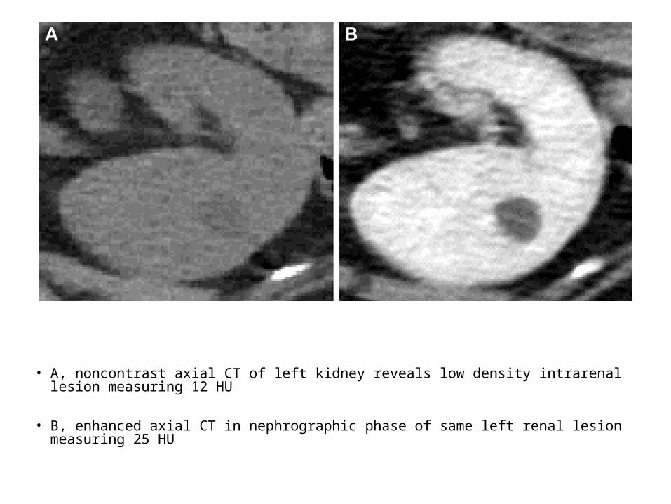
• A, noncontrast axial CT of left kidney reveals low density intrarenal lesion measuring 12 HU
• B, enhanced axial CT in nephrographic phase of same left renal lesion measuring 25 HU

Computerized Tomography• New CT techniques
• Thin (3 mm) overlapping sections improves the diagnosis of small renal lesions (less than 5 mm) and reduces the number of intermediate lesions detected
• delayed contrast enhanced CT improves diagnostic ability when post-contrast CT detects an incidental renal lesion
• Dual energy CT allows better detection of vascularity

Magnetic Resonance Imaging• Update on MRI of renal cysts
• MRI has a high sensitivity for the properties of simple cystic fluid, proteinaceous fluid and blood products
• Based on gadolinium enhanced MRI rates of malignancy were independently associated with mural wall irregularity (10 of 16 cases), mural nodules/masses (3 of 4), increased mural thickness (10 of 14) and intense mural enhancement (14 of 32)

• New MRI techniques
• DWMRI can be used to differentiate tissues with high and low cellular density since, compared to benign tissue, neoplastic tissue displays different permeability and osmolarity properties
• DWMRI requires no contrast ,hence can be used in patients with renal insufficiency
• DWMRI limitations are :1. No standardized technique2. Differing amounts of weighting /b value/3. Highly proteinaceous cystic lesions may be difficult to
distinguish from malignancies.
Magnetic Resonance Imaging

• A, T1 gadolinium enhanced axial MRI of right kidney demonstrates right interpolar lesion with enhancing wall nodule
• B, diffusion-weighted axial MRI of same lesion shows significant restricted diffusion, confirming suspicion of neoplasm. Lesion is considered Bosniak III

Ultrasound
• Update on US of renal cysts
• Standard renal US should not be used for initial evaluation of complex cystic renal masses largely because contrast enhancement, which is an important criterion for malignancy, cannot be assessed

Ultrasound• New US techniques• Contrast enhanced US newly emerged as a method of evaluating
complex renal cysts• CEUS is useful to detect tissue enhancement, including small
enhancing septa• Current evidence suggests that CEUS may be as good as or even
better than CT at detecting fine enhancing septa and tumor vascularity in complex cysts
• Park et al found that the diagnostic accuracy of CEUS and CT for malignant renal tumors was 90% and 74%, respectively.
• If there was a discrepancy in Bosniak criteria, the lesions tended to be upgraded by CEUS
• Limitations of CEUS are variable : microbubble contrast carries a black box warning ,operator dependent , interobserver variability ,location of the cysts , CEUS availability

• A, B-mode US of right renal cyst with dense internal echoes• B, CEUS reveals no enhancement within cyst

• A, B-mode US of right renal cyst with thick internal septations• B, CEUS demonstrates vivid irregular enhancement of internal septations
Biopsy of Renal Cysts
• Can be used to avoid unnecessary surgery ,a high degree of concordance (approximately 90%) between biopsy and final histological diagnosis for RCC
• Limitations : false-negative possibility , nondiagnostic biopsies , difficult reassessment , few studies investigate usefulness of biopsy for complex renal cysts

Surveillance of Renal Cysts• No surveillance protocol has been established for category
II and IIF cysts• Weibl et al reported that a minimum 5-years follow-up is
necessary to prove a lesion is benign• Israel and Bosniak reviewed 42 patients with Bosniak IIF
cysts (Avg. size 3.9 by 3.6 cm) with a mean follow-up of 5.8 years and found that :1. 6 (14.2%) had some form of progression 2. 36 cysts (86%) remained unchanged on CT 3. 3 (7.1%) decreased in size
• Median time to progression of 11 months to 4 years• The authors suggest a follow-up schedule of 6 months at
first, annually for 2 years and then biannually

DISCUSSION
• DW-MRI and CE-US offers a promise in better determination of lesion characteristics
• Biopsy techniques have been observed to be somewhat efficacious in diagnosis
• The importance of these new technologies lies in their ability to distinguish surgical from nonsurgical lesions

Limitations• Lack of prospective studies on renal cysts (retrospective
& clinically/statistically heterogeneous)
• It is possible that confounding and selection bias affected our conclusions
• Inclusion restricted to studies that were written in English, published in the last 15 years and had abstracts available
• We did not include any series on the use of PET-CT despite its potentiality to improve the evaluation of intermediate renal cysts

Results
• CT and MRI with and without contrast enhancement remain the gold standard to evaluate cystic lesions of the kidney
• diffusion-weighted MRI and contrast enhanced US have surfaced as new tools for assessment of complex cysts
• Image guided biopsy has increasingly been shown to be useful for evaluation of intermediate (Bosniak II and IIF) complex cysts
• few studies provide guidance on the duration and/or intensity of surveillance required for intermediate complex renal cysts
CONCLUSIONS
• New and enhanced techniques are in development and may be useful in the future management of complex renal cysts, there is a paucity of data regarding the value of these new techniques
• Future research should focus on surveillance of intermediate complex renal cysts, particularly on the ideal frequency and type of imaging required
• Randomized prospective studies should be performed