review article intradialytic hypotension and cardiac
TRANSCRIPT
Review ArticleIntradialytic Hypotension and Cardiac Remodeling:A Vicious Cycle
Chia-Ter Chao,1,2 Jenq-Wen Huang,3 and Chung-Jen Yen3,4
1Renal Division, Department of Internal Medicine, National Taiwan University Hospital, Jin-Shan Branch,51 Nan-Shih, Jin-Shan District, New Taipei City 208, Taiwan2Graduate Institute of Toxicology, National Taiwan University Medical School, Section 1, 1 Jen-Ai Road,Zhong-Zheng District, Taipei 100, Taiwan3Renal Division, Department of Internal Medicine, National Taiwan University Hospital, 7 Chung-Shan South Road,Zhong-Zheng District, Taipei 100, Taiwan4Department of Geriatrics and Gerontology, National Taiwan University Hospital, 7 Chung-Shan South Road,Zhong-Zheng District, Taipei 100, Taiwan
Correspondence should be addressed to Chung-Jen Yen; [email protected]
Received 24 April 2014; Accepted 19 September 2014
Academic Editor: John J. Gildea
Copyright © 2015 Chia-Ter Chao et al. This is an open access article distributed under the Creative Commons Attribution License,which permits unrestricted use, distribution, and reproduction in any medium, provided the original work is properly cited.
Hemodynamic instability during hemodialysis is a common but often underestimated issue in the nephrologist practice.Intradialytic hypotension, namely, a decrease of systolic or mean blood pressure to a certain level, prohibits the safe and smoothachievement of ultrafiltration and solute removal goal in chronic dialysis patients. Studies have elucidated the potential mechanismsinvolved in the development of Intradialytic hypotension, including excessive ultrafiltration and loss of compensatory mechanismsfor blood pressure maintenance. Cardiac remodeling could also be one important piece of the puzzle. In this review, we intendto discuss the role of cardiac remodeling, including left ventricular hypertrophy, in the development of Intradialytic hypotension.In addition, we will also provide evidence that a bidirectional relationship might exist between Intradialytic hypotension and leftventricular hypertrophy in chronic dialysis patients. A more complete understanding of the complex interactions in between couldassist the readers in formulating potential solutions for the reduction of both phenomena.
1. Introduction
Intradialytic hypotension (IDH) is a common scenario famil-iar to both patients under chronic hemodialysis and nephrol-ogists. It is defined as a decrease in systolic blood pressure(BP) by more than 20mmHg or decrease in mean arterialpressure (MAP) by 10mmHg, accompanied by symptomsincluding (but not limited to) abdominal discomfort, yawn-ing, sighing, nausea/vomiting,muscle cramping, restlessness,dizziness/fainting, or anxiety [1]. The incidence of IDH dur-ing each session of hemodialysis lies in somewhere between20% and 30% [1–3]. IDH, in the short term, impairs patients’quality of life by causing nuance symptoms and creates bar-riers to achieving adequate dialysis dose and ultrafiltration,while in the long term it leads to cardiovascular complications
(ischemic heart events, arrhythmia), more hospitalization,and higher mortality [4, 5].
2. Development of IDH
Risk factors for IDH have not been defined clearly from theliterature, but several issues have been held responsible. Forexample, demographic backgrounds andmedical comorbidi-ties play an important role in determining IDH susceptibility.Patients of advanced age (≧65), with diabetic nephropathy,cardiovascular illnesses, or autonomic dysfunction, are at riskfor developing IDH [1, 6–8]. Furthermore, clinical featuressuch as low blood pressure levels before hemodialysis session(<100mmHg), poor nutritional status (hypoalbuminemia),or severe anemia also predispose chronic dialysis patients to
Hindawi Publishing CorporationBioMed Research InternationalVolume 2015, Article ID 724147, 7 pageshttp://dx.doi.org/10.1155/2015/724147
2 BioMed Research International
IDHoccurrence [9]. Among these risk factors, cardiovascularcomorbidities, whether present before or after maintenancedialysis initiation, serve as an important step stone for thedevelopment or the perpetuation of subsequent IDH.
Most researchers concur that IDH frequently occursduring dialysis, when large volumes of fluid are removedwithin one session. Rapid ultrafiltration then fails to elicitcompensatory cardiovascular (CV) responses, such as vaso-constriction and rising cardiac output, while the combinationof inadequate peripheral vascular tone and plasma refillinginsufficiency leads to the drop of BP [3]. Clearly, four counter-active components are essential for the body’s defense againstIDH, that is, cardiac chronotropy and inotropy, plasma refill-ing, passive venoconstriction, and active arterioconstriction[3, 10]. Effective rise in heart rate in response to IDH could bean important component for countering acute hypotension.Among these four factors, the changes in heart function seemto be the major ones comparing with the others, since thelegacy of dysfunctional cardiac machinery could last evenafter dialysis sessions completion [11], while other factorswould not. Consequently, we will discuss in the following thepotential role of cardiac remodeling in end-stage renal disease(ESRD) patients and its relevancewith IDH. Furthermore, wewill also address the issue that IDHmight also lead to cardiacchanges, contributing to an ultimate downward spiral.
3. Cardiac Alterations in ESRD
3.1. Left Ventricular Hypertrophy (LVH). For patients withchronic kidney disease (CKD), the prevalence of LVHincreases progressively as renal function deteriorates, accom-panied by an elevation of pulse pressure [12]. Nearly three-fourths of chronic dialysis patients have LVH, based onechocardiographic findings [13–15]. These facts suggest thatcardiac structural alterations have already taken place duringearly renal function impairment, and the condition worsensfurther after dialysis initiation [16]. Advanced age, hyperten-sion, and increased arterial stiffness are significant predictorsfor LVH in general population and dialysis patients [13,17]. The pathogenic interplay of LVH of dialysis patientsencompasses three factors: high afterload, high preload, andmiscellaneous factors, such as renal failure per se [16, 18].The presence of LVH in dialysis patients correlates signif-icantly with subsequent CV events, with a dose-responserelationship. Zoccali et al. identified that dialysis patientswith LVH had a 2-3-fold higher mortality than those without[19]. Specifically, every 1 g/m2/month increase in LV massindex could lead to a 62% increase in the risk of CV events,applicable to both concentric or eccentric types [19, 20].Similarly, every 1 gram of LV mass reduction could translateinto 1% CV risk decrease [21]. This association betweenLVH and CV risk is potentially mediated by lethal atrial orventricular, but the exact pathway remains elusive [22–25].
3.2. Left Atrial Enlargement. As left atrium (LA) is positionedto sustain pressures from LV diastole, LA sizes/functions areoften touted as an indirect indicator of diastolic function[26]. LA volume index (LAVI) is identified as a morphologic
surrogate for LV diastolic dysfunction and an indicator forsubsequent cardiac remodeling [27, 28]. For dialysis patients,studies on LA functional/structural changes are far fewer[29]. Barberato and colleagues found that 37% of dialysispatients had LAVI higher than 35mL/m2, while othersidentified that ESRD patients have a progressive increase inLA sizes over time (11% over 1.5 years) [30, 31]. Furthermore,LA volumes changes over time are predictive of incident CVevents in dialysis patients, independent of LVmass index [32,33]. Consequently, the alterations of LA in dialysis patientsrepresent another underestimated issue.
3.3. Atrial Fibrillation (Afib). Cardiac rhythmic abnormali-ties, especially Afib, occur in 7–27% of all ESRD patients andincrease over higher dialysis vintage [34–36]. Thromboem-bolic events (cerebrovascular accidents) and the preloadreduction from Afib might precipitate hemodynamic alter-ations andmyocardial ischemia, leading to adverse prognosis[34]. Development of Afib in dialysis patients is associatedwith more hospitalization, CV events, and poorer survival[34]. Incident Afib also impairs renal prognosis in CKDpatients [37].
4. From Cardiac Remodeling to IDH:The Road In Between
Most patients, when they reach CKD stage 5, already carrya moderate to severe burden of CV illnesses, whether fromthe morbidities that cause their CKD (DM, hypertension)or from the imbalances of divalent ion homeostasis (CKD-MBD, mineral bone disease). The remodeling processesfound during initiation of dialysis could simply be thetrails left by the past injuries or the compensatory mech-anisms. Among these, cardiac remodeling is the predomi-nant manifestation and could contribute mechanistically tohemodynamic instability. Moreover, the sequels of cardiacremodeling might actively contribute to the development ofcomplications during dialysis, most important of all, IDH.It would then be necessary to discuss the role of cardiacremodeling, especially LVH, in the promotion of IDH fromtwo different points of view, that is, the passive one and theactive one.
4.1. LVH Sets the Background for IDH Development. Firstof all, LVH plays a permissive role in the development ofIDH. ESRD represents a combination of volume overload(fluid retention, the presence of arteriovenous shunt, ane-mia, etc.) and pressure overload (increasing arterial stiff-ness, atherosclerosis, etc.). However, the geometry of LVin ESRD mostly presents as concentric hypertrophy owingto pressure overload (from increased systemic resistance),despite the concurrent existence of significant volume com-ponent [38, 39]. Those with eccentric hypertrophy (fromincreased systolic and diastolic wall stress), on the contrary,are accompanied by coronary artery diseases, causing LVdilatation and later systolic dysfunction [40].Thesemechani-cal/morphologic subtypes (concentric versus eccentric) each
BioMed Research International 3
characterize different response patterns to fluid removalduring hemodialysis.
4.2. Concentric LVH. Current literature attributes the pre-disposing effect of concentric LVH on IDH to both hemo-dynamic factors and structural factors, such as valvulardegeneration. First, for patients with concentric LV, they areparticularly sensitive to abrupt changes in cardiac loadingstatuses, predisposing them to prominent BP fluctuationduring ultrafiltration [41]. This phenomenon stems fromthe fact that LV stiffening and reduction in complianceis associated with elevation of end-diastolic pressure. Asfilling of LV becomes difficult during dialysis (decreasingcirculating volume), even low levels of volume reductioncould translate into wide variation in cardiac output and thenBP. This has been found to be the major etiology of LVH-related IDH, that is, the diastolic dysfunction theory [3]. Also,concentric hypertrophy is often associated with progressivemyocardial fibrosis [42], further enhancing LV stiffness andthe downstream adverse sequences. Second, the presence ofLVH is frequently associated with the occurrence of aorticstenosis, as well as higher myocardial injury, represented byelevated plasma cardiac troponin I [43]. Prolonged aorticstenosis could expectedly lead to hemodynamic instability,precipitating IDH episodes.
4.3. Eccentric LVH. Eccentric hypertrophy, on the otherhand, is rarely discussed regarding its impact on IDH.Nonetheless, we propose two potential explanations linkingeccentric LVH and IDH occurrence. First of all, systolicfailure evolving from persistent eccentric LVH could beresponsible. Reportedly eccentric LVH demonstrates lessinfluence on IDH comparing with the concentric type, owingto their preservation of LV cavity and the correspondinglyhigher LV volume in the early phase of cardiac remodeling[40]. This minute advantage vanishes with the developmentof pump failure later, and the rapid change in loading statusesduring ultrafiltration will eventually lead to IDH in thesepatients. In this instance, systolic heart failure would be themain working hose behind the scene of IDH. Second, flow-mediated obstruction could also assist in IDH precipitation.Indeed, eccentric hypertrophy is often accompanied by asym-metric septal hypertrophy (ASH), caused by sympatheticsurge during ultrafiltration processes during dialysis [44].ASH, with the resultant sympathetic mediated hypercontrac-tility of the hypertrophic myocardium, might cause midsys-tolic obstruction at the level of LV outflow tract, servingas another pathway to IDH [44]. Furthermore, tachycardiacaused by sympathetic output might also contribute to IDHwith preload reduction.
4.4. LVH Proactively Induces IDH Occurrence. Finally, LVHactively contributes to IDH occurrence, through the induc-tion of myoischemia and arrhythmia. As the hypertrophiedmyocardium has to work against higher systemic resistanceduring LVH progression, the oxygen consumption rateincreases and sets the stage for ensuing ischemic myocardialdamages. This LVH-associated ischemia is often caused by
coronary microvascular dysfunction, which is a commonscenario in dialysis patients [45]. Furthermore, the reduc-tion in cardiac output from LV filling difficulty will drivethe heart rate higher for compensation purpose, while theaccompanied tachycardia also elevates myocardial oxygendemand. These ischemic changes could impair myocardialperformance and further lead to fibrosis.
Fatal/nonfatal arrhythmia could be another problem inthe LVH process. Myocardial ischemia from LVH couldenhance the arrhythmogenicity and potentially increase thechance of IDH [46]. LVH per se, by means of prolongationof action potential duration and refractory period, alongwith inherent nonuniform property within the involvedmyocardium, also potentiates the proarrhythmic phenotype[47]. Both factors could be important mediators for IDHepisodes.
5. IDH: Also a Precipitant forCardiac Remodeling?
Few researchers, if present, have addressed the issue ofIDH and its contribution to cardiac remodeling, besides itswell-known effect on long-term fistula outcomes. However,a diverse spectrum of putative causal relationships couldexist between IDH and cardiac remodeling, especially LVH.We would focus more on the occult effect of IDH in thedevelopment of LVH in the following section.
5.1. CoronaryHypoperfusion andMyocardial StunningTheory.Intuitively, IDH causes systemic hypoperfusion, includingcoronary vessels.This ischemic effect, though transient, couldpotentially cause myocardial fibrosis in the long term andthe subsequent LV kinetic dysregulation. A small-scale studyusing continuous intradialytic hemodynamic monitor didshow that IDH or intradialytic muscle cramping correlatedsignificantly with lower cardiac index and higher peripheralresistance [48].This myocardial injury applies to both ventri-cle and atrium, resulting in LA dilatation and LVH alike [30].The combination of coronary hypoperfusion and resultantmyocardial dysfunction culminates in an elevated mortalityin chronic dialysis patients. In addition, the intermittentreduction in fluid removal due to IDH also predisposespatients to chronic volume overload and eventually myocar-dial remodeling.
Furthermore, intermittent IDH produces a state called“HD-induced myocardial stunning” [49], which is causedby repeated ischemia and excessive ultrafiltration. Originallydescribed as a delayed recovery of regional myocardial con-tractile function after reperfusion despite the absence ofirreversible damage, the role ofmyocardial stunning has beensubsequently affirmed in chronic hemodialysis patients, asa prelude of heart failure. Myocardial stunning is usuallycharacterized by advanced age, higher ultrafiltration vol-umes, elevated cardiac troponin I, and, most importantly,presence of IDH [49]. Importantly, impaired calcium regu-lation, endothelial dysfunction, and the reperfusion periodafter IDH, which generates free radicals within myocardium,all contribute to this phenomenon [50]. The presence of
4 BioMed Research International
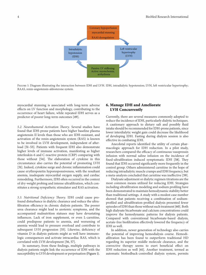
Intradialytichypotension
(IDH)
Left ventricular hypertrophy
(LVH)
Coronary hypoperfusionmyocardial stunning
RAAS dysregulation
Passive: LV stiffening Active: myoischemia,
arrhythmia
Figure 1: Diagram illustrating the interaction between IDH and LVH. IDH, intradialytic hypotension; LVH, left ventricular hypertrophy;RAAS, renin-angiotensin-aldosterone system.
myocardial stunning is associated with long-term adverseeffects on LV function and morphology, contributing to theoccurrence of heart failure, while repeated IDH serves as apredictor of poorer long-term outcomes [49].
5.2. Neurohumoral Activation Theory. Several studies havefound that IDH-prone patients have higher baseline plasmaangiotensin II levels than those who are IDH-resistant, andactivation of the renin-angiotensin system (RAS) is knownto be involved in LVH development, independent of after-load [51–53]. Patients with frequent IDH also demonstratehigher levels of immune activation, manifesting as higherinterleukin-6 and C-reactive protein (CRP) comparing withthose without [54]. The elaboration of cytokine in thiscircumstance also carries the potential of promoting LVH[51]. Indeed, cytokine surge and chronic inflammation couldcause erythropoietin hyporesponsiveness, with the resultantanemia, inadequate myocardial oxygen supply, and cardiacremodeling. Furthermore, IDH often occurred in the contextof dry-weight probing and intense ultrafiltration, which con-stitutes a strong sympathetic stimulator and RAS activation.
5.3. Nutritional Deficiency Theory. IDH could cause pro-found disturbance in dialytic clearance and reduce the ultra-filtration efficiency in chronic dialysis patients. The poorerurea clearance might lead to azotemic symptoms, and theaccompanied malnutrition statuses may have devastatinginfluences. Lack of iron supplement, or even L-carnitine,could predispose patients to anemia, and worsening ofanemia would lead to pressure overload and contribute tosubsequent LVH progression [55]. Likewise, deficiency ofvitamin D in dialysis patients might as well have immuno-logic consequences and activate intracardiac RAS, which iscorrelated with LVH development [56, 57].
In summary, from these findings, multiple pathways indialysis patients might link the occurrence of IDH with thesusceptibility to LVHdevelopment or perpetuation (Figure 1).
6. Manage IDH and AmeliorateLVH Concurrently
Currently, there are several measures commonly adopted toreduce the incidence of IDH, particularly dialytic techniques.A cautionary approach to dietary salt and possibly fluidintake should be recommended for IDH-prone patients, sincelower interdialytic weight gain could decrease the likelihoodof developing IDH. Fasting during dialysis session is alsoeffective in combating IDH.
Anecdotal reports identified the utility of certain phar-macologic approach for IDH reduction. In a pilot study,researchers compared the efficacy of continuous vasopressininfusion with normal saline infusion on the incidence offixed-ultrafiltration induced symptomatic IDH [58]. Theyfound that IDH occurred significantly more frequently in thecontrol group. Others administered carnitine in the hope ofreducing intradialyticmuscle cramps and IDH frequency, buta meta-analysis concluded that carnitine was ineffective [59].
Dialysate adjustment or dialytic regimen titrations are themost common means utilized for reducing IDH. Strategiesincluding ultrafiltration modeling and sodium profiling havebeen demonstrated tomaintain hemodynamic stability betterthan traditional settings. A study with a modest case numbershowed that patients receiving a combination of sodium-profiled and ultrafiltration-profiled dialysis presented fewerepisodes of IDH than thosewithout such treatment [60]. Bothhigh dialysate bicarbonate and calcium concentrations couldimprove the hemodynamic patterns for dialysis patients.Compared with conventional bicarbonate-based dialysis,acetate-free biofiltration effectively lowered the frequency ofIDH as well.
In addition, newer generation of technology also carriesthe potential of improving hemodialysis course. Hemodi-afiltration has been found to outperform traditional HDregarding its superior middle molecule clearance, and theconvective therapy seems to exert beneficial effect onIDH reduction as well [61]. Another treatment, termed asautomatic biofeedback-controlled dialysis system, permits
BioMed Research International 5
the adjustment of dialysate concentration and ultrafiltrationsettings according to body weight and sodium content.Several randomized trials assessing the biofeedback-basedcontrol system disclosed significant reduction in the fre-quency of IDH and nursing interventions among users [62,63]. Furthermore, the use of automated biofeedback dialysissignificantly reduced reversible LV regional wall motionabnormalities [64].This undoubtedly solidifies the intertwin-ing relationship between IDH and cardiac modeling.
7. Conclusion
IDH and myocardial remodeling, especially LVH, are closelyassociated with each other. LVH is an important determinantand etiology of IDH, as LVH not only paves the way towardfrequent IDH occurrence but also actively facilitates thedrop of BP during dialysis, through mechanisms includingarrhythmia and myocardial ischemia. Although IDH mightnot be the predominant cause of LVH in dialysis patients, itrepresents one potentially remediable factor, in light of thecurrent trend that focuses on treating LVH per se for ESRDpatients. It is truly difficult to disentangle the relationshipbetween each other, but a clearer understanding of thecomplex interactions between IDH and LVH might assist indevising useful strategies to avoid the occurrences of both.
Conflict of Interests
The authors have no relevant conflict of interests to declare inrelation to this paper.
Acknowledgments
The authors are grateful to the staffs of the Second CoreLaboratory of the Department of Medical Research ofNational TaiwanUniversityHospital, Taipei, Taiwan, for theirassistance, and the financial support from NTUH (Projectnos. 102-N2249 and 103-S2525).
References
[1] K/DOQIWorkgroup, “K/DOQI clinical practice guidelines forcardiovascular disease in dialysis patients,” American Journal ofKidney Disease, vol. 45, no. 4, supplement 3, pp. S1–S153, 2005.
[2] F. M. Van Der Sande, J. P. Kooman, and K. M. L. Leunissen,“Intradialytic hypotension—new concepts on an old problem,”Nephrology Dialysis Transplantation, vol. 15, no. 11, pp. 1746–1748, 2000.
[3] J. T. Daugirdas, “Pathophysiology of dialysis hypotension: anupdate,” American Journal of Kidney Diseases, vol. 38, no. 4, pp.S11–S17, 2001.
[4] T. Shoji, Y. Tsubakihara, M. Fujii, and E. Imai, “Hemodialysis-associated hypotension as an independent risk factor for two-year mortality in hemodialysis patients,” Kidney International,vol. 66, no. 3, pp. 1212–1220, 2004.
[5] J. K. Inrig, E. Z. Oddone, V. Hasselblad et al., “Association ofintradialytic blood pressure changes with hospitalization andmortality rates in prevalent ESRD patients,” Kidney Interna-tional, vol. 71, no. 5, pp. 454–461, 2007.
[6] C. Calvo, S. Maule, F. Mecca, R. Quadri, G.Martina, and P. Cav-allo Perin, “The influence of autonomic neuropathy onhypoten-sion during hemodialysis,” Clinical Autonomic Research, vol. 12,no. 2, pp. 84–87, 2002.
[7] M. Koremoto, N. Takahara, M. Takahashi et al., “Improvementof intradialytic hypotension in diabetic hemodialysis patientsusing vitamin E-bonded polysulfone membrane dialyzers,”Artificial Organs, vol. 36, no. 10, pp. 901–910, 2012.
[8] C.-T. Chao, Y.-F. Lin, H.-B. Tsai, V.-C. Wu, and W.-J. Ko,“Acute kidney injury network staging in geriatric postoperativeacute kidney injury patients: shortcomings and improvements,”Journal of the American College of Surgeons, vol. 217, no. 2, pp.240–250, 2013.
[9] A. Lacquaniti, D. Bolignano, S. Campo et al., “Malnutrition inthe elderly patient on dialysis,” Renal Failure, vol. 31, no. 3, pp.239–245, 2009.
[10] J. T. Daugirdas, “Dialysis hypotension: a hemodynamic analy-sis,” Kidney International, vol. 39, no. 2, pp. 233–246, 1991.
[11] R. N. Foley, P. S. Parfrey, G.M. Kent, J. D. Harnett, D. C.Murray,and P. E. Barre, “Long-term evolution of cardiomyopathy indialysis patients,” Kidney International, vol. 54, no. 5, pp. 1720–1725, 1998.
[12] E. Paoletti, D. Bellino, P. Cassottana, D. Rolla, and G. Cannella,“Left ventricular hypertrophy in nondiabetic predialysis CKD,”American Journal of Kidney Diseases, vol. 46, no. 2, pp. 320–327,2005.
[13] R. N. Foley, P. S. Parfrey, J. D. Harnett et al., “Clinical andechocardiographic disease in patients starting end-stage renaldisease therapy,”Kidney International, vol. 47, no. 1, pp. 186–192,1995.
[14] R. N. Foley, P. S. Parfrey, and M. J. Sarnak, “Clinical epidemi-ology of cardiovascular disease in chronic renal disease,” TheAmerican Journal of KidneyDiseases, vol. 32, pp. S112–S119, 1998.
[15] D. Levy, K.M.Anderson,D.D. Savage,W. B. Kannel, J. C. Chris-tiansen, and W. P. Castelli, “Echocardiographically detectedleft ventricular hypertrophy: prevalence and risk factors: theFramingham heart study,” Annals of Internal Medicine, vol. 108,no. 1, pp. 7–13, 1988.
[16] R. J. Glassock, R. Pecoits-Filho, and S. H. Barberato, “Leftventricular mass in chronic kidney disease and ESRD,” ClinicalJournal of the American Society of Nephrology, vol. 4, no. 1, pp.S79–S91, 2009.
[17] M. Brahimi, M. Dahan, H. Dabire, and B. I. Levy, “Impact ofpulse pressure on degree of cardiac hypertrophy in patients withchronic uraemia,” Journal of Hypertension, vol. 18, no. 11, pp.1645–1650, 2000.
[18] C. Zoccali, F. A. Benedetto, F. Mallamaci et al., “Prognosticimpact of the indexation of left ventricular mass in patientsundergoing dialysis,” Journal of the American Society of Nephrol-ogy, vol. 12, no. 12, pp. 2768–2774, 2001.
[19] C. Zoccali, F. A. Benedetto, F. Mallamaci et al., “Left ventricularmass monitoring in the follow-up of dialysis patients: prognos-tic value of left ventricular hypertrophy progression,” KidneyInternational, vol. 65, no. 4, pp. 1492–1498, 2004.
[20] G. M. London, B. Pannier, A. P. Guerin et al., “Alterations ofleft ventricular hypertrophy in and survival of patients receivinghemodialysis: follow-up of an interventional study,” Journal ofthe American Society of Nephrology, vol. 12, no. 12, pp. 2759–2767, 2001.
6 BioMed Research International
[21] F. O. B. Bonato, M. M. Lemos, J. L. Cassiolato, and M. E. F.Canziani, “Prevalence of ventricular arrhythmia and its asso-ciated factors in nondialyzed chronic kidney disease patients,”PLoS ONE, vol. 8, no. 6, Article ID e66036, 2013.
[22] P. M. Okin, C. N. Bang, K. Wachtell et al., “Relationship ofsudden cardiac death to new-onset atrial fibrillation in hyper-tensive patients with left ventricular hypertrophy,” Circulation:Arrhythmia and Electrophysiology, vol. 6, no. 2, pp. 243–251,2013.
[23] J. O. Burton, S. Korsheed, B. J. Grundy, and C. W. McIntyre,“Hemodialysis-induced left ventricular dysfunction is associ-ated with an increase in ventricular arrhythmias,” Renal Failure,vol. 30, no. 7, pp. 701–709, 2008.
[24] C.-T. Chao, C.-C. Hou, V.-C. Wu et al., “The impact of dial-ysis-requiring acute kidney injury on long-term prognosis ofpatients requiring prolonged mechanical ventilation: nation-wide population-based study,” PLoS ONE, vol. 7, no. 12, ArticleID e50675, 2012.
[25] J. J. Mourad, B. Pannier, J. Blacher et al., “Creatinine clearance,pulse wave velocity, carotid compliance and essential hyperten-sion,” Kidney International, vol. 59, no. 5, pp. 1834–1841, 2001.
[26] H. Miyoshi, Y. Oishi, Y. Mizuguchi et al., “Early predictorsof alterations in left atrial structure and function related toleft ventricular dysfunction in asymptomatic patients withhypertension,” Journal of the American Society of Hypertension,vol. 7, no. 3, pp. 206–215, 2013.
[27] T. S. M. Tsang, M. E. Barnes, B. J. Gersh, K. R. Bailey,and J. B. Seward, “Left atrial volume as a morphophysiologicexpression of left ventricular diastolic dysfunction and relationto cardiovascular risk burden,” American Journal of Cardiology,vol. 90, no. 12, pp. 1284–1289, 2002.
[28] F. M. Fouad, J. M. Slominski, and R. C. Tarazi, “Left ventriculardiastolic function in hypertension: relation to left ventricularmass and systolic function,” Journal of the American College ofCardiology, vol. 3, no. 6, pp. 1500–1506, 1984.
[29] S. H. Barberato and R. Pecoits Filho, “Valor prognostico doındice do volume do atrio esquerdo em pacientes em hemo-dialise,”Arquivos Brasileiros deCardiologia, vol. 88, pp. 643–650,2007.
[30] S. H. Barberato, M. Misocami, and R. Pecoits-Filho, “Asso-ciation between left atrium enlargement and intradialytichypotension: role of diastolic dysfunction in the hemodynamiccomplications during hemodialysis: CME,” Echocardiography,vol. 26, no. 7, pp. 767–771, 2009.
[31] G. Tripepi, F. Mattace-Raso, F. Mallamaci et al., “Biomarkers ofleft atrial volume: a longitudinal study in patients with end stagerenal disease,” Hypertension, vol. 54, no. 4, pp. 818–824, 2009.
[32] G. Tripepi, F. A. Benedetto, F. Mallamaci, R. Tripepi, L.Malatino, and C. Zoccali, “Left atrial volume in end-stage renaldisease: a prospective cohort study,” Journal of Hypertension,vol. 24, no. 6, pp. 1173–1180, 2006.
[33] G. Tripepi, F. A. Benedetto, F. Mallamaci, R. Tripepi, L.Malatino, and C. Zoccali, “Left atrial volume monitoring andcardiovascular risk in patients with end-stage renal disease: aprospective cohort study,” Journal of the American Society ofNephrology, vol. 18, no. 4, pp. 1316–1322, 2007.
[34] S. Genovesi, A. Vincenti, E. Rossi et al., “Atrial fibrillation andmorbidity and mortality in a cohort of long-term hemodialysispatients,”American Journal of Kidney Diseases, vol. 51, no. 2, pp.255–262, 2008.
[35] W. C. Winkelmayer, A. R. Patrick, J. Liu, M. A. Brookhart, andS. Setoguchi, “The increasing prevalence of atrial fibrillation
among hemodialysis patients,” Journal of the American Societyof Nephrology, vol. 22, no. 2, pp. 349–357, 2011.
[36] C.-T. Chao, V.-C. Wu, C.-F. Lai et al., “Advanced age affects theoutcome-predictive power of RIFLE classification in geriatricpatients with acute kidney injury,” Kidney International, vol. 82,no. 8, pp. 920–927, 2012.
[37] N. Bansal, D. Fan, C.-Y. Hsu, J. D. Ordonez, G. M. Marcus, andA. S. Go, “Incident atrial fibrillation and risk of end-stage renaldisease in adults with chronic kidney disease,” Circulation, vol.127, no. 5, pp. 569–574, 2013.
[38] C. Zoccali, F. Mallamaci, R. Maas et al., “Left ventricular hyper-trophy, cardiac remodeling and asymmetric dimethylarginine(ADMA) in hemodialysis patients,” Kidney International, vol.62, no. 1, pp. 339–345, 2002.
[39] M. A. Alpert, “Cardiac performance and morphology in end-stage renal disease,” The American Journal of the MedicalSciences, vol. 325, no. 4, pp. 168–178, 2003.
[40] D. Levy,M.G. Larson, R. S.Vasan,W.B.Kannel, andK.K. L.Ho,“Theprogression fromhypertension to congestive heart failure,”Journal of the AmericanMedical Association, vol. 275, no. 20, pp.1557–1562, 1996.
[41] G. M. London, A. P. Guerin, and S. J. Marchais, “Pathophysiol-ogy of left ventricular hypertrophy in dialysis patients,” BloodPurification, vol. 12, no. 4-5, pp. 277–283, 1994.
[42] C. W. Doering, J. E. Jalil, J. S. Janicki et al., “Collagen networkremodelling and diastolic stiffness of the rat left ventricle withpressure overload hypertrophy,” Cardiovascular Research, vol.22, no. 10, pp. 686–695, 1988.
[43] C. W. Chin, A. S. Shah, V. Vassiliou et al., “Left ventricularhypertrophy with strain and aortic stenosis,” Circulation, 2014.
[44] D. Bernardi, L. Bernini, G. Cini, S. Ghione, and I. Bonechi,“Asymmetric septal hypertrophy and sympathetic overactivityin normotensive hemodialyzed patients,” American Heart Jour-nal, vol. 109, no. 3, pp. 539–545, 1985.
[45] P. G. Camici, I. Olivotto, and O. E. Rimoldi, “The coronarycirculation and blood flow in left ventricular hypertrophy,”Journal of Molecular and Cellular Cardiology, vol. 52, no. 4, pp.857–864, 2012.
[46] D. Kozhevnikov, E. B. Caref, and N. El-Sherif, “Mechanismsof enhanced arrhythmogenicity of regional ischemia in thehypertrophied heart,” Heart Rhythm, vol. 6, no. 4, pp. 522–527,2009.
[47] R. Wolk, “Arrhythmogenic mechanisms in left ventricularhypertrophy,” Europace, vol. 2, no. 3, pp. 216–223, 2000.
[48] J. Kolb, T. M. Kitzler, T. Tauber, N. Morris, F. Skrabal, andP. Kotanko, “Proto-dialytic cardiac function relates to intra-dialytic morbid events,” Nephrology Dialysis Transplantation,vol. 26, no. 5, pp. 1645–1651, 2011.
[49] J. O. Burton, H. J. Jefferies, N. M. Selby, and C. W. McIntyre,“Hemodialysis-induced cardiac injury: determinants and asso-ciated outcomes,” Clinical Journal of the American Society ofNephrology, vol. 4, no. 5, pp. 914–920, 2009.
[50] M. Y. Zuidema and K. C. Dellsperger, “Myocardial stun-ning with hemodialysis: clinical challenges of the cardiorenalpatient,” Cardiorenal Medicine, vol. 2, pp. 125–133, 2012.
[51] G. Graziani, S. Finazzi, R. Mangiarotti et al., “Different cardio-vascular responses to hemodialysis-induced fluid depletion andblood pressure compliance,” Journal of Nephrology, vol. 23, no.1, pp. 55–61, 2010.
[52] E. Ritz, “Left ventricular hypertrophy in renal disease: Beyondpreload and afterload,” Kidney International, vol. 75, no. 8, pp.771–773, 2009.
BioMed Research International 7
[53] H. Schunkert, J.-I. Sadoshima, T. Cornelius et al., “AngiotensinII-induced growth responses in isolated adult rat hearts:evidence for load-independent induction of cardiac proteinsynthesis by angiotensin II,” Circulation Research, vol. 76, no.3, pp. 489–497, 1995.
[54] M. Tomita, D. Malhotra, S. Dheenan, J. I. Shapiro, W. L.Henrich, and T. J. Santoro, “A potential role for immuneactivation in hemodialysis hypotension,” Renal Failure, vol. 23,no. 5, pp. 637–649, 2001.
[55] T. Sakurabayashi, S. Miyazaki, Y. Yuasa et al., “L-carnitinesupplementation decreases the left ventricular mass in patientsundergoing hemodialysis,”Circulation Journal, vol. 72, no. 6, pp.926–931, 2008.
[56] P. Strozecki, A. Adamowicz, E. Nartowicz, G. Odrowaz-Sypniewska, Z. Włodarczyk, and J. Manitius, “Parathormon,calcium, phosphorus, and left ventricular structure and func-tion in normotensive hemodialysis patients,” Renal Failure, vol.23, no. 1, pp. 115–126, 2001.
[57] C.-T. Chao, C.-F. Lai, and J.-W. Huang, “Risk factors for her-pes zoster reactivation in maintenance hemodialysis patients,”European Journal of InternalMedicine, vol. 23, no. 8, pp. 711–715,2012.
[58] S. van der Zee, A. Thompson, R. Zimmerman et al., “Vaso-pressin administration facilitates fluid removal duringhemodialysis,” Kidney International, vol. 71, no. 4, pp. 318–324,2007.
[59] K. E. Lynch, H. I. Feldman, J. A. Berlin, J. Flory, C. G. Rowan,and S. M. Brunelli, “Effects of L-carnitine on dialysis-relatedhypotension and muscle cramps: a meta-analysis,” AmericanJournal of Kidney Diseases, vol. 52, no. 5, pp. 962–971, 2008.
[60] Y. L. Zhou, H. L. Liu, X. F. Duan, Y. Yao, Y. Sun, andQ. Liu, “Impact of sodium and ultrafiltration profiling onhaemodialysis-related hypotension,”Nephrology Dialysis Trans-plantation, vol. 21, no. 11, pp. 3231–3237, 2006.
[61] F. Locatelli, P. Altieri, S. Andrulli et al., “Hemofiltration andhemodiafiltration reduce intradialytic hypotension in ESRD,”Journal of the American Society of Nephrology, vol. 21, no. 10, pp.1798–1807, 2010.
[62] E. Mancini, E. Mambelli, M. Irpinia et al., “Prevention ofdialysis hypotension episodes using fuzzy logic control system,”Nephrology,Dialysis, andTransplantation, vol. 22, pp. 1420–1427,2007.
[63] C. Deziel, J. Bouchard, M. Zellweger, and F. Madore, “Impactof hemocontrol on hypertension, nursing interventions, andquality of life: a randomized, controlled trial,” Clinical Journalof the American Society of Nephrology, vol. 2, no. 4, pp. 661–668,2007.
[64] N. M. Selby, S. H. Lambie, P. G. Camici, C. S. Baker, and C. W.McIntyre, “Occurrence of regional left ventricular dysfunctionin patients undergoing standard and biofeedback dialysis,”American Journal of Kidney Diseases, vol. 47, no. 5, pp. 830–841,2006.

Submit your manuscripts athttp://www.hindawi.com
Stem CellsInternational
Hindawi Publishing Corporationhttp://www.hindawi.com Volume 2014
Hindawi Publishing Corporationhttp://www.hindawi.com Volume 2014
MEDIATORSINFLAMMATION
of
Hindawi Publishing Corporationhttp://www.hindawi.com Volume 2014
Behavioural Neurology
EndocrinologyInternational Journal of
Hindawi Publishing Corporationhttp://www.hindawi.com Volume 2014
Hindawi Publishing Corporationhttp://www.hindawi.com Volume 2014
Disease Markers
Hindawi Publishing Corporationhttp://www.hindawi.com Volume 2014
BioMed Research International
OncologyJournal of
Hindawi Publishing Corporationhttp://www.hindawi.com Volume 2014
Hindawi Publishing Corporationhttp://www.hindawi.com Volume 2014
Oxidative Medicine and Cellular Longevity
Hindawi Publishing Corporationhttp://www.hindawi.com Volume 2014
PPAR Research
The Scientific World JournalHindawi Publishing Corporation http://www.hindawi.com Volume 2014
Immunology ResearchHindawi Publishing Corporationhttp://www.hindawi.com Volume 2014
Journal of
ObesityJournal of
Hindawi Publishing Corporationhttp://www.hindawi.com Volume 2014
Hindawi Publishing Corporationhttp://www.hindawi.com Volume 2014
Computational and Mathematical Methods in Medicine
OphthalmologyJournal of
Hindawi Publishing Corporationhttp://www.hindawi.com Volume 2014
Diabetes ResearchJournal of
Hindawi Publishing Corporationhttp://www.hindawi.com Volume 2014
Hindawi Publishing Corporationhttp://www.hindawi.com Volume 2014
Research and TreatmentAIDS
Hindawi Publishing Corporationhttp://www.hindawi.com Volume 2014
Gastroenterology Research and Practice
Hindawi Publishing Corporationhttp://www.hindawi.com Volume 2014
Parkinson’s Disease
Evidence-Based Complementary and Alternative Medicine
Volume 2014Hindawi Publishing Corporationhttp://www.hindawi.com