review article immunotherapy for lung cancer: advances · pdf fileam j clin exp immunol...
TRANSCRIPT

Am J Clin Exp Immunol 2016;5(1):1-20www.ajcei.us /ISSN:2164-7712/AJCEI0026956
Review ArticleImmunotherapy for lung cancer: advances and prospects
Li Yang1,2, Liping Wang2, Yi Zhang1,2,3
1Biotherapy Center, 2Department of Oncology, The First Affiliated Hospital of Zhengzhou University, Zhengzhou, Henan 450052, P.R. China; 3School of Life Sciences, Zhengzhou University, Zhengzhou, Henan 450001, P.R. China
Received February 29, 2016; Accepted March 12, 2016; Epub March 23, 2016; Published March 30, 2016
Abstract: Lung cancer is the most commonly diagnosed cancer as well as the leading cause of cancer-related deaths worldwide. To date, surgery is the first choice treatment, but most clinically diagnosed cases are inoperable. While chemotherapy and/or radiotherapy are the next considered options for such cases, these treatment modali-ties have adverse effects and are sometimes lethal to patients. Thus, new effective strategies with minimal side effects are urgently needed. Cancer immunotherapy provides either active or passive immunity to target tumors. Multiple immunotherapy agents have been proposed and tested for potential therapeutic benefit against lung cancer, and some pose fewer side effects as compared to conventional chemotherapy and radiotherapy. In this article, we discuss studies focusing on interactions between lung cancer and the immune system, and we place an emphasis on outcome evidence in order to create a knowledge base well-grounded in clinical reality. Overall, this review highlights the need for new lung cancer treatment options, with much ground to be paved for future advances in the field. We believe that immunotherapy agents alone or with other forms of treatment can be recog-nized as next modality of lung cancer treatment.
Keywords: Lung cancer, immunology, immunotherapy, vaccines, checkpoint inhibitor
Introduction
Lung cancer is the most commonly diagnosed cancer and the leading cause of cancer-related deaths. Particularly, lung cancer has an esti-mated incidence of 1.6 million new cases every year [1]. Lung cancer is categorized into two major subtypes depending on their histological feature: non-small cell lung cancer (NSCLC) and small cell lung cancer (SCLC). NSCLC and SCLC constitute 85% and 15% of total lung cancer cases respectively [2].
Only 16.6% of lung cancer patients survive 5 years or more, with only 3.9% surviving in the metastatic setting [3]. Use of conventional ther-apeutic strategies has lots of unwanted side effects and drawbacks. For example, chances of missing micro metastasis and recurrence are common problems observed in surgically operated lung cancer patients; chemotherapy, radiotherapy, or concomitant chemo-radiother-apy for inoperable cases may not prevent recur-
rences. Recent introduction of molecular tar-geted therapies, including activating mutations of epidermal growth factor receptor (EGFR) and anaplastic lymphoma kinase (ALK) transloca-tions, in the metastatic setting led to improved treatment outcomes in selected subgroups of patients with advanced stage NSCLC [3]. Even with the latest advances, lung cancer prognosis remains dismal, and novel therapeutic appro- aches are needed.
In the last decade, there has been a better understanding of the interactions between immune cells and cancer cells, and the mecha-nisms that cancer evades the immune system, resulting in a new era of cancer immunotherapy protocols which overcomes the limitations of conventional therapeutic strategies [4]. Im- munotherapy represents a broad class of treat-ment modalities designed to elicit immune-mediated destruction of tumor cells [3]. In this review, we will provide a comprehensive review

A review of immunotherapy for lung cancer
2 Am J Clin Exp Immunol 2016;5(1):1-20
about the current understanding of immuno-therapy for lung cancer.
Lung cancer: specific immune responses
Lung cancer progression is a multi-step mecha-nism. Chronic inflammation mediated by ciga-rette smoking, chronic obstructive pulmonary disease, chronic bronchitis, and occupational exposure are some of the many factors that lead to an imbalance in cytokine secretions and inflammatory responses, which may favor the malignant transformation of normal epithe-lial cells [5]. Lung cancer employs several meth-ods to evade surveillance and elimination by the host immune system. Here in this section, we present a brief introduction of lung cancer immunology.
Innate immunity
Innate immunity is a nonspecific first-line of defense, involving natural killer (NK) cells, mac-rophages and neutrophils [6]. A chronic inflam-mation state activates innate immunity with subsequent release of cytokines, which may promote tumor destruction but can also lead to oncogenesis [7]. In addition, lung cancer cells can re-educate M1 macrophages to M2 macro-phages [8]. Sentinel cells from the innate branch of immunity may recognize relatively non-specific structurally preserved molecules, which are distinguishable from the host’s mol-ecules through Toll-like receptors on their surfaces.
Tumor microenvironment (TME)
The TME is facilitated by many distinct cell types, including endothelial cells and their pre-cursors, pericytes, myeloid-derived suppressor cells (MDSCs), cancer-associated fibroblasts (CAFs), tumor-associated macrophages (TAMs), T and B lymphocytes, NK cells, dendritic cells (DCs), neutrophils, eosinophils, basophils, and mast cells [9] (Figure 1). The TME along with its contents are crucial for driving malignant trans-formed cells into solid masses which include resistance to apoptosis, proliferation, invasion, angiogenesis and metastasis.
MDSCs comprise a major type of immunosup-pressive leukocyte population that inhibits host-protective anti-tumor responses. MDSCs are capable of suppressing multiple phases of the immune response by promoting tumor local
invasion and metastases by secreting factors such as vascular endothelial growth factor (VEGF), basic fibroblast growth factor (bFGF), and matrix metalloproteinases [10, 11]. MDSCs contribute to the development of an immuno-suppressive TME that blocks the action of cyto-toxic anti-tumor T effector cells. The TME also possesses the potential to induce regulatory T cell function by secreting tumor growth factor (TGF)-β and interleukin (IL)-10 and plays a major role in immune tolerance, resulting in a major obstacle to efficient cancer immunother-apy [12, 13].
Fibroblasts are distorted during cancer pro-gression. Particularly, CAFs are present in abundance in the tumor stroma of the TME, where they release the hepatocyte growth fac-tor (HGF). HGF released promotes proliferation and invasion by modulating the HGF/c-Met pathway. Some studies have supported the role of CAFs in angiogenesis via demonstrating the capacity of CAFs to secrete pro-angiogenic fac-tors like VEGF-A, platelet derived growth factor, and bFGF [14].
Similarly, macrophages with normal innate phagocytic functions are recruited to tumor cells and become TAMs. TAMs support tumor growth by promoting angiogenesis, immuno-suppression, invasion, and metastasis. Spe- cifically, TAMs mediate tumor growth through ILs (-6, -4, -13), leukocyte inhibitory factor, prostaglandin E2, CCL17, CCL22, and CCL24 [15-17].
Immunosurveillance
Immune system invasion and/or escape is essential for lung cancer propagation. Several mechanisms are proposed for immune inva-sion by lung cancer cells. Key mechanisms pro-posed are: (i) resistance of tumor cell lysis due to deficient expression of major histocompati-bility complex (MHC), (ii) expression of poorly immunogenic epitopes, (iii) release of immuno-suppressive cytokines, and (iv) T cell apoptosis. Alternatively, escape may result from the estab-lishment of an immunosuppressive state within the TME [18].
Cytotoxic CD8+ T cells present in the TME of lung cancer patients were observed to be less effective. Furthermore, these cells were found to be hyporesponsive to activation via the T cell receptor (TCR). This deficiency in T cells is due

A review of immunotherapy for lung cancer
3 Am J Clin Exp Immunol 2016;5(1):1-20
to a TCR signaling arrest partially dependent on membrane-associated TGF-β1 present in the TME. Alterations in IL-10-producing CD8+ T cells were observed in patients with early stage lung cancer compared to patients with advanced stage lung cancer. IL-10-producing CD8+ T cells were at significantly higher propor-tions in early stage lung cancer; such an obser-vation may provide some insights about slower tumor progression or a longer overall survival (OS). Changes in the proportion of CD8+ T cells subsets (naïve, effector/memory) in the tumor stroma also explain its alteration in lung cancer [19-21]. Functions of NK cells are also impaired in lung cancer. They possess less cytotoxicity along with a decreased ability to degranulate and produce interferon (IFN)-γ variation in the
the antigen presenting cell’s (APC’s)/tumor cell’s B7 surface molecules (CD80 or CD86).
In order to prevent autoimmunity, immune checkpoints are set in place. Activated CD8+ T cells express a protein receptor named cyto-toxic T-lymphocyte antigen-4 (CTLA-4), which also binds B7 with high affinity, limiting further T cell activation by CD28 [25, 26]. Programmed cell death-1 (PD-1) is another T cell surface receptor that upon binding its cognate ligand (Programmed cell death - ligand 1, PD-L1) in the APC/tumor cell, it inhibits the immune response (Figure 2). While CTLA-4’s action focuses on limiting the initiation of T cell activa-tion in the lymph nodes, PD-1 acts later by limit-ing T cell activity in the TME [25].
Immunotherapy options for lung cancer
Given this myriad of immunosuppressive tools, it is of little wonder that traditional immuno-therapy approaches have largely failed to eradi-cate lung cancer. Nevertheless, the fruits of this scientific effort are beginning to be realized in lung cancer. The aim of immunotherapy is to enhance the immune response specifically directed to the tumor. A description of current investigational immunotherapies for lung can-cer is provided in the following sections below and summarized in Figure 3.
Cytokines
The first immunotherapies developed for NS- CLC were recombinant cytokines, namely those secreted by Th1 cells, such as IL-2 and IFN.
Figure 1. The tumor microenvironment (TME) is facilitated by many distinct cell types, including endothelial cells and their precursors, pericytes, myeloid-de-rived suppressor cells (MDSCs), cancer-associated fibroblasts (CAFs), tumor-associated macrophages (TAMs), T and B lymphocytes, natural killer (NK) cells, dendritic cells (DCs), neutrophils, eosinophils, basophils, and mast cells.
NK cell receptors [22, 23]. Immature or dysfunctional DCs are also seen in lung cancer and are mediated by IL-10, VEGF, and TGF-β. These dysfunctional DCs are shown to have role in cancer migration, invasion, and epithelial-mesenchy-mal transition [24].
Immune checkpoints
Besides the antigen-MHC-TCR interaction, additional co-activation signals must also be present (Figure 2), namely interaction of the T cell’s CD28 molecule with
Figure 2. Improved therapeutic efficacy can be achieved by modulating immune checkpoints, in-cluding activation of effector cells by blocking CTLA4 or PD-1.

A review of immunotherapy for lung cancer
4 Am J Clin Exp Immunol 2016;5(1):1-20
Phase II trials were not suggestive of clinical benefit for human recombinant IL-2 administra-tion (with or without IFN) [27]. In fact, therapy was not well tolerated, yielding grade 3–4 car-diac and pulmonary toxicity. A phase II trial by Correale et al. showed that addition of IL-2 to chemotherapy (gemcitabine plus docetaxel) in patients with advanced NSCLC improved response rates (58.3% vs. 28.6%) with good tolerability [27, 28]. However, these findings were not replicated in a phase III randomized trial of IL-2 in combination with chemotherapy with a cisplatinum doublet [27, 29]. These results were further challenged by a subse-quent study reporting 20% partial response and 50% stable disease among 20 advanced NSCLC patients when IL-2 was administered with the pineal neurohormone melatonin [27, 30]. Blood concentration of IL-2 seems to follow a circadian pattern, which must be taken into account when defining a therapeutic strategy [31].
Cancer vaccine therapy
Cancer vaccine therapy for treatment of lung cancer has recently re-emerged as a potential therapeutic approach. Vaccine therapy of lung cancer is based on the fact that all the malig-nancies, including those found in lung cancer,
express either mutated proteins that can be recognized as foreign antigens, over-express normal proteins, or re-express fetal antigens not present in the normal, non-cancerous adult. These tumor associated antigens help to recog-nize malignant tissue as foreign particles, thus stimulating APCs [32]. Different vaccination strategies have been investigated for treating lung cancer (Table 1). Therapeutic lung cancer vaccines include whole cell vaccines and vac-cines directed against specific antigens [33]. Below, we discuss different therapeutic lung cancer vaccines.
Belagenpumatucel-L vaccine
Belagenpumatucel-L (Lucanix™) is an allogenic lung cancer tumor cell vaccine derived from four cancer cell lines, including SK-LU-1 (adeno-carcinoma), NCI-H460 (large cell carcinoma), NCI-H520 and Rh2 (squamous cell carcinoma), that are genetically modified. Particularly, each cell line is transfected with the antisense gene for TGF-β2, which decreases the expression of this immunosuppressive cytokine, thereby enhancing the immunogenicity of the vaccine [34, 35].
A randomized phase II trial in 75 patients with stage II–IV NSCLC was performed by Nemunaitis et al. [35]. Three different doses of belagenpu-matucel-L (1.25 × 10⁷, 2.5 × 10⁷, or 5.0 × 10⁷ cells) were tested in these patients. The drug showed a good safety profile at all three doses. The results showed that there was a dose-dependent survival advantage. OS of the two higher dose groups was significantly better than that of the low-dose patients (581 days vs. 252 days; P = 0.0069).
A double-blind, randomized, phase III study (STOP clinical trial) was conducted involving advanced NSCLC patients pretreated with a first line platinum-based chemotherapy and then treated with belagenpumatucel-L (2.5 × 10⁷ cells/intradermal monthly injection). The results revealed a median OS of 20.3 and 17.8 months in Lucanix™ and placebo groups, res- pectively [hazard ratio (HR) = 0.94; P = 0.594]. Although OS was larger, the STOP trial did not meet the primary endpoint. In addition, the results showed the improved OS in the non-adenocarcinoma and the stage IIIB/IV patients who were treated with this vaccine therapy within 12 weeks of finishing the initial chemo-therapy [4].
Figure 3. Immunotherapeutic strategies for lung can-cers: administration of immune checkpoint inhibi-tors, cancer vaccine therapy, adoptive transfer of im-mune cells generated in vitro, immunotherapy with dendritic cells (DCs), and recombinant cytokines.

A review of immunotherapy for lung cancer
5 Am J Clin Exp Immunol 2016;5(1):1-20
Table 1. Clinical trials of cancer vaccines for lung cancerAgents Study design N TNM staging ResultsBelagenpumatucel-L Phase II, randomized, 3 dose cohorts (1.25, 2.5, or 5 × 10⁷ cells/injection) 75 II–IV NSCLC Better OS in higher dose group, P = 0.0069
Phase III, randomized, double-blind, Belagenpumatucel-L vs. placebo (STOP trial)
532 IIIB-IV NSCLC Median OS: 20.3 months vs. 17.8 months, P = 0.594
TG4010Phase II, multicenter, randomized, TG4010 + chemotherapy vs. TG4010 until progression, followed by chemotherapy
65 IIIB-IV NSCLCMedian OS: 12.7 months vs. 14.9 months, 1-year survival rate: 53% vs. 60%
Phase IIB, multicenter, open-label, randomized, TG4010 + chemotherapy vs. chemotherapy
148 IIIB-IV NSCLC 6-month PFS: 43.2% vs. 35.1%
L-BLP 25 Phase I, 2 dose cohorts (20 or 200 mg) 17 IIIB-IV NSCLC Well tolerated, primary cellular immune response
Phase II, open-label, parallel-group, randomized. L-BLP25 + BSC vs. BSC 171 IIIB-IV NSCLC Well tolerated, OS: 17.4 months Vs. 13 months, 3-yr survival rate: 31%.
MAGE-A3 Phase II multicenter, double-blind, randomized, MAGE-A3 vs. placebo 182 IB/II NSCLC No significant improvement in PFS, OS
EGF Phase II, randomized, EGF vs. BSC 80 IIIB-IV NSCLC Median OS: 11.7 months vs. 5.33 months, well tolerated
Talactoferrin Phase II, randomized, double-blind. talactoferrin vs. placebo 110 IIIB-IV NSCLC ORR: 44% vs. 29%, P = 0.05
Phase II, randomized, double-blind. talactoferrin vs. placebo 100 IIIB-IV NSCLC Median OS: 13.7 months vs. 6.1 months
Phase III, multicenter, randomized, double-blind, talactoferrin vs. placebo (FORTIS trial)
742 IIIB-IV NSCLC OS: 7.49 months vs. 7.66 months, P = 0.6602
BEC2/BCG Phase III, randomized, open-labeled (SILVA study) 515 Limited stage SCLC No benefit for survival or quality of life
Racotumomab Phase III, multicenter, randomized, racotumomab vs. placebo 176 IIIB/IV NSCLC Median OS: 8.23 months vs. 6.8 months
GVAX Phase I/II 49 IIIB-IV NSCLC Stable disease in 7 casesNSCLC: Non-small cell of lung cancer; OS: Overall survival; PFS: Progression-free survival; BSC: Best supportive care; EGF: Epidermal growth factor; ORR: Objective response rate; SCLC: small cell lung cancer.

A review of immunotherapy for lung cancer
6 Am J Clin Exp Immunol 2016;5(1):1-20
Mucinous glycoprotein-1 (MUC1)
MUC1 is a tumor-associated antigen (TAA) that is commonly expressed in NSCLC, and often aberrantly expressed or glycosylated [36]. Two MUC1 vaccines, TG4010 and L-BLP25 have shown evidence of activity in clinical trials.
TG4010 vaccine: The TG4010 vaccine is a sus-pension of modified vaccinia Ankara (MVA strain) that expresses the MUC1 and IL-2 [37]. TG4010 has been designed to amplify a cellu-lar immune response directed against tumor cells expressing MUC1 [38].
A multicenter randomized phase II trial of 65 advanced stage (stage IIIB/IV) NSCLC patients with MUC1 antigen expression has explored two schedules of the combination of TG4010 with first-line chemotherapy. In arm 1, TG4010 was combined with cisplatin (100 mg/m2 day 1) and vinorelbine (25 mg/m2 day 1 and day 8). In arm 2, patients received subcutaneous injec-tion of TG4010 monotherapy until disease pro-gression followed by TG4010 plus the same chemotherapy regimen as in arm 1. The medi-an time to progression was 4.8 months, medi-an OS was 12.7 months, and the 1-year survival rate was 53%. In arm 2, only 2/14 evaluable patients responded, median OS was 14.9 months, and the 1-year survival rate was 60%. This regimen was not progressed further [39]. Further in a multicenter, open-label phase IIB randomized study, 148 untreated patients with MUC1 antigen expression in stage IIIB/IV NSCLC were randomized to receive up to six cycles of cisplatin-gemcitabine with or without TG4010. The results showed 6-month progres-sion-free survival (PFS) was 43.2% in the TG4010 plus chemotherapy group and 35.1% in the chemotherapy alone group. In addition, increased numbers of CD16+, CD56+, and CD69+ NK cells prior to treatment correlated negatively with OS. The data suggest that TG4010 augments the therapeutic effect of chemotherapy for advanced stage lung cancer [40].
A phase IIb/III trial of TG4010 (TIME clinical trial, NCT01383148) with first-line therapy in 1,000 patients with stage IV NSCLC is currently on-going. This is a randomized, double-blind, and placebo-controlled study comparing first-line therapy with or without TG4010. PFS and OS will be evaluated [41].
L-BLP25 (Tecemotide) vaccine: The BLP25 lipo-some vaccine (L-BLP25) consists of a 25 amino acid sequence that provides specificity to the exposed core peptide of MUC1 [42]. Even though MUC1 is also present in normal epithe-lial tissues, it differs structurally when expres- sed by malignant cells [43].
An initial phase I study in patients with NSCLC showed that the vaccine could be administered with minimal toxicity [44]. Survival patterns in patients with advanced NSCLC who received L-BLP25 were sufficiently encouraging to pro-ceed with a phase II randomized study. In addi-tion, an open-label, non-randomized phase I study combined with a double-blind, random-ized, placebo-controlled phase II study was conducted in Japanese patients with unresect-able stage III NSCLC after primary chemoradio-therapy. Their preliminary phase I safety data reported that L-BLP25 was well tolerated in Japanese patients, and the safety profile was consistent with that seen in previous studies.
In a phase II trial of L-BLP25, consisting of 171 patients with stage IIIB/IV NSCLC who are either stable or responding to fist-line chemo-therapy were randomly assigned to receive L-BLP25 plus best supportive care (BSC) or BSC alone. Patients in the L-BLP25 arm received a single intravenous dose of 300 mg/m2 cyclophosphamide followed by eight weekly subcutaneous immunizations with L-BLP25. Subsequent immunizations were administered at 6-week intervals. The results demonstrated a median survival time of 4.4 months longer for patients randomly assigned to the L-BLP25 arm than patients assigned to the BSC arm. Survival benefits were especially seen in patients with loco-regional advanced stage IIIB NSCLC. The drug was well tolerated with no adverse reactions observed during the study period [45]. This study was followed by a subse-quent update on survival analysis of these patients, which showed that the 3-year survival rate was 31% in patients receiving L-BLP25 plus BSC and 17% in those receiving BSC alone (P = 0.035) [45]. Furthermore, a subset of patients with locally advanced NSCLC showed a significantly better survival rate after vaccina-tion than those in the control group (30.6 months vs. 13.3 months) [46].
Three phase III trials are currently underway and in development in order to assess the effi-

A review of immunotherapy for lung cancer
7 Am J Clin Exp Immunol 2016;5(1):1-20
cacy of L-BLP25 in NSCLC. The first phase III trial (START trial) is a multicenter, randomized, double-blind placebo-controlled study which will enroll 1,513 stable or responding unresect-able stage IIIB patients with completion of first-line chemotherapy and/or radiotherapy. OS in the L-BLP25 group was 25.6 months, compared with 22.3 months in the placebo group (HR = 0.88, 95% CI: 0.75–1.03, P = 0.123). However, in the subgroup analysis, the survival of patients in the vaccination group was signifi-cantly better than that of those in the placebo group of patients receiving concurrent chemo-radiotherapy. In the sequential chemoradio-therapy group, OS was not significantly differ-ent (19.4 months in the vaccination group vs. 24.6 months in the placebo group). The reason for this result was unclear, but concurrent chemoradiotherapy may have induced a strong, nonspecific immune activation or antigen pre-sentation. The START2 trial (NCT02049151), which will enroll 35 patients with completed, concurrent chemoradiotherapy for unresect-able stage III NSCLC, is ongoing. The other phase III study of L-BLP25 cancer vaccine study for stage III unresectable NSCLC in the Asian population (INSPIRE, NCT01015443) has been terminated with an aim to evaluate efficacy and drug safety in 40 patients. The study is a multi-national, double-blind, placebo-controlled, ran-domized trial.
Melanoma-associated antigen (MAGE)-A3 vaccine
MAGE is a TAA, which is silent in normal tissues except for the testes [47]. MAGE-A3 is expressed in about 35% of NSCLC cases and may be associated with worse prognosis [48]. The MAGE-A3 vaccine is the first vaccine to be evaluated in the postoperative adjuvant setting, which suggests that operable patients with early-stage NSCLC may prove to be better candidates for this vaccination.
In a multicenter, double-blind, randomized, pla-cebo-controlled phase II study of the MAGE-A3 vaccine, efficacy was assessed in 182 resect-ed stage IB/II NSCLC patients. The patients were randomly assigned to MAGE-A3 treatment or placebo groups. There was a 25% relative risk reduction for relapse after a median post-resection period of 44 months, but there were no significant benefits for OS or PFS [49]. Based
on this result, a large-scale phase III trial, simi-lar in design to the previous phase II trial, has started in patients with completely resected stage IB–IIIA NSCLC (MAGRIT study; NCT- 00480025). The MAGRIT trail will include 2,270 patients with stage IB, II, or IIIA NSCLC. The pri-mary aim of the study is to assess the disease-free survival (DFS) and further evaluate adverse effects and the OS and DFS at 2, 3, 4, and 5-year intervals. However, the study was termi-nated following assessment of the lack of effi-cacy of the study’s product.
Epidermal growth factor (EGF) vaccine
The EGFR is a well-known oncogene. Over expression of EGFR by tumor cells is related to the aggressiveness of tumor cell growth [50]. The EGFR pathway is involved in cell prolifera-tion, apoptosis, angiogenesis, and metastasis. CIMAvax-EGF vaccine is composed of human recombinant EGF produced in yeast and chemi-cally conjugated to the P64K Neisseria menin-gitides recombinant protein produced in Escherichia coli. The vaccine is developed in Cuba and is already in practice for treatment of advanced stage lung cancer patients.
CIMAVax-EGF was tested in patients with advanced NSCLC who had finished first-line therapy and showed significant survival impro- vement in patients 60 years of age as com-pared with patients who did not receive the vaccine (median survival: 11.7 months vs. 5.33 months, P = 0.0124). The vaccine was well tol-erated, with no grade 3 or 4 adverse events reported [51]. An international phase III trial is ongoing which is to determine whether the recombinant human EGF cancer vaccine is safe, immunogenic, and effective in the treat-ment of stage IV NSCLC patients who are posi-tive in the selective EGF biomarker and wild type EGFR compared to standard treatment and supportive care (NCT02187367).
Talactoferrin
Talactoferrin is a recombinant human lactofer-rin isolated from Aspergillus niger var. awamori, which can suppress tumor growth through the recruitment of DCs into the intestinal lymphoid tissue, activating immune effector cells such as NK and CD8+ T cells [52].

A review of immunotherapy for lung cancer
8 Am J Clin Exp Immunol 2016;5(1):1-20
Table 2. Clinical trials of immune checkpoint inhibitors for lung cancerTarget Agents Study design N TNM staging ResultsCTLA-4 Ipilimumab Phase II, double-blind, randomized, Ipilimumab (phased and concurrent)
vs. placebo204 IIIB-IV NSCLC irPFS (5.7 and 5.5) months vs. 4.6 months, median OS (12.2 and 9.7)
months vs. 8.3 months
CTLA-4 Ipilimumab Phase II, multicenter, double-blind, randomized, Ipilimumab (phased and concurrent) vs. placebo
130 Extensive stage SCLC Phased group improved irPFS
CTLA-4 Tremelimumab Phase II, randomized 87 IIIB-IV NSCLC PFS at 3 months: no significant improvement
PD-1 Nivolumab Phase I, 3 dose cohorts (1, 3, 10 mg/kg) 76 IIIB-IV NSCLC Cumulative response rate: 18%
PD-1 Pembrolizumab Phase Ib 38 NSCLC ORR: 21%, median OS: under a year
PD-L1 MPDL3280A Phase I 53 NSCLC ORR: 23%
PD-L1 BMS-936559 Phase I, multicenter 75 IIIB-IV NSCLC ORR: 10%CTLA-4: Cytotoxic T-lymphocyte antigen-4; NSCLC: Non-small cell of lung cancer; OS: Overall survival; SCLC: small cell lung cancer; irPFS: Immune-related progression-free survival; PFS: Progression-free survival; PD-1: Programmed cell death-1; ORR: Objective response rate; PD-L1: Programmed cell death - lignad 1.
A review of immunotherapy for lung cancer
9 Am J Clin Exp Immunol 2016;5(1):1-20
In a placebo-controlled, randomized trial in 110 patients with stage IIIB/IV NSCLC, objective response rate (ORR) was significantly better in the talactoferrin-carboplatin/paclitaxel group than in the placebo-carboplatin/paclitaxel group (44% and 29%, respectively; P = 0.05). The difference in OS was also promising, although not statistically significant (10.4 months in the talactoferrin group and 8.5 months in the placebo group; P = 0.11) [53]. On the contrary, Parikh et al. showed that oral talactoferrin-α as monotherapy significantly increased OS compared with placebo (3.7 months vs. 6.1 months; one-tailed P = 0.04 log rank) [54]. The international, multicenter, ran-domized, double-blind phase III trials (FORTIS-M trial) failed to show improved survival after talactoferrin [55]. The FORTIS-C trial (NCT00706862), a randomized, double-blind study of talactoferrin combined with first-line chemotherapy, is currently ongoing.
Ganglioside vaccines
One of the earliest attempts at an antitumor vaccine used the GD3 ganglioside as an anti-gen and the Bacillus Calmette-Guérin (BCG) vaccine as an immunoadjuvant. It was used as a treatment option SCLC rather than NSCLC. After a very promising pilot study [56, 57], this BEC2/BCG vaccine did not provide survival or quality of life benefit in a phase III trial with 515 patients by the European Organization for the Research and Treatment of Cancer (Silva Study) [56, 58].
Racotumomab is a vaccine that consists of a monoclonal antibody (mAb) that mimics gangli-osides with a glycosylation pattern almost exclusive of neoplastic cells. A phase III trial by Alfonso et al. showed a median OS of 8.23 months in NSCLC patients treated with racotu-momab when compared to a median OS of 6.8 months in patients treated with a placebo, P = 0.004 [59].
GVAX
An autologous vaccine, named granulocyte macrophage colony-stimulating factor gene-transduced tumor vaccines (GVAX), was isolat-ed from 49 NSCLC patients in a phase I/II trial. Seven patients attained stable disease during 12 weeks or more following the first vaccina-
tion, but no patients attained remission (com-plete or partial) [56, 60].
DC-based therapies
DC-based vaccines work by administering acti-vated autologous DCs to the patient, producing a specific immune response against the neo-plasia. A phase III trial demonstrated a lower recurrence rate in patients treated with surgery with an adjuvant DC vaccine than in patients treated with surgery alone (10% vs. 25%, respectively) [61]. A translational study was conducted during the aforementioned trial in order to detect valid biomarkers for successful DC vaccine therapy such as reduction of macro-phage inflammatory protein-1α, increase of RANTES mRNA expression levels, increase of NK cell counts, and a normal CD4+/CD8+ ratio [61].
Immune checkpoint inhibitors
One of the most promising approaches in immunotherapy for lung cancer is to inhibit the immune checkpoints in order to harness an effective immune response against the tumor. In theory, immune checkpoint inhibitors should “remove the brakes” on most T cell-mediated immune responses. The current data on the activity of immune checkpoint inhibitors in lung cancer are reviewed below and in Table 2.
Anti-CTLA-4 checkpoint inhibitors
CTLA-4 is a member of the immunoglobulin superfamily. Once a cytotoxic T cell becomes active, it expresses CTLA-4 on its cell surface and then competes with the costimulatory mol-ecule CD28 for their mutually shared ligands, B7-1 (CD80) or B7-2 (CD86), on the APC. CTLA-4 regulation takes place in the early activation phase of immune induction, occurring in the regional lymph nodes at the level of the APC and unprimed T cell interaction. Lung cancer can stimulate abnormal expression of CTLA-4 in T cells, and these CTLA-4 aberrant T cells exhibit an anergic phenotype. Currently, two human mAbs to CTLA-4, tremelimumab and ipi-limumab, are being tested in lung cancer.
Ipilimumab: Ipilimumab is a mAb, designed to target CTLA-4, which inhibits T cell activation; the blockage of CTLA-4 leads to an increased immune response against tumor cells. The rationale of ipilimumab as an immunotherapy agent is based on the notion that blocking
A review of immunotherapy for lung cancer
10 Am J Clin Exp Immunol 2016;5(1):1-20
CTLA-4 may produce an increased immune response against tumor cells.
Two concurrent randomized phase II trials used ipilimumab in combination with chemotherapy (carboplatin/paclitaxel) for extensive stage SCLC (n =130) and advanced stage NSCLC (n = 204) [62, 63]. In the second study, patients were randomized to receive one of three regi-mens. All treatment arms received up to six cycles of paclitaxel and carboplatin. The first arm (early) received ipilimumab on day 1 of cycle 1–4 and a placebo for the remaining two cycles. The second arm received a placebo for the first two cycles and ipilimumab on day 1 of cycle 3–6 (delayed). The third arm (control) received only the placebo. Maintenance ipilim-umab was given in patients in the first two treatment groups once every 12 weeks until progression. In the delayed arm, the immune-related PFS (irPFS) was 5.7 months vs. 4.6 months (HR = 0.72; P = 0.05). In the early arm, no improvement in irPFS was seen (5.5 months vs. 4.6 months; HR = 0.81; P = 0.13). In the delayed group, a non-statistical improvement in OS was also seen (12.2 months vs. 8.3 months; HR = 0.87; P = 0.23). Although not sta-tistically significant, patients with squamous histology had longer OS (HR = 0.55; 95% CI: 0.27–1.12).
A larger phase III trial is currently being con-ducted, aiming specifically at the squamous subtype NSCLC (NCT01285609). Ipilimumab is also being studied in combination with EG- FR and ALK tyrosine kinase inhibitors (NCT- 01998126). The role of ipilimumab is also being investigated in additional SCLC trials (NCT01331525, NCT01450761, NCT020467- 33).
Tremelimumab: Tremelimumab is another hu- manized mAb that binds to CTLA-4 and thus inhibiting this immune checkpoint. In a random-ized phase II trial, 87 patients with locally advanced or metastatic NSCLC were enrolled. PFS at 3 months was not significantly improved by tremelimumab as compared with BSC, even though there was a 4.8% radiological response rate [64].
Anti-PD-1 checkpoint inhibitors
PD-1 is another immune checkpoint. Like CTLA-4, PD-1 is a surface receptor member of the
B7-CD28 superfamily. It is expressed on many cell types, including activated T cells, B cells, and NK cells [65]. Known ligands of PD-1 include PD-L1 (CD274, B7-H1) and PD-L2 (CD 273, B7-DC). The binding of PD-1 with PD-L1 or PD-L2 leads to decreased cytokine production, reduced proliferation and cell lysis. In many tumors, PD-1 is up-regulated in lymphocytes infiltrated in tumor, and many tumors have increased PD-L1 expression [66]. It is proposed that through this mechanism, tumors can induce T cell anergy and avoid the process- ing tumor antigens by APCs that lead to re- cognition.
Nivolumab: Nivolumab (formerly known as BMS-936558 or MDX1106b) is a human anti-body that inhibits PD-1 receptors expressed on activated T cells [67]. As PD-L1 is only expressed on selected tumor cells, the adverse effect of the drug is expected to be less than ipilimumab.
A phase I trial for nivolumab at three different doses (1, 3, and 10 mg/kg every 2 weeks) in NSCLC treatment refractory patients reported that the cumulative response rate (all doses) was 18% (14 of 76 patients) [68]. Follow-up of an expanded NSCLC cohort across all dosages showed a 1-year survival rate of 42%, a 2-year survival rate of 24%, and median OS of 9.9 months. Additional phase II studies are also ongoing, with ORR as their primary endpoint, testing nivolumab monotherapy as a third-line treatment in patients with advanced or meta-static squamous NSCLC, nivolumab plus ipilim-umab in advanced or metastatic solid tumors (including NSCLC), and nivolumab following azacitidine and entinostat vs. oral azacitidine in patients with recurrent metastatic NSCLC. Phase III trials of nivolumab vs. docetaxel in patients with NSCLC (NCT01642004, NCT- 01673867, NCT02041533, NCT02477826) have completed accrual, and results of these trials are eagerly anticipated.
Pembrolizumab: Pembrolizumab (formerly lam-brolizumab or MK-3475) is a mAb targeting PD-1 with significant antitumor activity in mela-noma [69]. In patients with NSCLC who were previously treated with two systemic regimens, MK-3475 was administered at 10 mg/kg every 3 weeks. In an interim analysis of 38 patients, the ORR was 21%, and most responses had occurred by 9 weeks [67]. New trials with pem-
A review of immunotherapy for lung cancer
11 Am J Clin Exp Immunol 2016;5(1):1-20
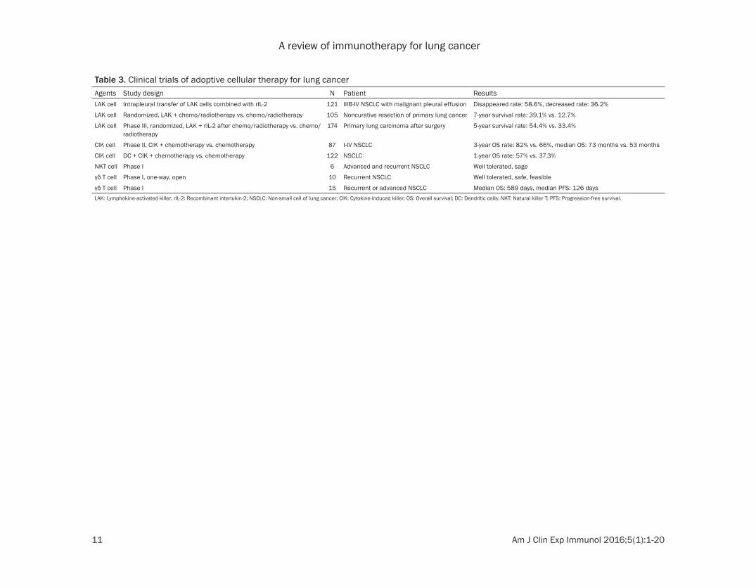
Table 3. Clinical trials of adoptive cellular therapy for lung cancerAgents Study design N Patient ResultsLAK cell Intrapleural transfer of LAK cells combined with rIL-2 121 IIIB-IV NSCLC with malignant pleural effusion Disappeared rate: 58.6%, decreased rate: 36.2%
LAK cell Randomized, LAK + chemo/radiotherapy vs. chemo/radiotherapy 105 Noncurative resection of primary lung cancer 7-year survival rate: 39.1% vs. 12.7%
LAK cell Phase III, randomized, LAK + rIL-2 after chemo/radiotherapy vs. chemo/radiotherapy
174 Primary lung carcinoma after surgery 5-year survival rate: 54.4% vs. 33.4%
CIK cell Phase II, CIK + chemotherapy vs. chemotherapy 87 I-IV NSCLC 3-year OS rate: 82% vs. 66%, median OS: 73 months vs. 53 months
CIK cell DC + CIK + chemotherapy vs. chemotherapy 122 NSCLC 1-year OS rate: 57% vs. 37.3%
NKT cell Phase I 6 Advanced and recurrent NSCLC Well tolerated, sage
γδ T cell Phase I, one-way, open 10 Recurrent NSCLC Well tolerated, safe, feasible
γδ T cell Phase I 15 Recurrent or advanced NSCLC Median OS: 589 days, median PFS: 126 daysLAK: Lymphokine-activated killer; rIL-2: Recombinant interlukin-2; NSCLC: Non-small cell of lung cancer; CIK: Cytokine-induced killer; OS: Overall survival; DC: Dendritic cells; NKT: Natural killer T; PFS: Progression-free survival.
A review of immunotherapy for lung cancer
12 Am J Clin Exp Immunol 2016;5(1):1-20
brolizumab for NSCLC have opened (NCT- 02220894, NCT02359019, NCT02402920, et al.).
Anti-PD-L1 checkpoint inhibitors
PD-L1 (also known as CD274 or B7-H1), the ligand for PD-1, is a member of the B7 super-family and is involved in the negative regulation of the immune response. PD-L1 is expressed in T and B cells, macrophages, and DCs and is up-regulated in a range of solid tumors, including NSCLC. PD-L1 expression has been reported to be associated with vascular invasion and high-er-grade differentiation [70]. PD-L1 expression is associated with increased macrophages, DCs, and inflammatory infiltrate. Given the key role PD-L1 has in lung cancer, the inhibition of PD-L1 is an attractive therapeutic approach.
MPDL3280A: MPDL3280A (MDX-1105) is a human mAb that targets PD-L1 and thus blocks PD-L1 from binding its receptors, including PD-1 and B7.1. In a phase I study of MPDL3280A in pre-treated patients with advanced NSCLC, the ORR was 23% [71]. Additional trials with MPDL3280A are ongoing. One trial is monitor-ing objective responses and safety in patients with PD-L1-positive locally advanced or meta-static NSCLC receiving MPDL3280A monother-apy. Another study is evaluating response rates and safety of MPDL3280A compared to docetaxel in patients with advanced or meta-static NSCLC in whom platinum therapy has failed.
BMS-936559: BMS-936559 is a high-affinity, fully human, PD-L1-specific IgG mAb [72]. In a phase I study, 5 of 49 evaluable NSCLC patients had an objective response; response duration ranged from 2.3+ months to 16.6+ months. Six of 49 patients had stable disease lasting 24 weeks, and 31% of patients had PFS at 24 weeks [73].
Adoptive cellular therapy
Adoptive cell transfer is a form of passive immunotherapy that involves identification, iso-lation, expansion, and subsequent re-infusion of autologous lymphocytes with anti-tumor activity into patients. This form of therapy has been used with or without administration of appropriate growth factors to enhance T cell survival and expansion in vivo. Such an ap-
proach also has the theoretical advantage in that identification and isolation of only a few tumor reactive lymphocytes is sufficient for therapy as these cells can be expanded signifi-cantly ex vivo prior to reinfusion. The genetic modification of isolated cells and the introduc-tion of TCRs with high avidity for tumor specific antigens also creates exciting therapeutic pos-sibilities. Table 3 shows the most important studies involving adoptive cellular therapy in lung cancer.
Lymphokine-activated killer (LAK) cells
LAK cells were first reported in 1982 [74]. In vitro, lymphocytes can be stimulated by IL-2 to kill tumor cells insensitive to CTLs or NK cells. The application of LAK cells in the treatment of advanced tumors can be traced back to 1985.
To enhance the power of LAK cells, patients receive recombinant IL-2 (rIL-2) during the treat-ment. Intrapleural transfer of autologous or allogeneic LAK cells combined with rIL-2 was used in the treatment of 121 patients with malignant effusion associated with advanced lung cancer. The effusion disappeared in 71 patients (58.6%) and was significantly decre- ased in 45 patients (36.2%). No serious side effects were observed [75]. In another clinical trial, 105 patients who had undergone non-curative resection of primary lung cancer were randomly divided into two groups. The group receiving rIL-2 and LAK cells combined with radiation therapy or chemotherapy showed a better 7-year survival rate than the control group, in which patients were treated by radia-tion therapy or chemotherapy alone [76]. A sim-ilar result was obtained in a randomized phase III study [77]. Because of the large amount of IL-2 associated with the clinical application of LAK cells, serious side effects have been re- ported such as capillary leak syndrome, which can lead to hypotension, oliguria, pulmonary edema, and dyspnea. These effects constitute an important obstacle limiting the development of LAK cells for clinical application.
Cytokine-induced killer (CIK) cells
CIK cells are generated from peripheral lym-phocytes by a cytokine cocktail of CD3 mAbs, IL-2, and IFN-γ. CIK cells possess a T cell-NK cell phenotype and MHC-independent antitu-

A review of immunotherapy for lung cancer
13 Am J Clin Exp Immunol 2016;5(1):1-20
mor action [78]. They possess an enhanced cytotoxicity and proliferate more than LAK cells.
In a phase II clinical study, autologous CIK cell immunotherapy improved the efficacy of con-ventional chemotherapy in advanced stage NSCLC patients [79]. DC-activated CIK cells enhanced antitumor effects, and chemothera-py combined with CIK/DC cells improved the clinical outcomes of advanced NSCLC patients [80]. Furthermore, combination treatment with CIK cells and endostatin or DC-based cancer vaccines may have a synergistic effect on improving clinical outcomes. In recent years, CIK cells have been widely used as immuno-therapy for many cancers because of their high proliferation rate and cytotoxic activity, espe-cially after activation by DCs [81].
Natural killer T (NKT) cells
NKT cells, which were first identified in 1986, share properties of both T cells and NK cells. NKT cells are characterized by the expre- ssion of unique invariant TCRs encoded by Vα24Jα18 in humans [82], and they can recog-nize α-galactosylceramide (α-GalCer) present-ed by CD1d. Invariant NKT (iNKT) cells can rap-idly produce large amounts of IFN-γ, which is a hallmark of inflammatory cytokines and is criti-cal for NK cells to attack MHC-negative tumors and for CTLs to kill MHC-positive tumors.
In a phase I study, activated NKT cell adminis-tration was well tolerated and performed safely with minor adverse effects in patients with advanced and recurrent NSCLC (n = 6). A clini-cal trial of iNKT cell-based immunotherapy showed that the infusion of ligand-pulsed APCs and/or activated iNKT cells was safe and well tolerated [83]. The administration of α-GalCer-pulsed APCs induced an activation of iNKT cells in the TME and augmented IFN-γ production by the α-GalCer-stimulated TILs. Another study investigated induced pluripotent stem (iPS)-derived NKT cells. iPS cells are developed into functional NKT cells in the presence of IL-7 and Flt3L in vitro. Although the function of iPS-derived NKT cells is well defined in vivo, further research is necessary before clinical appli- cation.
γδ T cells
Human γδ T cells comprise 1% to 10% of pe- ripheral blood T cells. Unlike αβ T cells, whose
activation requires antigen processing MHC-restricted peptides displayed by APCs, γδ T cells recognize tumor antigens directly through the γδ TCR and exhibit potent MHC-unrestricted lytic activity against microbial pathogens and tumors.
In a phase I clinical study, 10 patients with recurrent NSCLC were treated with intravenous infusion of autologous γδ T cells cultured with zoledronic acid and IL-2. γδ T cell therapy was given 3 to 12 times every 2 weeks. Median fol-low up of patients was 401 days. The regimen was well tolerated [84]. Recently a phase I study was conducted to evaluate the safety and potential anti-tumor effects of re-infusing ex vivo expanded γδ T cells in patients with recur-rent or advanced NSCLC. In this study, ex vivo expansion of γδ T cells from peripheral blood mononuclear cells was achieved by culturing with zoledronic acid (5 μM) and IL-2 (1000 IU/ml) for 14 days. Harvested cells, mostly γδ T cells, were given intravenously every 2 weeks without additional IL-2 for a total of six times. The cumulative number of transferred γδ T cells ranged from 2.6 to 45.1 × 10⁹ (median, 15.7×10⁹). Fifteen patients were treated, and an increase in the number of peripheral γδ T cells was observed with the increase in number of infusions. All patients remained alive during the study period with a median survival of 589 days and median PFS of 126 days. The drug was thus well tolerated [85].
Cytotoxic T lymphocytes (CTLs)
CTLs are CD8+ αβ T cells, which are the main force of anti-tumor immunity. These cells have the ability to recognize MHC class I molecules, present TAAs, and release granzymes and per-forins to lyse tumor cells. Hence, methods to increase the number of CTLs are being devel-oped. Re-infused CTLs are not effective because of the downregulation of MHC and co-stimulatory molecules. Under these circum-stances, there are no successful cases of treat-ment of lung cancer by CTL reinfusion. The identification of crucial factors potentially asso-ciated with the loss of MHC expression may provide a new direction for the development of immunotherapy strategies [86]. Injection of peptide vaccines, which are similar to TAAs, stimulate T cells, resulting in an increase in the number of CTLs available to attack tumor cells whose TAAs are positive.
A review of immunotherapy for lung cancer
14 Am J Clin Exp Immunol 2016;5(1):1-20
Tumor-infiltrating lymphocytes (TILs)
TILs, which were first identified in 1986, are iso-lated from tumor samples, draining lymph nodes, or malignant effusion. One specimen is processed into a single cell suspension that is exposed to high-dose IL-2 to increase the num-ber of lymphocytes and then re-infused into the patient.
Studies have shown the effectiveness of TIL therapy in NSCLC. High levels of intratumoral TILs are associated with a decreased risk of disease recurrence and improved DFS [87]. The infusion of in vitro expanded TILs derived from surgical samples is feasible and has been shown to prolong OS and control residual dis-ease in patients with advanced NSCLC. In 1996, TILs engineered by the IL-2 gene were re-infused into 10 advanced lung cancer patients with pleural effusions. The pleural effusions did not re-accumulate for at least 4 weeks in six patients, and the size of the original tumor decreased in one patient [88].
Re-infusion of TILs into lung cancer patients has certain advantages such as its specificity and safety. However, limitations include the dif-ficulty in obtaining samples from surgeries, adverse effects associated with the combina-tion of high-dose IL-2, and long culture periods (e.g., 5 weeks). In contrast to the modest suc-cess of cell transfer therapy for melanoma, clinical experience in lung cancer has been far from satisfactory [89].
Engineered T cell therapy
Adoptive T cell therapy with engineered T cells to target tumor antigens is an attractive and powerful strategy for cancer therapy. With fur-ther modifications in the laboratory and an increased number of clinical trials to test these approaches, engineered T cell therapy for can-cer may provide significant improvements to cancer immunotherapy.
Chimeric antigen receptor (CAR) T cell immu-notherapy
T cells genetically engineered with CAR vectors can specifically target the surface antigen of cancer cells and kill cancer them in an MHC-independent manner [90]. An objective tumor response was obtained for CAR T cells. CAR T cells were first translated for hematologic
malignancies because the antigen expression on hematologic cells was better understood and there were fewer barriers for honing in T cells on hematologic organs. The use of CAR T cells targeting CD19 has led to remarkable out-comes in the treatment of B cell malignances such as chronic lymphocytic leukemia, acute lymphoblastic leukemia, and other indolent lymphomas [91].
Zhou et al. constructed a CAR targeting EGFR on the cell membrane of T lymphocytes [92]. A xenogeneic mouse model of advanced lung metastatic (A549) cancer was established through tail vein injection in order to evaluate the functional activity of CAR-modified T cells. CAR-modified T cells were administered at days 3, 6, 9, 12, 15, and 18 after tumor injection. A549-derived tumor lung metastasis was sig-nificantly decreased when mice were treated with CAR-modified T cells. The mice treated with CAR-modified T cells had a very low tumor metastasis index (P < 0.05).
TCR-modified T cell immunotherapy
T cells express a heterodimeric αβ receptor on their surface called the TCR. This receptor rec-ognizes antigenic peptides presented by MHC proteins. Genes that encode the α- and β-chains of TCRs can be identified and isolated from the T cells of the rare patients who respond to tumors. These chains are then introduced into T cells, usually by means of viral or non-viral technologies. In this manner, large numbers of antigen-specific T cells can be rapidly generat-ed. The modified TCR-expressing T (TCR-T) cells respond to tumor cells expressing the target antigen.
An et al. designed a study to investigate the cytotoxicity of normal CD8+ T lymphocytes ret-rovirally transduced with Wilms tumor gene 1 (WT1) peptide-specific TCR genes against human lung cancer cells [93]. The results dem-onstrate the feasibility of adoptive immunother-apy with TCR-redirected T cell for the treatment of lung cancer.
Conclusions
Immunotherapies represent a novel approach to treat lung cancer and offer the potential for extended benefits even in advanced disease. At present, mAbs targeting immune check-

A review of immunotherapy for lung cancer
15 Am J Clin Exp Immunol 2016;5(1):1-20
points and antitumor vaccines are the most promising representatives of this treatment modality. An improvement in the understanding of the immune system in tumor immunosurveil-lance has resulted in the development of a new generation of immunotherapeutic agents. Many clinical trials are ongoing and will eventually give further insight into immunotherapy’s prop-er place in lung cancer treatment. Combination with chemotherapy, molecular-targeted thera-py, and other vaccine therapies could also be viable treatment options.
Acknowledgements
This study was supported by grants from the National Natural Science Foundation of China (No. 81171986, No. 81271815), Research Grant from the Ministry of Public Health (No. 201501004), International Cooperative Re- search Foundation of Henan Province (No. 162102410059), the Basic and Advanced Technology Research Foundation from Science and Technology Department of Henan Province (No. 112300410153, No. 122300410155), Funds for Creative Research Team of Henan Province, Creative Research Team of Higher Education of Henan Province and International Cooperative Research of Henan Province.
Disclosure of conflict of interest
None.
Address correspondence to: Dr. Yi Zhang, Biotherapy Center, The First Affiliated Hospital of Zhengzhou University, Zhengzhou, Henan 450052, P.R. China. Tel: +86 371 66295320; E-mail: [email protected]; Dr. Liping Wang, Department of Oncology, The First Affiliated Hospital of Zhengzhou University, Zhengzhou, Henan 450052, P.R. China. Tel: +86 371 66295553; E-mail: [email protected]
References
[1] Sundar R, Soong R, Cho BC, Brahmer JR, Soo RA. Brahmere, and Ross A. Soo. Immunothera-py in the treatment of non-small cell lung can-cer. Lung Cancer 2014; 85: 101-109.
[2] Lehtiö J, De Petris L. Lung cancer proteomics, clinical and technological considerations. J Proteomics 2010; 73: 1851-1863.
[3] Domingues D, Turner A, Silva MD, Marques DS, Mellidez JC, Wannesson L, Mountzios G, de Mello RA. Immunotherapy and lung cancer: current developments and novel targeted ther-apies. Immunotherapy 2014; 6: 1221-1235.
[4] Mostafa AA, Morris DG. Immunotherapy for Lung Cancer: Has it Finally Arrived? Front On-col 2014; 4: 288.
[5] Lee G, Walser TC, Dubinett SM. Chronic inflam-mation, chronic obstructive pulmonary dis-ease, and lung cancer. Curr Opin Pulm 2009; 15: 303-307.
[6] Iyengar P, Gerber DE. Locally advanced lung cancer: an optimal setting for vaccines and other immunotherapies. Cancer J 2013; 19: 247-262.
[7] Chow LM. Exploring novel immune-related tox-icities and endpoints with immune-checkpoint inhibitors in non-small cell lung cancer. Am Soc Clin Oncol Educ Book 2013; e280.
[8] Zielinski C, Knapp S, Mascaux C, Hirsch F. Ra-tionale for targeting the immune system through checkpoint molecule blockade in the treatment of non-small cell lung cancer. Ann Oncol 2013; 24: 1170-1179.
[9] Coussens LM, Werb Z. Infammation and can-cer. Nature 2002; 420: 860-867.
[10] Yang L, Huang J, Ren X, Gorska AE, Chytil A, Aakre M, Carbone DP, Matrisian LM, Richmond A, Lin PC, Moses HL. Abrogation of TGF beta signaling in mammary carcinomas recruits Gr-1+CD11b+ myeloid cells that promote metas-tasis. Cancer Cell 2008; 13: 23-35.
[11] Ye XZ, Yu SC, Bian XW. Contribution of myeloid-derived suppressor cells to tumor-induced im-mune suppression, angiogenesis, invasion and metastasis. J Genet Genomics 2010; 37: 423-430.
[12] Lesokhin AM Hohl TM, Kitano S, Cortez C, Hirschhorn-Cymerman D, Avogadri F, Rizzuto GA, Lazarus JJ, Pamer EG, Houghton AN, Merg-houb T, Wolchok JD. Monocytic CCR2 + My-eloid-Derived Suppressor Cells Promote Im-mune Escape by Limiting Activated CD8 T-cell Infiltration into the Tumor Microenvironment. Cancer Res 2012; 72: 876-886.
[13] Martin F, Apetoh L, Ghiringhelli F. Role of my-eloid-derived suppressor cells in tumor immu-notherapy. Immunotherapy 2012; 4: 43-57.
[14] Bremnes RM, Dønnem T, Al-Saad S, Al-Shibli K, Andersen S, Sirera R, Camps C, Marinez I, Bu-sund LT. The role of tumor stroma in cancer progression and prognosis: emphasis on carci-noma-associated fibroblasts and non-small cell lung cancer. J Thorac Oncol 2011; 6: 209-217.
[15] Bonde AK, Tischler Verena, Kumar S, Solter-mann A, Schwendener RA. Intratumoral mac-rophages contribute to epithelial-mesenchy-mal transition in solid tumors. BMC Cancer 2012; 12: 35.
[16] Wanga R, Zhang J, Chen S, Lu M, Luo X, Yao S, Liu S, Qin Y, Chen H. Tumor-associated macro-phages provide a suitable microenvironment
A review of immunotherapy for lung cancer
16 Am J Clin Exp Immunol 2016;5(1):1-20
for non-small lung cancer invasion and pro-gression. Lung Cancer 2011; 74: 188-196.
[17] Sawachi K, Shimada Y, Taniguchi H, Hirota K, Inagawa H, Kohchi C, Soma G, Makino K, Tera-da H. Cytotoxic effects of activated alveolar macrophages on lung carcinoma cells via cell-to-cell contact and nitric oxide. Anticancer Res 2010; 30: 3135-3141.
[18] Garrido C, Romero I, Berruguilla E, Cancela B, Algarra I, Collado A, García-Lora A, Garrido F. Immunotherapy eradicates metastases with reversible defects in MHC class I expression. Cancer Immunol Immunother 2011; 60: 1257-1268.
[19] Barnas JL, Simpson-Abelson MR, Yokota SJ, Kelleher RJ, Bankert RB. T cells and stromal fibroblasts in human tumor microenviron-ments represent potential therapeutic targets. Cancer Microenviron 2010; 3: 29-47.
[20] Zikos TA, Donnenberg AD, Landreneau RJ, Luketich JD, Donnenberg VS. Lung T-cell sub-set composition at the time of surgical resec-tion is a prognostic indicator in non-small cell lung cancer. Cancer Immunol Immunother 2011; 60: 819-827.
[21] Miotto D, Lo Cascio N, Stendardo M, Querzoli P, Pedriali M, De Rosa E, Fabbri LM, Mapp CE, Boschetto P. CD8+ T cells expressing IL-10 are associated with a favourable prognosis in lung cancer. Lung Cancer 2010; 69: 355-360.
[22] Rijavec M, Volarevic S, Osolnik K, Kosnik M, Korosec P. Natural killer T cells in pulmonary disorders. Respir Med 2011; 105: S20-25.
[23] Platonova S, Cherfils-Vicini J, Damotte D, Crozet L, Vieillard V, Validire P, André P, Dieu-Nosjean MC, Alifano M, Régnard JF, Fridman WH, Sautès-Fridman C, Cremer I. Profound co-ordinated alterations of intratumoral NK cell phenotype and function in lung carcinoma. Cancer Res 2011; 71: 5412-5422.
[24] Schneider T, Hoffmann H, Dienemann, Schna-bel PA, Enk AH, Ring S, Mahnke K. Non-small cell lung cancer induces an immunosuppres-sive phenotype of dendritic cells in tumor mi-croenvironment by upregulating B7-H3. J Tho-rac Oncol 2011; 6: 1162-1168.
[25] Zielinski C, Knapp S, Mascaux C, Hirsch F. Ra-tionale for targeting the immune system through checkpoint molecule blockade in the treatment of non-small cell lung cancer. Ann Oncol 2013; 24: 1170-1179.
[26] Declerck S, Vansteenkiste J. Immunotherapy for lung cancer: ongoing clinical trials. Future Oncol 2014; 10: 91105.
[27] Rijavec E, Genova C, Alama A, Barletta G, Sini C, Pronzato P, Coco S, Dal Bello MG, Savarino G, Truini A, Boccardo F, Grossi F. Role of immu-notherapy in the treatment of advanced non-small-cell lung cancer. Future Oncol 2014; 10: 79-90.
[28] Correale P, Tindara Miano S, Remondo C, Mi-gali C, Saveria Rotundo M, Macrì P, Tagliaferri P, Caraglia M, Gotti G, Francini G. Second-line treatment of non small cell lung cancer by bi-weekly gemcitabine and docetaxel +/- granulo-cyte-macrophage colony stimulating factor and low dose aldesleukine. Cancer Biol Ther 2009; 8: 497-502.
[29] Ridolfi L, Bertetto O, Santo A, Naglieri E, Lopez M, Recchia F, Lissoni P, Galliano M, Testore F, Porta C, Maglie M, Dall’agata M, Fumagalli L, Ridolfi R. Chemotherapy with or without low-dose interleukin-2 in advanced non-small cell lung cancer: results from a Phase III random-ized multicentric trial. Int J Oncol 2011; 39: 1011-1017.
[30] Lissoni P, Brivio F, Fumagalli L, Messina G, Vig-oré L, Parolini D, Colciago M, Rovelli F. Neuro-immunomodulation in medical oncology: appli-cation of psychoneuroimmunology with sub- cutaneous low-dose IL-2 and the pineal hor-mone melatonin in patients with untreatable metastatic solid tumors. Anticancer Res 2008; 28: 1377-1381.
[31] Mazzoccoli G, Sothern RB, Francavilla M, Gi-uliani F, Carughi S, Muscarella LA, Fazio VM, Parrella P, Vinciguerra M, Tarquini R. Hormone and cytokine circadian alteration in non-small cell LC patients. Int J Immunopathol Pharma-col 2012; 25: 691-702.
[32] Rosenberg SA. A new era for cancer immuno-therapy based on the genes that encode can-cer antigens. Immunity 1999; 10: 281-287.
[33] Mellstedt H, Vansteenkiste J, Thatcher N. Vac-cines for the treatment of non-small cell lung cancer: Investigational approaches and clini-cal experience. Lung Cancer 2011; 73: 11-17.
[34] Sandler A, Yi J, Dahlberg S, Kolb MM, Kolb MM, Wang L, Hambleton J, Schiller J, Johnson DH. Treatment outcomes by tumor histology in Eastern Cooperative Group Study E4599 of bevacizumab with paclitaxel/carboplatin for advanced non-small-cell lung cancer. J Thorac Oncol 2010; 5: 1416-1423.
[35] Nemunaitis J, Dillman RO, Schwarzenberger PO, Senzer N, Cunningham C, Cutler J, Tong A, Kumar P, Pappen B, Hamilton C, DeVol E, Ma-ples PB, Liu L, Chamberlin T, Shawler DL, Fakh-rai H. Phase II study of belagenpumatucel-L, a transforming factor beta-2 antisense gene-modified allogeneic tumor cell vaccine in non-small-cell lung cancer. J Clin Oncol 2006; 24: 4721-4730.
[36] Reck M, Vansteenkiste J, Brahmer JR. Target-ing the immune system for management of NSCLC: the revival? Curr Respir Care Rep 2013; 2: 22-39.
[37] Rochlitz C, Dreno B, Jantscheff P, Cavalli F, Squiban P, Acres B, Baudin M, Escudier B,

A review of immunotherapy for lung cancer
17 Am J Clin Exp Immunol 2016;5(1):1-20
Heinzerling L, Morant R, Herrmann R, Dietrich PY, Dummer R. Immunotherapy of metastatic melanoma by intratumoral injections of Vero cells producing human IL-2: phaseII random-ized study comparing two dose levels. Cancer Gene Ther 2002; 9: 289-295.
[38] Rochlitz C, Figlin R, Squiban P, Salzberg M, Pless M, Herrmann R, Tartour E, Zhao Y, Biz-ouarne N, Baudin M, Acres B. Phase I immuno-therapy with a modified vaccinia virus (MVA) expressing human MUC1 as antigen-specific immunotherapy in patients with MUC1-positive advanced cancer. J Gene Med 2003; 5: 690-699.
[39] Ramlau R, Quoix E, Rolski J, Pless M, Lena H, Lévy E, Krzakowski M, Hess D, Tartour E, Che-nard MP, Limacher JM, Bizouarne N, Acres B, Halluard C, Velu T. A phase II study of Tg4010 (Mva-Muc1-Il2) in association with chemother-apy in patients with stage III/IV Non-small cell lung cancer. J Thorac Oncol 2008; 3: 735-744.
[40] Quoix E, Ramlau R, Westeel V, Papai Z, Madro-szyk A, Riviere A, Koralewski P, Breton JL, Stoelben E, Braun D, Debieuvre D, Lena H, Buyse M, Chenard MP, Acres B, Lacoste G, Bastien B, Tavernaro A, Bizouarne N, Bonnefoy JY, Limacher JM. Therapeutic vaccination with TG4010 and first-line chemotherapy in ad-vanced non-small-cell lung cancer: a controlled phase 2B trial. Lancet Oncol 2011; 12: 1125-1133.
[41] Transgene. Phase IIB/III of TG4010 Immuno-therapy In Patients With Stage IV Non-Small Cell Lung Cancer (TIME). Available from: http://clinicaltrials.gov/show/NCT01383148. NLM identifier: NCT01383148. Accessed December 11, 2013.
[42] Powell E, Chow LQ. BLP-25 liposomal vaccine: a promising potential therapy in non-small-cell lung cancer. Expert Rev Respir Med 2008; 2: 37-45.
[43] Hall RD, Gray JE, Chiappori AA. Beyond the standard of care: a review of novel immuno-therapy trials for the treatment of lung cancer. Cancer Control 2013; 20: 22-31.
[44] Palmer M, Parker J, Modi S, et al. Phase I study of the BLP25 (MUC1 peptide) liposomal vac-cine for active specific immunotherapy in stage IIIB/IV non-small-cell lung cancer. Clin Lung Cancer 2001; 3: 49-57; discussion 58.
[45] Butts C, Murray N, Maksymiuk A, Goss G, Mar-shall E, Soulières D, Cormier Y, Ellis P, Price A, Sawhney R, Davis M, Mansi J, Smith C, Vergidis D, Ellis P, MacNeil M, Palmer M. Randomized phase IIB trial of BLP25 liposome vaccine in stage IIIB and IV non-small-cell lung cancer. J Clin Oncol 2005; 23: 6674-6681.
[46] Butts C, Maksymiuk A, Goss G, Soulières D, Marshall E, Cormier Y, Ellis PM, Price A, Sawh-
ney R, Beier F, Falk M, Murray N. Updated sur-vival analysis in patients with stage IIIB or IV non-small-cell lung cancer receiving BLP25 li-posome vaccine (L-BLP25): phase IIB random-ized, multicenter, open-label trial. J Cancer Res Clin Oncol 2011; 137: 1337-1342.
[47] Yoshida N, Abe H, Ohkuri T, Wakita D, Sato M, Noguchi D, Miyamoto M, Morikawa T, Kondo S, Ikeda H, Nishimura T. Expression of the MAGE-A4 and NY-ESO-1 cancer-testis antigens and T cell infiltration in non-small cell lung carcino-ma and their prognostic significance. Int J On-col 2006; 28: 1089-1098.
[48] Sienel W, Varwerk C, Linder A, Kaiser D, Tes-chner M, Delire M, Stamatis G, Passlick B. Melanoma associated antigen (MAGE)-A3 ex-pression in Stages I and II non-small cell lung cancer: results of a multi-center study. Eur J Cardiothorac Surg 2004; 25: 131-134.
[49] Vansteenkiste J, Zielinski M, Linder A, Dahab-reh J, Gonzalez EE, Malinowski W, Lopez-Brea M, Vanakesa T, Jassem J, Kalofonos H, Perde-us J, Bonnet R, Basko J, Janilionis R, Passlick B, Treasure T, Gillet M, Lehmann FF, Brichard VG. Adjuvant MAGE-A3 immunotherapy in re-sected non-small-cell lung cancer: Phase II randomized study results. J Clin Oncol 2013; 31: 2396-2403.
[50] Hirsch FR, Varella-Garcia M, Cappuzzo F. Pre-dictive value of EGFR and HER2 overexpres-sion in advanced non-small-cell lung cancer. Oncogene 2009; 28 Suppl 1: S32-37.
[51] Neninger Vinageras E, de la Torre A, Osorio Ro-dríguez M, Catalá Ferrer M, Bravo I, Mendoza del Pino M, Abreu Abreu D, Acosta Brooks S, Rives R, del Castillo Carrillo C, González Due-ñas M, Viada C, García Verdecia B, Crombet Ramos T, González Marinello G, Lage Dávila A. Phase II randomized controlled trial of an epi-dermal growth factor vaccine in advanced non-small-cell lung cancer. J Clin Oncol 2008; 26: 1452-1458.
[52] Varadhachary A, Wolf JS, Petrak K, O’Malley BW Jr, Spadaro M, Curcio C, Forni G, Pericle F. Oral lactoferrin inhibits growth of established tumors and potentiates conventional chemo-therapy. Int J Cancer 2004; 111: 398-403.
[53] Digumarti R, Wang Y, Raman G, Doval DC, Ad-vani SH, Julka PK, Parikh PM, Patil S, Nag S, Madhavan J, Bapna A, Ranade AA, Varad-hachary A, Malik R. A randomized, double-blind, placebo-controlled, phase II study of oral talactoferrin in combination with carboplatin and paclitaxel in previously untreated locally advanced or metastatic non-small cell lung cancer. J Thorac Oncol 2011; 6: 1098-1103.
[54] Parikh PM, Vaid A, Advani SH, Digumarti R, Madhavan J, Nag S, Bapna A, Sekhon JS, Patil S, Ismail PM, Wang Y, Varadhachary A, Zhu J,
A review of immunotherapy for lung cancer
18 Am J Clin Exp Immunol 2016;5(1):1-20
Malik R. Randomized, double-blind, placebo-controlled Phase II study of single-agent oral talactoferrin in patients with locally advanced or metastatic non-small-cell lung cancer that progressed after chemotherapy. J Clin Oncol 2011; 29: 4129-4136.
[55] Ramalingam S, Crawford J, Chang A, Manegold C, Perez-Soler R, Douillard JY, Thatcher N, Bar-lesi F, Owonikoko T, Wang Y, Pultar P, Zhu J, Malik R, Giaccone G. FORTIS-M Study Investi-gators. FORTIS-M Study Investigators. Talacto-ferrin alfa versus placebo in patients with re-fractory advanced non-small-cell lung cancer (FORTIS-M trial). Ann Oncol 2013; 24: 2875-2880.
[56] Hall RD, Gray JE, Chiappori AA. Beyond the standard of care: a review of novel immuno-therapy trials for the treatment of lung cancer. Cancer Control 2013; 20: 22-31.
[57] Grant SC, Kris MG, Houghton AN, Chapman PB. Long survival of patients with small cell lung cancer after adjuvant treatment with the anti-idiotypic antibody BEC2 plus Bacillus Cal- mette-Guérin. Clin Cancer Res 1999; 5: 1319-1323.
[58] Giaccone G, Debruyne C, Felip E, Chapman PB, Grant SC, Millward M, Thiberville L, D’addario G, Coens C, Rome LS, Zatloukal P, Masso O, Legrand C. Phase III study of adjuvant vaccina-tion with Bec2/bacille Calmette-Guerin in re-sponding patients with limited-disease small-cell lung cancer (European Organisation for Research and Treatment of Cancer 08971–08971B; Silva Study). J Clin Oncol 2005; 23: 6854-6864.
[59] Alfonso S, Valdes-Zayas A, Santiesteban ER, Flores YI, Areces F, Hernández M, Viada CE, Mendoza IC, Guerra PP, García E, Ortiz RA, de la Torre AV, Cepeda M, Pérez K, Chong E, Hernández AM, Toledo D, González Z, Mazorra Z, Crombet T, Pérez R, Vázquez AM, Macías AE. A randomized, multicenter, placebo-controlled clinical trial of racotumomab-alum vaccine as switch maintenance therapy in advanced non-small-cell-lung cancer patients. Clin Cancer Res 2014; 20: 3660-3671.
[60] Nemunaitis J, Jahan T, Ross H, Sterman D, Richards D, Fox B, Jablons D, Aimi J, Lin A, Hege K. Phase 1/2 trial of autologous tumor mixed with an allogeneic GVAX vaccine in ad-vanced-stage non-small-cell lung cancer. Can-cer Gene Ther 2006; 13: 555-562.
[61] Skachkova OV, Khranovska NM, Gorbach OI, Svergun NM, Sydor RI, Nikulina VV. Immuno-logical markers of anti-tumor dendritic cells vaccine efficiency in patients with non-small cell lung cancer. Exp Oncol 2013; 35: 109-113.
[62] Reck M, Bondarenko I, Luft A, Serwatowski P, Barlesi F, Chacko R, et al. Ipilimumab in combi-
nation with paclitaxel and carboplatin as fi- rst-line therapy in extensive-disease-small-cell lung cancer: results from a randomized, dou-ble-blind, multicenter phase 2 trial. Ann Oncol 2013; 24: 75-83.
[63] Lynch TJ, Bondarenko I, Luft A, Serwatowski P, Barlesi F, Chacko R, Sebastian M, Neal J, Lu H, Cuillerot JM, Reck M. Ipilimumab in combina-tion with paclitaxel and carboplatin as first-line treatment in stage IIIB/IV non-small cell lung cancer: results from a randomized, double-blind, multicenter phase II study. J Clin Oncol 2012; 30: 2046-2054.
[64] Creelan BC, Antonia SJ. Immunotherapy in LC: B7-bombers and other new developments. Se-min Respir Crit Care Med 2013; 34: 810-821.
[65] Keir ME, Butte MJ, Freeman GJ, Sharpe AH. PD-1 and its ligands in tolerance and immuni-ty. Annu Rev Immunol 2008; 26: 677-704.
[66] Ahmadzadeh M, Johnson LA, Heemskerk B, Wunderlich JR, Dudley ME, White DE, Rosen-berg SA. Tumor antigen-specific CD8 T cells in-filtrating the tumor express high levels of PD-1 and are functionally impaired. Blood 2009; 114: 1537-1544.
[67] PD-1 inhibitors raise survival in NSCLC. Cancer Discov 2014; 4: 6.
[68] Topalian SL, Hodi FS, Brahmer JR, Gettinger SN, Smith DC, McDermott DF, Powderly JD, Carvajal RD, Sosman JA, Atkins MB, Leming PD, Spigel DR, Antonia SJ, Horn L, Drake CG, Pardoll DM, Chen L, Sharfman WH, Anders RA, Taube JM, McMiller TL, Xu H, Korman AJ, Jure-Kunkel M, Agrawal S, McDonald D, Kollia GD, Gupta A, Wigginton JM, Sznol M. Safety, activi-ty, and immune correlates of anti-PD-1 anti-body in cancer. N Engl J Med 2012; 366: 2443-2454.
[69] Hamid O, Robert C, Daud A, Hodi FS, Hwu WJ, Kefford R, Wolchok JD, Hersey P, Joseph RW, Weber JS, Dronca R, Gangadhar TC, Patnaik A, Zarour H, Joshua AM, Gergich K, Elassaiss-Schaap J, Algazi A, Mateus C, Boasberg P, Tu-meh PC, Chmielowski B, Ebbinghaus SW, Li XN, Kang SP, Ribas A. Safety and tumor re-sponses with lambrolizumab (anti-PD-1) in melanoma. N Engl J Med 2013; 369: 134-144.
[70] Yang CY, Lin MW, Chang YL, Wu CT, Yang PC. Programmed cell death-ligand 1 expression in surgically resected stage I pulmonary adeno-carcinoma and its correlation with driver muta-tions and clinical outcomes. Eur J Cancer 2014; 50: 1361-1369.
[71] Herbst RS, Soria JC, Kowanetz M, Fine GD, Ha-mid O, Gordon MS, Sosman JA, McDermott DF, Powderly JD, Gettinger SN1, Kohrt HE, Horn L, Lawrence DP, Rost S, Leabman M, Xiao Y, Mo-katrin A, Koeppen H, Hegde PS, Mellman I, Chen DS, Hodi FS. Predictive correlates of re-

A review of immunotherapy for lung cancer
19 Am J Clin Exp Immunol 2016;5(1):1-20
sponse to the anti-PD-L1 antibody MPD-L3280A in cancer patients. Nature 2014; 515: 563-567.
[72] Brahmer JR, Tykodi SS, Chow LQ, Hwu WJ, To-palian SL, Hwu P, Drake CG, Camacho LH, Kauh J, Odunsi K, Pitot HC, Hamid O, Bhatia S, Martins R, Eaton K, Chen S, Salay TM, Alapar-thy S, Grosso JF, Korman AJ, Parker SM, Agraw-al S, Goldberg SM, Pardoll DM, Gupta A, Wig-ginton JM. Safety and activity of anti-PD-L1 antibody in patients with advanced cancer. N Engl J Med 2012; 366: 2455-2465.
[73] Brahmer JR, Tykodi SS, Chow LQ, Hwu WJ, To-palian SL, Hwu P, et al. Safety and activity of anti-PD-L1 antibody in patients with advanced cancer. N Engl J Med 2012; 366: 2455-2465.
[74] Grimm EA, Mazumder A, Zhang HZ, Rosenberg SA. Lymphokine-activated killer cell phenome-non. Lysis of natural killer-resistant fresh solid tumor cells by interleukin 2-activated autolo-gous human peripheral blood lymphocytes. J Exp Med 1982; 155: 1823-1841.
[75] Liu X, Li D, Zhang C, Ba D, Liu J, Wan T, Li Z, Jin Y, He Y. Treatment of 121 patients with malig-nant effusion due to advanced lung cancer by intrapleural transfer of autologous or alloge-neic LAK cells combined with rIL-2. Chin Med Sci J 1993; 8: 186-189.
[76] Kimura H, Yamaguchi Y. Adjuvant immunother-apy with interleukin 2 and lymphokine-activat-ed killer cells after noncurative resection of primary lung cancer. Lung Cancer 1995; 13: 31-44.
[77] Kimura H, Yamaguchi Y. A phase III random-ized study of interleukin-2 lymphokine-activat-ed killer cell immunotherapy combined with chemotherapy or radiotherapy after curative or noncurative resection of primary lung carcino-ma. Cancer 1997; 80: 42-49.
[78] Shi S, Wang R, Chen T, Song H, Chen L, Huang G. Combining antiangiogenic therapies with adoptive cell immunotherapy exerts better an-titumor effects in non-small cell lung cancer models. PLoS One 2013; 8: e65757.
[79] Li R, Wang C, Liu L, Du C, Cao S, Yu J, Wang SE, Hao X, Ren X, Li H. Autologous cytokine-in-duced killer cell immunotherapy in lung can-cer: a phase II clinical study. Cancer Immunol Immunother 2012; 61: 2125-2133.
[80] Yang L, Ren B, Li H, Yu J, Cao S, Hao X, Ren X. Enhanced antitumor effects of DC-activated CIKs to chemotherapy treatment in a single co-hort of advanced non-small-cell lung cancer patients. Cancer Immunol Immunother 2013; 62: 65-73.
[81] Zhong R, Han B, Zhong H. A prospective study of the efficacy of a combination of autologous dendritic cells, cytokine-induced killer cells, and chemotherapy in advanced non-small cell
lung cancer patients. Tumour Biol 2014; 35: 987-994.
[82] Fujii SI, Shimizu K, Okamoto Y, Kunii N, Na-kayama T, Motohashi S, Taniguchi M. NKT cells as an ideal anti-tumor immunotherapeutic. Front Immunol 2013; 4: 409.
[83] Motohashi S, Okamoto Y, Yoshino I, Nakayama T. Anti-tumor immune responses induced by iNKT cell-based immunotherapy for lung can-cer and head and neck cancer. Clin Immunol 2011; 140: 167-176.
[84] Nakajima J, Murakawa T, Fukami T, Goto S, Kaneko T, Yoshida Y, Takamoto S, Kakimi K. A phase I study of adoptive immunotherapy for recurrent non-small-cell lung cancer patients with autologous gammadelta T cells. Eur J Car-diothoac Surg 2010; 37: 1191-1197.
[85] Sakamoto M, Nakajima J, Murakawa T, Fukami T, Yoshida Y, Murayama T, Takamoto S, Matsu-shita H, Kakimi K. Adoptive immunotherapy for advanced non-small cell lung cancer using zoledronate-expanded gammadelta T cells: a phase I clinical study. J Immunother 2011; 34: 202-211.
[86] Baba T, Hanagiri T, Takenoyama M, Shiota H, Kuroda K, Shigematsu Y, Ichiki Y, Uramoto H, So T, Yasumoto K. Identification of a lung can-cer antigen evading CTL attack due to loss of human leukocyte antigen (HLA) class I expres-sion. Cancer Sci 2010; 101: 2115-2120.
[87] Kilic A, Landreneau RJ, Luketich JD, Pennathur A, Schuchert MJ. Density of tumor-infiltrating lymphocytes correlates with disease recur-rence and survival in patients with large non-smallcell lung cancer tumors. J Surg Res 2011; 167: 207-210.
[88] Li K, Zhang Q, Zhang Y, Yang J, Zheng J. T-cell-associated cellular immunotherapy for lung cancer. J Cancer Res Clin Oncol 2015; 141: 1249-1258.
[89] Goff SL, Smith FO, Klapper JA, Sherry R, Wun-derlich JR, Steinberg SM, White D, Rosenberg SA, Dudley ME, Yang JC. Tumor infiltrating lym-phocyte therapy for metastatic melanoma: analysis of tumors resected for TIL. J Immuno-ther 2010; 33: 840-847.
[90] Maus MV, Grupp SA, Porter DL, June CH. Anti-body-modified T cells: CARs take the front seat for hematologic malignancies. Blood 2014; 123: 2625-2635.
[91] Lee DW, Kochenderfer JN, Stetler-Stevenson M, Cui YK, Delbrook C, Feldman SA, Fry TJ, Orentas R, Sabatino M, Shah NN, Steinberg SM, Stroncek D, Tschernia N, Yuan C, Zhang H, Zhang L, Rosenberg SA, Wayne AS, Mackall CL. T cells expressing CD19 chimeric antigen re-ceptors for acute lymphoblastic leukaemia in children and young adults: a phase 1 dose-es-calation trial. Lancet 2014; 385: 517-528.

A review of immunotherapy for lung cancer
20 Am J Clin Exp Immunol 2016;5(1):1-20
[92] Zhou X, Li J, Wang Z, Chen Z, Qiu J, Zhang Y, Wang W, Ma Y, Huang N, Cui K, Li J, Wei YQ. Cellular Immunotherapy for Carcinoma Using Genetically Modified EGFR-Specific T Lympho-cytes. Neoplasia 2013; 15: 544-553.
[93] An J, Cai SW, Li Y, Zhang J. Cytotoxicity of T cells transduced with WT1 peptide-specific T-cell re-ceptor gene against human lung cancer cells in vitro. Nan Fang Yi Ke Da Xue Xue Bao 2014; 34: 1319-1323.