review article distinct characteristics of...
TRANSCRIPT
Review ArticleDistinct Characteristics of Mandibular Bone Collagen Relative toLong Bone Collagen: Relevance to Clinical Dentistry
Takashi Matsuura, Kentaro Tokutomi, Michiko Sasaki, Michitsuna Katafuchi,Emiri Mizumachi, and Hironobu Sato
Section of Fixed Prosthodontics, Department of Oral Rehabilitation, Fukuoka Dental College, 15-1 Tamura 2-Chome,Sawara-ku, Fukuoka 814-0193, Japan
Correspondence should be addressed to Takashi Matsuura; [email protected]
Received 12 February 2014; Accepted 19 March 2014; Published 10 April 2014
Academic Editor: Mitsuo Yamauchi
Copyright © 2014 Takashi Matsuura et al. This is an open access article distributed under the Creative Commons AttributionLicense, which permits unrestricted use, distribution, and reproduction in any medium, provided the original work is properlycited.
Bone undergoes constant remodeling throughout life. The cellular and biochemical mechanisms of bone remodeling vary in aregion-specific manner.There are a number of notable differences between the mandible and long bones, including developmentalorigin, osteogenic potential of mesenchymal stem cells, and the rate of bone turnover. Collagen, the most abundant matrix proteinin bone, is responsible for determining the relative strength of particular bones. Posttranslational modifications of collagen, such asintermolecular crosslinking and lysine hydroxylation, are the most essential determinants of bone strength, although the amountof collagen is also important. In comparison to long bones, the mandible has greater collagen content, a lower amount of maturecrosslinks, and a lower extent of lysine hydroxylation.The great abundance of immature crosslinks in mandibular collagen suggeststhat there is a lower rate of cross-link maturation. This means that mandibular collagen is relatively immature and thus morereadily undergoes degradation and turnover. The greater rate of remodeling in mandibular collagen likely renders more flexibilityto the bone and leaves it more suited to constant exercise. As reviewed here, it is important in clinical dentistry to understand thedistinctive features of the bones of the jaw.
1. Introduction
Bone is a dynamic tissue that undergoes constant remodelingin order to maintain a healthy skeleton. In clinical dentistry,jawbones frequently require surgical procedures, such asextraction of teeth, periodontal surgery, and implant surgerywith or without bone regeneration. Of the many regenerativeexperiments for bone, few have been tested in the jaw.Because of the unique properties of the jawbone tissue,dentists and dental researchers should be aware that the dataregarding other skeletal bones may not be entirely applicableto jawbones.
It is well recognized that the jawbone is remodeled fasterthan the other skeletal bones [1]. Jaw development is similarto that in other craniofacial bones but distinct from theaxial and appendicular skeleton. The jaw arises from neural
crest cells of the neuroectoderm germ layer rather thanthe mesoderm [2] and undergoes intramembranous, insteadof endochondral, ossification [3]. Skeletal diseases such ascherubism [4], hyperparathyroid jaw tumor syndrome [5],and bisphosphonate-related osteonecrosis [6] occur only inthe jaw. In case of ovariectomy andmalnutrition, it is reportedthat the rat mandible loses trabecular bone and mineraldensity at a lower rate than the tibiae do [7]. Mesenchymalstem cells or bone marrow stromal cells derived from the jawshow higher osteogenic potential and additional distinctivefeatures compared to other skeletal bones [8–12].
These distinctions owe partly to the unique charac-teristics of the jawbone matrix. It is important both forregenerative dental surgery and for maintenance of teethor implants thereafter that dentists and dental researchersare knowledgeable of the unique features of the jawbone
Hindawi Publishing CorporationBioMed Research InternationalVolume 2014, Article ID 769414, 9 pageshttp://dx.doi.org/10.1155/2014/769414
2 BioMed Research International
matrix. Though there are many bone matrix components,this review focuses on collagen, the most abundant matrixprotein in bone and a determinant of bone strength andquality [13]. Collagen biochemistry is not well characterizedin themaxilla; therefore, we focus on research findings for themandible. We will first describe the role of collagen in bonematrix organization.Wewill then compare the characteristicsof mandibular collagen to long bones to highlight the uniqueproperties of the jawbone matrix that are relevant to clinicaldentistry.
2. Role of Collagen on BoneMatrix Organization
Bone matrix consists mainly of a two-phase composite mate-rial: mineral and fibrillar collagen. Type I collagen comprisesapproximately 95% of the entire collagen content of bone.Theother types of collagen, such as types III [16] and V [17], are atlow levels and appear tomodulate the diameter of type I colla-gen fibrils [17].Mineral and fibrillar type I collagen are closelyassociated with each other; the latter functions as a three-dimensional template that organizes the former’s depositionand growth [18]. Bone acquires its durability against externalforces through this well-organized architectural arrangementbetween mineral and type I collagen fibrils.
The nature and extent of posttranslational modificationsof collagen, many of which are unique to collagen [19], arerelated to the organization of mineral and collagen fibrils[18]. One such modification, the intermolecular, covalentcrosslinking of collagen initiated by the enzymatic oxidativedeamination of specific lysine (Lys) and hydroxylysine (Hyl)residues by lysyl oxidase (LOX), contributes to bone strength.In fact, the inhibition of LOX activity by lathyrogens impairscrosslinking, which leads to decreased bone strength causedby increased solubility and abnormal structure of collagenfibrils [20, 21].
Another modification, enzymatic hydroxylation of spe-cific Lys residues by lysyl hydroxylase (LH), also can controlbone matrix organization.The Hyl serves as a site of glycosy-lation [22, 23], and the resultant glycosylated residues affectcollagen maturation [23–25], fibrillogenesis, and mineraliza-tion [22, 23]. In addition, this modification determines thepattern of intermolecular crosslinking of collagen. Amongthe 3 isoforms of LH (LH1, 2, and 3), LH2b, a spliced variantof LH2, catalyzes the hydroxylation of Lys residues in the C-or N-terminal, nontriple helical domain (i.e., the telopeptidedomain) of collagen, which then directs the subsequentcrosslinking towards the hydroxylysine (Hylald) pathway inmineralized tissues specifically [26]. Ectopic activation of theHylald pathway by overexpression of LH2b leads to defectivecollagen fibrillogenesis and matrix mineralization [27]. LH1catalyzes Lys hydroxylation in the triple helical domain(helical domain), while LH3has LHactivity and,more impor-tantly, galactosylhydroxylysine glucosyltransferase activity[22].
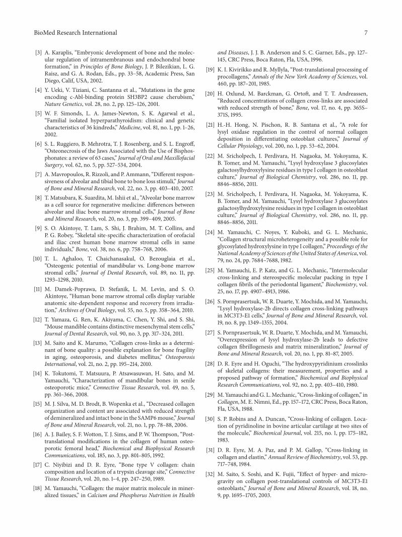
At the beginning of the bone-specific cross-linking path-way, the Hyl residue in the telopeptide domain (formed
by LH2b) is converted into an aldehyde (Hylald) by LOX(Figure 1). The iminium divalent intermolecular crosslinksare the first to form and they then mature into triva-lent crosslinks through condensation reactions (Figure 2).The pairing of Hylald with Hyl (formed by LH1) inthe helical domain of the neighboring molecule formsthe iminium crosslink, dehydrodihydroxylysinonorleucine(deH-DHLNL). By contrast, when the Hylald pairs with Lys,dehydrohydroxylysinonorleucine (deH-HLNL) is formed.The major mature crosslink, pyridinoline (Pyr), is a mat-urational product of deH-DHLNL, formed by any of thefollowing condensation reactions: (1) condensation of theirtwo keto-amines through elimination of a Hyl [28], (2)condensation of a keto-amine and a Hylald [29], or (3)condensation of a deH-DHLNL and its keto-amine [30]. Aminor, mature cross-link form is deoxypyridinoline (d-Pyr),a lysyl analog of Pyr, made up of twoHylald and one Lys in thehelical domain [31].
Because these cross-link condensation reactions are usu-ally spontaneous, turnover rate is an important factor inregulating cross-link maturation. For instance, periodontalligament collagen has low levels of mature cross-links dueto its high rate of turnover and, in turn, is more readilydegraded due to lack of stable cross-links [25]. As for bonecollagen, the levels of the major mature cross-link form, Pyr,versus the major immature cross-link form, deH-DHLNL,indicate the collagen maturation rate [32]. More recently, theresearch group of Yamauchi has demonstrated that the degreeof Hyl glycosylation also influences cross-link maturation[23].
The galactosylhydroxylysine glucosyltransferase activityof LH3 promotes the formation of glucosylgalactosyl (GG)-Hyl from galactosyl (G)-Hyl at the cross-linking site. G-or G free-deH-DHLNL can mature into G-Pyr or G free-Pyr, while GG-deH-DHLNL cannot mature into GG-Pyr.Suppression of galactosylhydroxylysine glucosyltransferaseactivity of LH3 decreases the speed of cross-link maturation,reduces the amount of both immature andmature crosslinks,increases the diameter of collagen fibrils, and impairs matrixmineralization.
Two articles have demonstrated disordered bone collagenin LOX or LH knock-out mice. Pischon et al. [33] reportedthat LOX knock-out mice showed a perinatal lethality andthat the craniofacial bone of the fetus at embryonic day18.5 exhibited fragility and thinner collagen fibrils. Theirosteoblast cultures revealed retard of osteoblastic differen-tiation and matrix mineralization. Takaluoma et al. [34]documented that LH1 knock-out mice were viable and fertilebut 15%of themwere led to sudden deathmainly due to aorticruptures. The femoral bone collagen of the adult LH1 knock-out mice showed a 75% and a 47% smaller amount of Hyland the major mature crosslink, Pyr, respectively, comparedto those of the adult wild-type mice. By contrast, the amountof a minor mature crosslink, d-Pyr, was 1284% greater,and thereby, total amount of Pyr and d-Pyr became 195%greater. Though collagen fibrils and matrix mineralizationwere not investigated, LH1 deficiency probably affects bonecollagen matrix. As described above, the posttranslational
BioMed Research International 3
Lys
Hyl Pyr
LH2b LOX Hyl(HEL)
Lys(HEL)
(TELO)
(TELO)
TELO
(Immature) (Mature)
Lysald Lysald-pathway in soft tissues
Hylald
Hylald
Hylald
deH-DHLNL (Major crosslinks)
deH-HLNL (Minor crosslinks)d-Pyr
Hylald-pathway in mineralized tissues
Figure 1: The collagen cross-linking pathway in mineralized tissues. The collagen cross-linking pathway in soft tissues arises from the Lysald
both at the C- and N-telopeptide domains (Lysald-pathway). In mineralized tissue, it does so from the Hylald mostly at the C-telopeptidedomain (Hylald-pathway). In mineralized tissues, the Lys residue at the C-telopeptide domain is converted into Hyl through the action ofLH2b, followed by the conversion of the Hyl into the Hylald through the action of LOX. Tomake the major crosslinks, the immature crosslink,deH-DHLNL, is first formed by pairing of the Hylald with the Hyl at the helical domain of the neighboring molecule (Hylald × Hyl), andthe mature crosslink, Pyr, is then formed by a spontaneous condensation reaction (Hylald × Hylald × Hyl). To make the minor crosslinks,the immature crosslink, deH-HLNL, is formed (Hylald × Lys), and then the mature crosslink, d-Pyr, is formed (Hylald × Hylald × Lys). Thevalue of Pyr/deH-DHLNL presents collagen maturation rate. Lysald : the aldehyde form of Lys; Hylald : the aldehyde form of Hyl; TELO: thetelopeptide domain; HEL: the helical domain.
modifications of collagen mediated by LOX and LHs havecritical roles on the organization of collagen fibrils and serveas a template for bone mineralization as well as matrixformation.
3. Collagen Content in the MandibleCompared to the Long Bones
The biomechanical roles for collagen in bone are related toboth the amount of collagen and its molecular stability andcrosslinking. Bailey et al. [35] found that age-related declinein collagen content is nonlinearly correlated to themaximumstress at failure and to the modulus of elasticity in bone fromhuman iliac crest. This does not mean that differences incollagen content are necessarily responsible for these changesbut only that they are associated with them. Indeed, in otherlocations such as the femoral head and neck, no changesin collagen content were detected [36]. Other changes, suchas the rate of turnover or degree of mineralization, mightaffect collagen content and themechanical properties of boneindependently. Despite such complex interactions, collagencontent is representative of the status of bone matrix.
We have published the only study comparing the collagencontent in the mandibular bone to long bones [37]. Wecalculated the collagen content in formalin-fixed humancortical bones from 44 cadavers at 3 different sites: thementalregion of mandible, the mesial neck of humerus, and themesial neck of femur.We found that the collagen content wassignificantly greater in the mandible (165.2𝜇g/mg of driedbone weight) than in the humerus (146.4 𝜇g/mg) and femur(139.5 𝜇g/mg). Silva et al. [15] compared the bone matrixand mechanical properties of the femur in male SAMP6mutant mice, a murine model of senile osteoporosis mice, tocontrol SAMR1 mice, a senescence-resistant mutant mouse.
They found that both demineralized and intact bones hadgreater reductions in mechanical strength in SAMP6 micecompared to the SAMR1 mice and that the cortical diaphysisalso had a smaller amount of collagen in the SAMP6mutants(97 𝜇g/mg of dried boneweight at 4months of age, 106 𝜇g/mgat 12 months of age) versus SAMR1 mutants (113 𝜇g/mg at 4months of age, 119𝜇g/mg at 12 months of age). We [14] alsoinvestigated the cortical mandibles in males from the samemutant mouse strains at 6 months of age and obtained datasimilar to that by Silva et al. [15]. The mandible possessed asmaller amount of collagen in SAMP6 (126.4 𝜇g/mg of driedbone weight) than in SAMR1 (149.5 𝜇g/mg). Collagen fiberswere also thinner in the SAMP6 mice (35.77 nm) than in theSAMR1mice (43.71 nm).Despite the difference of age analysisbetween the two studies, mandibular bone displayed a greateramount of collagen compared to the femoral bone in the twomouse models.
The calvaria, which has the same origin as the jawbones[2], has similar tendency in collagen content. The researchgroup of van den Bos et al. [38] investigated matrix composi-tion of calvaria and long bones (femur, tibia, ulna, and radius)in femalemice at 6months of age and showed that the calvaria(302 𝜇g/mg of dried bone weight) had a greater amount ofcollagen than the long bones (211𝜇g/mg).
As the collagen content mentioned above is calculatedbased on the value of hydroxyproline, it is mostly type I withtrace amounts of types III [16] and V [17]. The comparison oftype III or type V collagen content between themandible andlong bones has not been performed. Type III collagen is codis-tributed with type I collagen and is rich in Sharpey’s fibers atthe periodontal ligament and periosteum, penetrating to thebone [39]. In themandible, Sharpey’s fibers at the periodontalligament across the entire thickness of alveolar wall and thefibers at the periosteum also penetrate to the cortical bone butbecome fewer, fragmented, superficial, and shortened with
4 BioMed Research International
87 Hyl
HEL
TELO
N Or
Pyr
(Immature) (Mature)
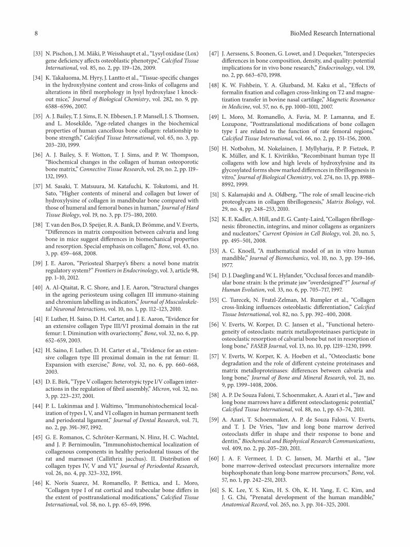
Collagen molecule
𝛼1
𝛼2
deH-DHLNL
Self-assembly of collagen molecules
C16
Hylald
Figure 2: Diagram of the formation andmaturation of themajor collagen crosslink inmineralized tissues.The procollagenmolecule secretedfrom the cell is processed by cleavages of both the N- and C-terminal propeptide extensions. The processed collagen molecules then self-assemble through clusters of charge and hydrophobicity of the triple helical domain of the molecule to form a fibril. The molecules in thefibril are then stabilized by extensive intermolecular crosslinking. The crosslink involves the Hylald at the 16C residue on the C-telopeptidedomain of the two 𝛼1 chains and the Hyl at the 87 residue on the triple helical domain of the two 𝛼1 chains or the single 𝛼1 and 𝛼2 chains.Theimmature crosslink, deH-DHLNL, is formed by pairing of the Hylald with the Hyl of the neighboring molecule. The mature crosslink, Pyr, isthen formed; it owes its origin to the two Hylald of 𝛼1 chains and the one Hyl of 𝛼1 or 𝛼2 chain. If the 87th residue on the helical domain isLys, the minor immature crosslink, deH-HLNL, is formed, and then the minor immature crosslink, d-Pyr, is made up. The solid and dottedlines represent 𝛼1 and 𝛼2 chains, respectively. Hylald: the aldehyde form of Hyl; HEL: the helical domain; TELO: the telopeptide domain; N:N-terminal telopeptide domain; C16: the 16C residue on the C-telopeptide domain; 87: the 87th residue on the helical domain.
age [40]. In the femur, it has been revealed that the periostealSharpey fibers are rich at the trochanter and neck regions andpenetrate to the cortical bone but decrease the density to thedistal portion [41]. Exercise increases the density [42], whileovariectomy decreases it [41]. Type V collagen assembleswith type I collagen into heterotypic fibrils [43]. The helicaldomain of type V collagen is buried within the fibril and typeI collagen molecules are present along the fibril surface. Theretained N-terminal domains of type V collagen are exposedat the surface and alter accretion of collagen molecules ontofibrils and then lateral growth. In bone, type V collagendoes not show so specific distribution that type III collagendoes. It shows a weak immunohistochemical staining in bonematrix [44], being preferably at the pericellular area but notin Sharpey’s fibers [45].
The collagen content is present at similar levels betweencortical and trabecular bones and between male and female[46]. Therefore, it is thought that the mandibular bonematrix including the trabecular bone is rich in collagen. Thephysiological basis of high collagen content in the mandibleis unclear. One possible explanation is a higher rate ofcollagen turnover in the mandibular bone [1]. It thereforehas properties of immature bone, which presumably have alow degree of mineralization resulting in a greater amountof collagen. Collagen fibrils contribute to bone flexibility,while mineral increases bone stiffness [13]. As a result,the mandible is more flexible than the long bones. Thismechanical property leaves the bone well adapted to the
constant,multidirectional forces associatedwith chewing andspeaking. Another possible explanation for the high abun-dance of mandibular collagen is the relatively low amount ofnoncollagenous proteins. Though the mineral and collagencontents usually show a negative correlation, a decreaseof collagen is occasionally compensated by an increase ofnoncollagenous proteins [47]. If the mandible has a smalleramount of noncollagenous proteins, it would then have agreater proportion of collagen. We will now further discussthe characteristics of the posttranslational modifications ofcollagen in the mandible.
4. Posttranslational Modifications ofCollagen in the Mandible Compared tothe Long Bones
There is no published data that directly compares collagencrosslinking between the mandible and long bones. In aprevious study [14], we compared collagen crosslinking in themandible of osteoporotic SAMP6 mice and control SAMR1mice. As shown inTable 1, compared to SAMR1mice, SAMP6mice showed a smaller amount of the most abundant imma-ture crosslink, deH-DHLNL (1.16 moles/mole of collagen inSAMP6, 1.30 moles/mole in SAMR1) but the same amountof the major mature crosslink, Pyr (0.34 moles/mole). Thetwo mouse models exhibited the same level of the othermeasurable crosslinks: the immature crosslink, deH-HLNL

BioMed Research International 5
Table 1: Collagen cross-links of the mandible and the femur from SAMmice.
Mouse Mandible (𝑛 = 6) Femur (𝑛 = 10)6 months 4 months 12 months
Immature crosslinksdeH-DHLNL SAMR1 1.30 ± 0.01 — —
SAMP6 1.16 ± 0.06 — —
deH-HLNL SAMR1 0.12 ± 0.01 — —SAMP6 0.12 ± 0.02 — —
Mature crosslinksPyr SAMR1 0.34 ± 0.02 0.62 ± 0.03 0.80 ± 0.02
SAMP6 0.34 ± 0.02 0.65 ± 0.02 0.84 ± 0.04
d-Pyr SAMR1 0.02 ± 0.01 0.028 ± 0.011 0.052 ± 0.007SAMP6 0.02 ± 0.00 0.030 ± 0.011 0.048 ± 0.009
Values show mean ± SD (mol/mol collagen). The data of the mandible and the femur was reported by Tokutomi et al. [14] and Silva et al. [15], respectively.
(0.12 moles/mole), and the mature crosslink, d-Pyr (0.02moles/mole). SAMP6 showed a smaller amount of totalcrosslinks (1.64 moles/mole in SAMP6, 1.79 moles/mole inSAMR1) due to a decrease in the most abundant crosslink,deH-DHLNL, and a higher rate of collagen maturation(Pyr/deH-DHLNL, 0.29 in SAMP6, 0.25 in SAMR1).
Silva et al. [15] also reported the amount of maturecrosslinks, Pyr and d-Pyr, in the femur of the same mousemodels at 4 months and 12 months of age. In this study, Pyrshowed similar levels between SAMP6 and SAMR1 mice ateach age, but the values were greater in the older animals asdid d-Pyr crosslinks (Table 1).
Becausewe [14] used the samemousemodels as Silva et al.[15], the data quantifying mature crosslinks can be comparedbetween the mandible and the femur. Although the mandiblewas tested at an older age than the femur (6 monthsversus 4 months of age, resp.), the amount of Pyr in themandible versus the femur of SAMP6 and SAMR1 mice was52% and 55% smaller, respectively. Similarly, the amountof d-Pyr is 67% smaller in SAMP6 mice and 71% smallerin SAMR1 mice. Given that aging increases the amountof mature crosslinks, it is likely that the mandible has alower amount of mature crosslinks. Although we cannotcompare immature crosslinks between mandible and longbones directly, an abundant quantity of deH-DHLNL, indeed,exists inmandible (Table 1).Thus, we speculate thatmandiblehas a higher rate of immature bone collagen. The high rate ofthe immature crosslinks allows for easy degradation of thematrix [25] and possibly a lower degree of mineralization[18]. These properties are associated with a high rate of boneturnover.
As for another important posttranslational modificationof collagen, Lys hydroxylation, we previously published acomparative study of the mandible, the humerus, and thefemur in formalin-fixed cadavers [37]. The extent of Lyshydroxylation (Hyl/Lys + Hyl) was lower in the mandible(11.9%) than in the humerus (14.8%) and the femur (13.7%).However, this data has some caveats. Because formalincrosslinks Lys and Hyl residues and causes formation of theirderivatives, the value of Lys and Hyl quantified by aminoacid analysis may have diminished during fixation [48].
We also note that Lys hydroxylation varies across differentregions of the same bone [49]. The lower Lys hydroxylationin the mandible has a number of potential implications formandible bone physiology. Bone in senile osteoporotic micehas impaired mechanical function correlated with increasedLys hydroxylation and decreases in collagen amount [14, 15]and in thickness of collagen fibrils [14]. These data are inaccordance with findings from other studies showing thatoverhydroxylation of Lys leads to impairment of collagenfibril formation and bone matrix organization [27, 50]. Thelower Lys hydroxylation in the mandible connotes thickercollagen fibrils, which accord with the greater amount ofcollagen.However, the collagen fibrillogenesis is complex andelaborate by not only its posttranslational modifications butalso other factors such as small leucine-rich proteoglycans[51], minor collagens, fibronectin, and integrins [52]. Thecollagen fibril thickness needs to be investigated.
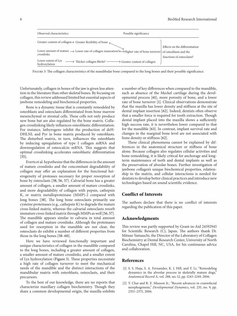
As shown in Figure 3, the differences in collagen char-acteristic between the normal and osteoporotic bones aresimilar to those between the mandibular and long bones.The greater amount of collagen, lower rate of cross-linkmaturation, and lower extent of Lys hydroxylation in themandible are suggestive of the higher rate of bone turnoverand greater bone flexibility. In fact, high turnover and greaterflexibility in mandibular bone are likely necessary to endurethe constant and multidirectional forces of routine activitieslike chewing and speaking. Notably, the force placed on themandible during mastication is almost twice as intense to theforce generated duringwalking [53, 54]. Further investigationwill test these hypotheses.
5. Relevance to Clinical Dentistry
It is important for dentists and dental researchers to under-stand the specific features of jaw physiology and its impacton the matrix of the jawbones. The jaw has interestingproperties related to its function and age-related changein bone volume. Aging is associated with atrophy but notfracture in the jaw. The most plausible explanation is that thejaw undergoes frequent exercise but is not weight bearing.
6 BioMed Research International
Observed characteristics Possible significance
Greater content of collagen Greater flexibility of boneEffects on the differentiation of osteoblasts and the functions of osteoclasts?
Lower amount of mature crosslinks
Lower rate of collagen maturation? Higher rate of bone turnover
Lower extent of Lys hydroxylation
Thicker collagen fibrils? Greater content of collagen
Figure 3: The collagen characteristics of the mandibular bone compared to the long bones and their possible significance.
Unfortunately, collagen in bones of the jaw is given less atten-tion in the literature than other skeletal bones. By focusing oncollagen, this review addressed limited but essential aspects ofjawbone remodeling and biochemical properties.
Bone is a dynamic tissue that is constantly remodeled byosteoblasts and osteoclasts differentiated from bone marrowmesenchymal or stromal cells. These cells not only producenew bone but are also regulated by the bone matrix. Colla-gen crosslinking likely influences osteoblastic differentiation.For instance, lathyrogens inhibit the production of deH-DHLNL and Pyr in bone matrix produced by osteoblasts.The disturbed matrix, in turn, influences the osteoblastsby inducing upregulation of type I collagen mRNA anddownregulation of osteocalcin mRNA. This suggests thatoptimal crosslinking accelerates osteoblastic differentiation[55].
Everts et al. hypothesize that the differences in the amountof mature crosslinks and the concomitant degradability ofcollagen may offer an explanation for the functional het-erogeneity of proteases necessary for proper resorption ofbone by osteoclasts [38, 56, 57]. Calvarial bone has a greateramount of collagen, a smaller amount of mature crosslinks,and more degradability of collagen with pepsin, cathepsinK, or matrix metalloproteinase (MMP)-2 compared withlong bones [38]. The long bone osteoclasts primarily usecysteine proteinases (e.g., cathepsin K) to degrade thematurecross-linked matrix, whereas the calvarial osteoclasts resorbimmature cross-linkedmatrix throughMMPs aswell [56, 57].The mandible appears similar to calvaria in total amountof collagen and mature crosslinks. Although the proteinasesused for resorption in the mandible are not clear, theosteoclasts do exhibit a number of different properties fromthose in the long bones [58–60].
Here we have reviewed functionally important andunique characteristics of collagen in the mandible comparedto the long bones, including a greater amount of collagen,a smaller amount of mature crosslinks, and a smaller extentof Lys hydroxylation (Figure 3). These properties necessitatea high rate of collagen turnover to meet the mechanicalneeds of the mandible and the distinct interactions of themandibular matrix with osteoblasts, osteoclasts, and theirprecursors.
To the best of our knowledge, there are no reports thatcharacterize maxillary collagen biochemistry. Though theyshare a common developmental origin, the maxilla exhibits
a number of key differences when compared to the mandible,such as absence of the Meckel cartilage during the devel-opmental process [61], more porosity of bone, and a lowerrate of bone turnover [1]. Clinical observations demonstratethat the maxilla has lower density and stiffness at the site ofdental implant insertion [62]. Indeed, dentists often observethat a smaller force is required for tooth extraction. Thoughdental implant placed into the maxilla shows a sufficientlyhigh success rate, it is nevertheless lower compared to thatfor the mandible [63]. In contrast, implant survival rate andchanges in the marginal bone level are not associated withbone density or stiffness [62].
These clinical phenomena cannot be explained by dif-ferences in the anatomical structure or stiffness of bonealone. Because collagen also regulates cellular activities andbone remodeling, it is likely critical for anchorage and long-term maintenance of teeth and dental implants as well asthe preservation of alveolar bones. Further investigation ofjawbone collagen’s unique biochemical properties, relation-ship to the matrix, and cellular interactions is needed fordentists to develop better clinical practices and introduce newtechnologies based on sound scientific evidence.
Conflict of Interests
The authors declare that there is no conflict of interestsregarding the publication of this paper.
Acknowledgments
This review was partly supported by Grant-in-Aid 24592945for Scientific Research (C), Japan. The authors thank Dr.Mitsuo Yamauchi, the Director of the Laboratory of CollagenBiochemistry at Dental Research Center, University of NorthCarolina, Chapel Hill, NC, USA, for his continuous adviceand collaboration.
References
[1] S. S. Huja, S. A. Fernandez, K. J. Hill, and Y. Li, “Remodelingdynamics in the alveolar process in skeletally mature dogs,”Anatomical Record A, vol. 288, no. 12, pp. 1243–1249, 2006.
[2] Y. Chai and R. E. Maxson Jr., “Recent advances in craniofacialmorphogenesis,” Developmental Dynamics, vol. 235, no. 9, pp.2353–2375, 2006.
BioMed Research International 7
[3] A. Karaplis, “Embryonic development of bone and the molec-ular regulation of intramembranous and endochondral boneformation,” in Principles of Bone Biology, J. P. Bilezikian, L. G.Raisz, and G. A. Rodan, Eds., pp. 33–58, Academic Press, SanDiego, Calif, USA, 2002.
[4] Y. Ueki, V. Tiziani, C. Santanna et al., “Mutations in the geneencoding c-Abl-binding protein SH3BP2 cause cherubism,”Nature Genetics, vol. 28, no. 2, pp. 125–126, 2001.
[5] W. F. Simonds, L. A. James-Newton, S. K. Agarwal et al.,“Familial isolated hyperparathyroidism: clinical and geneticcharacteristics of 36 kindreds,”Medicine, vol. 81, no. 1, pp. 1–26,2002.
[6] S. L. Ruggiero, B. Mehrotra, T. J. Rosenberg, and S. L. Engroff,“Osteonecrosis of the Jaws Associated with the Use of Bisphos-phonates: a review of 63 cases,” Journal of Oral andMaxillofacialSurgery, vol. 62, no. 5, pp. 527–534, 2004.
[7] A.Mavropoulos, R. Rizzoli, and P. Ammann, “Different respon-siveness of alveolar and tibial bone to bone loss stimuli,” Journalof Bone and Mineral Research, vol. 22, no. 3, pp. 403–410, 2007.
[8] T.Matsubara, K. Suardita, M. Ishii et al., “Alveolar bonemarrowas a cell source for regenerative medicine: differences betweenalveolar and iliac bone marrow stromal cells,” Journal of Boneand Mineral Research, vol. 20, no. 3, pp. 399–409, 2005.
[9] S. O. Akintoye, T. Lam, S. Shi, J. Brahim, M. T. Collins, andP. G. Robey, “Skeletal site-specific characterization of orofacialand iliac crest human bone marrow stromal cells in sameindividuals,” Bone, vol. 38, no. 6, pp. 758–768, 2006.
[10] T. L. Aghaloo, T. Chaichanasakul, O. Bezouglaia et al.,“Osteogenic potential of mandibular vs. Long-bone marrowstromal cells,” Journal of Dental Research, vol. 89, no. 11, pp.1293–1298, 2010.
[11] M. Damek-Poprawa, D. Stefanik, L. M. Levin, and S. O.Akintoye, “Human bone marrow stromal cells display variableanatomic site-dependent response and recovery from irradia-tion,” Archives of Oral Biology, vol. 55, no. 5, pp. 358–364, 2010.
[12] T. Yamaza, G. Ren, K. Akiyama, C. Chen, Y. Shi, and S. Shi,“Mousemandible contains distinctivemesenchymal stem cells,”Journal of Dental Research, vol. 90, no. 3, pp. 317–324, 2011.
[13] M. Saito and K. Marumo, “Collagen cross-links as a determi-nant of bone quality: a possible explanation for bone fragilityin aging, osteoporosis, and diabetes mellitus,” OsteoporosisInternational, vol. 21, no. 2, pp. 195–214, 2010.
[14] K. Tokutomi, T. Matsuura, P. Atsawasuwan, H. Sato, and M.Yamauchi, “Characterization of mandibular bones in senileosteoporotic mice,” Connective Tissue Research, vol. 49, no. 5,pp. 361–366, 2008.
[15] M. J. Silva, M. D. Brodt, B. Wopenka et al., “Decreased collagenorganization and content are associated with reduced strengthof demineralized and intact bone in the SAMP6mouse,” Journalof Bone and Mineral Research, vol. 21, no. 1, pp. 78–88, 2006.
[16] A. J. Bailey, S. F. Wotton, T. J. Sims, and P. W.Thompson, “Post-translational modifications in the collagen of human osteo-porotic femoral head,” Biochemical and Biophysical ResearchCommunications, vol. 185, no. 3, pp. 801–805, 1992.
[17] C. Niyibizi and D. R. Eyre, “Bone type V collagen: chaincomposition and location of a trypsin cleavage site,” ConnectiveTissue Research, vol. 20, no. 1–4, pp. 247–250, 1989.
[18] M. Yamauchi, “Collagen: the major matrix molecule in miner-alized tissues,” in Calcium and Phosphorus Nutrition in Health
and Diseases, J. J. B. Anderson and S. C. Garner, Eds., pp. 127–145, CRC Press, Boca Raton, Fla, USA, 1996.
[19] K. I. Kivirikko and R. Myllyla, “Post-translational processing ofprocollagens,” Annals of the New York Academy of Sciences, vol.460, pp. 187–201, 1985.
[20] H. Oxlund, M. Barckman, G. Ortoft, and T. T. Andreassen,“Reduced concentrations of collagen cross-links are associatedwith reduced strength of bone,” Bone, vol. 17, no. 4, pp. 365S–371S, 1995.
[21] H.-H. Hong, N. Pischon, R. B. Santana et al., “A role forlysyl oxidase regulation in the control of normal collagendeposition in differentiating osteoblast cultures,” Journal ofCellular Physiology, vol. 200, no. 1, pp. 53–62, 2004.
[22] M. Sricholpech, I. Perdivara, H. Nagaoka, M. Yokoyama, K.B. Tomer, and M. Yamauchi, “Lysyl hydroxylase 3 glucosylatesgalactosylhydroxylysine residues in type I collagen in osteoblastculture,” Journal of Biological Chemistry, vol. 286, no. 11, pp.8846–8856, 2011.
[23] M. Sricholpech, I. Perdivara, H. Nagaoka, M. Yokoyama, K.B. Tomer, and M. Yamauchi, “Lysyl hydroxylase 3 glucosylatesgalactosylhydroxylysine residues in type I collagen in osteoblastculture,” Journal of Biological Chemistry, vol. 286, no. 11, pp.8846–8856, 2011.
[24] M. Yamauchi, C. Noyes, Y. Kuboki, and G. L. Mechanic,“Collagen structural microheterogeneity and a possible role forglycosylated hydroxylysine in type I collagen,” Proceedings of theNational Academy of Sciences of theUnited States of America, vol.79, no. 24, pp. 7684–7688, 1982.
[25] M. Yamauchi, E. P. Katz, and G. L. Mechanic, “Intermolecularcross-linking and stereospecific molecular packing in type Icollagen fibrils of the periodontal ligament,” Biochemistry, vol.25, no. 17, pp. 4907–4913, 1986.
[26] S. Pornprasertsuk,W. R. Duarte, Y.Mochida, andM. Yamauchi,“Lysyl hydroxylase-2b directs collagen cross-linking pathwaysin MC3T3-E1 cells,” Journal of Bone and Mineral Research, vol.19, no. 8, pp. 1349–1355, 2004.
[27] S. Pornprasertsuk,W. R. Duarte, Y.Mochida, andM. Yamauchi,“Overexpression of lysyl hydroxylase-2b leads to defectivecollagen fibrillogenesis and matrix mineralization,” Journal ofBone and Mineral Research, vol. 20, no. 1, pp. 81–87, 2005.
[28] D. R. Eyre and H. Oguchi, “The hydroxypyridinium crosslinksof skeletal collagens: their measurement, properties and aproposed pathway of formation,” Biochemical and BiophysicalResearch Communications, vol. 92, no. 2, pp. 403–410, 1980.
[29] M.Yamauchi andG. L.Mechanic, “Cross-linking of collagen,” inCollagen, M. E. Nimni, Ed., pp. 157–172, CRCPress, Boca Raton,Fla, USA, 1988.
[30] S. P. Robins and A. Duncan, “Cross-linking of collagen. Loca-tion of pyridinoline in bovine articular cartilage at two sites ofthe molecule,” Biochemical Journal, vol. 215, no. 1, pp. 175–182,1983.
[31] D. R. Eyre, M. A. Paz, and P. M. Gallop, “Cross-linking incollagen and elastin,”Annual Review of Biochemistry, vol. 53, pp.717–748, 1984.
[32] M. Saito, S. Soshi, and K. Fujii, “Effect of hyper- and micro-gravity on collagen post-translational controls of MC3T3-E1osteoblasts,” Journal of Bone and Mineral Research, vol. 18, no.9, pp. 1695–1705, 2003.
8 BioMed Research International
[33] N. Pischon, J.M.Maki, P.Weisshaupt et al., “Lysyl oxidase (Lox)gene deficiency affects osteoblastic phenotype,” Calcified TissueInternational, vol. 85, no. 2, pp. 119–126, 2009.
[34] K. Takaluoma,M. Hyry, J. Lantto et al., “Tissue-specific changesin the hydroxylysine content and cross-links of collagens andalterations in fibril morphology in lysyl hydroxylase 1 knock-out mice,” Journal of Biological Chemistry, vol. 282, no. 9, pp.6588–6596, 2007.
[35] A. J. Bailey, T. J. Sims, E. N. Ebbesen, J. P.Mansell, J. S.Thomsen,and L. Mosekilde, “Age-related changes in the biochemicalproperties of human cancellous bone collagen: relationship tobone strength,” Calcified Tissue International, vol. 65, no. 3, pp.203–210, 1999.
[36] A. J. Bailey, S. F. Wotton, T. J. Sims, and P. W. Thompson,“Biochemical changes in the collagen of human osteoporoticbone matrix,” Connective Tissue Research, vol. 29, no. 2, pp. 119–132, 1993.
[37] M. Sasaki, T. Matsuura, M. Katafuchi, K. Tokutomi, and H.Sato, “Higher contents of mineral and collagen but lower ofhydroxylysine of collagen in mandibular bone compared withthose of humeral and femoral bones in human,” Journal of HardTissue Biology, vol. 19, no. 3, pp. 175–180, 2010.
[38] T. van denBos,D. Speijer, R. A. Bank,D. Bromme, andV. Everts,“Differences in matrix composition between calvaria and longbone in mice suggest differences in biomechanical propertiesand resorption. Special emphasis on collagen,” Bone, vol. 43, no.3, pp. 459–468, 2008.
[39] J. E. Aaron, “Periosteal Sharpey’s fibers: a novel bone matrixregulatory system?” Frontiers in Endocrinology, vol. 3, article 98,pp. 1–10, 2012.
[40] A. Al-Qtaitat, R. C. Shore, and J. E. Aaron, “Structural changesin the ageing periosteum using collagen III immuno-stainingand chromium labelling as indicators,” Journal of Musculoskele-tal Neuronal Interactions, vol. 10, no. 1, pp. 112–123, 2010.
[41] F. Luther, H. Saino, D. H. Carter, and J. E. Aaron, “Evidence foran extensive collagen Type III/VI proximal domain in the ratfemur: I. Diminution with ovariectomy,” Bone, vol. 32, no. 6, pp.652–659, 2003.
[42] H. Saino, F. Luther, D. H. Carter et al., “Evidence for an exten-sive collagen type III proximal domain in the rat femur: II.Expansion with exercise,” Bone, vol. 32, no. 6, pp. 660–668,2003.
[43] D. E. Birk, “TypeV collagen: heterotypic type I/V collagen inter-actions in the regulation of fibril assembly,”Micron, vol. 32, no.3, pp. 223–237, 2001.
[44] P. L. Lukinmaa and J. Waltimo, “Immunohistochemical local-ization of types I, V, and VI collagen in human permanent teethand periodontal ligament,” Journal of Dental Research, vol. 71,no. 2, pp. 391–397, 1992.
[45] G. E. Romanos, C. Schroter-Kermani, N. Hinz, H. C. Wachtel,and J. P. Bernimoulin, “Immunohistochemical localization ofcollagenous components in healthy periodontal tissues of therat and marmoset (Callithrix jacchus). II. Distribution ofcollagen types IV, V and VI,” Journal of Periodontal Research,vol. 26, no. 4, pp. 323–332, 1991.
[46] K. Noris Suarez, M. Romanello, P. Bettica, and L. Moro,“Collagen type I of rat cortical and trabecular bone differs inthe extent of posttranslational modifications,” Calcified TissueInternational, vol. 58, no. 1, pp. 65–69, 1996.
[47] J. Aerssens, S. Boonen, G. Lowet, and J. Dequeker, “Interspeciesdifferences in bone composition, density, and quality: potentialimplications for in vivo bone research,” Endocrinology, vol. 139,no. 2, pp. 663–670, 1998.
[48] K. W. Fishbein, Y. A. Gluzband, M. Kaku et al., “Effects offormalin fixation and collagen cross-linking on T2 and magne-tization transfer in bovine nasal cartilage,”Magnetic Resonancein Medicine, vol. 57, no. 6, pp. 1000–1011, 2007.
[49] L. Moro, M. Romanello, A. Favia, M. P. Lamanna, and E.Lozupone, “Posttranslational modifications of bone collagentype I are related to the function of rate femoral regions,”Calcified Tissue International, vol. 66, no. 2, pp. 151–156, 2000.
[50] H. Notbohm, M. Nokelainen, J. Myllyharju, P. P. Fietzek, P.K. Muller, and K. I. Kivirikko, “Recombinant human type IIcollagens with low and high levels of hydroxylysine and itsglycosylated forms showmarked differences in fibrillogenesis invitro,” Journal of Biological Chemistry, vol. 274, no. 13, pp. 8988–8992, 1999.
[51] S. Kalamajski and A. Oldberg, “The role of small leucine-richproteoglycans in collagen fibrillogenesis,” Matrix Biology, vol.29, no. 4, pp. 248–253, 2010.
[52] K. E. Kadler, A.Hill, andE.G.Canty-Laird, “Collagen fibrilloge-nesis: fibronectin, integrins, and minor collagens as organizersand nucleators,” Current Opinion in Cell Biology, vol. 20, no. 5,pp. 495–501, 2008.
[53] A. C. Knoell, “A mathematical model of an in vitro humanmandible,” Journal of Biomechanics, vol. 10, no. 3, pp. 159–166,1977.
[54] D. J. Daegling andW. L.Hylander, “Occlusal forces andmandib-ular bone strain: Is the primate jaw “overdesigned”?” Journal ofHuman Evolution, vol. 33, no. 6, pp. 705–717, 1997.
[55] C. Turecek, N. Fratzl-Zelman, M. Rumpler et al., “Collagencross-linking influences osteoblastic differentiation,” CalcifiedTissue International, vol. 82, no. 5, pp. 392–400, 2008.
[56] V. Everts, W. Korper, D. C. Jansen et al., “Functional hetero-geneity of osteoclasts: matrix metalloproteinases participate inosteoclastic resorption of calvarial bone but not in resorption oflong bone,” FASEB Journal, vol. 13, no. 10, pp. 1219–1230, 1999.
[57] V. Everts, W. Korper, K. A. Hoeben et al., “Osteoclastic bonedegradation and the role of different cysteine proteinases andmatrix metalloproteinases: differences between calvaria andlong bone,” Journal of Bone and Mineral Research, vol. 21, no.9, pp. 1399–1408, 2006.
[58] A. P. De Souza Faloni, T. Schoenmaker, A. Azari et al., “Jaw andlong bone marrows have a different osteoclastogenic potential,”Calcified Tissue International, vol. 88, no. 1, pp. 63–74, 2011.
[59] A. Azari, T. Schoenmaker, A. P. de Souza Faloni, V. Everts,and T. J. De Vries, “Jaw and long bone marrow derivedosteoclasts differ in shape and their response to bone anddentin,” Biochemical and Biophysical Research Communications,vol. 409, no. 2, pp. 205–210, 2011.
[60] J. A. F. Vermeer, I. D. C. Jansen, M. Marthi et al., “Jawbone marrow-derived osteoclast precursors internalize morebisphosphonate than long-bone marrow precursors,” Bone, vol.57, no. 1, pp. 242–251, 2013.
[61] S. K. Lee, Y. S. Kim, H. S. Oh, K. H. Yang, E. C. Kim, andJ. G. Chi, “Prenatal development of the human mandible,”Anatomical Record, vol. 265, no. 3, pp. 314–325, 2001.

BioMed Research International 9
[62] G. Bergkvist, K.-J. Koh, S. Sahlholm, E. Klintstrom, and C.Lindh, “Bone density at implant sites and its relationshipto assessment of bone quality and treatment outcome,” TheInternational Journal of Oral & Maxillofacial Implants, vol. 25,no. 2, pp. 321–328, 2010.
[63] P. K. Moy, D. Medina, V. Shetty, and T. L. Aghaloo, “Dentalimplant failure rates and associated risk factors,” InternationalJournal of Oral and Maxillofacial Implants, vol. 20, no. 4, pp.569–577, 2005.

Submit your manuscripts athttp://www.hindawi.com
ScientificaHindawi Publishing Corporationhttp://www.hindawi.com Volume 2014
CorrosionInternational Journal of
Hindawi Publishing Corporationhttp://www.hindawi.com Volume 2014
Polymer ScienceInternational Journal of
Hindawi Publishing Corporationhttp://www.hindawi.com Volume 2014
Hindawi Publishing Corporationhttp://www.hindawi.com Volume 2014
CeramicsJournal of
Hindawi Publishing Corporationhttp://www.hindawi.com Volume 2014
CompositesJournal of
NanoparticlesJournal of
Hindawi Publishing Corporationhttp://www.hindawi.com Volume 2014
Hindawi Publishing Corporationhttp://www.hindawi.com Volume 2014
International Journal of
Biomaterials
Hindawi Publishing Corporationhttp://www.hindawi.com Volume 2014
NanoscienceJournal of
TextilesHindawi Publishing Corporation http://www.hindawi.com Volume 2014
Journal of
NanotechnologyHindawi Publishing Corporationhttp://www.hindawi.com Volume 2014
Journal of
CrystallographyJournal of
Hindawi Publishing Corporationhttp://www.hindawi.com Volume 2014
The Scientific World JournalHindawi Publishing Corporation http://www.hindawi.com Volume 2014
Hindawi Publishing Corporationhttp://www.hindawi.com Volume 2014
CoatingsJournal of
Advances in
Materials Science and EngineeringHindawi Publishing Corporationhttp://www.hindawi.com Volume 2014
Smart Materials Research
Hindawi Publishing Corporationhttp://www.hindawi.com Volume 2014
Hindawi Publishing Corporationhttp://www.hindawi.com Volume 2014
MetallurgyJournal of
Hindawi Publishing Corporationhttp://www.hindawi.com Volume 2014
BioMed Research International
MaterialsJournal of
Hindawi Publishing Corporationhttp://www.hindawi.com Volume 2014
Nano
materials
Hindawi Publishing Corporationhttp://www.hindawi.com Volume 2014
Journal ofNanomaterials