review article antioxidant and anti-inflammatory...
TRANSCRIPT

Review ArticleAntioxidant and Anti-Inflammatory Activities of Berberine inthe Treatment of Diabetes Mellitus
Zheng Li,1 Ya-Na Geng,1 Jian-Dong Jiang,2 and Wei-Jia Kong1
1 Department of Pharmacology, Institute of Medicinal Biotechnology, Chinese Academy of Medical Sciencesand Peking Union Medical College, Beijing 100050, China
2 State Key Laboratory of Bioactive Natural Products and Function, Institute of Materia Medica, Chinese Academy of Medical Sciencesand Peking Union Medical College, Beijing 100050, China
Correspondence should be addressed to Jian-Dong Jiang; [email protected] and Wei-Jia Kong; [email protected]
Received 24 November 2013; Revised 30 December 2013; Accepted 2 January 2014; Published 11 February 2014
Academic Editor: Yao Tong
Copyright © 2014 Zheng Li et al.This is an open access article distributed under the Creative Commons Attribution License, whichpermits unrestricted use, distribution, and reproduction in any medium, provided the original work is properly cited.
Oxidative stress and inflammation are proved to be critical for the pathogenesis of diabetes mellitus. Berberine (BBR) is a naturalcompound isolated from plants such as Coptis chinensis and Hydrastis canadensis and with multiple pharmacological activities.Recent studies showed that BBR had antioxidant and anti-inflammatory activities, which contributed in part to its efficacyagainst diabetes mellitus. In this review, we summarized the antioxidant and anti-inflammatory activities of BBR as well as theirmolecular basis. The antioxidant and anti-inflammatory activities of BBR were noted with changes in oxidative stress markers,antioxidant enzymes, and proinflammatory cytokines after BBR administration in diabetic animals. BBR inhibited oxidative stressand inflammation in a variety of tissues including liver, adipose tissue, kidney and pancreas. Mechanisms of the antioxidant andanti-inflammatory activities of BBR were complex, which involved multiple cellular kinases and signaling pathways, such as AMP-activated protein kinase (AMPK), mitogen-activated protein kinases (MAPKs), nuclear factor erythroid-2-related factor-2 (Nrf2)pathway, and nuclear factor-𝜅B (NF-𝜅B) pathway. Detailed mechanisms and pathways for the antioxidant and anti-inflammatoryactivities of BBR still need further investigation. Clarification of these issues could help to understand the pharmacology of BBR inthe treatment of diabetes mellitus and promote the development of antidiabetic natural products.
1. Introduction
For decades, diabetes mellitus especially type 2 diabetes mel-litus (T2DM) has become a public health problem and threat-ened the people worldwide, not only inwestern countries, butalso in the developingworld like China.Thepathogenesis andpathophysiological processes of T2DM are extreme complexand remain to be controversial. However, a growing numberof evidences showed that oxidative stress and inflammationmight play important roles in the development of T2DM[1, 2].
In metabolic disorders, oxidative stress could be typicallyinduced by excessive nutritional factors like glucose and freefatty acids (FFA) [3, 4]. Oxidative stress could induce ordeteriorate insulin resistance and diabetes through multiplemechanisms. In the process of oxidative stress, excessivereactive oxygen species (ROS) were produced, mainly by
mitochondria [5]. ROS include superoxide anion (O2
−∙),hydroxyl radical (OH∙), hydrogen peroxide (H
2O2) [6].They
could cause damage and apoptosis of pancreatic islet 𝛽-cellsand reduction of insulin secretion [7]. ROS could activatecellular signaling pathways like c-jun N-terminal kinase(JNK), protein kinase C (PKC), and nuclear factor-𝜅B (NF-𝜅B) and then interfere with the insulin signaling pathway andcause insulin resistance [8–10]. In addition, oxidative stressalso contributes to the development of chronic complicationsof diabetes, such as diabetic nephropathy, retinopathy, andneuropathy [6].
Recently, results from laboratory studies as well as clin-ical investigations have proved that diabetes is in fact aninflammatory disease [2, 11].The importance of inflammationin insulin resistance, diabetes and complications has beencritically reviewed [12–16], concerning relevant proinflam-matory cytokines as well as cellular signaling pathways.
Hindawi Publishing CorporationEvidence-Based Complementary and Alternative MedicineVolume 2014, Article ID 289264, 12 pageshttp://dx.doi.org/10.1155/2014/289264
2 Evidence-Based Complementary and Alternative Medicine
In the pathogenesis of diabetes mellitus, proinflammatorycytokines such as tumor necrosis factor-𝛼 (TNF-𝛼) andinterleukin-6 (IL-6) could be produced by immunocytes(like macrophages) and adipocytes [12–14]. TNF-𝛼 and IL-6were important mediators of insulin resistance, as they couldinduce serine phosphorylation of insulin receptor substrate(IRS) through activation of JNK or NF-𝜅B pathway [14, 16].Furthermore, overproduction of TNF-𝛼 and IL-6 in pancreascould cause islet dysfunction and accelerate the progressionof diabetes [13].
There are a number of medications available in clinicto treat T2DM. In addition to chemical drugs, a numberof natural products or traditional Chinese medicine (TCM)formulas were reported to have antidiabetic or insulin-sensitizing activities, and some of them have been used inclinic with a long history [11, 17, 18]. Berberine (BBR) is a nat-ural compound isolated from plants such as Coptis chinensisand Hydrastis canadensis and with multiple pharmacologicalactivities [19]. The emerging role of BBR in modifying sugarand lipid metabolism has been verified in a large amount ofexperimental and clinical studies [20–23].
In general, BBR was safe and effective in the treatmentof patients with T2DM [19–21, 23, 24]. The glucose-loweringefficacy of BBR was close to those of metformin and rosigli-tazone [23, 24]. Notably, due to the low toxicity, BBR couldbe used in diabetic patients with chronic hepatitis [24].Beneficial effects of BBR were also observed in combatingdiabetic complications. Diabetes related endothelial dysfunc-tion, nephropathy, and neuropathy could be relieved afterBBR administration [21].
Mechanisms of BBR in restoring insulin sensitivity andlowering blood glucose included inhibition of mitochondrialfunction and activation of AMP-activated protein kinase(AMPK), regulation of islet function, modulation of gutmicroenvironment, and upregulation of insulin receptorexpression [20–22, 24–26]. Recent studies showed that BBRhad beneficial effects on cellular oxidative stress and inflam-mation, which could also play important roles in its activityagainst diabetes mellitus. In this paper, we reviewed theantioxidant and anti-inflammatory activities as well as theirmolecular basis of BBR in treating diabetes mellitus andinsulin resistance.
2. Antioxidant Activity and Mechanisms ofBBR in Treating Diabetes Mellitus
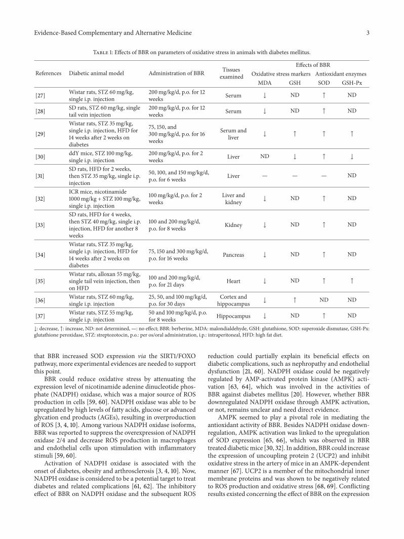
2.1. BBR Reduced Oxidative Stress in Diabetes Mellitus. Theinhibitory effect of BBR on oxidative stress was observed bothin cells culturedwith high glucose-containingmedia [38] andin a series of diabetic animal models (Table 1) [27–30, 32–37]. The antioxidant activity of BBR was revealed by changesof oxidative stress markers as well as antioxidant enzymes.Oxidative stress markers include malondialdehyde (MDA), aproduct of lipid peroxidation which increases during oxida-tive stress [39], as well as glutathione (GSH), which oftendeclines during oxidative stress [40]. GSH is an antioxidantitself and is a substrate of glutathione peroxidase (GSH-Px)in the clearance of peroxides [40]. In addition to GSH-Px,
another well-known antioxidant enzyme, superoxide dismu-tase (SOD), is also involved to evaluate the inhibitory effect ofBBR on oxidative stress. Antioxidant enzyme is a part of theantioxidant defensemechanisms, which helps tomaintain thebalance of redox in organisms and could be damaged in thepathogenesis of diabetes mellitus [41].
As summarized in Table 1, the majority of reports [27–30,32–37] supported the antioxidant activity of BBR in diabeticanimal models, which were induced by streptozotocin (STZ)or alloxan injection with or without high fat diet (HFD)feeding. The relief of oxidative stress by BBR was noted bychanges of oxidative stress markers as well as antioxidantenzymes. In general, BBR administration decreased MDAcontent and increased the contents of SOD, GSH and GSH-Px, which would help to scavenge excessive free radicals andovercome oxidative stress [40, 41]. In only one of the reports[31], the effect of BBR on oxidative stress seemed not to beobvious. However, in this report [31], the contents of MDAand SOD of the diabetic animals did not have statisticallysignificant difference as compared with those of the normalcontrol animals, inconsistent with the results from otherreports [27–30, 32–37]. In another report [30], BBR treatmentgreatly upregulated the mRNA level of SOD but decreasedGSH and GSH-Px contents in diabetic mice. In this report[30], while SODwas observed to be reduced in diabetic mice,the amounts of GSH and GSH-Px were increased, probablydue to the acute stage of experimental diabetes mellitus [52],which usually occurred within 3-4 weeks of STZ injection[53, 54]. So, it seems that BBR could regulate the GSH/GSH-Px antioxidant mechanism differentially at different stages ofdiabetes mellitus.
The antioxidant activity of BBR was associated with itsinhibitory effect on the development of diabetes mellitus andinsulin resistance induced by STZ/alloxan + HFD in animals[32–35]. Furthermore, BBR inhibited oxidative stress in avariety of tissues, such as the serum [27–29], liver [29, 30, 32],kidney [32, 33], pancreas [34], heart [35] and central nervesystem [36, 37]. The inhibitory effect of BBR on oxidativestress was associated with its activity against renal injury[27, 28, 33], pancreatic islet dysfunction [34], and memoryimpairment [36] in diabetic animals.
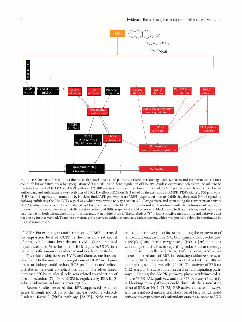
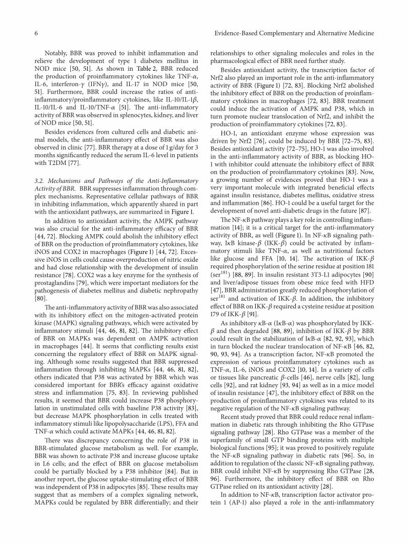
2.2. Mechanisms and Pathways of BBR against OxidativeStress. Molecular mechanisms of BBR in reducing oxidativestress seem to be related with multiple cellular pathways andneed further investigation. The schematic illustration of thepathways from available data was presented in Figure 1.
It was reported that BBR scavenged superoxide freeradicals directly in vitro in a system containing alkalinedimethyl sulfoxide (DMSO) [55].ThemRNA expression levelof SOD could be upregulated by BBR in diabetic mice, and itplayed a role in BBR’s activity against oxidative stress [30, 32].Also, BBR was reported to increase the expression level ofsirtuin 1 (SIRT1) [56], a deacetylase with multiple biologicalactivities and antioxidant activity [57]. In oxidative stress,SIRT1 could induce deacetylation of the forkhead box O(FOXO) transcription factors and increased the transcriptionof their target genes, which included SOD [58]. It is possible
Evidence-Based Complementary and Alternative Medicine 3
Table 1: Effects of BBR on parameters of oxidative stress in animals with diabetes mellitus.
References Diabetic animal model Administration of BBR Tissuesexamined
Effects of BBROxidative stress markers Antioxidant enzymes
MDA GSH SOD GSH-Px
[27] Wistar rats, STZ 60mg/kg,single i.p. injection
200mg/kg/d, p.o. for 12weeks Serum ↓ ND ↑ ND
[28] SD rats, STZ 60mg/kg, singletail vein injection
200mg/kg/d, p.o. for 12weeks Serum ↓ ND ↑ ND
[29]
Wistar rats, STZ 35mg/kg,single i.p. injection, HFD for14 weeks after 2 weeks ondiabetes
75, 150, and300mg/kg/d, p.o. for 16weeks
Serum andliver ↓ ↑ ↑ ↑
[30] ddY mice, STZ 100mg/kg,single i.p. injection
200mg/kg/d, p.o. for 2weeks Liver ND ↓ ↑ ↓
[31]SD rats, HFD for 2 weeks,then STZ 35mg/kg, single i.p.injection
50, 100, and 150mg/kg/d,p.o. for 6 weeks Liver — — — ND
[32]ICR mice, nicotinamide1000mg/kg + STZ 100mg/kg,single i.p. injection
100mg/kg/d, p.o. for 2weeks
Liver andkidney ↓ ND ↑ ND
[33]
SD rats, HFD for 4 weeks,then STZ 40mg/kg, single i.p.injection, HFD for another 8weeks
100 and 200mg/kg/d,p.o. for 8 weeks Kidney ↓ ND ↑ ND
[34]
Wistar rats, STZ 35mg/kg,single i.p. injection, HFD for14 weeks after 2 weeks ondiabetes
75, 150 and 300mg/kg/d,p.o. for 16 weeks Pancreas ↓ ND ↑ ND
[35]Wistar rats, alloxan 55mg/kg,single tail vein injection, thenon HFD
100 and 200mg/kg/d,p.o. for 21 days Heart ↓ ND ↑ ↑
[36] Wistar rats, STZ 60mg/kg,single i.p. injection
25, 50, and 100mg/kg/d,p.o. for 30 days
Cortex andhippocampus ↓ ↑ ND ND
[37] Wistar rats, STZ 55mg/kg,single i.p. injection
50 and 100mg/kg/d, p.o.for 8 weeks Hippocampus ↓ ND ↑ ND
↓: decrease, ↑: increase, ND: not determined, —: no effect; BBR: berberine, MDA: malondialdehyde, GSH: glutathione, SOD: superoxide dismutase, GSH-Px:glutathione peroxidase, STZ: streptozotocin, p.o.: per os/oral administration, i.p.: intraperitoneal, HFD: high fat diet.
that BBR increased SOD expression via the SIRT1/FOXOpathway, more experimental evidences are needed to supportthis point.
BBR could reduce oxidative stress by attenuating theexpression level of nicotinamide adenine dinucleotide phos-phate (NADPH) oxidase, which was a major source of ROSproduction in cells [59, 60]. NADPH oxidase was able to beupregulated by high levels of fatty acids, glucose or advancedglycation end products (AGEs), resulting in overproductionof ROS [3, 4, 10]. Among various NADPH oxidase isoforms,BBR was reported to suppress the overexpression of NADPHoxidase 2/4 and decrease ROS production in macrophagesand endothelial cells upon stimulation with inflammatorystimuli [59, 60].
Activation of NADPH oxidase is associated with theonset of diabetes, obesity and arthrosclerosis [3, 4, 10]. Now,NADPH oxidase is considered to be a potential target to treatdiabetes and related complications [61, 62]. The inhibitoryeffect of BBR on NADPH oxidase and the subsequent ROS
reduction could partially explain its beneficial effects ondiabetic complications, such as nephropathy and endothelialdysfunction [21, 60]. NADPH oxidase could be negativelyregulated by AMP-activated protein kinase (AMPK) acti-vation [63, 64], which was involved in the activities ofBBR against diabetes mellitus [20]. However, whether BBRdownregulated NADPH oxidase through AMPK activation,or not, remains unclear and need direct evidence.
AMPK seemed to play a pivotal role in mediating theantioxidant activity of BBR. Besides NADPH oxidase down-regulation, AMPK activation was linked to the upregulationof SOD expression [65, 66], which was observed in BBRtreated diabeticmice [30, 32]. In addition, BBR could increasethe expression of uncoupling protein 2 (UCP2) and inhibitoxidative stress in the artery of mice in an AMPK-dependentmanner [67]. UCP2 is a member of the mitochondrial innermembrane proteins and was shown to be negatively relatedto ROS production and oxidative stress [68, 69]. Conflictingresults existed concerning the effect of BBR on the expression
4 Evidence-Based Complementary and Alternative Medicine
BBR
NADPH oxidase AMPK activation
UCP2
?
SIRT1/FOXO
SOD PI3K/Akt activation
Nrf2 nuclear
P38 activation
MAPK inhibition inhibition
Expression of
Rho GTPase inhibition activation
AP-1 pathway inhibition
??
??
?
?
O
O N+
OCH3
OCH3
mRNA ↑ mRNA ↓IKK-𝛽 PPAR𝛾
translocation ↑ and degradation ↓
mRNA ↑ NF-𝜅Bactivation ↓
GSH ↑SOD activity ↑
NQO-1 expression ↑
HO-1expression ↑
proinflammatory cytokines ↓
ROS production ↓
Oxidative stress ↓Inflammation ↓
Cl−
I𝜅B-𝛼 phosphorylation
Figure 1: Schematic illustration of the molecular mechanisms and pathways of BBR in reducing oxidative stress and inflammation. (1) BBRcould inhibit oxidative stress by upregulation of SOD, UCP2 and downregulation of NADPH oxidase expression, which was possible to bemediated by the SIRT1/FOXOorAMPKpathway. (2) BBR administration induced the activation of theNrf2 pathway, whichwas crucial for theantioxidant and anti-inflammatory activities of BBR.The effect of BBRonNrf2 relied on the activation ofAMPK, PI3K/Akt, andP38 pathways.(3) BBR could suppress inflammation by blocking theMAPKpathways in anAMPK-dependentmanner, inhibiting the classicNF-𝜅B signalingpathway; inhibiting the Rho GTPase pathway, which was proved to play a role in NF-𝜅B regulation, and attenuating the transcription activityof AP-1, which was possible to be mediated by PPAR𝛾 activation. The black lines/boxes and red lines/boxes indicate pathways and moleculesinvolved in the antioxidant or anti-inflammatory activity of BBR, respectively. Red boxes with black frame indicate pathways and moleculesresponsible for both antioxidant and anti-inflammatory activities of BBR.The symbols of “?” indicate possible mechanisms and pathways thatneed to be further verified.There was a vicious cycle between oxidative stress and inflammation, which was possibly able to be terminated byBBR administration.
of UCP2. For example, in another report [70], BBR decreasedthe expression level of UCP2 in the liver in a rat modelof nonalcoholic fatty liver disease (NAFLD) and reducedhepatic steatosis. Whether or not BBR regulates UCP2 in atissue-specific manner is unknown and needs more study.
The relationship betweenUCP2 and diabetesmellitus wascomplex. On the one hand, upregulation of UCP2 in adiposetissue or kidney could reduce ROS production and relievediabetes or relevant complication; but on the other hand,increased UCP2 in islet 𝛽-cells was related to reduction ofinsulin secretion [71]. How UCP2 is regulated by BBR in 𝛽-cells is unknown and needs investigation.
Recent studies revealed that BBR suppressed oxidativestress through induction of the nuclear factor erythroid-2-related factor-2 (Nrf2) pathway [72–75]. Nrf2 was an
antioxidant transcription factor mediating the expression ofantioxidant enzymes like NADPH quinine oxidoreductase-1 (NQO-1) and heme oxygenase-1 (HO-1) [76]; it had awide range of activities in regulating redox state and energymetabolism in cells [76]. Now, Nrf2 is recognized as animportant mediator of BBR in reducing oxidative stress, asblocking Nrf2 abolishes the antioxidant activity of BBR inmacrophages and nerve cells [72–75]. The activity of BBR onNrf2 relied on the activation of several cellular signaling path-ways including the AMPK pathway, phosphatidylinositol 3-kinase (PI3K)/Akt pathway, and the P38 pathway (Figure 1),as blocking these pathways could diminish the stimulatingeffect of BBR on Nrf2 [72–75]. BBR activated these pathways,and then induced nuclear translocation of Nrf2 which couldactivate the expression of antioxidant enzymes, increase SOD
Evidence-Based Complementary and Alternative Medicine 5
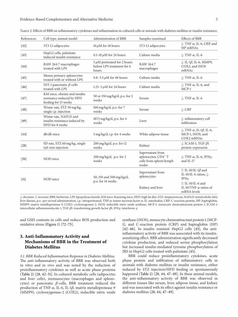
Table 2: Effects of BBR on inflammatory cytokines and inflammation in cultured cells or animals with diabetes mellitus or insulin resistance.
References Cell type, animal model Administration of BBR Samples examined Effects of BBR
[42] 3T3-L1 adipocytes 10𝜇M for 18 hours 3T3-L1 adipocytes ↓: TNF-𝛼, IL-6, CRP, andHP mRNAs
[43] HepG2 cells, palmitateinduced insulin resistance 0.1–10 𝜇M for 24 hours Culture media ↓: TNF-𝛼, IL-6
[44] RAW 264.7 macrophagestreated with LPS
5 𝜇M pretreated for 2 hoursbefore LPS treatment for 6hours
RAW 264.7macrophages
↓: IL-1𝛽, IL-6, MMP9,COX2, and iNOSmRNAs
[45] Mouse primary splenocytestreated with or without LPS 0.8–3.3 𝜇M for 48 hours Culture media ↓: TNF-𝛼, IL-6
[46] NIT-1 pancreatic 𝛽-cellstreated with LPS 1.25–5 𝜇M for 24 hours Culture media ↓: TNF-𝛼, IL-6, and
MCP-1
[47]KMmice, obesity and insulinresistance induced by HFDfeeding for 13 weeks
50 or 150mg/kg/d, p.o. for 2weeks Serum ↓: TNF-𝛼, IL-6
[48] Wistar rats, STZ 50mg/kg,single i.p. injection
100mg/kg/d, p.o. for 7weeks Serum ↓: CRP
[49]Wistar rats, NAFLD andinsulin resistance induced byHFD for 8 weeks
187.5mg/kg/d, p.o. for 4weeks Liver ↓: inflammatory cell
infiltration
[44] db/db mice 5mg/kg/d, i.p. for 4 weeks White adipose tissue↓: TNF-𝛼, IL-1𝛽, IL-6,MCP-1, iNOS, andCOX2 mRNAs
[28] SD rats, STZ 60mg/kg, singletail vein injection
200mg/kg/d, p.o. for 12weeks Kidney ↓: ICAM-1, TGF-𝛽1
protein expression
[50] NOD mice 200mg/kg/d, p.o. for 2weeks
Supernatant fromsplenocytes, CD4+ Tcells from spleen/lymphnodes
↓: TNF-𝛼, IL-6, IFN𝛾,and IL-17
[51] NOD mice 50, 150 and 500mg/kg/d,p.o. for 14 weeks
Supernatant fromsplenocytes
↑: IL-10/IL-1𝛽 andIL-10/IL-6 ratios; ↓:IFN𝛾
Kidney and liver↑: IL-10/IL-6 andIL-10/TNF-𝛼 ratios ofmRNA levels
↓: decrease, ↑: increase; BBR: berberine, LPS: lipopolysaccharide, KMmice: Kunmingmice, HFD: high fat diet, STZ: streptozotocin, NAFLD: nonalcoholic fattyliver disease, p.o.: per os/oral administration, i.p.: intraperitoneal, TNF-𝛼: tumor necrosis factor-𝛼, IL: interleukin, CRP: C-reaction protein, HP: haptoglobin,MMP9: matrix metalloprotease 9, COX2: cyclooxygenase-2, iNOS: inducible nitric oxide synthase, MCP-1: monocyte chemoattractant protein 1, ICAM-1:intercellular adhesionmolecule-1, TGF-𝛽1: transforming growth factor-𝛽1, IFN𝛾: interferon-𝛾.
and GSH contents in cells and reduce ROS production andoxidative stress (Figure 1) [72–75].
3. Anti-Inflammatory Activity andMechanisms of BBR in the Treatment ofDiabetes Mellitus
3.1. BBR Reduced Inflammation Response in Diabetes Mellitus.The anti-inflammatory activity of BBR was observed bothin vitro and in vivo and was noted by the reduction ofproinflammatory cytokines as well as acute phase proteins(Table 2) [28, 42–51]. In cultured metabolic cells (adipocytesand liver cells), immunocytes (macrophages and spleno-cytes) or pancreatic 𝛽-cells, BBR treatment reduced theproduction of TNF-𝛼, IL-6, IL-1𝛽, matrix metalloprotease 9(MMP9), cyclooxygenase-2 (COX2), inducible nitric oxide
synthase (iNOS),monocyte chemoattractant protein 1 (MCP-1), and C-reaction protein (CRP) and haptoglobin (HP)[42–46]. In insulin resistant HepG2 cells [43], the anti-inflammatory activity of BBR was associated with its insulin-sensitizing effect. BBR administration significantly decreasedcytokine production, and reduced serine phosphorylationbut increased insulin-mediated tyrosine phosphorylation ofIRS in HepG2 cells treated with palmitate [43].
BBR could reduce proinflammatory cytokines, acutephase protein and infiltration of inflammatory cells inanimals with diabetes mellitus or insulin resistance, eitherinduced by STZ injection/HFD feeding or spontaneouslyhappened (Table 2) [28, 44, 47–49]. In these animal models,the anti-inflammatory activity of BBR was observed indifferent tissues like serum, liver, adipose tissue, and kidneyand was associated with its effect against insulin resistance ordiabetes mellitus [28, 44, 47–49].
6 Evidence-Based Complementary and Alternative Medicine
Notably, BBR was proved to inhibit inflammation andrelieve the development of type 1 diabetes mellitus inNOD mice [50, 51]. As shown in Table 2, BBR reducedthe production of proinflammatory cytokines like TNF-𝛼,IL-6, interferon-𝛾 (IFN𝛾), and IL-17 in NOD mice [50,51]. Furthermore, BBR could increase the ratios of anti-inflammatory/proinflammatory cytokines, like IL-10/IL-1𝛽,IL-10/IL-6 and IL-10/TNF-𝛼 [51]. The anti-inflammatoryactivity of BBRwas observed in splenocytes, kidney, and liverof NOD mice [50, 51].
Besides evidences from cultured cells and diabetic ani-mal models, the anti-inflammatory effect of BBR was alsoobserved in clinic [77]. BBR therapy at a dose of 1 g/day for 3months significantly reduced the serum IL-6 level in patientswith T2DM [77].
3.2. Mechanisms and Pathways of the Anti-InflammatoryActivity of BBR. BBR suppresses inflammation through com-plex mechanisms. Representative cellular pathways of BBRin inhibiting inflammation, which apparently shared in partwith the antioxidant pathways, are summarized in Figure 1.
In addition to antioxidant activity, the AMPK pathwaywas also crucial for the anti-inflammatory efficacy of BBR[44, 72]. Blocking AMPK could abolish the inhibitory effectof BBR on the production of proinflammatory cytokines, likeiNOS and COX2 in macrophages (Figure 1) [44, 72]. Exces-sive iNOS in cells could cause overproduction of nitric oxideand had close relationship with the development of insulinresistance [78]. COX2 was a key enzyme for the synthesis ofprostaglandins [79], which were important mediators for thepathogenesis of diabetes mellitus and diabetic nephropathy[80].
The anti-inflammatory activity of BBRwas also associatedwith its inhibitory effect on the mitogen-activated proteinkinase (MAPK) signaling pathways, which were activated byinflammatory stimuli [44, 46, 81, 82]. The inhibitory effectof BBR on MAPKs was dependent on AMPK activationin macrophages [44]. It seems that conflicting results existconcerning the regulatory effect of BBR on MAPK signal-ing. Although some results suggested that BBR suppressedinflammation through inhibiting MAPKs [44, 46, 81, 82],others indicated that P38 was activated by BBR which wasconsidered important for BBR’s efficacy against oxidativestress and inflammation [75, 83]. In reviewing publishedresults, it seemed that BBR could increase P38 phosphory-lation in unstimulated cells with baseline P38 activity [83],but decrease MAPK phosphorylation in cells treated withinflammatory stimuli like lipopolysaccharide (LPS), FFA andTNF-𝛼 which could activate MAPKs [44, 46, 81, 82].
There was discrepancy concerning the role of P38 inBBR-stimulated glucose metabolism as well. For example,BBR was shown to activate P38 and increase glucose uptakein L6 cells; and the effect of BBR on glucose metabolismcould be partially blocked by a P38 inhibitor [84]. But inanother report, the glucose uptake-stimulating effect of BBRwas independent of P38 in adipocytes [85].These results maysuggest that as members of a complex signaling network,MAPKs could be regulated by BBR differentially; and their
relationships to other signaling molecules and roles in thepharmacological effect of BBR need further study.
Besides antioxidant activity, the transcription factor ofNrf2 also played an important role in the anti-inflammatoryactivity of BBR (Figure 1) [72, 83]. Blocking Nrf2 abolishedthe inhibitory effect of BBR on the production of proinflam-matory cytokines in macrophages [72, 83]. BBR treatmentcould induce the activation of AMPK and P38, which inturn promote nuclear translocation of Nrf2, and inhibit theproduction of proinflammatory cytokines [72, 83].
HO-1, an antioxidant enzyme whose expression wasdriven by Nrf2 [76], could be induced by BBR [72–75, 83].Besides antioxidant activity [72–75], HO-1 was also involvedin the anti-inflammatory activity of BBR, as blocking HO-1 with inhibitor could attenuate the inhibitory effect of BBRon the production of proinflammatory cytokines [83]. Now,a growing number of evidences proved that HO-1 was avery important molecule with integrated beneficial effectsagainst insulin resistance, diabetes mellitus, oxidative stressand inflammation [86]. HO-1 could be a useful target for thedevelopment of novel anti-diabetic drugs in the future [87].
TheNF-𝜅B pathway plays a key role in controlling inflam-mation [14]; it is a critical target for the anti-inflammatoryactivity of BBR, as well (Figure 1). In NF-𝜅B signaling path-way, I𝜅B kinase-𝛽 (IKK-𝛽) could be activated by inflam-matory stimuli like TNF-𝛼, as well as nutritional factorslike glucose and FFA [10, 14]. The activation of IKK-𝛽required phosphorylation of the serine residue at position 181(ser181) [88, 89]. In insulin resistant 3T3-L1 adipocytes [90]and liver/adipose tissues from obese mice feed with HFD[47], BBR administration greatly reduced phosphorylation ofser181 and activation of IKK-𝛽. In addition, the inhibitoryeffect of BBR on IKK-𝛽 required a cysteine residue at position179 of IKK-𝛽 [91].
As inhibitory 𝜅B-𝛼 (I𝜅B-𝛼) was phosphorylated by IKK-𝛽 and then degraded [88, 89], inhibition of IKK-𝛽 by BBRcould result in the stabilization of I𝜅B-𝛼 [82, 92, 93], whichin turn blocked the nuclear translocation of NF-𝜅B [46, 82,90, 93, 94]. As a transcription factor, NF-𝜅B promoted theexpression of various proinflammatory cytokines such asTNF-𝛼, IL-6, iNOS and COX2 [10, 14]. In a variety of cellsor tissues like pancreatic 𝛽-cells [46], nerve cells [82], lungcells [92], and rat kidney [93, 94] as well as in a mice modelof insulin resistance [47], the inhibitory effect of BBR on theproduction of proinflammatory cytokines was related to itsnegative regulation of the NF-𝜅B signaling pathway.
Recent study proved that BBR could reduce renal inflam-mation in diabetic rats through inhibiting the Rho GTPasesignaling pathway [28]. Rho GTPase was a member of thesuperfamily of small GTP binding proteins with multiplebiological functions [95]; it was proved to positively regulatethe NF-𝜅B signaling pathway in diabetic rats [96]. So, inaddition to regulation of the classic NF-𝜅B signaling pathway,BBR could inhibit NF-𝜅B by suppressing Rho GTPase [28,96]. Furthermore, the inhibitory effect of BBR on RhoGTPase relied on its antioxidant activity [28].
In addition to NF-𝜅B, transcription factor activator pro-tein 1 (AP-1) also played a role in the anti-inflammatory
Evidence-Based Complementary and Alternative Medicine 7
activity of BBR [97, 98]. Like NF-𝜅B, AP-1 was critical forthe development of inflammation [99]. Administration ofBBR to macrophages or epithelial cells greatly attenuated theDNA binding activity of AP-1 and reduced the production ofcytokines like MCP-1 and COX2 [97, 98].
There were reports that the transcription stimulatingactivity of AP-1 andNF-𝜅B could be inhibited by activation ofperoxisome proliferator-activated receptor 𝛾 (PPAR𝛾) [100–102]. BBR was reported to reduce the production of proin-flammatory cytokines partially through PPAR𝛾 activationin macrophages and intestine [103, 104]. It is possible thatPPAR𝛾 activation may contribute in part to the inhibitionof AP-1 and NF-𝜅B by BBR (Figure 1); direct experimentalevidences are needed to support this presumption. However,the effect of BBR on PPAR𝛾 remains uncertain. Some reportsshowed that BBR treatment could activate PPAR𝛾 [103, 104]or increase its expression in adipose tissues [105, 106], butothers suggested that BBR downregulated the expression ofPPAR𝛾 in adipocytes [107, 108]. The explanation for thedifference is still unavailable.
4. Discussion
As a herbal compound, BBR was first reported to haveglucose-lowering efficacy in 1986 in diabetic animals [109].Then, in 1988, BBR was found to reduce the blood glucoselevel in patients with T2DM [110].Themolecular pharmacol-ogy of BBR in treating diabetesmellitus and insulin resistancewas intensively studied in recent years and was reviewedin detail elsewhere [20–23]. Here in the present review, wesummarized the activity of BBR against diabetes mellitusfrom a different point of view: the inhibitory effects of BBRon oxidative stress and inflammation.
From the view of metabolic disorders, inflammation andoxidative stress closely relates each other [14, 111, 112]. NF-𝜅B is a master regulator of both inflammation reaction andoxidative stress [10, 14, 111, 112]. Oxidative stress could stim-ulate the production of proinflammatory cytokines such asTNF-𝛼 and IL-6 in the adipose tissue [3]. On the other hand,proinflammatory cytokines could also increase the amountof ROS in cells and promote oxidative stress [113]. It wasobvious that a vicious cycle existed between oxidative stressand inflammation, which, in collaboration, could deteriorateinsulin resistance [14, 111]. BBR could reduce oxidative stressand inflammation through some of the common cellularsignaling pathways, such as theAMPKpathway andNrf2/HOpathway [72, 83]. It was rational to infer that BBR adminis-tration could terminate the vicious cycle between oxidativestress and inflammation. The inhibitory effect of BBR on theoxidative-inflammatory loop needs to be further studied.
BBR suppressed oxidative stress and inflammationthrough multiple mechanisms. In addition to what wasmentioned above, recent studies indicated that the anti-inflammatory activity of BBR was also associated with itsbeneficial effects in the gut [114–116]. Due to the possiblelow bioavailability [117], high concentration of BBR in thegut after oral administration could modulate the structure ofgut microbiota, resulting in the enrichment of short-chain
fatty acid (SCFA)-producing bacteria in the gut [114]. SCFAcould improve intestinal barrier function and preventinflammation by blocking exogenous antigen or endotoxinto enter into the blood [118]. This theory was supported bythe observations that BBR could ameliorate intestinal barrierdamage induced by TNF-𝛼 or LPS in cultured human colonmonolayer or animal models [119–121].
On the other hand, while BBR seemed poorly absorbedin the gut, recent pharmacokinetic study [122] suggested thatthe concentrations of BBR (together with its metabolites) inorgans (like the liver and kidney) were significantly higherthan its blood concentration after oral administration inrats.This finding could partially explain the pharmacologicalactivities of BBR in various tissues despite its low bloodconcentration [117, 122]. Our previous work showed that themetabolites of BBR could activate AMPK, as well [123]. Itwill be interesting to investigate if BBR metabolites have anyantioxidant or anti-inflammatory activity against diabetesmellitus.
Some of the key issues of BBR in reducing oxidativestress and inflammation still need to be further studied.For example, conflicting results in the regulation of UCP2,MAPKs, and PPAR𝛾 by BBR need to be clarified. Theclinical outcome and significance of the antioxidant and anti-inflammatory activities of BBR in treating diabetes mellitusneed to be investigated. In addition, recent studies showedthat BBRhad beneficial effects against endoplasmic reticulum(ER) stress in insulin resistance and islet 𝛽-cell dysfunction[124, 125]. ER stress, which could be induced by ROS andinflammation [126, 127], is a key factor for the pathogenesis ofdiabetes mellitus and has become an important therapeutictarget in recent years [128]. Further investigation of theinfluence of BBR on ER stress and its relationship to oxidativestress/inflammation will help to clarify the pharmacology ofBBR against diabetes mellitus and promote the research anddevelopment of antidiabetic natural products.
5. Conclusion
In summary, natural compound BBR has antioxidant andanti-inflammatory activities which might contribute in partto its therapeutic efficacies against diabetes mellitus andinsulin resistance. Multiple cellular kinases as well as sig-naling pathways such as AMPK, MAPKs, Nrf2/HO pathway,and NF-𝜅B pathway (Figure 1) were verified to be pivotal forBBR in reducing oxidative stress and inflammation. Becauseof the increased interest in BBR’s clinical uses in the past10 years, the molecular details for the antioxidant and anti-inflammatory activities of BBR merit further investigation.
Abbreviations
BBR: CerberineSIRT1: Sirtuin 1FOXO: Forkhead box ONADPH: Nicotinamide adenine dinucleotide phosphateSOD: Superoxide dismutaseAMPK: AMP-activated protein kinasePI3K: Phosphatidylinositol 3-kinase
8 Evidence-Based Complementary and Alternative Medicine
Nrf2: Nuclear factor erythroid-2-relatedfactor-2
UCP2: Uncoupling protein 2GSH: GlutathioneNQO-1: NADPH quinine oxidoreductase-1HO-1: Heme oxygenase-1iNOS: Inducible nitric oxide synthaseCOX2: Cyclooxygenase-2ROS: Reactive oxygen speciesMAPK: Mitogen-activated protein kinaseIKK-𝛽: I𝜅B kinase-𝛽I𝜅B-𝛼: Inhibitory 𝜅B-𝛼NF-𝜅B: Nuclear factor-𝜅BAP-1: Activator protein 1PPAR𝛾: Peroxisome proliferator-activated
receptor 𝛾.
Conflict of Interests
The authors have no conflict of interests in this paper.
Acknowledgment
This work was supported by the National Mega-Project forInnovative Drugs (2012ZX09301-002-001-015).
References
[1] J. L. Evans, B. A. Maddux, and I. D. Goldfine, “The molecularbasis for oxidative stress-induced insulin resistance,” Antioxi-dants and Redox Signaling, vol. 7, no. 7-8, pp. 1040–1052, 2005.
[2] M. Y. Donath and S. E. Shoelson, “Type 2 diabetes as aninflammatory disease,” Nature Reviews Immunology, vol. 11, no.2, pp. 98–107, 2011.
[3] S. Furukawa, T. Fujita,M. Shimabukuro et al., “Increased oxida-tive stress in obesity and its impact onmetabolic syndrome,”TheJournal of Clinical Investigation, vol. 114, no. 12, pp. 1752–1761,2004.
[4] D. Bonnefont-Rousselot, “Glucose and reactive oxygen species,”Current Opinion in Clinical Nutrition andMetabolic Care, vol. 5,no. 5, pp. 561–568, 2002.
[5] L. C. Alberici, A. E. Vercesi, and H. C. F. Oliveira, “Mitochon-drial energy metabolism and redox responses to hypertriglyc-eridemia,” Journal of Bioenergetics and Biomembranes, vol. 43,no. 1, pp. 19–23, 2011.
[6] P. Rosen, P. P. Nawroth, G. King, W. Moller, H.-J. Tritschler,and L. Packer, “The role of oxidative stress in the onset andprogression of diabetes and its complications: a summary of acongress series sponsored by UNESCO-MCBN, the Americandiabetes association and the German diabetes society,” Dia-betes/Metabolism Research and Reviews, vol. 17, no. 3, pp. 189–212, 2001.
[7] J. L. Evans, I. D. Goldfine, B. A. Maddux, and G. M. Grodsky,“Are oxidative stress—activated signaling pathways mediatorsof insulin resistance and 𝛽-cell dysfunction?” Diabetes, vol. 52,no. 1, pp. 1–8, 2003.
[8] H. Kaneto, G. Xu, N. Fujii, S. Kim, S. Bonner-Weir, and G.C. Weir, “Involvement of c-Jun N-terminal kinase in oxidativestress-mediated suppression of insulin gene expression,” Journalof Biological Chemistry, vol. 277, no. 33, pp. 30010–30018, 2002.
[9] V. Scivittaro, M. B. Ganz, and M. F. Weiss, “AGEs induceoxidative stress and activate protein kinase C-𝛽(II) in neonatalmesangial cells,” American Journal of Physiology, vol. 278, no. 4,pp. F676–F683, 2000.
[10] A. Goldin, J. A. Beckman, A. M. Schmidt, and M. A. Creager,“Advanced glycation end products: sparking the developmentof diabetic vascular injury,” Circulation, vol. 114, no. 6, pp. 597–605, 2006.
[11] W. Xie and L. Du, “Diabetes is an inflammatory disease:evidence from traditional Chinesemedicines,”Diabetes, Obesityand Metabolism, vol. 13, no. 4, pp. 289–301, 2011.
[12] M. Crook, “Type 2 diabetes mellitus: a disease of the innateimmune system? An update,” Diabetic Medicine, vol. 21, no. 3,pp. 203–207, 2004.
[13] M. Y. Donath, “Targeting inflammation in the treatment oftype 2 diabetes,” Diabetes, Obesity and Metabolism, vol. 15,supplement 3, pp. 193–196, 2013.
[14] A. Gratas-Delamarche, F. Derbre, S. Vincent, and J. Cillard,“Physical inactivity, insulin resistance, and the oxidative-inflammatory loop,” Free Radical Research, vol. 48, no. 1, pp. 93–108, 2014.
[15] F.Mahmoud andE. Al-Ozairi, “Inflammatory cytokines and therisk of cardiovascular complications in type 2 diabetes,”DiseaseMarkers, vol. 35, no. 4, pp. 235–241, 2013.
[16] P. S. Patel, E. D. Buras, and A. Balasubramanyam, “The role ofthe immune system in obesity and insulin resistance,” Journal ofObesity, Article ID 616193, 2013.
[17] H.-Y. Hung, K. Qian, S. L. Morris-Natschke, C.-S. Hsu, and K.-H. Lee, “Recent discovery of plant-derived anti-diabetic naturalproducts,” Natural Product Reports, vol. 29, no. 5, pp. 580–606,2012.
[18] J. Yin, H. Zhang, and J. Ye, “Traditional Chinese medicine intreatment of metabolic syndrome,” Endocrine, Metabolic andImmune Disorders, vol. 8, no. 2, pp. 99–111, 2008.
[19] J. Yao,W. Kong, and J. Jiang, “Learning from berberine: treatingchronic diseases through multiple targets,” Science China LifeScience, 2013.
[20] J. Yin, J. Ye, andW. Jia, “Effects and mechanisms of berberine indiabetes treatment,” Acta Pharmaceutica Sinica B, vol. 2, no. 4,pp. 327–334, 2012.
[21] M. Zhang and L. Chen, “Berberine in type 2 diabetes therapy:a new perspective for an old antidiarrheal drug,” Acta Pharma-ceutica Sinica B, vol. 2, no. 4, pp. 379–386,, 2012.
[22] Y. Liu, L. Zhang, H. Song, and G. Ji, “Update on berberine innonalcoholic Fatty liver disease,” Evidence-Based Complemen-tary and Alternative Medicine, vol. 2013, Article ID 308134, 8pages, 2013.
[23] H. Dong, N. Wang, L. Zhao, and F. Lu, “Berberine in the treat-ment of type 2 diabetes mellitus: a systemic review and meta-analysis,”Evidence-BasedComplementary andAlternativeMedi-cine, vol. 2012, Article ID 591654, 12 pages, 2012.
[24] H. Zhang, J. Wei, R. Xue et al., “Berberine lowers blood glucosein type 2 diabetes mellitus patients through increasing insulinreceptor expression,” Metabolism, vol. 59, no. 2, pp. 285–292,2010.
[25] W.-J. Kong, H. Zhang, D.-Q. Song et al., “Berberine reducesinsulin resistance through protein kinase C-dependent up-regulation of insulin receptor expression,” Metabolism, vol. 58,no. 1, pp. 109–119, 2009.
[26] H. Zhang, W.-J. Kong, Y.-Q. Shan et al., “Protein kinase Dactivation stimulates the transcription of the insulin receptor
Evidence-Based Complementary and Alternative Medicine 9
gene,” Molecular and Cellular Endocrinology, vol. 330, no. 1-2,pp. 25–32, 2010.
[27] W.-H. Liu, Z.-Q. Hei, H. Nie et al., “Berberine ameliorates renalinjury in streptopzotocin-induced diabetic rats by suppressionof both oxidative stress and aldose reductase,” Chinese MedicalJournal, vol. 121, no. 8, pp. 706–712, 2008.
[28] X. Xie, X. Chang, L. Chen et al., “Berberine ameliorates experi-mental diabetes-induced renal inflammation and fibronectin byinhibiting the activation of RhoA/ROCK signaling,” Molecularand Cellular Endocrinology, vol. 381, pp. 56–65, 2013.
[29] J.-Y. Zhou and S.-W. Zhou, “Protective effect of berberineon antioxidant enzymes and positive transcription elongationfactor b expression in diabetic rat liver,” Fitoterapia, vol. 82, no.2, pp. 184–189, 2011.
[30] T. Lao-ong,W. Chatuphonprasert, N. Nemoto, and K. Jarukam-jorn, “Alteration of hepatic glutathione peroxidase and super-oxide dismutase expression in streptozotocin-induced diabeticmice by berberine,” Pharmaceutical Biology, vol. 50, no. 8, pp.1007–1012, 2012.
[31] Y. Wang, T. Campbell, B. Perry, C. Beaurepaire, and L. Qin,“Hypoglycemic and insulin-sensitizing effects of berberinein high-fat diet- and streptozotocin-induced diabetic rats,”Metabolism, vol. 60, no. 2, pp. 298–305, 2011.
[32] W. Chatuphonprasert, T. Lao-Ong, and K. Jarukamjorn,“Improvement of superoxide dismutase and catalase in strepto-zotocin-nicotinamide-induced type 2-diabetes in mice byberberine and glibenclamide,” Pharmaceutical Biology, 2013.
[33] D.Wu,W.Wen, C.-L. Qi et al., “Ameliorative effect of berberineon renal damage in rats with diabetes induced by high-fat dietand streptozotocin,” Phytomedicine, vol. 19, no. 8-9, pp. 712–718,2012.
[34] J. Zhou, S. Zhou, J. Tang et al., “Protective effect of berberineon beta cells in streptozotocin- and high-carbohydrate/high-fatdiet-induced diabetic rats,” European Journal of Pharmacology,vol. 606, no. 1–3, pp. 262–268, 2009.
[35] L.-Q. Tang,W.Wei, L.-M.Chen, and S. Liu, “Effects of berberineon diabetes induced by alloxan and a high-fat/high-cholesteroldiet in rats,” Journal of Ethnopharmacology, vol. 108, no. 1, pp.109–115, 2006.
[36] P. Bhutada, Y. Mundhada, K. Bansod et al., “Protection ofcholinergic and antioxidant system contributes to the effect ofberberine ameliorating memory dysfunction in rat model ofstreptozotocin-induced diabetes,” Behavioural Brain Research,vol. 220, no. 1, pp. 30–41, 2011.
[37] H. K. Moghaddam, T. Baluchnejadmojarad, M. Roghani etal., “Berberine ameliorate oxidative stress and astrogliosis inthe hippocampus of STZ-induced diabetic rats,” MolecularNeurobiology, 2013.
[38] W. Liu, P. Liu, S. Tao et al., “Berberine inhibits aldose reductaseand oxidative stress in rat mesangial cells cultured under highglucose,”Archives of Biochemistry and Biophysics, vol. 475, no. 2,pp. 128–134, 2008.
[39] D. Del Rio, A. J. Stewart, and N. Pellegrini, “A review ofrecent studies on malondialdehyde as toxic molecule andbiologicalmarker of oxidative stress,”Nutrition,Metabolism andCardiovascular Diseases, vol. 15, no. 4, pp. 316–328, 2005.
[40] I. Ceballos-Picot, V. Witko-Sarsat, M. Merad-Boudia et al.,“Glutathione antioxidant system as a marker of oxidative stressin chronic renal failure,” Free Radical Biology and Medicine, vol.21, no. 6, pp. 845–853, 1996.
[41] A. C. Maritim, R. A. Sanders, and J. B. Watkins III, “Diabetes,oxidative stress, and antioxidants: a review,” Journal of Biochem-ical and Molecular Toxicology, vol. 17, no. 1, pp. 24–38, 2003.
[42] B.-H. Choi, I.-S. Ahn, Y.-H. Kim et al., “Berberine reduces theexpression of adipogenic enzymes and inflammatorymoleculesof 3T3-L1 adipocyte,”Experimental andMolecularMedicine, vol.38, no. 6, pp. 599–605, 2006.
[43] T. Lou, Z. Zhang, Z. Xi et al., “Berberine inhibits inflammatoryresponse and ameliorates insulin resistance in hepatocytes,”Inflammation, vol. 34, no. 6, pp. 659–667, 2011.
[44] H. W. Jeong, K. C. Hsu, J.-W. Lee et al., “Berberine sup-presses proinflammatory responses through AMPK activationin macrophages,” American Journal of Physiology - Endocrinol-ogy and Metabolism, vol. 296, no. 4, pp. E955–E964, 2009.
[45] W.-C. Lin and J.-Y. Lin, “Five bitter compounds display dif-ferent anti-inflammatory effects through modulating cytokinesecretion using mouse primary splenocytes in vitro,” Journal ofAgricultural and Food Chemistry, vol. 59, no. 1, pp. 184–192, 2011.
[46] Y. Wang, “Attenuation of berberine on lipopolysaccharide-induced inflammatory and apoptosis responses in 𝛽-cells viaTLR4-independent JNK/NF-𝜅B pathway,” Pharmaceutical Biol-ogy, 2013.
[47] W. Shang, J. Liu, X. Yu, and J. Zhao, “Effects of berberineon serum levels of inflammatory factors and inflammatorysignaling pathway in obese mice induced by high fat diet,”Zhongguo Zhongyao Zazhi, vol. 35, no. 11, pp. 1474–1477, 2010.
[48] Y. Chen, Y. Wang, J. Zhang, C. Sun, and A. Lopez, “Berber-ine improves glucose homeostasis in streptozotocin-induceddiabetic rats in association with multiple factors of insulinresistance,” ISRN Endocrinology, vol. 2011, Article ID 519371,2011.
[49] L.-J. Xing, L. Zhang, T. Liu, Y.-Q. Hua, P.-Y. Zheng, and G. Ji,“Berberine reducing insulin resistance by up-regulating IRS-2mRNA expression in nonalcoholic fatty liver disease (NAFLD)rat liver,” European Journal of Pharmacology, vol. 668, no. 3, pp.467–471, 2011.
[50] G. Cui, X. Qin, Y. Zhang, Z. Gong, B. Ge, and Y. Q. Zang,“Berberine differentially modulates the activities of ERK, p38MAPK, and JNK to suppressTh17 andTh1 T cell differentiationin type 1 diabeticmice,” Journal of Biological Chemistry, vol. 284,no. 41, pp. 28420–28429, 2009.
[51] W.-H. Chueh and J.-Y. Lin, “Protective effect of isoquinolinealkaloid berberine on spontaneous inflammation in the spleen,liver and kidney of non-obese diabetic mice through downreg-ulating gene expression ratios of pro-/anti-inflammatory andTh1/Th2 cytokines,” Food Chemistry, vol. 131, no. 4, pp. 1263–1271, 2012.
[52] J. B. Majithiya and R. Balaraman, “Time-dependent changes inantioxidant enzymes and vascular reactivity of aorta in strepto-zotocin-induced diabetic rats treatedwith curcumin,” Journal ofCardiovascular Pharmacology, vol. 46, no. 5, pp. 697–705, 2005.
[53] C. Hill, A. Flyvbjerg, H. Grønbæk et al., “The renal expressionof transforming growth factor-𝛽 isoforms and their receptors inacute and chronic experimental diabetes in rats,”Endocrinology,vol. 141, no. 3, pp. 1196–1208, 2000.
[54] H. Mulder, B. Ahren, and F. Sundler, “Islet amyloid polypeptide(amylin) and insulin are differentially expressed in chronicdiabetes induced by streptozotocin in rats,”Diabetologia, vol. 39,no. 6, pp. 649–657, 1996.
[55] A. Shirwaikar, A. Shirwaikar, K. Rajendran, and I. S. R. Punitha,“In vitro antioxidant studies on the benzyl tetra isoquinoline
10 Evidence-Based Complementary and Alternative Medicine
alkaloid berberine,” Biological and Pharmaceutical Bulletin, vol.29, no. 9, pp. 1906–1910, 2006.
[56] X. Zhu, X. Guo, G. Mao et al., “Hepatoprotection of berberineagainst hydrogen peroxide-induced apoptosis by upregulationof Sirtuin 1,” Phytotherapy Research, vol. 27, no. 3, pp. 417–421,2013.
[57] A. Salminen, K. Kaarniranta, and A. Kauppinen, “Crosstalkbetween oxidative stress and SIRT1: impact on the agingprocess,” International Journal of Molecular Sciences, vol. 14, no.2, pp. 3834–3859, 2013.
[58] A. van der Horst, L. G. J. Tertoolen, L. M. M. De Vries-Smits,R. A. Frye, R. H. Medema, and B. M. T. Burgering, “FOXO
4
is acetylated upon peroxide stress and deacetylated by thelongevity protein hSir𝑆𝐼𝑅𝑇1,”The Journal of Biological Chemistry,vol. 279, no. 28, pp. 28873–28879, 2004.
[59] L. K. Sarna, N. Wu, S.-Y. Hwang, Y. L. Siow, and O. Karmin,“Berberine inhibits NADPH oxidase mediated superoxideanion production in macrophages,” Canadian Journal of Physi-ology and Pharmacology, vol. 88, no. 3, pp. 369–378, 2010.
[60] F. Cheng, Y. Wang, J. Li et al., “Berberine improves endothe-lial function by reducing endothelial microparticles-mediatedoxidative stress in humans,” International Journal of Cardiology,vol. 167, no. 3, pp. 936–942, 2013.
[61] S. P. Gray, E. Di Marco, J. Okabe et al., “NADPH oxidase 1plays a key role in diabetesmellitus-accelerated atherosclerosis,”Circulation, vol. 127, no. 18, pp. 1888–1902, 2013.
[62] J.-M. Li and A. M. Shah, “ROS generation by nonphagocyticNADPH oxidase: potential relevance in diabetic nephropathy,”Journal of the American Society of Nephrology, vol. 14, supple-ment 3, pp. S221–S226, 2003.
[63] A. A. Eid, B. M. Ford, K. Block et al., “AMP-activated ProteinKinase (AMPK) negatively regulates Nox4-dependent activa-tion of p53 and epithelial cell apoptosis in diabetes,”The Journalof Biological Chemistry, vol. 285, no. 48, pp. 37503–37512, 2010.
[64] S. Wang, M. Zhang, B. Liang et al., “AMPK𝛼2 Deletion causesaberrant expression and activation of NAD(P)H Oxidase andconsequent endothelial dysfunction in vivo: role of 26S protea-somes,” Circulation Research, vol. 106, no. 6, pp. 1117–1128, 2010.
[65] D. Kukidome, T. Nishikawa, K. Sonoda et al., “Activation ofAMP-activated protein kinase reduces hyperglycemia-inducedmitochondrial reactive oxygen species production and pro-motes mitochondrial biogenesis in human umbilical veinendothelial cells,” Diabetes, vol. 55, no. 1, pp. 120–127, 2006.
[66] Z. Xie, J. Zhang, J.Wu, B. Viollet, andM.-H. Zou, “Upregulationof mitochondrial uncoupling protein-2 by the AMP-Activatedprotein kinase in endothelial cells attenuates oxidative stress indiabetes,” Diabetes, vol. 57, no. 12, pp. 3222–3230, 2008.
[67] Q. Wang, M. Zhang, B. Liang, N. Shirwany, Y. Zhu, and M.-H.Zou, “Activation ofAMP-activated protein kinase is required forberberine-induced reduction of atherosclerosis inmice: the roleof uncoupling protein 2,” PLoS ONE, vol. 6, no. 9, Article IDe25436, 2011.
[68] A. Negre-Salvayre, C. Hirtz, G. Carrera et al., “A role foruncoupling protein-2 as a regulator of mitochondrial hydrogenperoxide generation,”TheFASEB Journal, vol. 11, no. 10, pp. 809–815, 1997.
[69] D. Arsenijevic, H. Onuma, C. Pecqueur et al., “Disruption of theuncoupling protein-2 gene in mice reveals a role in immunityand reactive oxygen species production,” Nature Genetics, vol.26, no. 4, pp. 435–439, 2000.
[70] Q.-H. Yang, S.-P. Hu, Y.-P. Zhang et al., “Effect of berberineon expressions of uncoupling protein-2 mRNA and protein
in hepatic tissue of non-alcoholic fatty liver disease in rats,”Chinese Journal of Integrative Medicine, vol. 17, no. 3, pp. 205–211, 2011.
[71] B. M. de Souza, T. S. Assmann, L. M. Kliemann, J. L. Gross, L.H. Canani, and D. Crispim, “The role of uncoupling protein 2(UCP2) on the development of type 2 diabetes mellitus and itschronic complications,”Arquivos Brasileiros de Endocrinologia eMetabologia, vol. 55, no. 4, pp. 239–248, 2011.
[72] C.Mo, L.Wang, J. Zhang et al., “The crosstalk betweenNrf2 andAMPK signal pathways is important for the anti-inflammatoryeffect of berberine in LPS-stimulated macrophages and endo-toxin-shocked mice,” Antioxidants & Redox Signaling, 2013.
[73] Y.-Y. Hsu, C.-S. Chen, S.-N. Wu, Y.-J. Jong, and Y.-C. Lo,“Berberine activates Nrf2 nuclear translocation and protectsagainst oxidative damage via a phosphatidylinositol 3-kinase/Akt-dependent mechanism in NSC34 motor neuron-like cells,”European Journal of Pharmaceutical Sciences, vol. 46, no. 5, pp.415–425, 2012.
[74] Y. Y. Hsu, Y. T. Tseng, and Y. C. Lo, “Berberine, a natural anti-diabetes drug, attenuates glucose neurotoxicity and promotesNrf2-related neurite outgrowth,” Toxicology and Applied Phar-macology, vol. 272, no. 3, pp. 787–796, 2013.
[75] J. Bae, D. Lee, Y. K. Kim, M. Gil, J. Y. Lee, and K. J.Lee, “Berberine protects 6-hydroxydopamine-induced humandopaminergic neuronal cell death through the induction ofheme oxygenase-1,” Molecules and Cells, vol. 35, no. 2, pp. 151–157, 2013.
[76] E. E. Vomhof-Dekrey andM. J. Picklo Sr, “TheNrf2-antioxidantresponse element pathway: a target for regulating energymetabolism,” Journal of Nutritional Biochemistry, vol. 23, no. 10,pp. 1201–1206, 2012.
[77] Y. Zhang, X. Li, D. Zou et al., “Treatment of type 2 diabetesand dyslipidemia with the natural plant alkaloid berberine,”TheJournal of Clinical Endocrinology and Metabolism, vol. 93, no. 7,pp. 2559–2565, 2008.
[78] M. Perreault and A. Marette, “Targeted disruption of induciblenitric oxide synthase protects against obesity-linked insulinresistance in muscle,” Nature Medicine, vol. 7, no. 10, pp. 1138–1143, 2001.
[79] R. N. DuBois, S. B. Abramson, L. Crofford et al., “Cyclooxyge-nase in biology and disease,”The FASEB Journal, vol. 12, no. 12,pp. 1063–1073, 1998.
[80] A. Mima, “Inflammation and oxidative stress in diabetic neph-ropathy: new insights on its inhibition as new therapeutictargets,” Journal of Diabetes Research, vol. 2013, Article ID248563, 8 pages, 2013.
[81] Q.Wang, J. Qi, R. Hu, Y. Chen, A. Kijlstra, and P. Yang, “Effect ofberberine on proinflammatory cytokine production by ARPE-19 cells following stimulation with tumor necrosis factor-𝛼,”Investigative Ophthalmology & Visual Science, vol. 53, no. 4, pp.2395–2402, 2012.
[82] L. Jia, J. Liu, Z. Song et al., “Berberine suppresses amyloid-beta-induced inflammatory response in microglia by inhibitingnuclear factor-kappaB and mitogen-activated protein kinasesignalling pathways,” Journal of Pharmcy and Pharmacology,vol. 64, no. 10, pp. 1510–1521, 2012.
[83] D. Lee, J. Bae, Y. K. Kim et al., “Inhibitory effects of berberineon lipopolysaccharide-induced inducible nitric oxide synthaseand the high-mobility group box 1 release in macrophages,”Biochemical and Biophysical Research Communications, vol. 431,no. 3, pp. 506–511, 2013.

Evidence-Based Complementary and Alternative Medicine 11
[84] Z. Cheng, T. Pang, M. Gu et al., “Berberine-stimulated glucoseuptake in L6 myotubes involves both AMPK and p38 MAPK,”Biochimica et Biophysica Acta, vol. 1760, no. 11, pp. 1682–1689,2006.
[85] L. Zhou, Y. Yang, X. Wang et al., “Berberine stimulates glucosetransport through a mechanism distinct from insulin,” Meta-bolism, vol. 56, no. 3, pp. 405–412, 2007.
[86] J. F. Ndisang, “Role of heme oxygenase in inflammation, insu-lin-signalling, diabetes and obesity,”Mediators of Inflammation,vol. 2010, Article ID 359732, 2010.
[87] Y. Son, J. H. Lee, H. T. Chung, and H. O. Pae, “Therapeutic rolesof heme oxygenase-1 in metabolic diseases: curcumin and res-veratrol analogues as possible Inducers of heme oxygenase-1,”Oxidative Medicine and Cellular Longevity, vol. 2013, Article ID639541, 12 pages, 2013.
[88] F. Mercurio, H. Zhu, B. W. Murray et al., “IKK-1 and IKK-2:cytokine-activated I𝜅B kinases essential for NF-𝜅B activation,”Science, vol. 278, no. 5339, pp. 860–866, 1997.
[89] M. Karin, “Positive and negative regulation of I𝜅B kinaseactivity through IKK𝛽 subunit phosphorylation,” Science, vol.284, no. 5412, pp. 309–313, 1999.
[90] P. Yi, F.-E. Lu, L.-J. Xu, G. Chen, H. Dong, and K.-F. Wang,“Berberine reverses free-fatty-acid-induced insulin resistancein 3T3-L1 adipocytes through targeting IKK𝛽,” World Journalof Gastroenterology, vol. 14, no. 6, pp. 876–883, 2008.
[91] M. K. Pandey, B. Sung, A. B. Kunnumakkara, G. Sethi, M. M.Chaturvedi, and B. B. Aggarwal, “Berberine modifies cysteine179 of I𝜅B𝛼 kinase, suppresses nuclear factor-𝜅B-regulatedantiapoptotic gene products, and potentiates apoptosis,”CancerResearch, vol. 68, no. 13, pp. 5370–5379, 2008.
[92] C.-H. Lee, J.-C. Chen, C.-Y. Hsiang, S.-L. Wu, H.-C. Wu, andT.-Y. Ho, “Berberine suppresses inflammatory agents-inducedinterleukin-1𝛽 and tumor necrosis factor-𝛼 productions via theinhibition of I𝜅B degradation in human lung cells,” Pharmaco-logical Research, vol. 56, no. 3, pp. 193–201, 2007.
[93] Q. Jiang, P. Liu, X. Wu et al., “Berberine attenuates lipopoly-saccharide-induced extracelluar matrix accumulation andinflammation in rat mesangial cells: involvement of NF-𝜅Bsignaling pathway,” Molecular and Cellular Endocrinology, vol.331, no. 1, pp. 34–40, 2011.
[94] X. Wan, X. Chen, L. Liu et al., “Berberine ameliorates chronickidney injury caused by atherosclerotic renovascular diseasethrough the suppression of NF𝜅B signaling pathway in rats,”PLoS ONE, vol. 8, no. 3, Article ID e59794, 2013.
[95] J. Shi and L.Wei, “Rho kinases in cardiovascular physiology andpathophysiology: the effect of fasudil,” Journal of CardiovascularPharmacology, vol. 62, no. 4, pp. 341–354, 2013.
[96] X. Xie, J. Peng, X. Chang et al., “Activation of RhoA/ROCKregulates NF-𝜅B signaling pathway in experimental diabeticnephropathy,” Molecular and Cellular Endocrinology, vol. 369,no. 1-2, pp. 86–97, 2013.
[97] A. Remppis, F. Bea, H. J. Greten et al., “Rhizoma coptidisinhibits LPS-induced MCP-1/CCL2 production in murinemacrophages via an AP-1 and NF B-dependent pathway,”Mediators of Inflammation, vol. 2010, Article ID 194896, 8 pages,2010.
[98] C.-L. Kuo, C.-W. Chi, and T.-Y. Liu, “The anti-inflammatorypotential of berberine in vitro and in vivo,” Cancer Letters, vol.203, no. 2, pp. 127–137, 2004.
[99] H. B. Schonthaler, J. Guinea-Viniegra, andE. F.Wagner, “Target-ing inflammation bymodulating the Jun/AP-1 pathway,”Annalsof the Rheumatic Diseases, vol. 70, no. 1, pp. i109–i112, 2011.
[100] M. Ricote, A. C. Li, T. M. Willson, C. J. Kelly, and C. K. Glass,“The peroxisome proliferator-activated receptor-𝛾 is a negativeregulator of macrophage activation,” Nature, vol. 391, no. 6662,pp. 79–82, 1998.
[101] P. Delerive, F. Martin-Nizard, G. Chinetti et al., “Peroxisomeproliferator-activated receptor activators inhibit thrombin-induced endothelin-1 production in human vascular endothe-lial cells by inhibiting the activator protein-1 signaling pathway,”Circulation Research, vol. 85, no. 5, pp. 394–402, 1999.
[102] V. Pasceri, H. D.Wu, J. T. Willerson, and E. T. H. Yeh, “Modula-tion of vascular inflammation in vitro and in vivo by peroxisomeproliferator-activated receptor-𝛾 activators,” Circulation, vol.101, no. 3, pp. 235–238, 2000.
[103] F. L. Chen, Z. H. Yang, Y. Liu et al., “Berberine inhibits theexpression of TNFalpha,MCP-1, and IL-6 in AcLDL-stimulatedmacrophages through PPARgamma pathway,” Endocrine, vol.33, no. 3, pp. 331–337, 2008.
[104] A.-W. Feng, W. Gao, G.-R. Zhou et al., “Berberine amelioratesCOX-2 expression in rat small intestinal mucosa partiallythrough PPAR𝛾 pathway during acute endotoxemia,” Interna-tional Immunopharmacology, vol. 12, no. 1, pp. 182–188, 2012.
[105] J. Zhou and S. Zhou, “Berberine regulates peroxisome pro-liferator-activated receptors and positive transcription elon-gation factor b expression in diabetic adipocytes,” EuropeanJournal of Pharmacology, vol. 649, no. 1–3, pp. 390–397, 2010.
[106] G.-S. Li, X.-H. Liu, H. Zhu et al., “Berberine-improved visceralwhite adipose tissue insulin resistance associated with alteredsterol regulatory element-binding proteins, liver X receptors,and peroxisome proliferator-activated receptors transcriptionalprograms in diabetic hamsters,” Biological and PharmaceuticalBulletin, vol. 34, no. 5, pp. 644–654, 2011.
[107] J. Yang, J. Yin, H. Gao et al., “Berberine improves insulin sen-sitivity by inhibiting fat store and adjusting adipokines profilein human preadipocytes and metabolic syndrome patients,”Evidence-based Complementary and Alternative Medicine, vol.2012, Article ID 363845, 9 pages, 2012.
[108] C. Huang, Y. Zhang, Z. Gong et al., “Berberine inhibits 3T3-L1 adipocyte differentiation through the PPAR𝛾 pathway,”Biochemical andBiophysical ResearchCommunications, vol. 348,no. 2, pp. 571–578, 2006.
[109] Q. M. Chen and M. Z. Xie, “Studies on the hypoglycemic effectof Coptis chinensis and berberine,” Acta Pharmaceutica Sinica,vol. 21, no. 6, pp. 401–406, 1986.
[110] Y. X. Ni, “Therapeutic effect of berberine on 60 patients withtype II diabetes mellitus and experimental research,” Zhong XiYi Jie He Za Zhi, vol. 8, no. 12, pp. 711–713, 1988.
[111] J. Lugrin, N. Rosenblatt-Velin, R. Parapanov, and L. Liaudet,“The role of oxidative stress during inflammatory processes,”Biological Chemistry, 2013.
[112] A. Munoz and M. Costa, “Nutritionally mediated oxidativestress and inflammation,” Oxidative Medicine and CellularLongevity, vol. 2013, Article ID 610950, 2013.
[113] S. S. Soskic, B. D. Dobutovic, E. M. Sudar et al., “Regulationof inducible Nitric Oxide synthase (iNOS) and its potentialrole in insulin resistance, diabetes and heart failure,” OpenCardiovascular Medicine Journal, vol. 5, no. 1, pp. 153–163, 2011.
[114] X. Zhang, Y. Zhao, M. Zhang et al., “Structural changes of gutmicrobiota during berberine-mediated prevention of obesityand insulin resistance in high-fat diet-fed rats,” PLoS ONE, vol.7, no. 8, Article ID e42529, 2012.
[115] W. Xie, D. Gu, J. Li, K. Cui, and Y. Zhang, “Effects and actionmechanisms of berberine and rhizoma coptidis on gutmicrobes

12 Evidence-Based Complementary and Alternative Medicine
and obesity in high-fat diet-fed C57BL/6Jmice,” PLoS ONE, vol.6, no. 9, Article ID e24520, 2011.
[116] J. Han, H. Lin, and W. Huang, “Modulating gut microbiotaas an anti-diabetic mechanism of berberine,” Medical ScienceMonitor, vol. 17, no. 7, pp. RA164–RA167, 2011.
[117] Y. Q. Shan, Y. P. Zhu, J. Pang et al., “Tetrandrine potentiates thehypoglycemic efficacy of berberine by inhibiting P-glycoproteinfunction,” Biological and Pharmaceutical Bulletin, vol. 36, no. 10,pp. 1562–1569, 2013.
[118] L. Peng, Z.-R. Li, R. S. Green, I. R. Holzman, and J. Lin,“Butyrate enhances the intestinal barrier by facilitating tightjunction assembly via activation of AMP-activated proteinkinase in Caco-2 cell monolayers,” Journal of Nutrition, vol. 139,no. 9, pp. 1619–1625, 2009.
[119] Q. Zhang, X.-L. Piao, X.-S. Piao, T. Lu, D. Wang, and S. W.Kim, “Preventive effect of Coptis chinensis and berberine onintestinal injury in rats challenged with lipopolysaccharides,”Food and Chemical Toxicology, vol. 49, no. 1, pp. 61–69, 2011.
[120] M. Amasheh, A. Fromm, S. M. Krug et al., “TNF𝛼-inducedand berberine-antagonized tight junction barrier impairmentvia tyrosine kinase, Akt and NF𝜅B signaling,” Journal of CellScience, vol. 123, no. 23, pp. 4145–4155, 2010.
[121] L. Gu, N. Li, J. Gong, Q. Li, W. Zhu, and J. Li, “Berberineameliorates intestinal epithelial tight-junction damage anddown-regulates myosin light chain kinase pathways in a mousemodel of endotoxinemia,” Journal of Infectious Diseases, vol.203, no. 11, pp. 1602–1612, 2011.
[122] X. S. Tan, J. Y. Ma, R. Feng et al., “Tissue distribution ofberberine and its metabolites after oral administration in rats,”PLoS ONE, vol. 8, no. 10, Article ID e77969, 2013.
[123] Y. X. Wang, W. J. Kong, Y. H. Li et al., “Synthesis andstructure-activity relationship of berberine analogues in LDLRup-regulation and AMPK activation,” Bioorganic & MedicinalChemistry, vol. 20, no. 22, pp. 6552–6558, 2012.
[124] Z.-S. Wang, F.-E. Lu, L.-J. Xu, and H. Dong, “Berberine reducesendoplasmic reticulum stress and improves insulin signal trans-duction inHepG2 cells,”Acta Pharmacologica Sinica, vol. 31, no.5, pp. 578–584, 2010.
[125] S. Wu, F. E. Lu, H. Dong et al., “Effect of berberine on theendoplasmic reticulum stress-related apoptosis in pancreatic 𝛽-cells,” Chinese Journal of Diabetes, vol. 21, no. 6, pp. 544–547,2013.
[126] X. Xue, J.-H. Piao, A. Nakajima et al., “Tumor necrosis factor𝛼 (TNF𝛼) induces the unfolded protein response (UPR) in areactive oxygen species (ROS)-dependent fashion, and theUPRcounteracts ROS accumulation by TNF𝛼,” Journal of BiologicalChemistry, vol. 280, no. 40, pp. 33917–33925, 2005.
[127] S. Hanada, M. Harada, H. Kumemura et al., “Oxidative stressinduces the endoplasmic reticulum stress and facilitates inclu-sion formation in cultured cells,” Journal of Hepatology, vol. 47,no. 1, pp. 93–102, 2007.
[128] C. Evans-Molina, M. Hatanaka, and R. G. Mirmira, “Lostin translation: endoplasmic reticulum stress and the declineof 𝛽-cell health in diabetes mellitus,” Diabetes, Obesity andMetabolism, vol. 15, supplement 3, pp. 159–169, 2013.

Submit your manuscripts athttp://www.hindawi.com
Stem CellsInternational
Hindawi Publishing Corporationhttp://www.hindawi.com Volume 2014
Hindawi Publishing Corporationhttp://www.hindawi.com Volume 2014
MEDIATORSINFLAMMATION
of
Hindawi Publishing Corporationhttp://www.hindawi.com Volume 2014
Behavioural Neurology
EndocrinologyInternational Journal of
Hindawi Publishing Corporationhttp://www.hindawi.com Volume 2014
Hindawi Publishing Corporationhttp://www.hindawi.com Volume 2014
Disease Markers
Hindawi Publishing Corporationhttp://www.hindawi.com Volume 2014
BioMed Research International
OncologyJournal of
Hindawi Publishing Corporationhttp://www.hindawi.com Volume 2014
Hindawi Publishing Corporationhttp://www.hindawi.com Volume 2014
Oxidative Medicine and Cellular Longevity
Hindawi Publishing Corporationhttp://www.hindawi.com Volume 2014
PPAR Research
The Scientific World JournalHindawi Publishing Corporation http://www.hindawi.com Volume 2014
Immunology ResearchHindawi Publishing Corporationhttp://www.hindawi.com Volume 2014
Journal of
ObesityJournal of
Hindawi Publishing Corporationhttp://www.hindawi.com Volume 2014
Hindawi Publishing Corporationhttp://www.hindawi.com Volume 2014
Computational and Mathematical Methods in Medicine
OphthalmologyJournal of
Hindawi Publishing Corporationhttp://www.hindawi.com Volume 2014
Diabetes ResearchJournal of
Hindawi Publishing Corporationhttp://www.hindawi.com Volume 2014
Hindawi Publishing Corporationhttp://www.hindawi.com Volume 2014
Research and TreatmentAIDS
Hindawi Publishing Corporationhttp://www.hindawi.com Volume 2014
Gastroenterology Research and Practice
Hindawi Publishing Corporationhttp://www.hindawi.com Volume 2014
Parkinson’s Disease
Evidence-Based Complementary and Alternative Medicine
Volume 2014Hindawi Publishing Corporationhttp://www.hindawi.com