review article 3d-printed biopolymers for tissue...
TRANSCRIPT
Review Article3D-Printed Biopolymers for Tissue Engineering Application
Xiaoming Li,1 Rongrong Cui,1 Lianwen Sun,1 Katerina E. Aifantis,2
Yubo Fan,1 Qingling Feng,3 Fuzhai Cui,3 and Fumio Watari4
1 Key Laboratory for Biomechanics andMechanobiology ofMinistry of Education, School of Biological Science andMedical Engineering,Beihang University, Beijing 100191, China
2 College of Engineering, University of Arizona, Tucson, AZ 85721, USA3 State Key Laboratory of New Ceramic and Fine Processing, Tsinghua University, Beijing 100084, China4Department of Biomedical Materials and Engineering, Graduate School of Dental Medicine, Hokkaido University,Sapporo 060-8586, Japan
Correspondence should be addressed to Xiaoming Li; [email protected] and Yubo Fan; [email protected]
Received 21 January 2014; Accepted 18 March 2014; Published 24 April 2014
Academic Editor: Nicholas Dunne
Copyright © 2014 Xiaoming Li et al. This is an open access article distributed under the Creative Commons Attribution License,which permits unrestricted use, distribution, and reproduction in any medium, provided the original work is properly cited.
3D printing technology has recently gained substantial interest for potential applications in tissue engineering due to the ability ofmaking a three-dimensional object of virtually any shape from a digital model. 3D-printed biopolymers, which combine the 3Dprinting technology and biopolymers, have shown great potential in tissue engineering applications and are receiving significantattention, which has resulted in the development of numerous research programs regarding thematerial systemswhich are availablefor 3D printing.This review focuses on recent advances in the development of biopolymermaterials, including natural biopolymer-based materials and synthetic biopolymer-based materials prepared using 3D printing technology, and some future challenges andapplications of this technology are discussed.
1. Introduction
Tissue engineering has been an area of immense research inrecent years because of its vast potential in the repair or re-placement of damaged tissues and organs [1, 2]. The presentreviewwill focus on scaffolds as they are one of the threemostimportant factors, including seed cells, growth factors, andscaffolds in tissue engineering [3].
According to Hutmacher [4] a scaffold should satisfy thefollowing criteria: (1) it should be bioresorbable and biocom-patible with a controllable degradation and resorption rateto match cell/tissue growth in vitro/vivo; (2) it should havesuitable surface chemistry for cell attachment, proliferation,and differentiation; (3) it should be three-dimensional andhighly porous with an interconnected porous network for cellgrowth, flow transport of nutrients, and metabolic waste; (4)it should have proper mechanical properties to match thetissues at the site of implantation [5].
Randomprocesses such as foaming [6], emulsification [7]solvent casting, particle/salt leaching [8, 9], freeze drying [10],thermally induced phase separation [11], and electrospinning[12, 13] have been used for the manufacturing of tissue engi-neering scaffolds. Onemajor drawback is the fact that porousscaffolds fabricated by random processes cannot be producedwith complete control of the geometrical parameters, such aspore size, pore interconnection size, and porosity.
Moreover scaffolds with tailored porosity for specificdefects are difficult to manufacture with most of these ap-proaches. Such scaffolds can be designed and fabricated usingthree-dimensional printing (3DP), which is becoming popu-lar due to the ability to directly print porous scaffolds withdesigned shape, controlled chemistry, and interconnectedporosity [14].
3D printing proposes an effectivemeans to assemble all ofthese necessary components through the use of biomate-rials, printing techniques, and even cell delivery methods.
Hindawi Publishing CorporationInternational Journal of Polymer ScienceVolume 2014, Article ID 829145, 13 pageshttp://dx.doi.org/10.1155/2014/829145
2 International Journal of Polymer Science
Laser/UV/binderPowder/liquid
Recycling
Product in the beginningof 3D printing process
Product in the middle of 3D printing process
The final product
Scanning
Data in 3D form Slicing up the date
Data in 2D form
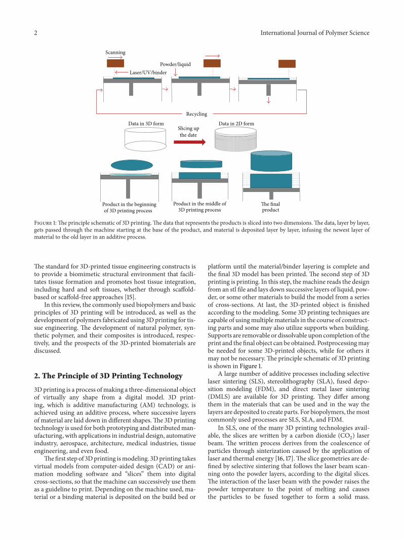
Figure 1:The principle schematic of 3D printing.The data that represents the products is sliced into two dimensions.The data, layer by layer,gets passed through the machine starting at the base of the product, and material is deposited layer by layer, infusing the newest layer ofmaterial to the old layer in an additive process.
The standard for 3D-printed tissue engineering constructs isto provide a biomimetic structural environment that facili-tates tissue formation and promotes host tissue integration,including hard and soft tissues, whether through scaffold-based or scaffold-free approaches [15].
In this review, the commonly used biopolymers and basicprinciples of 3D printing will be introduced, as well as thedevelopment of polymers fabricated using 3D printing for tis-sue engineering. The development of natural polymer, syn-thetic polymer, and their composites is introduced, respec-tively, and the prospects of the 3D-printed biomaterials arediscussed.
2. The Principle of 3D Printing Technology
3D printing is a process ofmaking a three-dimensional objectof virtually any shape from a digital model. 3D print-ing, which is additive manufacturing (AM) technology, isachieved using an additive process, where successive layersof material are laid down in different shapes.The 3D printingtechnology is used for both prototyping and distributedman-ufacturing, with applications in industrial design, automativeindustry, aerospace, architecture, medical industries, tissueengineering, and even food.
Thefirst step of 3Dprinting ismodeling. 3Dprinting takesvirtual models from computer-aided design (CAD) or ani-mation modeling software and “slices” them into digitalcross-sections, so that the machine can successively use themas a guideline to print. Depending on the machine used, ma-terial or a binding material is deposited on the build bed or
platform until the material/binder layering is complete andthe final 3D model has been printed. The second step of 3Dprinting is printing. In this step, themachine reads the designfrom an stl file and lays down successive layers of liquid, pow-der, or some other materials to build the model from a seriesof cross-sections. At last, the 3D-printed object is finishedaccording to the modeling. Some 3D printing techniques arecapable of usingmultiplematerials in the course of construct-ing parts and some may also utilize supports when building.Supports are removable or dissolvable upon completion of theprint and the final object can be obtained. Postprocessingmaybe needed for some 3D-printed objects, while for others itmay not be necessary.The principle schematic of 3D printingis shown in Figure 1.
A large number of additive processes including selectivelaser sintering (SLS), stereolithography (SLA), fused depo-sition modeling (FDM), and direct metal laser sintering(DMLS) are available for 3D printing. They differ amongthem in the materials that can be used and in the way thelayers are deposited to create parts. For biopolymers, themostcommonly used processes are SLS, SLA, and FDM.
In SLS, one of the many 3D printing technologies avail-able, the slices are written by a carbon dioxide (CO
2) laser
beam. The written process derives from the coalescence ofparticles through sinterization caused by the application oflaser and thermal energy [16, 17].The slice geometries are de-fined by selective sintering that follows the laser beam scan-ning onto the powder layers, according to the digital slices.The interaction of the laser beam with the powder raises thepowder temperature to the point of melting and causesthe particles to be fused together to form a solid mass.
International Journal of Polymer Science 3
The sintered material forms the object and the powder sup-ports it during its manufacturing [18]. In the final stage ofthe SLS process the object is removed from the powder envi-ronment and it is cleaned from the remaining powder at-tached to its walls [17].
In SLA, thin successive layers are photocrosslinked byultraviolet or visible light that induces photopolymerizationof a reactive system according to a sliced CAD model. Thetechnique requires a liquid photocrosslinkable resin withdefined viscosity properties and, for that reason, reactive ornonreactive diluents are typically used. SLA can generate alarge number of widely differing 3D structures in a repro-ducible waywith precise control over the finalmicrostructureand geometry [19]. In addition, nonlinear scaffold geometriescan be produced.
In FDM, like other RP methods, one layer at a time isprinted but typically the material is directly deposited on asurface where it is desired. Extrusion through a nozzle resultsin a cylindrical coiled morphology of each layer [20]. FDMuses a small temperature-controlled extruder to force out athermoplastic filament material and deposit the semimoltenpolymer onto a platform in a layer by layer process. Themonofilament is moved by two rollers and acts as a piston todrive the semimolten extrudate. At the end of each finishedlayer, the base platform is lowered and the next layer isdeposited.
3. Materials Fabricated by3D Printing for Tissue Engineering
In the search for alternatives to conventional treatment strate-gies for the repair or replacement of missing or mal-functioning human tissues and organs, promising solutionshave been explored through tissue engineering approaches.Biomaterials-based scaffolds have played a pivotal role in thisquest. At present, biomaterials have been widely used in skin[21, 22], cartilage [23], bone [24, 25], tendons [26], vessels[27, 28], nerves [29], bladder [30], and liver [31] tissue engi-neering.When designing a polymeric scaffold, a combinationof biological and engineering requisites is considered withinan application-specific manner. Material selection for tissueengineering applications is based on several important factorsincluding biocompatibility, degradability, surface characteris-tics, processability, and mechanical properties [32].
Both natural and synthetic polymers have been used forbiomedical applications. Natural polymers, such as collagen[33], chondroitin sulphate [34, 35], chitin [36, 37], and chi-tosan [38, 39], are widely used for tissue engineering andorgan regeneration, since they facilitate cell attachment andmaintenance of differentiation. Synthetic polymers such aspoly(𝜀-caprolactone) (PCL), poly(lactic acid) (PLA), poly(glycolic acid) (PGA), poly(lactic-co-glycolic acid) (PLGA),and other synthetic polymers can provide extreme versatilityregarding the control of their physicochemical properties andare generally easy to process into tissue engineering scaffolds[40–42]. Synthesis of these polymers can be tailored to yielda specific molecular weight, chemical structure, end groupchemistry, and composition (homopolymers, copolymers,
and polymer blends) in terms of tissue response. Further-more, the biodegradation time of synthetic polymers makesthem more attractive over natural ones [43].
Among the polymers used in tissue engineering, poly(𝛼-hydroxy esters) (such as PLA, PGA, and PLGA) haveattracted extensive attention for a variety of biomedicalapplications. Besides, PCL has been widely utilized as a tissueengineering scaffold. Blends and block copolymers of PCLwith other poly(𝛼-hydroxy esters) (such as poly(L-lactic acid-co-𝜀-caprolactone) (PLLACL) or poly(D,L-lactic acid-co-𝜀-caprolactone) (PDLLACL)) have been used to produce pol-ymers with tailored properties [44–46]. In order to increasewettability, biocompatibility, or softness of bioresorbable pol-ymers, blends and copolymers with nondegradable poly(ethylene glycol) (PEG) have been developed, such as blockcopolymers of PEG with poly(L-lactide) (PLLA), PLGA, andPCL [46, 47].
3.1. Natural Polymer and Its Composites Fabricated by 3DPrinting Technology. Gelatin, the main component of hy-drolyzed collagen, has been given much attention due to itsnatural origins in the extracellular matrix (ECM) and its abil-ity to suspend cells in a gel at low temperatures [15]. Yan andothers manually printed a liver tissue construct made of gel-atin and chitosan mixed with hepatocytes followed by glu-taraldehyde fixation [15, 48].
Research that explores natural polymers (such as starch)with water-based binders for use in direct 3D printing meth-ods has shown promising results and can be combined withsynthetic polymers for the desired biodegradable andmechanical properties. Starch-based polymers allow for in-creased degradation time and consequently expanded poros-ity as cellular integration increases, which is optimal for bonetissue engineering. A unique blend of starch-based polymerpowders (cornstarch, dextran, and gelatin) was developed forthe 3DP process by Lam et al. [49]. The scaffold propertieswere characterized by scanning electron microscopy (SEM),differential scanning calorimetry (DSC), porosity analysis,and compression tests. The analysis and tests demonstratedthat porous 3D scaffolds created by a new blend of materialsthrough 3DP were achievable. Starch and cellulose compos-ites were shown to be biocompatible, which has been utilizedin polymers designed for controlled drug delivery [50, 51] andbone cement [52, 53]. Salmoria et al. present the rapid fabrica-tion of starch-cellulose and cellulose acetate scaffolds by SLSand the evaluation of the laser power, laser scan speed, and thepolymer particle size influence on the scaffold properties [54].The specimens with small particle sizes presented a higherdegree of sintering and a significant level of closed pores, asindicated by the density measurements and mechanical tests,while the mechanical properties of specimens prepared withlarger particles presented lower values of elastic modulus andtensile strength because of the low degree of sintering andlimited number of unions. The results obtained showed thatit is possible to fabricate biopolymer scaffold structures usingstarch-cellulose and cellulose acetate using SLS by processoptimization based on the adjustment of laser power and scanspeed. Specimens prepared with small particle size exhibit
4 International Journal of Polymer Science
satisfactory mechanical properties and level of porosity forthe design and fabrication of scaffolds with potential use intissue engineering and drug delivery [54].
The application of 3D printing in tissue engineering hasenabled new methods for the printing of cells and matrixmaterials to fabricate tissue-analogous structures [55–57].The practicality of using 3D printing to fabricate cell-ladenconstructs was demonstrated, where cells were localized asintended and the cell viability of the fabricated constructs washigh.
Gelatin methacrylate was used to fabricate via the pro-posed projection stereolithography (PSL) platform, whichcan be used to design intricate 3D structure that can be engi-neered to mimic the microarchitecture of tissues, based onCAD [58]. Variation of the structure and prepolymer con-centration enabled tailoring the mechanical properties of thescaffolds. A dynamic cell seeding method was utilized toimprove the coverage of the scaffold throughout its thickness.The results demonstrated that the interconnectivity of poresallowed for uniform human umbilical vein endothelial cells(HUVECs) distribution and proliferation in the scaffolds,leading to high cell density and confluence at the end of theculture period. Moreover, immunohistochemistry resultsshowed that cells seeded on the scaffold maintained theirendothelial phenotype, demonstrating the biological func-tionality of the microfabricated GelMA scaffolds.
In 2014 Billiet et al. [59] reported the 3D printing ofgelatin methacrylamide cell-laden tissue-engineered con-structs with high cell viability for liver tissue engineering.They used VA-086 as a photoinitiator with enhanced bio-compatibility compared to the conventional Irgacure 2959. Aparametric study on the printing of gelatins was performedin order to characterize and compare construct architectures.The parameters including hydrogel building block concen-tration, the printing temperature, the printing pressure, theprinting speed, and the cell density were analyzed in depthand optimized. The scaffolds can be designed having a 100%interconnected pore network in the gelatin concentrationrange of 10–20w/v%. Control over the deposited stranddimensions can be guaranteed due to the physical propertiesof gelatin methacrylamide hydrogels and machine operatingparameters. High viability (>97%) constructs displaying amaintained expression of liver specific functions were ob-tained using the VA-086 photoinitiator.
3.2. Synthetic Biopolymers andTheirComposites Fabricated by 3D Printing
3.2.1. Synthetic Biopolymers Fabricated by 3D Printing.Among several tissues that are being actively researched,bone is one of themost widely studied, due to its critical func-tions in everyday life. When bone experiences disease ortrauma, the defective portion often needs to be surgicallyremoved [60]. Bone is able to self-regenerate; however, regen-eration is limited to a distance of a few millimeters fromhealthy bone. Thereafter, a graft used to replace the removedportion of bone in order to restore functionality is required[61]. Autografting, which is the gold standard in filling bone
defects, has known disadvantages, such as donor site mor-bidity, greater scar formation, and increased surgery times,as well as limited donor sites [62]. To enhance bone regen-eration bone defects must be filled with an artificial porousspacer allowing the ingrowth of blood vessels and bone butrestricting soft tissue ingrowth. In general, it is agreed thatthe porous network should consist of interconnected poreswith a diameter in the range of 50–1000𝜇m [5]. Becauseof the obvious advantage of 3D printing, many researchersstudy the potential of 3D printing biopolymers for bone tissueengineering.
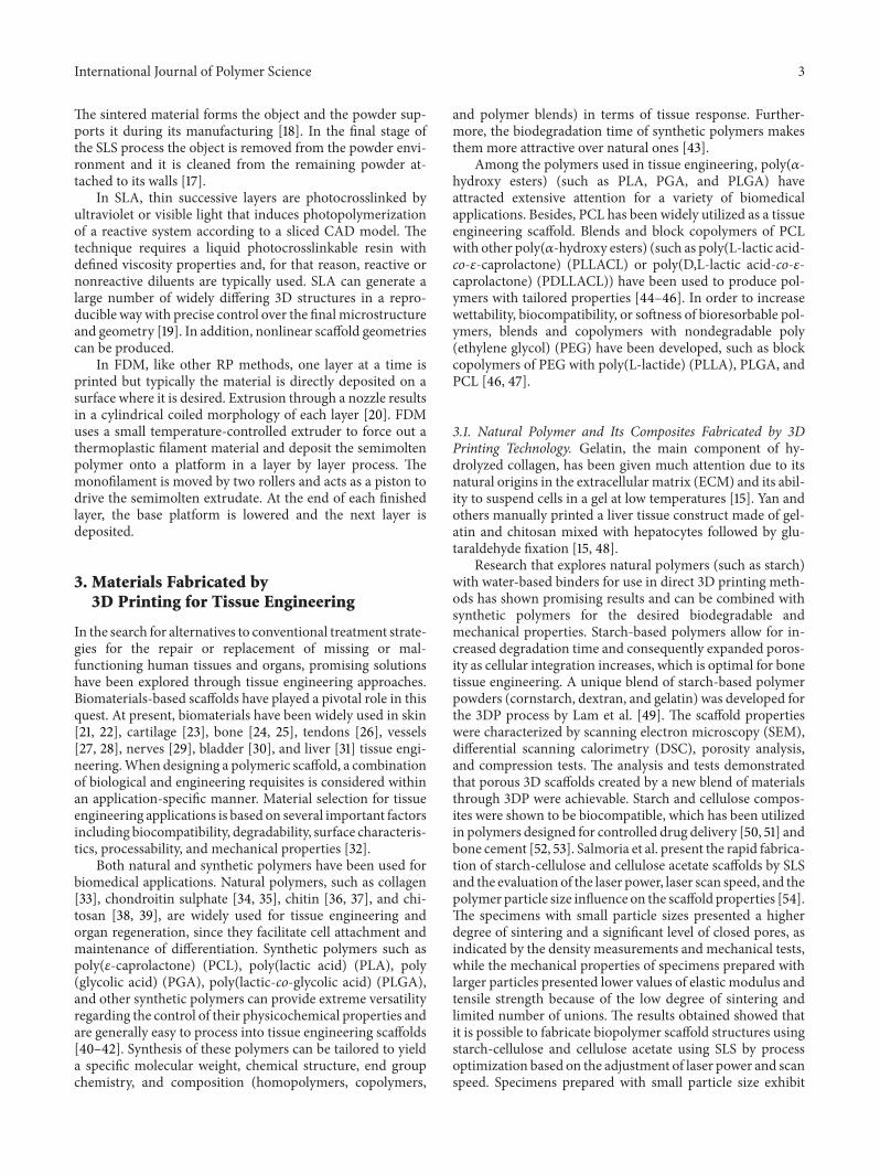
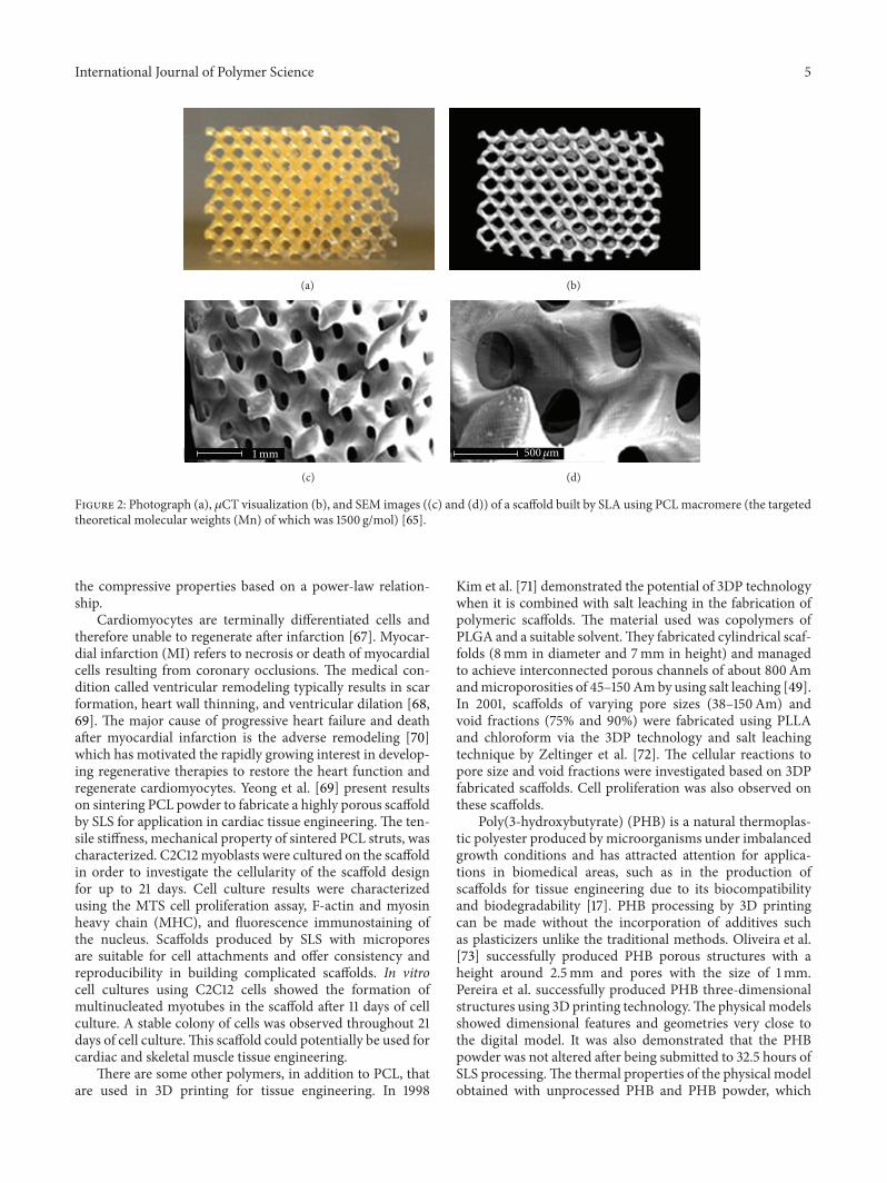
Themost commonly used polymer for 3D porous scaffoldis PCL, which, despite its good biocompatibility and process-ability, is rather hydrophobic leading to limited cell-scaffoldinteractions. Further, PCL is semicrystalline which togetherwith its hydrophobicity and low water absorbing capacity,resulted in very slow degradation kinetics, which is regardedas a soft and hard tissue compatible bioresorbable material[43, 63]. Sudarmadji et al. chose PCL to fabricate 3D porousscaffolds using SLS [64]. Mathematical relations correlatingscaffold porosity and compressive stiffness readings were for-mulated and compiled. In addition, cytotoxicity assessmentwas conducted to evaluate the toxicity of the fabricated PCLscaffolds [46]. The porosities, compressive stiffnesses, andyield strengths of the scaffolds varied in the ranges 40–84%,2.74–55.95MPa, and 0.17–5.03MPa, respectively. This rangeof stiffness closely matches that of cancellous bone in themaxillofacial region. Besides, the chosen mode of fabricationfor the PCL scaffolds has been proven to be feasible, as itis evident from the results of the cytotoxicity assessment.Elomaa et al. prepared and applied a photocrosslinkablePLC-based resin to build a porous scaffold using solvent-free SLA [65] (Figure 2). Photocrosslinkablemacromers wereprepared by methacrylating three-armed oligomers withmethacrylic anhydride. Photocrosslinked networks had ahigh gel content, which indicates a high degree of crosslink-ing. Since macromers were heated above the melting temper-ature to obtain the suitable viscosity, no solvent was needed.No material shrinkage was observed after extraction anddrying of the scaffolds due to the absence of solvent. Thescaffolds accurately represented the structure modeled bycomputer-aided design.The average porosity was 70.5±0.8%,and the average pore size was 465 𝜇m. The interconnectivityof the pores was high, indicating a great potential for thesestructures in cell seeding and implanting. PCLwas developedas a filament modeling material to produce porous scaffolds,made of layers of directionally aligned microfilaments, usingthis computer-controlled extrusion and deposition processby Zein et al. in 2002 [66]. The PCL scaffolds were producedwith a range of channel size 160–700mm, filament diameter260–370mm, and porosity 48–77% and regular geometricalhoneycomb pores, depending on the processing parameters.The scaffolds of different porosity also exhibited a pattern ofcompressive stress-strain behavior characteristics of poroussolids under such loading. The compressive stiffness rangedfrom 4 to 77MPa, yield strength from 0.4 to 3.6MPa, andyield strain from 4% to 28%. Analysis of the measured datashows a high correlation between the scaffold porosity and
International Journal of Polymer Science 5
(a) (b)
1mm
(c)
500𝜇m
(d)
Figure 2: Photograph (a), 𝜇CT visualization (b), and SEM images ((c) and (d)) of a scaffold built by SLA using PCLmacromere (the targetedtheoretical molecular weights (Mn) of which was 1500 g/mol) [65].
the compressive properties based on a power-law relation-ship.
Cardiomyocytes are terminally differentiated cells andtherefore unable to regenerate after infarction [67]. Myocar-dial infarction (MI) refers to necrosis or death of myocardialcells resulting from coronary occlusions. The medical con-dition called ventricular remodeling typically results in scarformation, heart wall thinning, and ventricular dilation [68,69]. The major cause of progressive heart failure and deathafter myocardial infarction is the adverse remodeling [70]which has motivated the rapidly growing interest in develop-ing regenerative therapies to restore the heart function andregenerate cardiomyocytes. Yeong et al. [69] present resultson sintering PCL powder to fabricate a highly porous scaffoldby SLS for application in cardiac tissue engineering. The ten-sile stiffness, mechanical property of sintered PCL struts, wascharacterized. C2C12myoblasts were cultured on the scaffoldin order to investigate the cellularity of the scaffold designfor up to 21 days. Cell culture results were characterizedusing the MTS cell proliferation assay, F-actin and myosinheavy chain (MHC), and fluorescence immunostaining ofthe nucleus. Scaffolds produced by SLS with microporesare suitable for cell attachments and offer consistency andreproducibility in building complicated scaffolds. In vitrocell cultures using C2C12 cells showed the formation ofmultinucleated myotubes in the scaffold after 11 days of cellculture. A stable colony of cells was observed throughout 21days of cell culture.This scaffold could potentially be used forcardiac and skeletal muscle tissue engineering.
There are some other polymers, in addition to PCL, thatare used in 3D printing for tissue engineering. In 1998
Kim et al. [71] demonstrated the potential of 3DP technologywhen it is combined with salt leaching in the fabrication ofpolymeric scaffolds. The material used was copolymers ofPLGA and a suitable solvent.They fabricated cylindrical scaf-folds (8mm in diameter and 7mm in height) and managedto achieve interconnected porous channels of about 800Amandmicroporosities of 45–150Amby using salt leaching [49].In 2001, scaffolds of varying pore sizes (38–150Am) andvoid fractions (75% and 90%) were fabricated using PLLAand chloroform via the 3DP technology and salt leachingtechnique by Zeltinger et al. [72]. The cellular reactions topore size and void fractions were investigated based on 3DPfabricated scaffolds. Cell proliferation was also observed onthese scaffolds.
Poly(3-hydroxybutyrate) (PHB) is a natural thermoplas-tic polyester produced by microorganisms under imbalancedgrowth conditions and has attracted attention for applica-tions in biomedical areas, such as in the production ofscaffolds for tissue engineering due to its biocompatibilityand biodegradability [17]. PHB processing by 3D printingcan be made without the incorporation of additives suchas plasticizers unlike the traditional methods. Oliveira et al.[73] successfully produced PHB porous structures with aheight around 2.5mm and pores with the size of 1mm.Pereira et al. successfully produced PHB three-dimensionalstructures using 3D printing technology.The physical modelsshowed dimensional features and geometries very close tothe digital model. It was also demonstrated that the PHBpowder was not altered after being submitted to 32.5 hours ofSLS processing.The thermal properties of the physical modelobtained with unprocessed PHB and PHB powder, which
6 International Journal of Polymer Science
underwent printing sets, did not show a significant differencebetween them.This corroborated the powder analysis, whichindicated that the reuse of remaining material from a 32.15hours-SLS did not affect the reproducibility of the process[17].
Recently, a functionalized aliphatic polyester based onPCL, namely, poly(hydroxymethylglycolide-co-𝜀-caprolac-tone) (PHMGCL) which possesses significantly higher hy-drophilicity due to its hydroxyl groups attached to the back-bone, resulting in a significant increase in human mesenchy-mal stem cell adhesion, proliferation, and differentiation ascompared to PCL, was developed [74]. To evaluate the in vivobiodegradation and biocompatibility of 3D-printed scaffoldsbased on PHMGCL, which has enhanced hydrophilicity,increased degradation rate, and improved cell-material inter-actions as compared to its counterpart PCL, 3D scaffoldsbased on this polymer were prepared by means of fiberdeposition (melt plotting) [75].Thebiodegradation and tissuebiocompatibility of PHMGCL and PCL scaffolds after subcu-taneous implantation in Balb/cmice were investigated.The invitro enzymatic degradation of these scaffolds was also inves-tigated in lipase solutions. It was shown that PHMGCL 3Dscaffolds lost more than 60% of their weight within 3 monthsof implantation while PCL scaffolds showed no weight lossin this time frame. The molecular weight (Mw) of PHMGCLdecreased significantly after 3 months of implantation, whilethemolecular weight of PCLwas unchanged in this period. Invitro enzymatic degradation showed that PHMGCL scaffoldswere degraded within 50 h, while the degradation time forPCL scaffolds of similar structure was longer. A normalforeign body response to both scaffold types characterized bythe presence of macrophages, lymphocytes, and fibrosis wasobserved with a more rapid onset in PHMGCL scaffolds.Theextent of tissue-scaffold interactions as well as vascularizationwas shown to be higher for PHMGCL scaffolds comparedto PCL ones. Therefore, the fast degradable PHMGCL whichshowed good biocompatibility is a promising biomaterial forbone and cartilage tissue engineering [75].
3.2.2. Synthetic Biopolymer-Based Composites Fabricated by3D Printing. Griffith and others bound a mixed biodegrad-able polymer powder of 25% PLLA and 75% PLGA usingchloroform as a binder to print a branched liver constructwith internal architecture [76].This design took host implan-tation into consideration by creating an artery- and vein-likeinlet and outlet [15]. Organogenesis of liver tissue using 3D-printed PLLA/PLGA scaffolds has been investigated in vitro[14]. It was shown that culturing a mixture of hepatocytesand endothelial cells on a channeled biodegradable scaffoldresults in the desired tissue structure intrinsically.
Pati et al. [77] successfully printed 3D cell-laden con-structs using the principle of hybrid structure fabrication,using PEG and PCL as materials. They printed an ear-shapedconstruct with a complex porous structure using the processdescribed above. The use of sacrificial layer technologyallowed the stacking of complicated structures regardless ofgeometrical shape. To fabricate the ear-shaped construct, aCADmodel was generated from a computerized tomography
(CT) scanned image of an ear; both the main framework andsacrificial parts were designed. A code generation process wasthen carried out via amotion programgenerator developed inhouse. In particular, two different hydrogels were printed intothe framework, because native human ears consist of bothcartilage and fat tissues.
Two photocrosslinkable hydrogel biopolymers, poly(ethylene glycol) dimethacrylate (PEG-DMA,MW 1000) andpoly(ethylene glycol) diacrylate (PEG-DA, MW 3400), wereused as the primary scaffold materials to prepare multimate-rials [78]. Multimaterial scaffolds were fabricated by includ-ing controlled concentrations of fluorescently labeled dex-tran, fluorescently labeled bioactive PEG, or bioactive PEGin different regions of the scaffold. The presence of the flu-orescent component in specific regions of the scaffold wasanalyzed with fluorescent microscopy, while human dermalfibroblast cells were seeded on top of the fabricated scaffoldswith selective bioactivity and phase contrast microscopyimages were used to show specific localization of cells in theregions patterned with bioactive PEG. Multimaterial spatialcontrol was successfully demonstrated. In addition, the equi-librium swelling behavior of the two biopolymers after SLfabrication was determined and used to design constructswith the specified dimensions at the swollen state. The useof multimaterial SL and the relative ease of conjugatingdifferent bioactive ligands or growth factors to PEG allow forthe fabrication of tailored three-dimensional constructs withspecified spatially controlled bioactivity.
The majority of current protocols utilize water-insolublephotoinitiators that are incompatiblewith live cell-fabricationand ultraviolet (UV) light that is damaging to the cellularDNA. Various studies have reported the use of water-solubledimethacrylatedpoly(ethyleneglycol)(PEG-DMA) to createstructured, cell-containing hydrogels by stereolithography[79]. Dhariwala et al. were the first to successfully encapsulate(Chinese hamster ovary) cells in PEG-DMA hydrogels, usingSLA [80]. Later, PEG-DMA constructs containing encap-sulated human dermal fibroblasts [36] and PEG-diacrylategel structures containing marrow stromal cells [81] werereported. In these cases, large numbers of cells could be cap-sulated at high densities (several millions of cells per mL)[82]. Lin et al. [79] reported the development of a visible light-based PSL system (VL-PSL), using lithium phenyl-2,4,6-trimethylbenzoylphosphinate (LAP) as the initiator and pol-yethylene glycol diacrylate (PEGDA) as the monomer, toproduce hydrogel scaffolds with specific shapes and internalarchitectures. Furthermore, live human adipose-derived stemcells (hADSCs) were suspended in PEGDA/LAP solutionduring the PSL process and were successfully incorpo-rated within the fabricated hydrogel scaffolds. hADSCs inPEG scaffolds showed high viability (>90%) for up to 7days after fabrication as revealed by live/dead staining.Scaffolds with porous internal architecture retained highercell viability and activity than solid scaffolds, likely becauseof the increased oxygen and nutrients exchange into theinterior of the scaffolds. The VLPSL can be applicableas an efficient and effective tissue engineering technol-ogy for point-of-care tissue repair in the clinic. Three-dimensional biodegradable PEG/PDLLA hydrogel structures
International Journal of Polymer Science 7
were prepared by Seck et al. using SLA at high reso-lutions [83]. The porous hydrogel structures had a well-defined pore network architecture, with a narrow pore sizedistribution and high interconnectivity of the pores.The resinand the built structures were compatible with cells. Uponseeding, human mesenchymal stem cells attached to thesurfaces of the hydrogel structures and showed a spreadmor-phology. After five days in culture, proliferation of the cellswas observed. These hydrogel structures could therefore beused in tissue engineering, drug delivery, cell transplantation,and other biomedical applications.
ABS is not widely used in medical devices in comparisonto materials such as PCL and PLA which offer greater nativebiocompatibility [20]. Applications involving biological sys-tems require materials that can minimize protein and anyother biomolecule adhesion during flow. It is therefore ofgreat interest to chemically modify the surface of ABS toengineer hydrophilicity and enable biocompatibility. Surfacemodification, specifically by the grafting of PEG, has longbeen shown to be a promising strategy to increase the bio-compatibility ofmaterials [20, 84].McCullough andYadavalli[20] examined the use of ABS as a core material for the con-struction of microdevices. A method to fabricate water-tightmicrofluidic devices using chemical dissolution via acetoneis shown to render a porous FDM ABS device imperviousto water flow between layers, while preserving the structuralfidelity of printed microstructures down to 250𝜇m. A strat-egy is then presented that can enable the formation of a stable,biocompatible surface of ABS by the photoinduced graftingof PEG groups that improves the biocompatibility of the ABSby reducing the biofouling behavior. Surface characterizationand protein adhesion studies are presented that demonstratethat thismodifiedABS represents a versatilematerial that canbe used in fused deposition modeling to form microfluidicchannels resistant to biofouling, thereby broadening therange of possible uses for ABS based FDM in microdeviceand lab-on-a-chip type applications. The grafting caused thecontact angle of the surface to be lowered from 77.58∘ fornative ABS samples to below 40∘ for ABS-g-PEG and reducedthe adhesion of the protein BSA. The results clearly indicatethat sealing an FDM ABS surface with an acetone treatmentcan be achieved with minimal effect on the device and thegrafting of PEGMA onto ABS is a viable method to increasesurface hydrophilicity and biocompatibility.
Currently, composites of polymers and bioactive inor-ganic materials are being developed with the aim of increas-ing the mechanical scaffold stability and improving tis-sue interaction. By combining biodegradable polymers andbioactive ceramics, such as hydroxyapatite (HA) and trical-cium phosphate (TCP), calcium phosphate (CaP) compositescaffolds were made by 3D printing. Nanosized osteoconduc-tive calcium phosphates (Ca-Ps), including HA, tricalciumphosphate (TCP), and substituted HA and TCP, have gainedmuch recognition in biomaterials development due to theirsmall size, high surface area to volume ratio, and biomimeticsimilarity to natural bone structure when combined withbiopolymers such as collagen, PLLA, and chitosan [17, 85, 86].
Hydroxyapatite (Ca10(PO4)
6(OH)2) has attracted much
interest due to its chemical similarity to the calcium phos-phate mineral present in biological hard tissues [87, 88]. HAhas been used for a variety of biomedical applications, suchas a matrix for controlled drug release and a carrier materialin bone tissue engineering [89]. Recently, nanosized hydrox-yapatite (nHA) has been highlighted due to its advantageousfeatures over conventionalmicrosizedmaterials. nHAhas thepotential to act as a carrier of therapeutic agents, enablingcontrolled drug release extracellularly or intracellularly, andat the same time it has high absorbability in the body for theregeneration of hard tissue [90, 91].
PCL/HA composite has attracted a great interest for thebone tissue engineering application. Wiria et al. researchedthe use of biocomposite materials, consisting of PCL andHA,to fabricate tissue engineering scaffolds via the SLS technol-ogy in 2007 [92]. Simulated body fluid (SBF) samples showthe formation of hydroxy carbonate apatite, as a result ofsoaking HA in a SBF environment. Cell culture experimentsshowed that Saos-2 cells were able to live and replicate on thefabricated scaffolds. The results show the favorable potentialof PCL/HA biocomposites as tissue engineering scaffolds thatare fabricated via SLS. Eosoly et al. [93] studied PCL/HAcomposite scaffolds in the same year. In their study HA andPCL, which were considered suitable for hard tissue engi-neering purposes, were used in a weight ratio of 30 : 70. Fourparameters, namely, laser fill power, outline laser power, scanspacing, and part orientation, were investigated according toa central composite design. A model of the effects of theseparameters on the accuracy and mechanical properties of thefabricated parts was developed. The compressive modulusand yield strength of the fabricated microstructures with adesigned relative density of 0.33 varied between 0.6 and 2.3,0.1 and 0.6MPa, respectively. The mechanical behavior wasstrongly dependent on themanufacturing direction. Eshraghiand Das [94] studied the experimental characterization ofthe compressive mechanical properties of PCL-HA com-posite scaffolds prepared by SLS technology for bone tissueengineering. In their study, they further establish the abilityof SLS to manufacture PCL-HA composite scaffolds withnear-full density in designed solid regions for bone tissueengineering.Themechanical properties of the PCL-HA com-posites showed improvement over that of pure PCL.They alsodemonstrate that the mechanical properties of these scaffoldscan be predicted before manufacturing with high accuracy.A direct extension of being able to predict the mechan-ical properties of composite materials at any filler load-ing in combination with a direct fabrication method with thecapability to produce complex anatomic parts is the abilityto custom design both the material properties and the an-atomical shape of tissue-engineered constructs for bothpatient and site-specific recovery strategies.
A poly(D,L-lactide)/nanosized hydroxyapatite (PDLLA/nHA) composite resin was prepared and used to fabricatecomposite films and computer designed porous scaffolds byRonca et al. [95] using SLA,mixing varying quantities of nHApowder and a liquid photoinitiator into a photocrosslink-able PDLLA-diacrylate resin. The influence of nHA on therheological and photochemical properties of the resins was
8 International Journal of Polymer Science
(a) (b)
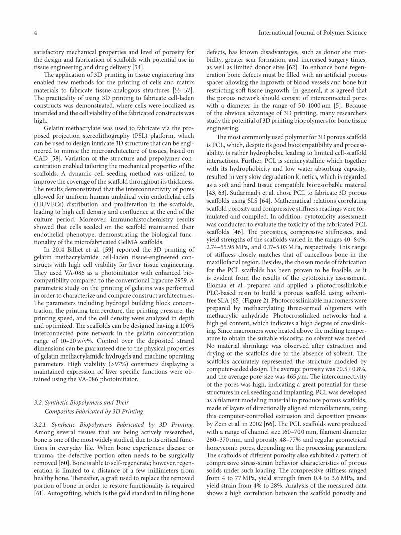
Figure 3:Three dimensionally interconnected controlled porosity PP-TCP composite scaffoldswith different internal architecture using FDMprocess [101].
investigated, with the materials being characterized withrespect to their mechanical, thermal, and morphologicalproperties after postpreparation curing. In the cured com-posites stiffness was observed to increase with increasingconcentration of nanoparticles. With increasing ceramiccomponent the resins became more viscous, and NMP wasadded as a nonreactive diluent to decrease the viscosity andallow processing by stereolithography. SEM images showedexposed ceramic particles on the pore surface, allowinginteraction between the bone-forming nHA and cells.
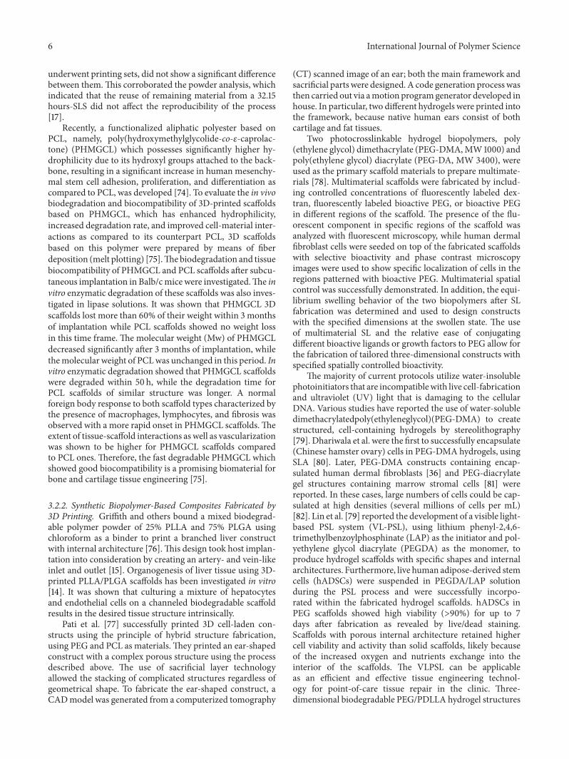
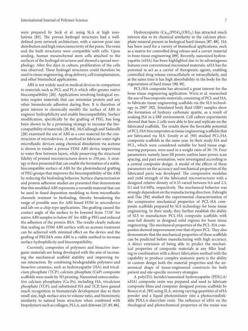
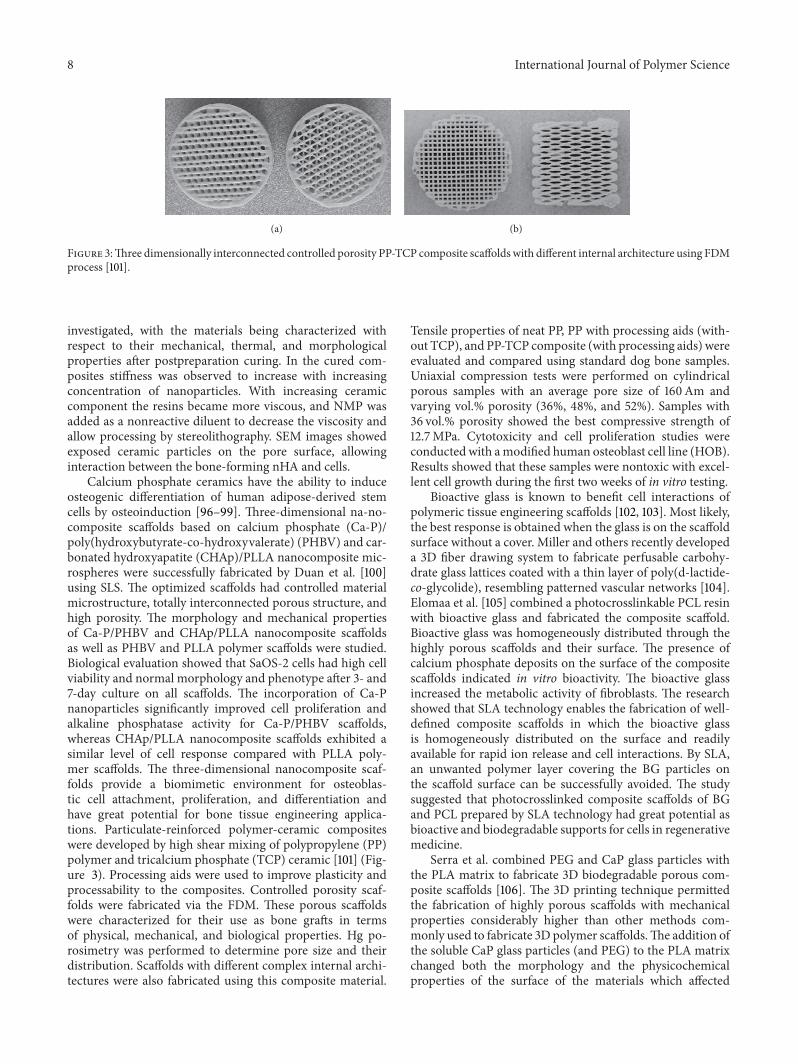
Calcium phosphate ceramics have the ability to induceosteogenic differentiation of human adipose-derived stemcells by osteoinduction [96–99]. Three-dimensional na-no-composite scaffolds based on calcium phosphate (Ca-P)/poly(hydroxybutyrate-co-hydroxyvalerate) (PHBV) and car-bonated hydroxyapatite (CHAp)/PLLA nanocomposite mic-rospheres were successfully fabricated by Duan et al. [100]using SLS. The optimized scaffolds had controlled materialmicrostructure, totally interconnected porous structure, andhigh porosity. The morphology and mechanical propertiesof Ca-P/PHBV and CHAp/PLLA nanocomposite scaffoldsas well as PHBV and PLLA polymer scaffolds were studied.Biological evaluation showed that SaOS-2 cells had high cellviability and normal morphology and phenotype after 3- and7-day culture on all scaffolds. The incorporation of Ca-Pnanoparticles significantly improved cell proliferation andalkaline phosphatase activity for Ca-P/PHBV scaffolds,whereas CHAp/PLLA nanocomposite scaffolds exhibited asimilar level of cell response compared with PLLA poly-mer scaffolds. The three-dimensional nanocomposite scaf-folds provide a biomimetic environment for osteoblas-tic cell attachment, proliferation, and differentiation andhave great potential for bone tissue engineering applica-tions. Particulate-reinforced polymer-ceramic compositeswere developed by high shear mixing of polypropylene (PP)polymer and tricalcium phosphate (TCP) ceramic [101] (Fig-ure 3). Processing aids were used to improve plasticity andprocessability to the composites. Controlled porosity scaf-folds were fabricated via the FDM. These porous scaffoldswere characterized for their use as bone grafts in termsof physical, mechanical, and biological properties. Hg po-rosimetry was performed to determine pore size and theirdistribution. Scaffolds with different complex internal archi-tectures were also fabricated using this composite material.
Tensile properties of neat PP, PP with processing aids (with-out TCP), and PP-TCP composite (with processing aids) wereevaluated and compared using standard dog bone samples.Uniaxial compression tests were performed on cylindricalporous samples with an average pore size of 160Am andvarying vol.% porosity (36%, 48%, and 52%). Samples with36 vol.% porosity showed the best compressive strength of12.7MPa. Cytotoxicity and cell proliferation studies wereconducted with amodified human osteoblast cell line (HOB).Results showed that these samples were nontoxic with excel-lent cell growth during the first two weeks of in vitro testing.
Bioactive glass is known to benefit cell interactions ofpolymeric tissue engineering scaffolds [102, 103]. Most likely,the best response is obtained when the glass is on the scaffoldsurface without a cover. Miller and others recently developeda 3D fiber drawing system to fabricate perfusable carbohy-drate glass lattices coated with a thin layer of poly(d-lactide-co-glycolide), resembling patterned vascular networks [104].Elomaa et al. [105] combined a photocrosslinkable PCL resinwith bioactive glass and fabricated the composite scaffold.Bioactive glass was homogeneously distributed through thehighly porous scaffolds and their surface. The presence ofcalcium phosphate deposits on the surface of the compositescaffolds indicated in vitro bioactivity. The bioactive glassincreased the metabolic activity of fibroblasts. The researchshowed that SLA technology enables the fabrication of well-defined composite scaffolds in which the bioactive glassis homogeneously distributed on the surface and readilyavailable for rapid ion release and cell interactions. By SLA,an unwanted polymer layer covering the BG particles onthe scaffold surface can be successfully avoided. The studysuggested that photocrosslinked composite scaffolds of BGand PCL prepared by SLA technology had great potential asbioactive and biodegradable supports for cells in regenerativemedicine.
Serra et al. combined PEG and CaP glass particles withthe PLA matrix to fabricate 3D biodegradable porous com-posite scaffolds [106]. The 3D printing technique permittedthe fabrication of highly porous scaffolds with mechanicalproperties considerably higher than other methods com-monly used to fabricate 3D polymer scaffolds.The addition ofthe soluble CaP glass particles (and PEG) to the PLA matrixchanged both the morphology and the physicochemicalproperties of the surface of the materials which affected
International Journal of Polymer Science 9
cell behavior. Surface properties were also assessed, showingthat the incorporation of glass particles increased both theroughness and the hydrophilicity of the scaffolds. Mechanicaltests indicated that compression strength is dependent onthe scaffold geometry and the presence of glass. Prelimi-nary cell response was studied with primary mesenchymalstem cells (MSC) and revealed that CaP glass improvedcell adhesion. Overall, the results showed the suitability ofthe technique/materials combination to develop 3D porousscaffolds and their initial biocompatibility, with both beingvaluable characteristics for tissue engineering applications.
4. Prospects and Conclusions
By the year of 2014, 3D printing technology has been studiedby biotechnology firms and academia for possible use in tis-sue engineering applications in which organs and body partsare built using inkjet techniques. One of the main advantagesof 3D printing is that it allows the manufacturing of objectshaving complex geometries and intricate internal structure,which can be designed according to the needs of individualpatients using their 3D medical scan data. A great prospectfor biomedical applications, especially for tissue engineeringapplications, has been shown. Both natural and syntheticpolymers have been developed for tissue engineering via the3D printing technology, and numerous additional materialsare being developed. Fibers and particles have been combinedwith polymers to fabricate materials with better bioactivityand biocompatibility, as well as physical and chemical prop-erties.
For a wider application of 3D printing, themoral problemrelated to the 3D-printed organ for medical applicationshould be further studied, the cost of 3D printers should beless, and more materials systems which are available for 3Dprinting should be developed. A further study on the mech-anism of cell attachment, differentiation, and growth withinthe 3D-printed materials should be carried on as well.
With the development of nanotechnology, materials suchas nanotubes [107–110], nano-structured particles [111], na-nofibers [112, 113], and other nanosized materials [114–116]have been fabricated.Nanomaterials have specialmechanical,electrical, magnetic, optical, chemical, and other biologicalproperties due to their high aspect ratio and surface area [117–119]. Many nanomaterial surfaces exemplify high (bio- andcyto-) compatibility, by promoting protein adsorption andenhancing subsequent cellular adhesion and tissue growthmore than on conventional flat implant surfaces such astitanium, ceramics, and biopolymers [114]. Nanocompositesattract more attention because of their potential combinationof properties from both the nanomaterials and the hostmaterials matrix [108, 120–122]. It is promising to incorporatenanomaterials in 3D printing. The nanomaterials can beintroduced into 3D printing in the following way: (1) premix-ing the nanomaterials into the host matrix before 3D printingand (2) introducing the nanomaterials at the intermittentstoppages of the 3D-printed host matrix [123]. All in all, 3Dprinting will help to expand the application of nanomaterialsfor tissue engineering.
Conflict of Interests
The authors declare that there is no conflict of interestsregarding the publication of this paper.
Acknowledgments
The authors acknowledge the financial support from theNational Basic Research Program of China (973 Program,no. 2011CB710901), the National Natural Science Founda-tion of China (no. 31370959), Fok Ying Tong EducationFoundation (no. 141039), Beijing Natural Science Foundation(no. 7142094), the Beijing Nova Program (no. 2010B011),Program for New Century Excellent Talents (NCET) inUniversity from Ministry of Education of China, State KeyLaboratory of New Ceramic and Fine Processing (TsinghuaUniversity), International Joint ResearchCenter of AerospaceBiotechnology andMedical Engineering, Ministry of Scienceand Technology of China, and the 111 Project (no. B13003).
References
[1] X. M. Li, Y. Huang, L. S. Zheng et al., “Effect of substratestiffness on the functions of rat bonemarrow and adipose tissuederived mesenchymal stem cells in vitro,” Journal of BiomedicalMaterials Research A, vol. 102A, pp. 1092–1101, 2014.
[2] X. Li, Y. Fan, and F. Watari, “Current investigations into carbonnanotubes for biomedical application,” Biomedical Materials,vol. 5, no. 2, Article ID 022001, 2010.
[3] S. P. Nukavarapu and D. L. Dorcemus, “Osteochondral tissueengineering: current strategies and challenges,” BiotechnologyAdvances, vol. 31, pp. 706–721, 2013.
[4] D. W. Hutmacher, “Scaffolds in tissue engineering bone andcartilage,” Biomaterials, vol. 21, no. 24, pp. 2529–2543, 2000.
[5] A. Butscher, M. Bohner, S. Hofmann, L. Gauckler, and R.Muller, “Structural and material approaches to bone tissueengineering in powder-based three-dimensional printing,”ActaBiomaterialia, vol. 7, no. 3, pp. 907–920, 2011.
[6] L. M. Mathieu, T. L. Mueller, P.-E. Bourban, D. P. Pioletti, R.Muller, and J.-A. E. Manson, “Architecture and properties ofanisotropic polymer composite scaffolds for bone tissue engi-neering,” Biomaterials, vol. 27, no. 6, pp. 905–916, 2006.
[7] M. Bohner, G. H. Van Lenthe, S. Grunenfelder, W. Hirsiger,R. Evison, and R. Muller, “Synthesis and characterization ofporous 𝛽-tricalcium phosphate blocks,” Biomaterials, vol. 26,no. 31, pp. 6099–6105, 2005.
[8] M. Stoppato, S. Vahabzadeh, and A. Bandyopadhyay, “Bone tis-sue engineering using 3D printing,” Bioactive and CompatiblePolymers, vol. 28, pp. 16–32, 2013.
[9] H. Cao and N. Kuboyama, “A biodegradable porous compositescaffold of PGA/𝛽-TCP for bone tissue engineering,” Bone, vol.46, no. 2, pp. 386–395, 2010.
[10] X. Li andQ. Feng, “Porous poly-L-lactic acid scaffold reinforcedby chitin fibers,” Polymer Bulletin, vol. 54, no. 1-2, pp. 47–55,2005.
[11] H. D. Kim, E. H. Bae, I. C. Kwon, R. R. Pal, J. D. Nam, and D. S.Lee, “Effect of PEG-PLLA diblock copolymer on macroporousPLLA scaffolds by thermally induced phase separation,” Bioma-terials, vol. 25, no. 12, pp. 2319–2329, 2004.
10 International Journal of Polymer Science
[12] C. P. Grey, S. T. Newton, G. L. Bowlin et al., “Gradient fiberelectrospinning of layered scaffolds using controlled transitionsin fiber diameter,” Biomaterials, vol. 34, pp. 4993–5006, 2013.
[13] Y. Jun Lee, J.-H. Lee, H.-J. Cho et al., “Electrospun fibers immo-bilized with bone forming peptide-1 derived from BMP7 forguided bone regeneration,”Biomaterials, vol. 34, pp. 5059–5069,2013.
[14] S. Bose, S.Vahabzadeh, andA. Bandyopadhyay, “Bone tissue en-gineering using 3D printing,”Materials Today, vol. 16, pp. 496–504, 2013.
[15] D. Jack Richards, Y. Tan, J. Jia et al., “3D printing for tissue engi-neering,” Israel Journal of Chemistry, vol. 53, pp. 805–814, 2013.
[16] A. Sachdeva, S. Singh, and V. S. Sharma, “Investigating surfaceroughness of parts produced by SLS process,” InternationalJournal of Advanced Manufacturing Technology, vol. 64, pp.1505–1516, 2012.
[17] T. F. Pereira, M. F. Oliveira, I. A. Maia et al., “3D printing ofpoly(3-hydroxybutyrate) porous structures using selective lasersintering,”Macromolecular Symposia, vol. 319, pp. 64–73, 2012.
[18] K. Senthilkumaran, P. M. Pandey, and P. V. M. Rao, “Influenceof building strategies on the accuracy of parts in selective lasersintering,” Materials and Design, vol. 30, no. 8, pp. 2946–2954,2009.
[19] B. Wendel, D. Rietzel, F. Kuhnlein, R. Feulner, G. Hulder, andE. Schmachtenberg, “Additive processing of polymers,”Macro-molecular Materials and Engineering, vol. 293, pp. 799–809,2008.
[20] E. J. McCullough and V. K. Yadavalli, “Surface modification offused deposition modeling ABS to enable rapid prototypingof biomedical microdevices,” Journal of Materials ProcessingTechnology, vol. 213, pp. 947–954, 2013.
[21] S. Gautam, C.-F. Chou, A. K. Dinda et al., “Surfacemodificationof nanofibrouspolycaprolactone/gelatin composite scaffold bycollagen type I grafting for skin tissue engineering,” MaterialsScience and Engineering C, vol. 34, pp. 402–409, 2014.
[22] F. Groeber, M. Holeiter, M. Hampel, S. Hinderer, and K.Schenke-Layland, “Skin tissue engineering—in vivo and in vitroapplications,”AdvancedDrugDelivery Reviews, vol. 63, no. 4, pp.352–366, 2011.
[23] W.-J. Li, R. Tuli, C. Okafor et al., “A three-dimensional nanofi-brous scaffold for cartilage tissue engineering using humanmesenchymal stem cells,” Biomaterials, vol. 26, no. 6, pp. 599–609, 2005.
[24] A. I. Rodrigues, M. E. Gomes, I. B. Leonor et al., “Bioactivestarch-based scaffolds and human adipose stem cells are a goodcombination for bone tissue engineering,” Acta Biomaterialia,vol. 8, pp. 3765–3776, 2012.
[25] X. Li, Q. Feng, W. Wang, and F. Cui, “Chemical characteristicsand cytocompatibility of collagen-based scaffold reinforced bychitin fibers for bone tissue engineering,” Journal of BiomedicalMaterials Research B: Applied Biomaterials, vol. 77, no. 2, pp.219–226, 2006.
[26] S. Sahoo, L.-T. Ang, J. Cho-Hong Goh, and S.-L. Toh, “Bioac-tive nanofibers for fibroblastic differentiation of mesenchymalprecursor cells for ligament/tendon tissue engineering applica-tions,” Differentiation, vol. 79, no. 2, pp. 102–110, 2010.
[27] J. Hu, X. Sun, H. Ma, C. Xie, Y. E. Chen, and P. X. Ma, “Porousnanofibrous PLLA scaffolds for vascular tissue engineering,”Biomaterials, vol. 31, no. 31, pp. 7971–7977, 2010.
[28] A. Hasan, A. Memic, N. Annabi et al., “Electrospun scaffoldsfor tissue engineering of vascular graft,” Acta Biomaterial, vol.10, pp. 11–25, 2014.
[29] L. Ghasemi-Mobarakeh, M. P. Prabhakaran, M. Morshed, M.-H. Nasr-Esfahani, and S. Ramakrishna, “Electrospun poly(𝜀-caprolactone)/gelatin nanofibrous scaffolds for nerve tissueengineering,” Biomaterials, vol. 29, no. 34, pp. 4532–4539, 2008.
[30] M. A. Pattison, S. Wurster, T. J. Webster, and K. M. Haberstroh,“Three-dimensional, nano-structured PLGA scaffolds for blad-der tissue replacement applications,” Biomaterials, vol. 26, no.15, pp. 2491–2500, 2005.
[31] F. Chen,M. Tian,D. Zhang et al., “Preparation and characteriza-tion of oxidized alginate covalently cross-linked galactosylatedchitosan scaffold for liver tissue engineering,”Materials Scienceand Engineering C, vol. 32, no. 2, pp. 310–320, 2012.
[32] B.-S. Kim, I.-K. Park, T. Hoshiba et al., “Design of artificialextracellular matrices for tissue engineering,” Progress in Poly-mer Science (Oxford), vol. 36, no. 2, pp. 238–268, 2011.
[33] X. Li, Q. Feng, X. Liu, W. Dong, and F. Cui, “Collagen-basedimplants reinforced by chitin fibres in a goat shank bone defectmodel,” Biomaterials, vol. 27, no. 9, pp. 1917–1923, 2006.
[34] M. C. Serrano, S. Nardecchia, C. Garcıa-Rama et al., “Chon-droitin sulphate-based 3D scaffolds containing MWCNTs fornervous tissue repair,” Biomaterials, vol. 35, pp. 1543–1551, 2014.
[35] L. Zhao, H.-J. Gwon, Y.-M. Lim et al., “Hyaluronic acid/chondroitin sulfate-based hydrogel prepared bygamma irradi-ation technique,” Carbohydrate Polymers, vol. 102, pp. 598–605,2014.
[36] X. Li, X. Liu, W. Dong et al., “In vitro evaluation of porouspoly(L-lactic acid) scaffold reinforced by chitin fibers,” Journalof Biomedical Materials Research B: Applied Biomaterials, vol.90, no. 2, pp. 503–509, 2009.
[37] H. Tamura, T. Furuike, S. V. Nair, and R. Jayakumar, “Biomed-ical applications of chitin hydrogel membranes and scaffolds,”Carbohydrate Polymers, vol. 84, no. 2, pp. 820–824, 2011.
[38] A. Islam, T. Yasin, and I. Ur Rehman, “Synthesis of hybridpolymer networks of irradiated chitosan/poly(vinyl acohol) forbiomedical applications,” Physics and Chemistry, vol. 96, pp.115–119, 2014.
[39] X. Li, Q. Feng, Y. Jiao, and F. Cui, “Collagen-based scaffoldsreinforced by chitosan fibres for bone tissue engineering,”Polymer International, vol. 54, no. 7, pp. 1034–1040, 2005.
[40] I. Sousa, A. Mendes, and P. J. Bartolo, “PCL scaffolds with col-lagen bioactivator for applications in Tissue Engineering,”Procedia Engineering, vol. 59, pp. 279–284, 2013.
[41] G. Chen, J. Tanaka, and T. Tateishi, “Osteochondral tissue engi-neering using a PLGA-collagen hybrid mesh,”Materials Scienceand Engineering C, vol. 26, no. 1, pp. 124–129, 2006.
[42] C. Zhang, N. Sangaj, Y. Hwang, A. Phadke, C.-W. Chang, and S.Varghese, “Oligo(trimethylene carbonate)-poly(ethylene glycol)-oligo(trimethylene carbonate) triblock-based hydrogels for car-tilage tissue engineering,” Acta Biomaterialia, vol. 7, no. 9, pp.3362–3369, 2011.
[43] H. Sun, L. Mei, C. Song, X. Cui, and P. Wang, “The in vivo deg-radation, absorption and excretion of PCL-based implant,” Bi-omaterials, vol. 27, no. 9, pp. 1735–1740, 2006.
[44] E. D. Boland, B. D. Coleman, C. P. Barnes, D. G. Simpson, G.E.Wnek, and G. L. Bowlin, “Electrospinning polydioxanone forbiomedical applications,” Acta Biomaterialia, vol. 1, no. 1, pp.115–123, 2005.
[45] M. J. Smith, M. J. McClure, S. A. Sell et al., “Suture-reinforcedelectrospunpolydioxanone-elastin small-diameter tubes for usein vascular tissue engineering: a feasibility study,” Acta Bioma-terialia, vol. 4, no. 1, pp. 58–66, 2008.
International Journal of Polymer Science 11
[46] M. Singh, F. Kurtis Kasper, and A. G. Mikos, CHAPTER II.6.3tissue engineering scaffolds. SECTION II.6 Applications ofbiomaterials in functional tissue engineering 1138–1159.
[47] B. Baroli, “Hydrogels for tissue engineering and delivery oftissue-inducing substances,” Journal of Pharmaceutical Sciences,vol. 96, no. 9, pp. 2197–2223, 2007.
[48] Y. Yan, X. Wang, Y. Pan et al., “Fabrication of viable tissue-engineered constructs with 3D cell-assembly technique,” Bio-materials, vol. 26, no. 29, pp. 5864–5871, 2005.
[49] C. X. F. Lam, X. M. Mo, S. H. Teoh, and D. W. Hutmacher,“Scaffold development using 3D printing with a starch-basedpolymer,” Materials Science and Engineering C, vol. 20, no. 1-2,pp. 49–56, 2002.
[50] M. G. Duarte, D. Brunnel, M. H. Gil, and E. Schacht, “Micro-capsules prepared from starch derivatives,” Journal of MaterialsScience: Materials in Medicine, vol. 8, no. 5, pp. 321–323, 1997.
[51] C. Elvira, J. F.Mano, J. San Roman, and R. L. Reis, “Starch-basedbiodegradable hydrogels with potential biomedical applicationsas drug delivery systems,” Biomaterials, vol. 23, no. 9, pp. 1955–1966, 2002.
[52] I. R.Matthew, R.M. Browne, J.W. Frame, andB.G.Millar, “Sub-periosteal behaviour of alginate and cellulose wound dressingmaterials,” Biomaterials, vol. 16, no. 4, pp. 275–278, 1995.
[53] I. Espigares, C. Elvira, J. F. Mano, B. Vazquez, J. San Roman,and R. L. Reis, “New partially degradable and bioactive acrylicbone cements based on starch blends and ceramic fillers,”Biomaterials, vol. 23, no. 8, pp. 1883–1895, 2002.
[54] G. V. Salmoria, P. Klauss, R. A. Paggi, L. A. Kanis, and A. Lago,“Structure and mechanical properties of cellulose based scaf-folds fabricated by selective laser sintering,”Polymer Testing, vol.28, no. 6, pp. 648–652, 2009.
[55] K. Jakab, C. Norotte, F. Marga, K. Murphy, G. Vunjak-Novak-ovic, and G. Forgacs, “Tissue engineering by self-assembly andbio-printing of living cells,” Biofabrication, vol. 2, Article ID022001, 2010.
[56] F. Marga, K. Jakab, C. Khatiwala et al., “Toward engineeringfunctional organ modules by additive manufacturing,” Biofab-rication, vol. 4, Article ID 022001, 2012.
[57] C. Norotte, F. S. Marga, L. E. Niklason, and G. Forgacs,“Scaffold-free vascular tissue engineering using bioprinting,”Biomaterials, vol. 30, no. 30, pp. 5910–5917, 2009.
[58] R.Gauvin, Y.-C.Chen, J.W. Lee et al., “Microfabrication of com-plex porous tissue engineering scaffolds using 3D projectionstereolithography,” Biomaterials, vol. 33, no. 15, pp. 3824–3834,2012.
[59] T. Billiet, E. Gevaert, and T. De Schryver, “The 3D printing ofgelatinmethacrylamide cell-laden tissue-engineered constructswith high cell viability,” Biomaterials, vol. 35, pp. 49–62, 2014.
[60] X. M. Li, X. H. Liu, G. P. Zhang et al., “Repairing 25mm bonedefect using fibres reinforced scaffolds aswell as autograft bone,”Bone, vol. 43, article S94, 2008.
[61] J. Christophe Fricain, S. Schlaubitz, C. Le Visage et al., “Anano-hydroxyapatite—pullulan/dexran polysaccharide compositemacroporous materials for bone tissue engineering,” Bioma-terials, vol. 34, pp. 2947–2959, 2013.
[62] X. M. Li, L. Wang, Y. B. Fan, Q. L. Feng, and F. Z. Cui, “Bio-compatibility and toxicity of nanoparticles and nanotubes,”Journal of Nanomaterials, vol. 2012, Article ID 548389, 19 pages,2012.
[63] H. L. Khor, K. W. Ng, J. T. Schantz et al., “Poly (𝜀-caprolactone)films as a potential substrate for tissue engineering an epidermalequivalent,”Materials Science and Engineering C, vol. 20, no. 1-2,pp. 71–75, 2002.
[64] N. Sudarmadji, J. Y. Tan, K. F. Leong, C. K. Chua, and Y. T.Loh, “Investigation of the mechanical properties and porosityrelationships in selective laser-sintered polyhedral for function-ally graded scaffolds,” Acta Biomaterialia, vol. 7, no. 2, pp. 530–537, 2011.
[65] L. Elomaa, S. Teixeira, R. Hakala, H. Korhonen, D. W. Grijpma,and J. V. Seppala, “Preparation of poly(𝜀-caprolactone)-basedtissue engineering scaffolds by stereolithography,” Acta Bioma-terialia, vol. 7, no. 11, pp. 3850–3856, 2011.
[66] I. Zein, D.W.Hutmacher, K. C. Tan, and S.H. Teoh, “Fused dep-osition modeling of novel scaffold architectures for tissueengineering applications,” Biomaterials, vol. 23, no. 4, pp. 1169–1185, 2002.
[67] M. Shin, O. Ishii, T. Sueda, and J. P. Vacanti, “Contractile cardiacgrafts using a novel nanofibrousmesh,” Biomaterials, vol. 25, no.17, pp. 3717–3723, 2004.
[68] M. A. Laflamme and C. E. Murry, “Regenerating the heart,”Na-ture Biotechnology, vol. 23, no. 7, pp. 845–856, 2005.
[69] W. Y. Yeong, N. Sudarmadji, H. Y. Yu et al., “Porous polyca-prolactone scaffold for cardiac tissue engineering fabricated byselective laser sintering,” Acta Biomaterialia, vol. 6, no. 6, pp.2028–2034, 2010.
[70] A. A. Kocher, M. D. Schuster, M. J. Szabolcs et al., “Neovascu-larization of ischemic myocardium by human bone-marrow-derived angioblasts prevents cardiomyocyte apoptosis, reducesremodeling and improves cardiac function,” Nature Medicine,vol. 7, no. 4, pp. 430–436, 2001.
[71] S. S. Kim, H. Utsunomiya, J. A. Koski et al., “Survival andfunction of hepatocytes on a novel three-dimensional syntheticbiodegradable polymer scaffold with an intrinsic network ofchannels,” Annals of Surgery, vol. 228, no. 1, pp. 8–13, 1998.
[72] J. Zeltinger, J. K. Sherwood, D. A. Graham, R.Mueller, and L. G.Griffith, “Effect of pore size and void fraction on cellular adhe-sion, proliferation, and matrix deposition,” Tissue Engineering,vol. 7, no. 5, pp. 557–572, 2001.
[73] M. F. Oliveira, I. A. Maia, P. Y. Noritomi et al., “Building tis-sue engineering Scaffolds utilizing rapid prototyping,” RevistaMateria, vol. 12, p. 373, 2007.
[74] H. Seyednejad, T. Vermonden, N. E. Fedorovich et al., “Synthe-sis and characterization of hydroxyl-functionalized caprolac-tone copolymers and their effect on adhesion, proliferation, anddifferentiation of human mesenchymal stem cells,” Biomacro-molecules, vol. 10, no. 11, pp. 3048–3054, 2009.
[75] H. Seyednejad, D. Gawlitta, R. V. Kuiper et al., “In vivo bio-compatibility and biodegradation of 3D-printed porous scaf-folds based on a hydroxyl-functionalized poly(𝜀-caprolactone),”Biomaterials, vol. 33, no. 17, pp. 4309–4318, 2012.
[76] L. G. Griffith, B. Wu, M. J. Cima, M. J. Powers, B. Chaignaud,and J. P. Vacanti, “In vitro organogenesis of liver tissue,” Annalsof the New York Academy of Sciences, vol. 831, pp. 382–397, 1997.
[77] F. Pati, J.-H. Shim, J.-S. Lee et al., “3D printing of cell-laden con-structs for heterogeneous tissue regeneration,” ManufacturingLetters, vol. 1, pp. 49–53, 2013.
[78] K. Arcaute, B. Mann, and R. Wicker, “Stereolithography of spa-tially controlled multi-material bioactive poly(ethylene glycol)scaffolds,” Acta Biomaterialia, vol. 6, no. 3, pp. 1047–1054, 2010.
12 International Journal of Polymer Science
[79] H. Lin, D. Zhang, P. G. Alexander et al., “Application of visiblelight-based projection stereolithography for live cell-scaffoldfabrication with designed architecture,” Biomaterials, vol. 34,pp. 331–339, 2013.
[80] B. Dhariwala, E. Hunt, and T. Boland, “Rapid prototyping oftissue-engineering constructs, using photopolymerizable hy-drogels and stereolithography,” Tissue Engineering, vol. 10, no.9-10, pp. 1316–1322, 2004.
[81] Y. Lu, G. Mapili, G. Suhali et al., “Adigitalmicro-mirrordevice-based system for the microfabrication of complex, spatiallypatterned tissue engineering scaffolds,” Journal of BiomedicalMaterials Research Part A, vol. 77A, no. 2, pp. 396–405, 2006.
[82] F. P. W. Melchels, J. Feijen, and D. W. Grijpma, “A review onstereolithography and its applications in biomedical engineer-ing,” Biomaterials, vol. 31, no. 24, pp. 6121–6130, 2010.
[83] T. M. Seck, F. P. W. Melchels, J. Feijen, and D. W. Grijpma,“Designed biodegradable hydrogel structures prepared bystereolithography using poly(ethylene glycol)/poly(d,l-lactide)-based resins,” Journal of Controlled Release, vol. 148, no. 1, pp.34–41, 2010.
[84] D. L. Elbert and J. A. Hubbell, “Surface treatments of polymersfor biocompatibility,” Annual Review of Materials Science, vol.26, no. 1, pp. 365–394, 1996.
[85] C. Liu, Z. Han, and J. T. Czernuszka, “Gradient collagen/na-nohydroxyapatite composite scaffold: development and charac-terization,” Acta Biomaterialia, vol. 5, no. 2, pp. 661–669, 2009.
[86] C. Xianmiao, L. Yubao, Z. Yi, Z. Li, L. Jidong, and W. Hua-nan, “Properties and in vitro biological evaluation of nano-hydroxyapatite/chitosan membranes for bone guided regener-ation,” Materials Science and Engineering C, vol. 29, no. 1, pp.29–35, 2009.
[87] J. Christophe Fricain, S. Schlaubitz, C. Le Visage et al., “A nano-hydroxyapatite e Pullulan/dextran polysaccharide compositemacroporous material for bone tissue engineering,” Biomateri-als, vol. 34, pp. 2947–2959, 2013.
[88] Y. Deng, H.Wang, L. Zhang et al., “In situ synthesis and in vitrobiocompatibility of needle-like nano-hydroxyapatiteinagar-gelatinco-hydrogel,”Materials Letters, vol. 104, pp. 8–12, 2013.
[89] M.N. Salimi andA. Anuar, “Characterizations of biocompatibleand bioactive hydroxyapatite particles,” Procedia Engineering,vol. 53, pp. 192–196, 2013.
[90] M. Suto, E. Nemoto, S. Kanay et al., “Nanohydroxyapatiteincreases BMP-2 expression via a p38 MAP kinase dependentpathway in periodontal ligament cells,”Archives of Oral Biology,vol. 58, no. 8, pp. 1021–1028, 2013.
[91] H. Wang, Y. Li, Y. Zuo, J. Li, S. Ma, and L. Cheng, “Bi-ocompatibility and osteogenesis of biomimetic nano-hydrox-yapatite/polyamide composite scaffolds for bone tissue engi-neering,” Biomaterials, vol. 28, no. 22, pp. 3338–3348, 2007.
[92] F. E. Wiria, K. F. Leong, C. K. Chua, and Y. Liu, “Poly-𝜀-capro-lactone/hydroxyapatite for tissue engineering scaffold fabrica-tion via selective laser sintering,” Acta Biomaterialia, vol. 3, no.1, pp. 1–12, 2007.
[93] S. Eosoly, D. Brabazon, S. Lohfeld, and L. Looney, “Selective la-ser sintering of hydroxyapatite/poly-𝜀-caprolactone scaffolds,”Acta Biomaterialia, vol. 6, no. 7, pp. 2511–2517, 2010.
[94] S. Eshraghi and S. Das, “Micromechanical finite-elementmodeling and experimental characterization of the compres-sive mechanical properties of polycaprolactone-hydroxyapatitecomposite scaffolds prepared by selective laser sintering forbone tissue engineering,” Acta Biomaterialia, vol. 8, no. 8, pp.3138–3143, 2012.
[95] A. Ronca, L. Ambrosio, and D. W. Grijpma, “Preparation of de-signed poly(D, L-lactide)/nanosized hydroxyapatite compositestructures by stereolithography,” Acta Biomaterialia, vol. 9, pp.5989–5996, 2013.
[96] X. Li, H. Liu, X. Niu et al., “Osteogenic differentiation of humanadipose-derived stem cells induced by osteoinductive calciumphosphate ceramics,” Journal of Biomedical Materials ResearchB: Applied Biomaterials, vol. 97, no. 1, pp. 10–19, 2011.
[97] X. M. Li, X. H. Liu, M. Uo, Q. L. Feng, F. Z. Cui, and F. Watari,“Investigation on the mechanism of the osteoinduction for cal-cium phosphate,” Bone, vol. 43, pp. S111–S112, 2008.
[98] R. A. Surmenev, M. A. Surmeneva, and A. A. Ivanova, “Signif-icance of calcium phosphate coatings for the enhancement ofnew bone osteogenesis—a review,” Acta Biomaterialia, vol. 10,pp. 557–579, 2014.
[99] X. Li, C. A. van Blitterswijk, Q. Feng, F. Cui, and F. Watari, “Theeffect of calcium phosphate microstructure on bone-relatedcells in vitro,” Biomaterials, vol. 29, no. 23, pp. 3306–3316, 2008.
[100] B. Duan, M. Wang, W. Y. Zhou, W. L. Cheung, Z. Y. Li, and W.W. Lu, “Three-dimensional nanocomposite scaffolds fabricatedvia selective laser sintering for bone tissue engineering,” ActaBiomaterialia, vol. 6, no. 12, pp. 4495–4505, 2010.
[101] S. J. Kalita, S. Bose, H. L. Hosick, and A. Bandyopadhyay,“Development of controlled porosity polymer-ceramic com-posite scaffolds via fused deposition modeling,” Materials Sci-ence and Engineering C, vol. 23, no. 5, pp. 611–620, 2003.
[102] D. Xynos, A. J. Edgar, L. D. K. Buttery, L. L. Hench, and J. M.Polak, “Gene-expression profiling of human osteoblasts fol-lowing treatment with the ionic products of Bioglass 45S5dissolution,” Biomedical Materials Research, vol. 55, pp. 151–157,2001.
[103] G. Jell andM.M. Stevens, “Gene activation by bioactive glasses,”Journal ofMaterials Science:Materials inMedicine, vol. 17, no. 11,pp. 997–1002, 2006.
[104] J. S. Miller, K. R. Stevens,M. T. Yang et al., “Rapid casting of pat-terned vascular network for perfusable engineered three-dimensional tissues,” Nature Materials, vol. 11, pp. 768–774,2012.
[105] L. Elomaa, A. Kokkari, T. Narhi et al., “Porous 3Dmodeled scaf-folds of bioactive glass and photocrosslinkable poly(e-capro-lactone) by stereolithography,” Composites Science and Technol-ogy, vol. 74, pp. 99–106, 2013.
[106] T. Serra, J. A. Planell, and M. Navarro, “High-resolution PLA-based composite scaffolds via 3-D printing technology,” ActaBiomaterialia, vol. 9, pp. 5521–5530, 2013.
[107] X. Li, H. Liu, X. Niu et al., “The use of carbon nanotubesto induce osteogenic differentiation of human adipose-derivedMSCs in vitro and ectopic bone formation in vivo,”Biomaterials,vol. 33, no. 19, pp. 4818–4827, 2012.
[108] G. Ciofani, S. Del Turco, G. Graziana Genchia et al., “Trans-ferrin-conjugated boron nitride nanotubes: protein grafting,characterization, and interaction with human endothelial cells,”International Journal of Pharmaceutics, vol. 436, pp. 444–453,2012.
International Journal of Polymer Science 13
[109] X. Li, H. Gao, M. Uo et al., “Maturation of osteoblast-like SaoS2induced by carbon nanotubes,” Biomedical Materials, vol. 4,Article ID 015005, 2009.
[110] Y. Yang, Q. He, L. Duan, Y. Cui, and J. Li, “Assembled alginate/chitosan nanotubes for biological application,” Biomaterials,vol. 28, no. 20, pp. 3083–3090, 2007.
[111] G. E. Poinern, R. K. Brundavanam, N. Mondinos, and Z.-T.Jiang, “Synthesis and characterisation of nanohydroxyapatiteusing an ultrasound assisted method,” Ultrasonics Sonochem-istry, vol. 16, no. 4, pp. 469–474, 2009.
[112] V. Vamvakaki, K. Tsagaraki, and N. Chaniotakis, “Carbon na-nofiber-based glucose biosensor,” Analytical Chemistry, vol. 78,no. 15, pp. 5538–5542, 2006.
[113] L. Wu, J. Lei, X. Zhang, and H. Ju, “Biofunctional nanocompos-ite of carbon nanofiber with water-soluble porphyrin for highlysensitive ethanol biosensing,” Biosensors and Bioelectronics, vol.24, no. 4, pp. 644–649, 2008.
[114] X. M. Li, R. R. Cui, W. Liu et al., “The use of nano-scaledfibers or tubes to improve biocompatibility and bioactivityof biomedical materials,” Journal of Nanomaterials, vol. 2013,Article ID 728130, 16 pages, 2013.
[115] X. M. Li and Q. L. Feng, “Dynamic rheological behaviors ofthe bone scaffold reinforced by chitin fibres,” Materials ScienceForum, vol. 475–479, pp. 2387–2390, 2005.
[116] S. Zhang, L. Chen, Y. Jiang et al., “Bi-layer collagen/micropo-rouselectrospunnanofiber scaffold improves the osteochondralregeneration,” Acta Biomaterialia, vol. 9, pp. 7236–7247, 2013.
[117] X. Li, H. Gao, M. Uo et al., “Effect of carbon nanotubes on cel-lular functions in vitro,” Journal of Biomedical MaterialsResearch A, vol. 91, no. 1, pp. 132–139, 2009.
[118] X. Li, X. Liu, J. Huang, Y. Fan, and F.-Z. Cui, “Biomedical in-vestigation of CNT based coatings,” Surface and Coatings Tech-nology, vol. 206, no. 4, pp. 759–766, 2011.
[119] X. M. Li, L. Wang, Y. B. Fan, Q. L. Feng, F. Z. Cui, and F. Watari,“Nanostructured scaffolds for bone tissue engineering,” Journalof Biomedical Materials Research Part A, vol. 101A, pp. 2424–2435, 2013.
[120] S.-H. Hsu, H.-J. Tseng, and Y.-C. Lin, “The biocompatibilityand antibacterial properties of waterborne polyurethane-silvernanocomposites,” Biomaterials, vol. 31, no. 26, pp. 6796–6808,2010.
[121] W. Wang, Y. Zhu, F. Watari et al., “Carbon nanotubeshydrox-yapatite nanocomposites fabricated by spark plasma sinteringfor bonegraft applications,”Applied Surface Science, vol. 262, pp.194–199, 2012.
[122] X. M. Li, Y. Yang, Y. B. Fan, Q. L. Feng, F. Z. Cui, and F. Watari,“Biocomposites reinforced by fibers or tubes, as scaffolds fortissue engineering or regenerativemedicine,” Journal of Biomed-ical Materials Research Part A, vol. 102A, pp. 1580–1594, 2014.
[123] T. A. Campbell and S. O. Ivanova, “3D printing of multifunc-tional nanocomposites,” Nano Today, vol. 8, pp. 119–120, 2013.

Submit your manuscripts athttp://www.hindawi.com
ScientificaHindawi Publishing Corporationhttp://www.hindawi.com Volume 2014
CorrosionInternational Journal of
Hindawi Publishing Corporationhttp://www.hindawi.com Volume 2014
Polymer ScienceInternational Journal of
Hindawi Publishing Corporationhttp://www.hindawi.com Volume 2014
Hindawi Publishing Corporationhttp://www.hindawi.com Volume 2014
CeramicsJournal of
Hindawi Publishing Corporationhttp://www.hindawi.com Volume 2014
CompositesJournal of
NanoparticlesJournal of
Hindawi Publishing Corporationhttp://www.hindawi.com Volume 2014
Hindawi Publishing Corporationhttp://www.hindawi.com Volume 2014
International Journal of
Biomaterials
Hindawi Publishing Corporationhttp://www.hindawi.com Volume 2014
NanoscienceJournal of
TextilesHindawi Publishing Corporation http://www.hindawi.com Volume 2014
Journal of
NanotechnologyHindawi Publishing Corporationhttp://www.hindawi.com Volume 2014
Journal of
CrystallographyJournal of
Hindawi Publishing Corporationhttp://www.hindawi.com Volume 2014
The Scientific World JournalHindawi Publishing Corporation http://www.hindawi.com Volume 2014
Hindawi Publishing Corporationhttp://www.hindawi.com Volume 2014
CoatingsJournal of
Advances in
Materials Science and EngineeringHindawi Publishing Corporationhttp://www.hindawi.com Volume 2014
Smart Materials Research
Hindawi Publishing Corporationhttp://www.hindawi.com Volume 2014
Hindawi Publishing Corporationhttp://www.hindawi.com Volume 2014
MetallurgyJournal of
Hindawi Publishing Corporationhttp://www.hindawi.com Volume 2014
BioMed Research International
MaterialsJournal of
Hindawi Publishing Corporationhttp://www.hindawi.com Volume 2014
Nano
materials
Hindawi Publishing Corporationhttp://www.hindawi.com Volume 2014
Journal ofNanomaterials