reversible signal changes in mr diffusion-weighted imaging in
TRANSCRIPT
From the 1Department of Neurology, Landseed Hospital, Tao-Yuan, Taiwan, 2Department of Radiology, Taipei MedicalUniversity-Shuang Ho Hospital, Taipei, Taiwan, 3Departmentof Neurology, Southern Illinois University, Springfield, Illinois,United States, 4Department of Neurology, National TaiwanUniversity Hospital, Taipei, Taiwan.Received October 19, 2009. Revised November 11, 2009. Accepted March 22, 2010.
Correspondence to: Yu-Wei Chen, MD. Department ofNeurology, Landseed Hospital, No. 77, Kung-Tai Road, Ping-Jen City, 324, Tao-Yuan, Taiwan.E-mail: [email protected]
42
Acta Neurologica Taiwanica Vol 20 No 1 March 2011
Reversible Signal Changes in MR Diffusion-Weighted Imaging in a Patient with Status Epilepticus
Kuo-Ying Lee1, Chi-Jen Chen2, Yen-Yi Peng3, Yu-Wei Chen1,4
Abstract-Background: Abnormality in diffusion-weighted magnetic resonance imaging representing early changes of
acute ischemic lesions in human and animal models of focal status epilepticus has been reported to cor-relate with clinical outcome.
Case Report: We reported a 35 year-old woman with initial status epilepticus, probably related to previoushead injury with traumatic intracerebral hemorrhage. The presenting MRI showed reversible hyperin-tensity lesions on DWI, which is probably corresponding to the epileptogenic lesion. Similar abnormali-ties in the splenium as a remote effect were demonstrated in this case.
Conclusion: The atrophic changes in the splenium and right parietal lobe in the follow-up MRI scans weresupposed to correlate with the following neurological sequelae.
Key Words: status epilepticus, diffusion magnetic resonance imaging, corpus callosum
Acta Neurol Taiwan 2011;20:42-46
INTRODUCTION
Diffusion-weighted imaging (DWI) is a recentlyintroduced magnetic resonance imaging (MRI) tech-nique that allows relative measurement of water diffu-sion in brain parenchyma. With this technique it is pos-sible to measure the apparent diffusion coefficient(ADC) of water. DWI becomes a noninvasive tool forthe early detection of acute ischemic lesions in humanand animal models of focal status epilepticus. In ratmodels of kainate-induced status epilepticus, the ADCmapping decreased by 7-30% about 5-24 hours postic-tally. Similar changes may last for 1-3 days and com-
pletely resolve 9 days later(1-3). Some data on humanswith periictal changes on DWI suggest that there aresignificant correlations among the areas of diffusionabnormalities, increased perfusion, and electro-cortico-graphic abnormalities(4-6). We presented a patient withreversible signal changes in DWI after status epilepti-cus.
CASE REPORT
A 35-year-old woman was brought to theEmergency Department in Landseed Hospital for acutesudden of unconsciousness and long-lasting convulsion.
Based on the history reported by her family, she present-ed initially with tonic-clonic convulsion of the left limbs,followed by generalized convulsion for more than 30minutes. When she was aged 20, she had a traumaticintracerebral hemorrhage with sequelae of mild clumsi-ness of the left limbs. She could perform daily activitiesindependently and had a fair work performance despitethe neurologic deficits. There was no past history ofseizure prior to this admission.
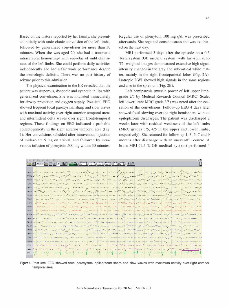
The physical examination in the ER revealed that thepatient was stuporous, dyspneic and cyanotic in lips withgeneralized convulsion. She was intubated immediatelyfor airway protection and oxygen supply. Post-ictal EEGshowed frequent focal paroxysmal sharp and slow waveswith maximal activity over right anterior temporal areasand intermittent delta waves over right frontotemporalregions. Those findings on EEG indicated a probableepileptogenicity in the right anterior temporal area (Fig.1). Her convulsions subsided after intravenous injectionof midazolam 5 mg on arrival, and followed by intra-venous infusion of phenytoin 500 mg within 30 minutes.
Regular use of phenytoin 100 mg q8h was prescribedafterwards. She regained consciousness and was extubat-ed on the next day.
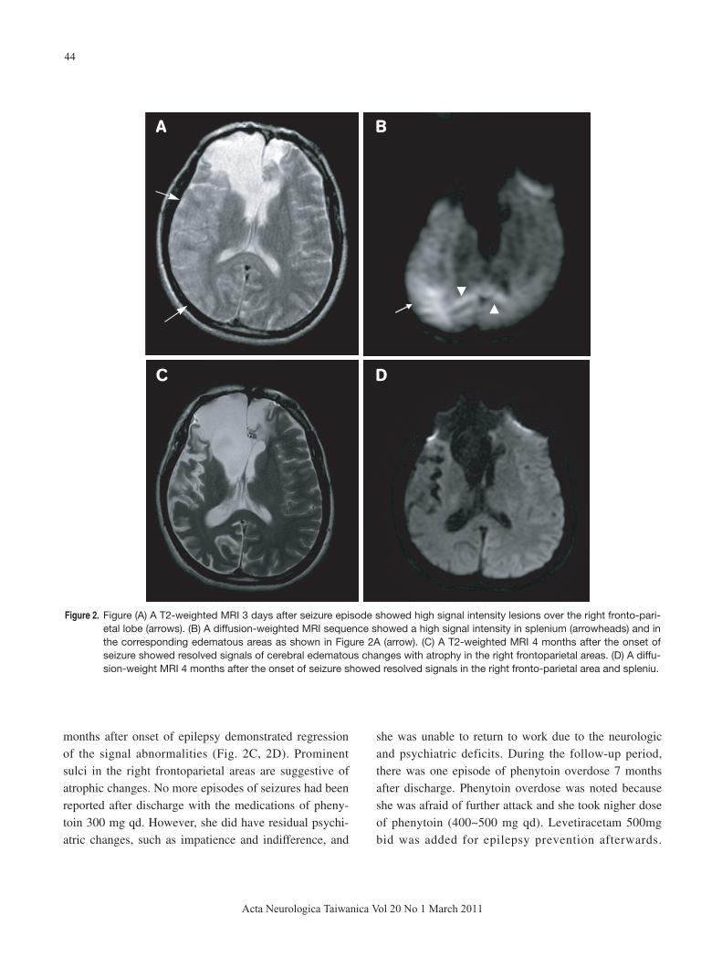
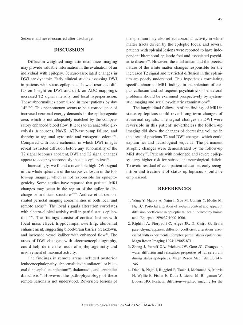
MRI performed 3 days after the episode on a 0.5Tesla system (GE medical system) with fast-spin echoT2 -weighted images demonstrated extensive high signalintensity changes in the gray and subcortical white mat-ter, mainly in the right frontoparietal lobes (Fig. 2A).Isotropic DWI showed high signals in the same regionsand also in the splenium (Fig. 2B).
Left hemiparesis (muscle power of left upper limb:grade 2/5 by Medical Research Council (MRC) Scale,left lower limb: MRC grade 3/5) was noted after the ces-sation of the convulsions. Follow-up EEG 4 days latershowed focal slowing over the right hemisphere withoutepileptiform discharges. The patient was discharged 2weeks later with residual weakness of the left limbs(MRC grades 3/5, 4/5 in the upper and lower limbs,respectively). She returned for follow-up 1, 3, 5, 7 and 9months after discharge with an uneventful course. Abrain MRI (1.5-T, GE medical system) performed 4
43
Acta Neurologica Taiwanica Vol 20 No 1 March 2011
Figure 1. Post-ictal EEG showed focal paroxysmal epileptiform sharp and slow waves with maximum activity over right anteriortemporal area.
44
Acta Neurologica Taiwanica Vol 20 No 1 March 2011
months after onset of epilepsy demonstrated regressionof the signal abnormalities (Fig. 2C, 2D). Prominentsulci in the right frontoparietal areas are suggestive ofatrophic changes. No more episodes of seizures had beenreported after discharge with the medications of pheny-toin 300 mg qd. However, she did have residual psychi-atric changes, such as impatience and indifference, and
she was unable to return to work due to the neurologicand psychiatric deficits. During the follow-up period,there was one episode of phenytoin overdose 7 monthsafter discharge. Phenytoin overdose was noted becauseshe was afraid of further attack and she took nigher doseof phenytoin (400~500 mg qd). Levetiracetam 500mgbid was added for epilepsy prevention afterwards.
A B
C D
Figure 2. Figure (A) A T2-weighted MRI 3 days after seizure episode showed high signal intensity lesions over the right fronto-pari-etal lobe (arrows). (B) A diffusion-weighted MRI sequence showed a high signal intensity in splenium (arrowheads) and inthe corresponding edematous areas as shown in Figure 2A (arrow). (C) A T2-weighted MRI 4 months after the onset ofseizure showed resolved signals of cerebral edematous changes with atrophy in the right frontoparietal areas. (D) A diffu-sion-weight MRI 4 months after the onset of seizure showed resolved signals in the right fronto-parietal area and spleniu.
45
Acta Neurologica Taiwanica Vol 20 No 1 March 2011
Seizure had never occurred after discharge.
DISCUSSION
Diffusion-weighted magnetic resonance imagingmay provide valuable information in the evaluation of anindividual with epilepsy. Seizure-associated changes inDWI are dynamic. Early clinical studies assessing DWIin patients with status epilepticus showed restricted dif-fusion (bright on DWI and dark on ADC mapping),increased T2 signal intensity, and local hyperperfusion.These abnormalities normalized in most patients by day14(1-3,6,7). This phenomenon seems to be a consequence ofincreased neuronal energy demands in the epileptogenicarea, which is not adequately matched by the compen-satory enhanced blood flow. It leads to an anaerobic gly-colysis in neurons, Na+/K+ ATP-ase pump failure, andthereby to regional cytotoxic and vasogenic edema(8).Compared with acute ischemia, in which DWI imagesreveal restricted diffusion before any abnormality of theT2 signal becomes apparent, DWI and T2 signal changesappear to occur synchronously in status epilepticus(8).
Interestingly, we found a reversible high DWI signalin the whole splenium of the corpus callosum in the fol-low-up imaging, which is not responsible for epilepto-genicity. Some studies have reported that periictal MRIchanges may occur in the region of the epileptic dis-charge or in distant structures(7-9). Andrew et al. demon-strated periictal imaging abnormalities in both local andremote areas(8). The local signals alteration correlateswith electro-clinical activity well in partial status epilep-ticus(10). The findings consist of cortical lesions withfocal mass effect, hippocampal swelling, abnormalenhancement, suggesting blood-brain barrier breakdown,and increased vessel caliber with enhanced flow(8). Theareas of DWI changes, with electroencephalography,could help define the focus of epileptogenicity andinvolvement of maximal activity.
The findings in remote areas included posteriorleukoencephalopathy, abnormalities in unilateral or bilat-eral diencephalon, splenium(8), thalamus(11), and cerebellardiaschisis(9). However, the pathophysiology of theseremote lesions is not understood. Reversible lesions of
the splenium may also reflect abnormal activity in whitematter tracts driven by the epileptic focus, and severalpatients with splenial lesions were reported to have inde-pendent bitemporal epileptic foci and associated psychi-atric disease(8). However, the mechanism and the precisenature of the white matter changes responsible for theincreased T2 signal and restricted diffusion in the spleni-um are poorly understood. This hypothesis correlatingspecific abnormal MRI findings in the splenium of cor-pus callosum and subsequent psychiatric or behavioralproblems should be examined prospectively by system-atic imaging and serial psychiatric examinations(8).
The longitudinal follow-up of the findings of MRI instatus epilepticus could reveal long-term changes ofabnormal signals. The signal changes in DWI werereversible in this patient; nevertheless the follow-upimaging did show the changes of decreasing volume inthe areas of previous T2 and DWI changes, which couldexplain her and neurological sequelae. The permanentatrophic changes were demonstrated by the follow-upMRI study(12). Patients with prolonged and severe epilep-sy carry higher risk for subsequent neurological deficit.To avoid residual effects, patient education, early recog-nition and treatment of status epilepticus should beemphasized.
REFERENCES
1. Wang Y, Majors A, Najm I, Xue M, Comair Y, Modic M,
Ng TC. Postictal alteration of sodium content and apparent
diffusion coefficient in epileptic rat brain induced by kainic
acid. Epilepsia 1996;37:1000-1006.
2. Righini A, Pierpaoli C, Alger JR, Di Chiro G. Brain
parenchyma apparent diffusion coefficient alterations asso-
ciated with experimental complex partial status epilepticus.
Magn Reson Imaging 1994;12:865-871.
3. Zhong J, Petroff OA, Prichard JW, Gore JC. Changes in
water diffusion and relaxation properties of rat cerebrum
during status epilepticus. Magn Reson Med 1993;30:241-
246.
4. Diehl B, Najm I, Ruggieri P, Tkach J, Mohamed A, Morris
H, Wyllie E, Fisher E, Duda J, Lieber M, Bingaman W,
Luders HO. Postictal diffusion-weighted imaging for the
localization of focal epileptic areas in temporal lobe epilep-
sy. Epilepsia 2001;42:21-28.
5. Diehl B, Najm I, Ruggieri P, Foldvary N, Mohamed A,
Tkach J, Morris H, Barnett G, Fisher E, Duda J, Luders
HO. Periictal diffusion-weighted imaging in a case of
lesional epilepsy. Epilepsia 1999;40:1667-1671.
6. Yogarajah M, Duncan JS. Diffusion-based magnetic reso-
nance imaging and tractography in epilepsy. Epilepsia
2008;49:189-200.
7. Toledo M, Munuera J, Sueiras M, Rovira R, Alvarez-Sabin
J, Rovira A. MRI findings in aphasic status epilepticus.
Epilepsia 2008;49:1465-1469.
8. Cole AJ. Status epilepticus and periictal imaging. Epilepsia
2004;45:72-77.
9. Calistri V, Caramia F, Bianco F, Fattapposta F, Pauri F,
Bozzao L. Visualization of evolving status epilepticus with
diffusion and perfusion MR imaging. Am J Neuroradiol
2003;24:671-673.
10. Di Bonaventura C, Bonini F, Fattouch J, Mari F, Petrucci S,
Carni M, Tinelli E, Pantano P, Bastianello S, Maraviglia B,
Manfredi M, Prencipe M, Giallonardo AT. Diffusion-
weighted magnetic resonance imaging in patients with par-
tial status epilepticus. Epilepsia 2009;50:45-52.
11. Katramados AM, Burdette D, Patel SC, Schultz LR,
Gaddam S, Mitsias PD. Periictal diffusion abnormalities of
the thalamus in partial status epilepticus. Epilepsia 2009;
50:265-275.
12. Gong G, Shi F, Concha L, Beaulieu C, Gross DW. Insights
into the sequence of structural consequences of convulsive
status epilepticus: A longitudinal MRI study. Epilepsia
2008;49:1941-1945.
46
Acta Neurologica Taiwanica Vol 20 No 1 March 2011