reversal of subjective temporal order due to sensory and
TRANSCRIPT

1
Reversal of subjective temporal order due to sensory and motor
integrations
Shigeru Kitazawa1-3, Shunjiro Moizumi1, Ayami Okuzumi1, Fumine Saito1, Satoshi
Shibuya4, Toshimitsu Takahashi1-3, Makoto Wada1-3 and Shinya Yamamoto3
1 Department of Neurophysiology, Juntendo University Graduate School of Medicine,
Tokyo, Japan
2 CREST, Japan Science and Technology Corporation, Tokyo, Japan
3 Neuroscience Research Institute, National Institute of Advanced Industrial Science and
Technology
4 Department of Integrative Physiology, Kyorin University School of Medicine, Tokyo,
Japan
Correspondence should be addressed to S.K.

2
Abstract
It is generally accepted that the brain can resolve the order of two stimuli that are
separated in time by 20-50 ms. This applies to temporal order judgment of two tactile
stimuli, delivered one to each hand, as long as the arms are uncrossed. However,
crossing the arms caused misreporting (that is, inverting) of the temporal order. The
reversal was not due to simple confusion of hands, because correct judgment was
recovered at longer intervals (e.g., 1.5 s). When the stimuli were delivered to the tips of
sticks held in each hand, the judgment was altered by crossing the sticks without
changing the spatial locations of the hands. We recently found that temporal order
judgments of tactile stimuli are sometimes reversed by visual distractors and by
saccadic eye movements. The results suggest that tactile stimuli are ordered in time only
after they are referred to relevant locations in space, where multisensory (visual, tactile
and proprioceptive) and motor (saccade-related) signals converge. Results from
functional imaging generally support this idea.
Because performance of congenitally blind people in tactile temporal order
judgment is much superior to the sighted and is never impaired by crossing the arms
(Roeder et al., 2004), we finally suggest that our integrity of multisensory signals in
space is only achieved at the cost of continuity in time.

3
1. Introduction
Ordering sensory signals is a critical step for deciphering symbols like in hearing
speeches, reading books and reading Braille or sign language. In fact, poor readers are
reported to be impaired in ordering sensory signals in multiple sensory modalities
(Tallal et al., 1998, May et al., 1988, Laasonen et al., 2001, Habib, 2000). In this chapter
we raise a question of how the brain orders successive events.
An ideal observer in physics is able to read his watch on the occurrence of each event
no matter how small the interval of the events is. The brain is no way an ideal observer,
but it is often assumed that there is a decision mechanism that plays a role much similar
to the ideal observer in physics (Sternberg and Knoll, 1973). The decision mechanism
was hypothesized to yield a judgment that a signal A preceded another signal B
according to the temporal difference of their arrival times (TA and TB). The probability of
the judgment that A preceded B was hypothesized to be determined by a monotonically
increasing decision function G of the arrival-time difference (TB- TA). If there is such a
decision mechanism in the brain, where is it located?
To maximize the temporal resolution in ordering the signals A and B, it is
advantageous to locate such a decision mechanism as near as possible to the entrance of
these signals, before the physical order of the two signals is obscured (Dennett and
Kinsbourne, 1992). For example, timing in the 10 microseconds range is actually
detected by the neurons in the medial superior olive that receive convergent inputs
directly from the left and the right cochlear nuclei. However, we do not regard this as an
example of the decision mechanism of temporal order, because these two successive
sounds, delivered one to each ear, with an interval of 10 microseconds lead to
perception of a single sound, not two successive sounds. The timing information in the

4
medial superior olive is used for localizing a single sound source, rather than for
ordering two successive sounds. In this chapter, we deal with the problem of how the
brain orders two signals when they are subjectively perceived as two distinct events.
Interestingly, the threshold for detecting non-simultaneity of two sensory signals
varies among sensory modalities, but the threshold for judging their temporal order
seems to be independent of sensory modalities (Hirsh and Sherrick, 1961, Pöppel, 1997,
although see Spence et al., 2003). This suggests that there is a general decision
mechanism that orders sensory events that originated from different sensory organs.
In the history of vision research, studies of illusions have much contributed to
exploring the neural mechanisms of vision. Likewise, several anomalous experiences in
time have provided us with insights on how conscious perceptions develop in time
(Dennett and Kinsbourne, 1992). Here we focus on more recent illusions, reversals in
subjective temporal order due to arm crossing (Yamamoto and Kitazawa, 2001a, Shore
et al., 2002) and during peri-saccadic periods (Morrone et al., 2005) in the hope of
shedding light on whether and where the hypothetical decision mechanism is located
and how it orders sensory signals in time.
2. Reversal of temporal order due to crossings of the arms
We have been studying how the brain orders successive sensory signals that were
delivered one to each hand (Yamamoto et al., 2005, Yamamoto and Kitazawa, 2001b,
Yamamoto and Kitazawa, 2001a, Wada et al., 2004). Signals A and B in Fig. 1
correspond to those from the right hand and those from the left hand, respectively.
Signals from each hand first reaches the contralateral primary sensory cortex that is
arranged somatotopically. If the signals from the two hands reach the decision

5
mechanism while they are represented in the somatotopical coordinate but not in the
spatial coordinate, the judgment would never be affected by the spatial position of the
arms. To test which is the case, we examined temporal order judgment with the arms
crossed and uncrossed. Seated subjects were asked to close their eyes and to judge the
temporal order of successive mechanical stimuli, one delivered to the right hand and the
other to the left hand, and to respond by extending the index finger of the hand that
received the first (or, in half of the experiments, the second) stimulus. In the uncrossed
condition, the order-judgment probability that the right hand was stimulated first was
closely approximated by a monotonic sigmoid function (a cumulative density function
of a Gaussian distribution) (Yamamoto and Kitazawa, 2001a, Wada et al., 2004). This
basically agrees with the decision model of (Sternberg and Knoll, 1973), and the
temporal resolution that was defined as the standard deviation of the Gaussian function
in our studies (approximately corresponds to the 84%-correct interval)1 was 52 ms for
the right-handed subjects (Wada et al., 2004).
When the arms were crossed, to our surprise, many subjects reported inverted
judgment at intervals of around 100-200 ms. In the most apparent case (Fig. 2A), the
subject’s report was completely inverted when the stimulation interval was 100-200 ms.
The correct judgment was restored as the interval approached 1500 ms, clearly
indicating that the inverted judgment was not caused by a trivial confusion in
distinguishing between the two hands. As a result, the response curve of the subject
became N-shaped with a peak and a trough, which was clearly distinct from the
conventional non-decreasing sigmoid function as hypothesized in the decision model
(Sternberg and Knoll, 1973). Because the degree of reversal varied considerably across 1 Our temporal resolution can be converted to the conventional just noticeable difference that yields 75% correct responses by multiplying 0.68.
6
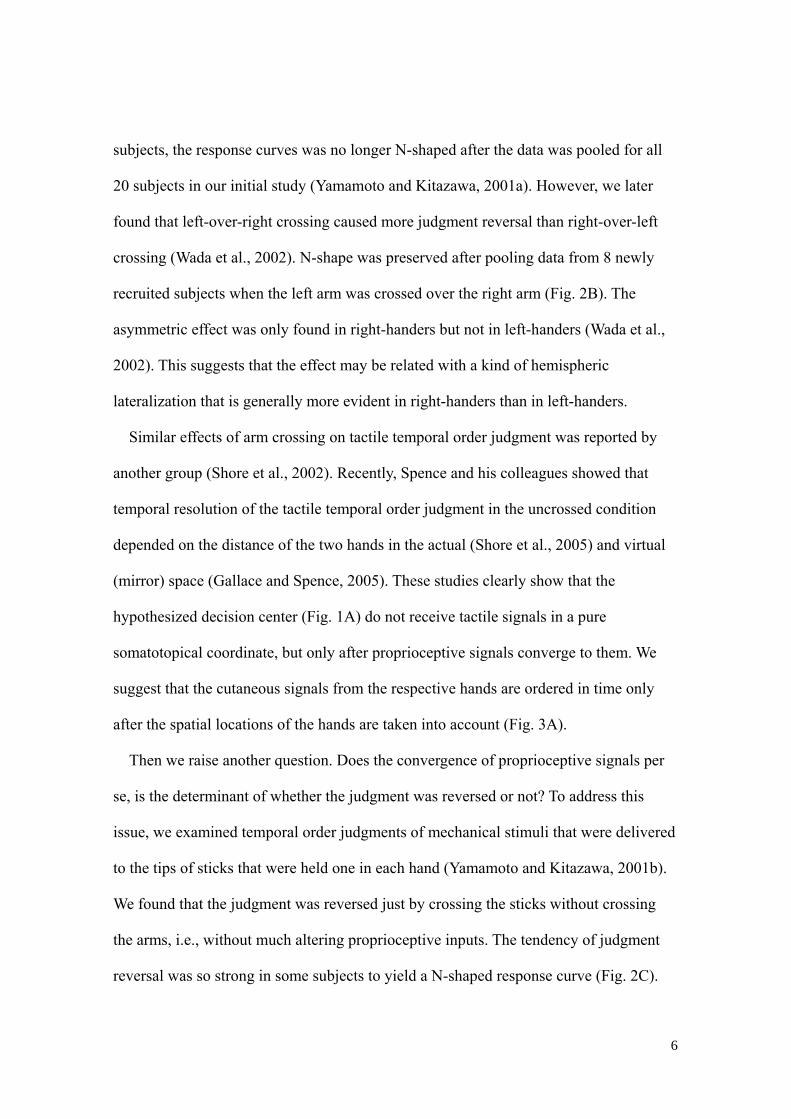
subjects, the response curves was no longer N-shaped after the data was pooled for all
20 subjects in our initial study (Yamamoto and Kitazawa, 2001a). However, we later
found that left-over-right crossing caused more judgment reversal than right-over-left
crossing (Wada et al., 2002). N-shape was preserved after pooling data from 8 newly
recruited subjects when the left arm was crossed over the right arm (Fig. 2B). The
asymmetric effect was only found in right-handers but not in left-handers (Wada et al.,
2002). This suggests that the effect may be related with a kind of hemispheric
lateralization that is generally more evident in right-handers than in left-handers.
Similar effects of arm crossing on tactile temporal order judgment was reported by
another group (Shore et al., 2002). Recently, Spence and his colleagues showed that
temporal resolution of the tactile temporal order judgment in the uncrossed condition
depended on the distance of the two hands in the actual (Shore et al., 2005) and virtual
(mirror) space (Gallace and Spence, 2005). These studies clearly show that the
hypothesized decision center (Fig. 1A) do not receive tactile signals in a pure
somatotopical coordinate, but only after proprioceptive signals converge to them. We
suggest that the cutaneous signals from the respective hands are ordered in time only
after the spatial locations of the hands are taken into account (Fig. 3A).
Then we raise another question. Does the convergence of proprioceptive signals per
se, is the determinant of whether the judgment was reversed or not? To address this
issue, we examined temporal order judgments of mechanical stimuli that were delivered
to the tips of sticks that were held one in each hand (Yamamoto and Kitazawa, 2001b).
We found that the judgment was reversed just by crossing the sticks without crossing
the arms, i.e., without much altering proprioceptive inputs. The tendency of judgment
reversal was so strong in some subjects to yield a N-shaped response curve (Fig. 2C).
7
When the arms were crossed without crossing the sticks, there was a tendency of
judgment reversal. Finally, when the sticks were crossed in addition to crossing the arms,
the judgment curve returned to a normal sigmoid as when neither of the arms nor the
sticks were crossed. We obtained similar results when subjects held L-shaped sticks
instead of the straight ones (Yamamoto et al., 2005) and when the subjects manipulated
tools in the virtual reality (Moizumi et al., 2004). When the tip or the action point of
each tool was located in the same hemispace as the hand that held the tool, response
curves were ordinary sigmoid with temporal resolutions of about 60 ms. On the other
hand, the curves showed clear tendency of judgment reversal at moderately short
intervals, when the tips were located in the contralateral hemispaces.
These results with real and virtual tools2 show that temporal order judgment of
tactile stimuli does not necessarily depend on the spatial positions of the hands per se.
When the subjects were required to judge the order of mechanical stimuli that were
delivered to the tips of tools in hands, judgments depended on the spatial positions of
the tips of the tools irrespective of whether the arms were crossed or uncrossed. We
suggest that signals from the skin mechanoreceptors of the right and the left hands are
ordered in time after they are referred to a relevant location in space whether it is the
hand itself or the tip of the tool in the hand (Fig. 3A).
It is well known in choice reaction time tasks that response latencies to right-side
stimuli are shorter when the responses are made with the right than the left hand when
the arms are uncrossed (Simon et al., 1970, Riggio et al., 1986). When the arms are
crossed, in contrast, response latencies to right-side stimuli are shorter when the
responses are made with the left hand that is now spatially compatible with the 2 In experiments with straight sticks and virtual tools, they were actually used for making responses, but L-shaped sticks were used as pure sensing tools and subjects used their foot for their response.

8
right-side stimuli. Riggio et al. (1986) used sticks to examine choice reaction time after
crossing response goals without crossing the hands, and showed that reaction time is
shorter when the location of the response goal, not that of the hand per se, is compatible
with the side of stimulus. This phenomenon, known as stimulus-response compatibility,
may seem to be closely related with the judgment reversal due to arm- or stick-crossing.
However, this is not likely because stimulus-response compatibility is observed
irrespective of whether the response goals are crossed or not. On the other hand,
reaction latencies are generally longer when the response goals (or the hands) are
crossed than when they are uncrossed (Riggio et al., 1986, Yamamoto and Kitazawa,
2001a). We suggest that tactile signals of one hand is initially mapped to the response
goal (the tip of a stick, or the hand itself) ipsilateral to the anatomical side of the hand,
and it takes time to remap the signals to the correct (contralateral) goal when the
response goals are crossed. We later propose a hypothesis that explains judgment
reversal by taking the process of remapping into account.
Studies of Iriki and colleagues give us some implications for neural mechanisms that
underlie the referral of tactile signals to the hands or tips of tools. They showed in
monkey that visual receptive fields of bimodal intraparietal neurons extended along a
rake in hand (Iriki et al., 1996) and were projected even to an action point in the video
monitor (Iriki et al., 2001). We infer that activation of such bimodal neurons in response
to tactile stimuli would associate the tactile stimuli to the visual receptive fields of these
neurons.
3. Motion projection hypothesis in temporal order judgments
The results so far strongly suggested that tactile temporal order judgment involves

9
spatial representations in the brain. To test if this was the case, we examined which
neural circuits is actually activated during the temporal order judgments (Takahashi et
al., 2004). Event-related functional magnetic resonance imaging (fMRI) was used while
right-handed subjects (n = 12) judged the temporal order of two successive stimuli
delivered one to each hand. Each subject was laid in a 3T magnetic resonance scanner
(GE Medical Systems) with their eyes closed and with the index finger of each hand
placed on a non-magnetic eight-pin braille stimulator (a 4 by 2 array with inter-pin
intervals of 3 mm) fixed on the thigh. In each trial, right-then-left or left-then-right
stimuli were delivered with a fixed stimulus onset asynchrony (SOA) by sticking out
1-7 pins from each stimulator. The subject was required to judge the temporal order of
the stimuli in one condition (TOJ condition), and to compare the numerosity of the pins
in the control condition. In both conditions, each stimulator served as a response button.
The subject was required in a forced choice manner to push the ‘second’ button in the
TOJ condition, and to push the button with the greater number of pins in the numerosity
judgment condition. The SOA (50-100 ms) and the difference in the number of pins
(2-3) were chosen for each subject so that correct response rate of approximately 80%
was achieved in both tasks. Greater activation was elicited bilaterally during the
temporal order judgment than during the numerosity judgment task in the following
areas: bilateral posterior part of the middle temporal gyri (BA 37), bilateral premotor
cortices (BA 6), bilateral inferior frontal gyri (BA 44), bilateral inferior parietal cortices
(BA 40), and the left primary and secondary somatosensory cortices. The activation
around the middle temporal gyri overlapped with the areas that were reported to respond
to biological motion (Puce et al., 1998, Puce and Perrett, 2003) and the most anterior
part of the putative MT and MST (Dukelow et al., 2001). From the results we propose

10
that temporal order of tactile signals are ordered in time by combining representation of
‘motion’ in the temporal cortex with the spatial representation of the stimuli in the
prefrontal and parietal cortices (Motion projection hypothesis, Fig. 3B). In the
hypothesis, we assume that successive tactile stimuli evokes a kind of motion signal
(such as tactile apparent motion) in the motion related areas in the brain, and they are
also represented in spatial representations such as those in the parietal cortex. Then the
order of two tactile events is reconstructed by integrating the motion signal with the two
spatial locations of tactile stimuli.
4. Evidence of contribution of motion signals in tactile temporal order judgment
The motion projection hypothesis assumes that a motion signal, which is created from
successive tactile stimuli, is critically important for reconstructing temporal order of the
tactile stimuli. From the hypothesis we predict that tactile temporal order judgment
would be affected by concurrent presentation of visual signals that elicit apparent
motion and converge on the motion areas in the brain.
Experiment 1: Does visual apparent motion alter tactile temporal order judgment?
Methods
Subjects Six naive subjects (2 men and 4 women; 30 ± 10 years old, mean ± SD)
participated in this experiment. They were all strongly right-handed according to the
Edinburgh Inventory (Oldfield, 1971).
Apparatus and task procedures Each subject sat in a chair and placed each hand on a
tactile stimulator (Dot View DV-1; KGS, Saitama, Japan) that consisted of a matrix of
24 × 32 pins placed at 3-mm intervals with the arms uncrossed. Each pin (round head,
1.3 mm in diameter) could be raised by an independent piezoelectric actuator that

11
produced 0.127 N within 5 ms from the onset of a command signal from a PC
(Dimension 8200; Dell, Round Rock, TX). A tilted (45 degrees) 17-inch CRT monitor
(CPD-G220; Sony, Tokyo, Japan) was used to present visual stimuli in a mirror (20 × 26
cm) placed above the tactile stimulator so that visual stimuli were presented on their
fingers (Figs. 4A, B). A masking board (30 × 43 cm) was positioned just below the
mirror to occlude vision of the subject’s hands and arms.
Two tactile stimuli were delivered to the third finger pad of the two hands, one to
each hand. Each stimulus was delivered by simultaneously raising a 10 × 8 pin array
under each finger pad. The distance between the third fingers was 18 cm. After the two
10 × 8 pin arrays were raised one by one with a stimulus onset asynchrony (SOA) that
was randomly assigned for each trial from 24 values (–1500, –900, –600, –400, –300,
–200, –150, –100, –80, –60, –30, –10, 10, 30, . . ., 1500 ms), all 160 pins were lowered
simultaneously at 1 s after the delivery of the second stimulus. Positive values indicate
that the right hand was stimulated first and vice versa.
Simultaneously with the delivery of two tactile stimuli, visual stimuli appeared in two
small squares (1.5 × 1.5 cm) placed at 18-cm intervals (center to center) so that they
appeared in a randomized order on the tips of the third fingers (Fig. 4B). The visual
stimuli appeared in the order identical (congruent condition) or opposite (incongruent
condition) to the order of tactile stimuli. Two lighted squares were turned off at 1 s after
the delivery of the second visual and tactile stimuli. During the experiments, subjects
were instructed to gaze at a fixation point at the middle of the two squares. Subjects
were also required to ignore visual stimuli and make a forced choice between the two
orders of tactile stimuli. White noise (80 dB) was played through headphones placed
over the subjects’ plugged ears to mask the sound of the stimulator, although the
12
stimulator was basically silent. Thus, subjects could only feel the tactile stimulation and
see the visual stimuli.
After the second stimulus was delivered, the subjects were required to report the
order of tactile stimulation, by pressing a button under the index finger of the hand that
was judged as stimulated later than the other. For each trial, subjects were forced to
choose one of the two responses as soon as possible after the delivery of the second
stimulus. All response data were recorded and stored on the PC for off-line analyses.
Each subject participated in one experiment that consisted of 384 trials in which two
visual stimuli (congruent and incongruent) were assigned 8 times each for 24 SOAs, in
a pseudorandom order.
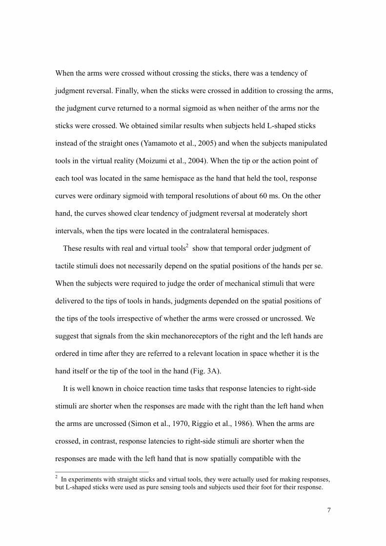
Data analysis The response data were sorted by SOA to calculate the order–judgment
probabilities that the right hand was stimulated earlier than the left hand under the
congruent (pcong) and the incongruent (pincong) conditions. The order–judgment
probability under the congruent condition (pcong) was fitted by a cumulative density
function of a Gaussian distribution:
min2
2
minmax 2)(
exp2
1)()( pdd
pptpcong
congt
congcong +⎟
⎟⎠
⎞⎜⎜⎝
⎛ −−×−= ∫ ∞−
τσ
τσπ
, (1)
where t, dcong, σcong, pmax, and pmin denote the SOA, the size of horizontal transition, the
time constant, and the upper and lower asymptotes of the judgment probability,
respectively. Matlab (optimization toolbox) was used for the fitting to minimize
Pearson’s chi-square statistics (df=19), which reflected the discrepancy between the
sampled order–judgment probability (24 data points) and the prediction, using the
four-parameter model. The time constant (σcong) is an effective measure of temporal
order resolution.

13
The order–judgment probability under the incongruent condition (pincong) was
assumed to be flipped from the order–judgment probability under the congruent
condition (pcong) in a manner formulated as follows:
)()}(1{)}(1){()( tptftptftp congrconglincong −+−= , (2)
cdtAtff
rr +⎟⎟⎠
⎞⎜⎜⎝
⎛ −−= 2
2
2)(exp)(
σ, (3)
cdtAtff
ll +⎟⎟⎠
⎞⎜⎜⎝
⎛ −−= 2
2
2)(exp)(
σ, (4)
where fr denotes the flip probability of judgment from right-hand-first to left-hand-first
and fl from left-hand-first to right-hand-first. We estimated five parameters in the flip
probabilities that followed the Gaussian functions shown in Equations 3 and 4: the peak
flip amplitude of the Gaussian functions (Ar and Al), the size of the horizontal transition
(d), the time window of the flip (σf), and a constant (c). Matlab (optimization toolbox)
was used for the fitting to minimize Pearson’s chi-square statistics (df=18).
Results
When the spatial direction of visual stimuli was congruent with that of visual
stimuli, the order-judgment probability (pcong) that the right hand was stimulated first
(open circles, Fig. 4C) was closely approximated by a monotonic sigmoid function (Eq.
1, solid curves in Fig. 4C and D, r2 = 0.94 and r2 = 0.91, respectively). The temporal
resolution (σcong) was 82 ms for the pooled data (Fig. 4D) and comparable (mean ± S.E.
= 71 ± 6 ms, n = 6) to those in our previous studies (Wada et al., 2004, mean ± S.E. =
52 ± 4 ms, Yamamoto and Kitazawa, 2001a, 71 ± 6 ms) in which there was no
concurrent visual stimuli.

14
However, when the direction of visual stimuli was incongruent to that of tactile
stimuli, many subjects reported inverted judgments at SOAs less than about 300 ms (red
dots in Fig. 4C, D). In the most apparent case (Fig. 4C), the response curve became
N-shaped even though the arms were not crossed. The N-shaped response curve seems
to be similar to those obtained when the arms were crossed (e.g., Fig. 2A), though the
reversal under the visual distraction peaked at ~100 ms in contrast to ~200 ms under the
previous crossed-arm condition without any visual stimuli.
Discussion
The results clearly show that the subjects were unable to ignore visual stimuli when the
visual stimuli were incongruent to tactile stimuli. This supports the prediction from the
motion projection hypothesis that visual motion stimuli would interfere with the tactile
temporal order judgment. However, it may be argued that the visual interference can be
explained in terms of the “ventriloquism effect”, one-to-one mislocalization of a tactile
stimulus to the location of visual stimulation, rather than the visual apparent motion.
Although the effect was originally used to describe the mislocalization of a sound
source to the location of visual stimulation (Howard and Templeton, 1966), tactile
stimuli can also be referred to the location of concurrent visual stimuli (Botvinick and
Cohen, 1998, Spence et al., 2004, Armel and Ramachandran, 2003). If such one-to-one
mislocalization were the cause of judgment reversal, the cross-modal interaction should
have been independent of the SOA, because each tactile stimulus was always
accompanied with a simultaneous visual stimulus. However, the judgment reversal was
peaked at the SOA of ~100 ms, subsided as the SOA became longer and was no longer
apparent when the SOA was longer than 400 ms. Thus, our findings do not support
major involvement of the ventriloquism effect in the visuotactile interaction. On the

15
other hand, a sensation of motion evoked by successive visual stimuli (apparent motion)
has a peak at the interstimulus interval of ~100 ms or shorter and decays over several
hundreds of milliseconds to disappear at ~1 s (Soto-Faraco et al., 2004). This prediction
was supported by the results that the reversal of judgment under the incongruent
condition decayed and disappeared in a Gaussian manner with a standard deviation of
~150 ms (Fig. 4). Thus the present results support the motion projection hypothesis that
motion signals are integrated to the spatial representation of two events for ordering the
two events in time.
5. Reversal of subjective temporal order due to saccades
Recently, Morrone et al. (2005) reported that subjective temporal order of two
successive visual stimuli was reversed when the stimuli were delivered during
perisaccadic periods. Subjects were required to make a 30-degree horizontal saccade
from a left fixation point to a right target (Fig. 5A), and judge the order of two green
horizontal bands that were presented for a brief period (8 ms) in succession, one in the
top and the other at the bottom of the red screen. When the bands were presented during
a post-saccadic period (more than 100 ms after the saccade), the response curve was
sigmoid (Fig. 5B) and the exemplified subject responded correctly when the SOA was
as small as 25 ms. However, when the visual stimuli were presented just prior to the
saccade onset (within a time window between -70 and -30 ms), the response curve
became N-shaped with complete reversal at stimulation intervals of -50 and +50 ms,
and with recovery at longer intervals (Fig. 5C). Although stimulus intervals that yielded
the peak reversal was longer in the tactile temporal order judgment in the crossed-arm
condition (100~200 ms) than in the judgment reversal prior to the saccade (~50 ms),

16
this is clearly the second case of anomaly that cannot be explained in terms of the
classic temporal order decision mechanism with a non-decreasing decision function (Fig.
1).
Experiment 2: Replication of an experiment reported by Morrone et al.
As a control, we examined whether a saccade reverses subjective temporal order of
visual stimuli, by replicating the experiments reported by Morrone et al. (2005).
Methods
Subjects Six right-handed subjects participated in the experiments. Four were naive to
the purpose of the experiment and the others were two of the co-authors.
Apparatus and task procedures Each subject sat with their head rested on a chin rest,
facing a 24 inch CRT monitor (HM204-DA, Iiyama, Tokyo, Japan) that was placed 45
cm apart from the eyes. The subject was required to fixate on a target in the left of the
red monitor screen, and to make a visually guided saccade to another target that
appeared 24° to the right of the fixation target. The fixation target was extinguished
simultaneously to the appearance of the target for a saccade. Two visual stimuli, green
horizontal bars at the top and at the bottom of the monitor was briefly (10 ms) presented
in succession with an SOA pseudo-randomly chosen from 16 (-150, -100, -90, -75, -60,
-50, -25, -15, 15, 25, 50, 60, 75, 90, 100 and 150 ms) in which positive SOA represented
top-first stimuli. The first visual stimulus was delivered after the target onset with an
interval chosen from four (20, 50, 100 and 500 ms). The subject was required to judge
the order of visual stimuli (top-first or bottom-first) and to respond by pressing one of
two buttons in a forced choice manner. During the task trials, gaze positions were
measured at 500 Hz by an eye tracker (Eyelink2, SR Research, Ontario, Canada). The
17
experiment was controlled by Experiment Builder (SR Research, Ontario, Canada).
Each subject participated in one experiment that consisted of 128 trials in which 16
SOAs were assigned 2 times each for 4 visual delays, in a pseudorandom order.
Analysis After each experiment, the onset of each saccadic eye movement, which was
automatically detected by a built-in program of Eyelink2, was inspected one by one to
be acknowledged, corrected or discarded. Based on the registered onsets of saccade, the
delay of stimulus presentation was defined as the interval between the saccade onset and
the middle of the timings of successive visual stimuli. Positive delays generally
correspond to post-saccade presentation and vice versa, although in some conditions the
first and the second stimulus could be presented before and after the saccade-onset,
respectively. Trials with pre-saccadic delays between –150 ms to 0 ms (pre-saccadic
trials, n = 52), and those with post-saccadic delays larger than 100 ms (post-saccadic
trials, n = 178) were used for further analysis. The response data were sorted by SOA to
calculate the order–judgment probabilities that the top bar was presented earlier than the
bottom bar in the pre- (ppre) and post- (ppost) saccadic trials. The order–judgment
probability in the post-saccadic trials (ppost) was fitted by a cumulative density function
of a Gaussian distribution. The order–judgment probability in the pre-saccadic trials
(ppre) was assumed to be flipped from the order–judgment probability in the
post-saccadic trials (ppost) in a manner similar to that formulated as Eqs. 3 and 4. The
degree of judgment reversal was quantified by calculating the maximal discrepancy
between the sigmoid fitting in the pre-saccadic trials and the flip model fitting in the
post-saccadic trials.
Results and Comments
The probability of top-first judgment in the post-saccadic trials (ppost) was well fitted by

18
a sigmoid function with a temporal resolution of 46 ms (Fig. 6A, black curve and dots).
On the other hand, subjects often made inverted judgments in the pre-saccadic trials at
stimulation intervals of ~50 ms and the order–judgment probability in the pre-saccadic
trials (ppre) was N-shaped (Fig. 6A, red curve and dots). The results generally agreed
with those of Morrone et al. (2005), though the degree of judgment reversal (0.38) was
not as strong as that (1.0) in the previous report. There are several factors that might
explain the quantitative difference. First, we set a wider time window (-150 to 0 ms)
than that (-70 to -30 ms) in Morrone et al. (2005) to increase the number of trials. The
strongest reversal may be observed in the narrower time window. Second, the brightness
of the green bar was not matched with the brightness of the red background so precisely
in our experiment as in Morrone et al. (2005). Third, we presented pooled data from six
subjects including four naive subjects, but those from a single expert subject was shown
in Morrone et al. (2005).
Experiment 3: Does saccade reverse subjective temporal order of tactile stimuli?
If a temporal order judgment mechanism is shared by signals of different modalities,
saccades would reverse temporal order judgments of not only visual stimuli but also
those of other sensory modalities. We thus examined whether saccades reverse temporal
order judgments of tactile stimuli.
Methods
Subjects Five right-handed subjects participated in the experiments. Four were naive
to the purpose of the experiment and the other was one of the authors.
Apparatus and task procedures Each subject sat with their head rested on a chin rest,
facing a 24 inch CRT monitor (HM204DA; Iiyama, Tokyo, Japan). The subjects were

19
required to make a visually guided saccade (24°) from a fixation target in the left to a
target in the right, and to judge the order of successive tactile stimuli that were delivered
one to each hand (finger pad of the index finger) at various timing. The second tactile
stimulus was delivered after the target onset with an interval chosen from five (20, 50,
100, 200 and 500 ms). The left hand was placed 30 cm above the right hand on the
midsagittal plane so that they are positioned at neutral positions relative to the saccades.
In each trial, tactile stimuli were delivered with a stimulus onset asynchrony (SOA)
chosen from ten (-500, -200, -100, -50, -20, 20, 50, 100, 200 and 500 ms) in a
pseudo-random manner. Positive SOAs represented top-first (left-hand-first) stimuli.
The subjects were asked to judge which hand was stimulated second and to respond by
pressing one of the two foot switches. During the task trials, gaze positions were
measured at 500 Hz by an eye tracker (Eyelink2). One experiment consisted of 150
trials in which 10 SOAs were assigned 3 times each for 5 stimulus onset delays, in a
pseudorandom order. Each subject participated in two experiments. As a control, the
subjects were required to judge the temporal order of tactile stimuli while fixating on a
fixation point at the middle of the two hands. Each subject participated in one fixation
experiment that consisted of 100 trials in which 10 SOAs were assigned 10 times in a
pseudorandom order.
Analysis Data were analyzed in a manner similar to that in Experiment 2. After
inspecting each eye movement, the delay of stimulus presentation was defined as the
interval between the saccade onset and the middle of the timings of successive tactile
stimuli. Trials with pre-saccadic delays between –100 ms to 0 ms (pre-saccadic trials, n
= 201) and those with post-saccadic delays larger than 100 ms (post-saccadic trials, n =
281) were used for further analysis. Data under the fixation condition (500 trials) was

20
also analyzed as a control. The response data were sorted by SOA to calculate the
order–judgment probabilities that the top hand (left hand) was stimulated earlier than
the bottom hand (right hand) in the pre-saccadic trials (ppre), in the post-saccadic trials
(ppost) and in the fixation control (pfix). The order–judgment probability in the
post-saccadic trials (ppost), and in the fixation control (pfix) was fitted by a cumulative
density function of a Gaussian distribution. The order–judgment probabilities in the
pre-saccadic trials (ppre) were assumed to be flipped from the order–judgment
probability in the post-saccadic trials (ppost) and in the fixation control (pfix) in a manner
similar to that formulated as Eqs. 3 and 4. To evaluate whether the judgment in the test
condition was reversed from the control condition, we tested the null hypothesis that the
peak flip amplitudes in the flip model (Al and Ar) were zero and that the remaining
parameter is only c in the flip model (Eqs. 3 and 4). If the goodness-of-fit test using the
Pearson’s χ2 statistic produced P < 0.05, we judged that the reversal was significant
(Yamamoto et al., 2005).
Results and Comments
The probability of top-first judgment in the post-saccadic trials (ppost) was well fitted
by a sigmoid function with a temporal resolution of 97 ms (Fig. 6B, black curve and
open circles; r2=0.96). When the stimuli were delivered just prior to the onset of the
saccade (within 100 ms, red dots in Figs. 6B and C), the order–judgment probability in
the pre-saccadic trials (ppre) was N-shaped after averaging data from the five subjects.
The judgment reversal as compared with the post-saccadic trials (around 0.2 at SOAs of
-50 and 50 ms) was significant (χ2 (8) = 19.1; p = 0.0143).
However, the lower asymptote of the sigmoid in the post-saccadic period was much
larger than zero (Fig. 6B) and the temporal resolution (97 ms) was worse than that

21
obtained for visual temporal order judgment in the present study (46 ms) and those
reported in the arms uncrossed condition in previous studies (Yamamoto and Kitazawa,
2001a, Wada et al., 2004). To see if this was due to the vertical arrangement of the
hands, we examined responses when the subjects fixated on a fixation target (Fig. 6C,
inset). The order–judgment probability (pfix) was closely fitted by the sigmoid function
with asymptotes of zero and one, and the temporal resolution was 59 ms (Figure 6C,
black curve), comparable to those reported in the previous studies (Yamamoto and
Kitazawa, 2001a, Wada et al., 2004). The results show that vertical arrangement of the
hands do not interfere with the tactile temporal order judgments. When the
order-judgment probability in the pre-saccadic period was compared with that in the
fixation control (Fig. 6C), the effect of judgment reversal was more prominent (0.33, χ2
(8) = 36.9; p = 0.000012).
The results show that the reversal effect in the pre-saccadic period extends to tactile
temporal order judgments, and further support the idea that multimodal brain areas are
involved in ordering sensory events in time. There was some additional effect in the
post-saccadic period when tactile stimuli were used. The time course of recovery in the
post-saccadic period remains to be elucidated.
6. Hypotheses for explaining judgment reversals
As shown, there have been two conditions that reverse subjective temporal order of
successive stimuli delivered with a moderately short stimulation interval. One is the
reversal due to arm crossing (Yamamoto and Kitazawa, 2001a) and the other is the
reversal during perisaccadic periods (Morrone et al., 2005). At present, it is not certain

22
whether the underlying reversal mechanisms are common in these phenomena, but we
present two hypotheses that would possibly explain both.
Hypothesis 1: Reversal occurs due to inverted motion signals
Assuming the motion projection hypothesis, subjective temporal order is reconstructed
by integrating spatial representation of two signals with a motion signal created from
successive stimuli. Thus the subjective temporal order can be reversed if the motion
signal was erroneously inverted.
Is the inverted ‘motion’ generated by delivering tactile stimuli to the crossed hands?
Observations of saccadic eye movements to somatosensory targets (Groh and Sparks,
1996) provide a clue to answer this question. In their experiments, vibrotactile stimuli
were delivered to the hands (somatosensory targets), which were concealed beneath a
barrier. Interestingly, the trajectories of many saccades curved markedly when the arms
were crossed. Many saccades began in the direction of the wrong target with an onset
latency of ~200 ms, curved toward the correct one in midflight and reached the correct
one ~400 ms after the delivery of tactile stimulus. Their results indicate that the spatial
position of the stimulus delivered to the crossed hand (1 in Figure 7A, Physical events)
was initially mapped to the wrong hand before the onset of the curved saccade (1’ in Fig.
7A) and is remapped to the correct hand before the end of the curved saccade (1’’, ~400
ms after the touch). Thus, when two successive stimuli are delivered one to each crossed
hand with an SOA of 100 ms (i.e. right hand then left hand, Physical events in Fig. 7B),
it is likely that the first right-hand-stimulus is mapped to the left hand (1’, wrong hand)
and the second left-hand-stimulus (2) is mapped to the right hand (2’, wrong hand)
before these inverted mappings are remapped to the correct hands. We suggest that a
leftward motion signal (Motion area in the brain, Fig. 7B), i.e. an inverted motion vector,

23
is generated from the initial inverted mappings (1’ and 2’). It may be argued that
another rightward (correct) motion signal can be generated by the signals after
remapping. However, timing of remapping would not be so sharp as the initial wrong
mapping after an additional processing time that the generated motion signal might be
weaker if it were generated at all. Assuming the motion projection hypothesis, spatially
represented two tactile stimuli represented in space are integrated with the inverted
motion signal (integration), then the inverted experience (1’’ then 2”, subjective
experience) is reconstructed in a postdictive manner.
Is the ‘motion’ inverted during a perisaccade period?
It is generally accepted that saccadic suppression predominantly affects the
magnocellular visual system (Burr et al., 1994) and is particularly powerful in the
motion domain (Ilg and Hoffmann, 1993, Burr et al., 1982). The saccadic suppression
thus leads to absence of motion signal that we hypothesized to be critically important in
reconstructing the temporal order. This saccadic suppression of motion signal would
explain why the proportion of correct judgment reduces during perisaccadic periods.
Although it may seem difficult to explain the reversal, Thiele et al. (2002) reported that
30~40% of neurons in the middle temporal and middle superior temporal cortical areas
in monkeys reversed their direction tuning during saccadic eye movements (But see
Price et al., 2005). Because our results in functional imaging (Takahashi et al., 2004)
showed that motion areas in the temporal lobe are activated during the tactile temporal
order judgment, reversal of preferred direction in these areas would result in the reversal
of temporal order judgments not only in the visual (Morrone et al., 2005) but also in the
tactile temporal order judgments.
Hypothesis 2: Reversal occurs due to backward referral with a slowing neural
24
clock (Morrone et al., 2005)
In addition to the temporal inversion, Morrone et al. (2005) found that a physical
100 ms is perceived as ~50 ms during the perisaccadic period. Assuming that there is a
neural clock that count the subjective duration, this compression indicates that the
frequency of the clock was halved, or in other words, the clock cycle was doubled. To
explain the temporal inversion, Morrone et al. (2005) further assumed after Libet (Libet,
2004, Libet et al., 1979) that any event is referred backward in time by a certain number
of clock cycles. When the clock cycle is doubled, backward referral is doubled too.
Thus, if the first stimulus is given during the normal cycle (S1 in Fig. 8A) and the
second stimulus is given during the doubled cycle (S2 in Fig. 8A), the second stimulus is
referred backward twice as large as the backward referral of the first stimulus (broken
arrows in Fig. 8B), resulting in the reversal of subjective temporal order (S2’ and S1’ in
Fig. 8B).
Although the idea is intriguing, there occurs a contradiction. When a subjective time
is plotted against the physical time to illustrate a subjective time-line (Fig. 8B), the
slope of the time-line should be inversely related with the cycle of the neural clock.
When the slope is halved, subjective duration is halved with the clock cycle doubled.
When the slope is zero, the neural clock stops with a cycle of infinity. Thus the clock
cycle can be calculated as the inverse of the slope of subjective time-line as shown by
the broken curve in Fig. 8A. This is apparently different from the empirical data (solid
curve) from which we started.
Before saccades, perception of space is compressed (Ross et al., 1997) and visual
receptive fields of neurons in the parietal cortex move dynamically (Duhamel et al.,
1992, Kusunoki and Goldberg, 2003). It is plausible that a neural clock is decelerated
25
when there is such a massive computational load as mapping receptive fields to relevant
locations upon each saccade. When successive stimuli are delivered to the crossed
hands, mapping of the signals to the wrong hands and remapping to the correct hands
would take a massive resource as in those associated with saccades and may decelerate
the neural clock. Deceleration of the neural clock would lead to the temporal inversion,
though causal relationship between the slowing and inversion remains elusive at
present.
7. Supports from the studies in the blind
Röder et al. (2004) examined temporal order judgments of successive taps to the
hands in the late and congenitally blind people. They found that the temporal resolution
of the congenitally blind people was much better than the control subjects: the 75%
correct threshold was ~20 ms for the congenitally blind subjects whereas ~50 ms in the
control subject with normal vision. But the late blind was no better than the control
subjects. They further found that the congenitally blind people showed no trace of
judgment reversal after crossing the arms, but temporal order judgments of the late
blind were impaired as in the control subjects. These results show that tactile signals are
ordered in time without being referred to visual spatial representation in the congenital
blind who did not have any chance to associate tactile signals with visual inputs. On the
other hand, the late blind was no better than the control subjects, showing that such
association of tactile signals with visual coordinate is maintained once it is achieved
during infancy. The congenitally blind people should be also free from temporal
inversion due to saccades, because they are not able to voluntarily initiate saccades or to
track their outstretched thumb in a self-induced movement (Kompf and Piper, 1987).

26
More recently, Kobor et al. (2006) reported that subjects showed much reduced
deficits in tactile temporal order judgments when the arms were crossed behind their
arms than when they are crossed in front. This shows that tactile signals are not referred
to a spatial location where we cannot see, and the lack of visuo-tactile association is
advantageous for ordering events in time.
These studies suggest that our brain achieves an integrated representation of space
across sensory modalities at the cost of continuous flow of time in the brain. Subjective
time may go back and forth every time signals are integrated into a unified
representation of space after each movement of the eyes and the arms. The discontinuity,
or temporal reversal, may cause little trouble in our daily life, as long as it occurs during
the short peri-saccadic period in processing visual and tactile signals that are separated
by 50 ms. Even the reversal in the two hundred milliseconds range would be left
unnoticed, as long as it occurs with touches to both hands only when we cross our arms.
However, the reversal would cause a more serious problem, if it occurs in the auditory
domain. Recent findings suggest that people with dyslexia, with learning deficits for
reading, show evident deficits in phonological processing and have difficulty in
detection of modulation at rather low rates (2-10 Hz) similar to those seen at the syllable
level in speech (Demonet et al., 2004, Goswami et al., 2002). This suggests that
temporal reversal with auditory signals would cause a serious problem, when it occurs
in the range from 100 ms (10 Hz) to 500 ms (2 Hz), which is just longer than the 50 ms
during the peri-saccadic period and coincides with the range of reversal due to arm
crossing. Because dyslexics show diverse deficits in motion processing (Stein and
Walsh, 1997, Wilmer et al., 2004, Eden et al., 1996) and in ocular saccades (Fischer and
Hartnegg, 2000, Biscaldi et al., 2000), we suggest from our motion projection
27
hypothesis that temporal order would be disorganized beyond a physiological range in
dyslexics (Habib, 2000). It remain to be tested in the future as to whether temporal
reversal occurs with auditory signals during the peri-saccadic period, and whether the
degree of temporal reversal is greater in dyslexics.

28
Notes
Satoshi Shibuya, Toshimitsu Takahashi and Shigeru Kitazawa conducted Experiment 1.
Toshimitsu Takahashi, Ayami Okuzumi, Fumine Saito, Shunjiro Moizumi and Shigeru
Kitazawa conducted Experiments 2 and 3.

29
References Armel, K. C. & Ramachandran, V. S. (2003) Projecting sensations to external objects:
evidence from skin conductance response. Proc Biol Sci, 270, 1499-506. Biscaldi, M., Fischer, B. & Hartnegg, K. (2000) Voluntary saccadic control in dyslexia.
Perception, 29, 509-21. Botvinick, M. & Cohen, J. (1998) Rubber hands 'feel' touch that eyes see. Nature, 391,
756. Burr, D. C., Holt, J., Johnstone, J. R. & Ross, J. (1982) Selective depression of motion
sensitivity during saccades. J Physiol, 333, 1-15. Burr, D. C., Morrone, M. C. & Ross, J. (1994) Selective suppression of the
magnocellular visual pathway during saccadic eye movements. Nature, 371, 511-3.
Demonet, J. F., Taylor, M. J. & Chaix, Y. (2004) Developmental dyslexia. Lancet, 363, 1451-60.
Dennett, D. & Kinsbourne, M. (1992) Time and observer: the where and when of consciousness in the brain. Behav Brain Res, 15, 183-247.
Duhamel, J. R., Colby, C. L. & Goldberg, M. E. (1992) The updating of the representation of visual space in parietal cortex by intended eye movements. Science, 255, 90-2.
Dukelow, S. P., Desouza, J. F., Culham, J. C., Van Den Berg, A. V., Menon, R. S. & Vilis, T. (2001) Distinguishing subregions of the human MT+ complex using visual fields and pursuit eye movements. J Neurophysiol, 86, 1991-2000.
Eden, G. F., Vanmeter, J. W., Rumsey, J. M., Maisog, J. M., Woods, R. P. & Zeffiro, T. A. (1996) Abnormal processing of visual motion in dyslexia revealed by functional brain imaging. Nature, 382, 66-9.
Fischer, B. & Hartnegg, K. (2000) Effects of visual training on saccade control in dyslexia. Perception, 29, 531-42.
Gallace, A. & Spence, C. (2005) Visual capture of apparent limb position influences tactile temporal order judgments. Neurosci Lett, 379, 63-8.
Goswami, U., Thomson, J., Richardson, U., Stainthorp, R., Hughes, D., Rosen, S. & Scott, S. K. (2002) Amplitude envelope onsets and developmental dyslexia: A new hypothesis. Proc Natl Acad Sci U S A, 99, 10911-6.
Groh, J. M. & Sparks, D. L. (1996) Saccades to somatosensory targets. I. behavioral characteristics. J Neurophysiol, 75, 412-27.
Habib, M. (2000) The neurological basis of developmental dyslexia: an overview and working hypothesis. Brain, 123 Pt 12, 2373-99.

30
Hirsh, I. J. & Sherrick, C. E., Jr. (1961) Perceived order in different sense modalities. J Exp Psychol, 62, 423-32.
Howard, I. P. & Templeton, W. B. (1966) Human Spatial Orientation, New York, Wiley. Ilg, U. J. & Hoffmann, K. P. (1993) Motion perception during saccades. Vision Res, 33,
211-20. Iriki, A., Tanaka, M. & Iwamura, Y. (1996) Coding of modified body schema during
tool use by macaque postcentral neurones. Neuroreport, 7, 2325-30. Iriki, A., Tanaka, M., Obayashi, S. & Iwamura, Y. (2001) Self-images in the video
monitor coded by monkey intraparietal neurons. Neurosci Res, 40, 163-73. Kobor, I., Furedi, L., Kovacs, G., Spence, C. & Vidnyanszky, Z. (2006) Back-to-front:
improved tactile discrimination performance in the space you cannot see. Neurosci Lett, 400, 163-7.
Kompf, D. & Piper, H. F. (1987) Eye movements and vestibulo-ocular reflex in the blind. J Neurol, 234, 337-41.
Kusunoki, M. & Goldberg, M. E. (2003) The time course of perisaccadic receptive field shifts in the lateral intraparietal area of the monkey. J Neurophysiol, 89, 1519-27.
Laasonen, M., Service, E. & Virsu, V. (2001) Temporal order and processing acuity of visual, auditory, and tactile perception in developmentally dyslexic young adults. Cogn Affect Behav Neurosci, 1, 394-410.
Libet, B. (2004) Mind time. The temporal factor in consciousness, London, Harvard university press.
Libet, B., Wright, E. W., Jr., Feinstein, B. & Pearl, D. K. (1979) Subjective referral of the timing for a conscious sensory experience: a functional role for the somatosensory specific projection system in man. Brain, 102, 193-224.
May, J. G., Williams, M. C. & Dunlap, W. P. (1988) Temporal order judgements in good and poor readers. Neuropsychologia, 26, 917-24.
Moizumi, S., Yamamoto, S. & Kitazawa, S. (2004) Sensation at the tips of tools in the virtual reality. Abstract Viewer/Itinerary Planner. Society for Neuroscience, 2004. Online, Program No. 321.4.
Morrone, M. C., Ross, J. & Burr, D. (2005) Saccadic eye movements cause compression of time as well as space. Nat Neurosci, 8, 950-4.
Oldfield, R. C. (1971) The assessment and analysis of handedness: the Edinburgh inventory. Neuropsychologia, 9, 97-113.
Pöppel, E. (1997) A hierarchical model of temporal perception. . Trends Cogn Sci, 1, 56-61.

31
Price, N. S., Ibbotson, M. R., Ono, S. & Mustari, M. J. (2005) Rapid processing of retinal slip during saccades in macaque area MT. J Neurophysiol, 94, 235-46.
Puce, A., Allison, T., Bentin, S., Gore, J. & Mccarthy, G. (1998) Temporal cortex activation in humans viewing eye and mouth movements. J Neurosci, 18, 2188-2199.
Puce, A. & Perrett, D. (2003) Electrophysiology and brain imaging of biological motion. Philos Trans R Soc Lond B Biol Sci, 358, 435-45.
Riggio, L., Gawryszewski, L. & Umilta, C. (1986) What is crossed in crossed-hand effect? Acta Psychol, 62, 89-100.
Roder, B., Rosler, F. & Spence, C. (2004) Early vision impairs tactile perception in the blind. Curr Biol, 14, 121-4.
Ross, J., Morrone, M. C. & Burr, D. C. (1997) Compression of visual space before saccades. Nature, 386, 598-601.
Shore, D. I., Gray, K., Spry, E. & Spence, C. (2005) Spatial modulation of tactile temporal-order judgments. Perception, 34, 1251-62.
Shore, D. I., Spry, E. & Spence, C. (2002) Confusing the mind by crossing the hands. Brain Res Cogn Brain Res, 14, 153-63.
Simon, J. R., Hinrichs, J. V. & Craft, J. L. (1970) Auditory S-R compatibility: reaction time as a function of ear-hand correspondence and ear-response-location correspondence. J Exp Psychol, 86, 97-102.
Soto-Faraco, S., Spence, C. & Kingstone, A. (2004) Cross-modal dynamic capture: congruency effects in the perception of motion across sensory modalities. J Exp Psychol Hum Percept Perform, 30, 330–345.
Spence, C., Baddeley, R., Zampini, M., James, R. & Shore, D. I. (2003) Multisensory temporal order judgments: when two locations are better than one. Percept Psychophys, 65, 318-28.
Spence, C., Pavani, F., Maravita, A. & Holmes, N. (2004) Multisensory contributions to the 3-D representation of visuotactile peripersonal space in humans: evidence from the crossmodal congruency task. J Physiol Paris, 98, 171-89.
Stein, J. & Walsh, V. (1997) To see but not to read; the magnocellular theory of dyslexia. Trends Neurosci, 20, 147-52.
Sternberg, S. & Knoll, R. (1973) The perception of temporal order: fundamental issues and a general model. IN KORNBLUM, S. (Ed.) Attention and Performance. New York, Academic Press.
Takahashi, T., Shibuya, S., Kowatari, Y., Yamamoto, M. & Kitazawa, S. (2004) Neural correlates of temporal order judgment - An fMRI study. Society for

32
Neuroscience, 2004. Online, 2004 Abstract Viewer/Itinerary Planner. Washington, DC, Program No. 321.2.
Tallal, P., Merzenich, M. M., Miller, S. & Jenkins, W. (1998) Language learning impairments: integrating basic science, technology, and remediation. Exp Brain Res, 123, 210-9.
Thiele, A., Henning, P., Kubischik, M. & Hoffmann, K. P. (2002) Neural mechanisms of saccadic suppression. Science, 295, 2460-2.
Wada, M., Yamamoto, M. & Kitazawa, S. (2002) Effects of handedness on the temporal order judgment of successive hand stimuli. Society for Neuroscience, 2002. Online, 2002 Abstract Viewer/Itinerary Planner. Washington, DC, Program No. 673.19.
Wada, M., Yamamoto, S. & Kitazawa, S. (2004) Effects of handedness on tactile temporal order judgment. Neuropsychologia, 42, 1887-95.
Wilmer, J. B., Richardson, A. J., Chen, Y. & Stein, J. F. (2004) Two visual motion processing deficits in developmental dyslexia associated with different reading skills deficits. J Cogn Neurosci, 16, 528-40.
Yamamoto, S. & Kitazawa, S. (2001a) Reversal of subjective temporal order due to arm crossing. Nat Neurosci, 4, 759-65.
Yamamoto, S. & Kitazawa, S. (2001b) Sensation at the tips of invisible tools. Nat Neurosci, 4, 979-80.
Yamamoto, S., Moizumi, S. & Kitazawa, S. (2005) Referral of tactile sensation to the tips of L-shaped sticks. J Neurophysiol, 93, 2856-63.

33
Legends
Figure 1. A decision mechanism of temporal order judgment. The probability of the
judgment that a signal A preceded anoher signal B has been hypothesized to be a
non-decreasing function (G) of the interval of the arrival times (TB-TA) (e.g., Sternberg
and Knoll, 1973).
Figure 2. Reversal of subjective temporal order due to arm (A, B) and stick (C) crossing.
A: The judgment probability that the right hand was stimulated earlier than the left hand
is plotted against the stimulation interval. A positive interval indicates that the right
hand was stimulated first. The response curve was a classical sigmoid when the arms
were uncrossed (black circles and curves), whereas it became N-shaped in the
exemplified subject when the arms were crossed (red dots and curves). The results were
basically similar when the subject was required to respond by making saccadic eye
movements to the right or left, rather than by extending the corresponding index fingers.
B: Pooled data from 8 naive subjects who crossed the left hand over the right hand in
the crossed-arm condition (red dots and curves). The N-shape is still apparent. C: Data
from a subject with an apparent judgment reversal when the sticks were crossed without
crossing the arms (red dots and curve). Stimulation was delivered to the tip of each stick,
and the subject was required to judge which of the two tips was stimulated first. Panel A
was reproduced from Yamamoto and Kitazawa (2001a) by permission from Macmillan
Publishers Ltd: Nature Neuroscience (Yamamoto and Kitazawa, 2001a), copyright
(2005).

34
Figure 3. Models of tactile temporal order judgment. A: A schema showing that signals
from the skin mechanoreceptors of the right and the left hands are ordered in time after
they are referred to a relevant location in space whether it is the hand itself or the tip of
the tool in the hand. B: Motion projection hypothesis. Based on the results of a
functional imaging study (Takahashi et al., 2004), a motion signal was added to the
model in A. See text for further explanations.
Figure 4. Judgment reversal in tactile temporal order due to incongruent presentation of
visual stimuli (Experiment 1). A: Apparatus. B: Visual stimuli were presented in a
mirror so that they appear at the tips of the middle fingers to which tactile stimuli were
delivered in succession. Each visual stimulus was presented simultaneously to each
tactile stimulus, but the order was congruent or incongruent in a pseudo-random manner.
C: Data from a subject with the most apparent reversal. The response curve was sigmoid
when the visual stimuli were congruent to the tactile stimuli (black circles and curve),
whereas it became N-shaped when they were incongruent. D: Data pooled from six
subjects.
Figure 5. Temporal inversion during a perisaccadic period (Morrone et al., 2005). A:
Two horizontal bars appeared in succession at the top and the bottom of the screen
while a subject made saccade from a fixation point (left dot) to a target (right dot). B:
Judgment during a post-saccadic period. Stimuli were presented more than 100 ms after
the saccade. Top-first judgment probability is plotted against the stimulus separation.
The response yielded an ordinary sigmoid curve. C: Temporal inversion during a
pre-saccadic period (-70 ms to -30 ms). The response curve was N-shaped with the peak
reversals at -50 and +50 ms. Data from a single subject. Adapted by permission from
35
Macmillan Publishers Ltd: Nature Neuroscience (Morrone et al., 2005), copyright
(2005).
Figure 6. Perisaccadic reversal of subjective temporal order in visual (A) and tactile (B)
temporal order judgments. A: Visual temporal order judgments were often inverted
during a pre-saccadic period within 150 ms prior to the saccade onset (red dots and an
N-shaped curve), but not in a post-saccadic period (open circles and a sigmoid curve)
more than 100 ms after the saccade onset. Data from six subjects. B, C: Tactile temporal
order judgments during pre- (red dots in B and C; within 100 ms prior to the onset) and
post-saccadic (open circles in B; more than 100 ms after the onset) periods, and during
fixation (filled circles in C). Data from five subjects.
Figure 7. An account of reversal in tactile temporal order judgment due to arm crossing
based on curved somatosensory saccades and the motion projection hypothesis. A: a
dynamic representation of a single tactile stimulus delivered to the right hand in the
arm-crossed condition (1 in Physical events). From the data of curved somatosensory
saccades (Groh and Sparks, 1996), we infer that the signal is initially mapped to the
wrong hand (1’ in Space in the brain) and remapped to the correct hand (1’’) in about
0.3~0.4 s after the stimulus. B: an account of temporal reversal due to arm crossing
when two stimuli are delivered in succession with an SOA of 0.1 s (Physical events).
We infer that both stimuli are initially mapped to the wrong hands and an inverted
motion signal is generated from these. In the spatial representation, the physical order of
the two signals is obscured by remapping. The two events (tactile stimuli) in space is
integrated with the inverted motion signal and an inverted experience is constructed

36
therefrom.
Figure 8. An account of perisaccadic temporal inversion based on slowing of a neural
clock and backward referral of a conscious experience in time. The basic idea is adapted
from Morrone et al. (2005). A: an example change in the cycle time of a neural clock
that measures subjective duration. In analogy with the time course of compression of
subjective duration in a peri-saccadic period (Morrone et al., 2005), it is assumed that
the cycle time increases from 1 to 2 and decreases back to 1 with a peak at time zero.
The curve is borrowed from the density function of a normal distribution with a
standard deviation of 0.1 s (thick curve). Broken curves show a time course of the clock
cycle calculated from the slope of the subjective time-line in B. Note a clear
discrepancy between the two curves. B: Reversal in the subjective temporal order due to
backward referral of conscious experience. A hypothetical subjective time (y-axis) is
plotted against the physical time (x-axis). A thick diagonal line shows y = x,
representing a condition that the subjective time exactly matches the physical time
without delay. We assumed, as an example, that a conscious experience is referred
backward in time by 0.5 s (Libet et al., 1979, Libet, 2004) when the cycle of the neural
clock is one (a single dotted arrow at S1). When the clock cycle is doubled, the
backward referral should be as large as 1 s (two dotted arrows at S2). This results in the
reversal of temporal order in the subjective time-line (S2’ and S1’).

1
G(TB-TA)
Signal A
Signal B
Arrival Time (TA)
Arrival Time (TB )
Judgment
Kitazawa et al. Figure 1

-1500 -900 -600 -300 0 300 600 900 15000
0.5
1
-1500 -900 -600 -300 0 300 600 900 1500
1
0.8
0.6
0.4
0.2
0
交差すると 判断が逆転する時間差がある
-1500 -900 -600 -300 0 300 600 900 15000
0.2
0.4
0.6
0.8
1
Kitazawa et al. Figure 2
B
C
Stimulation Interval (ms)
Stimulation Interval (ms)
Stimulation Interval (ms)

Somatotopical map
Spatial map
DecisionMechanism
Kitazawa et al. Figure 3
Somatotopical map
Integration
MotionSpatial map
Reconstruction
1 2
1 2
B

–1500 –1000 –500 0 500 1000 15000
0.5
1
Rig
ht H
and
Firs
t
�–1500 �–1000 �–500 0 500 1000 15000
0.5
1
Rrig
ht H
and
Firs
t
Right Hand FirstLeft Hand First
Left Hand First Right Hand First
Stimulus Onset Asynchrony (ms)
Stimulus Onset Asynchrony (ms)
C
D
Kitazawa et al. Figure 4

Kitazawa et al. Figure 5

Pro
bof
Top
Firs
tA
B
-400 -300 -200 -100 0 100 200 300 400 500-5000
0.5
1
Pro
bof
Top
Firs
t
-500 -400 -300 -200 -100 0 100 200 300 400 5000
0.5
1
Pro
b of
Top
Firs
t
Stimulus Onset Asynchrony (ms) Top First
C
Fixation Pre-saccade
Post- Pre-saccade
Post- Pre-saccade
-500 -400 -300 -200 -100 0 100 200 300 400 5000
0.5
1
Kitazawa et al. Figure 6

1
Time
0
Physical Events Space in the brain
11’
0.3~0.4
1’’
A
Time
0
Physical Events Space in the brain
0.1Motion
in the brain
11’
SubjectiveExperience
1’’
2’’
2
integration
Recon-struction~0.6
B
2’
Kitazawa et al. Figure 7

1
-0.5 0.5-4-3-2-101234
Nor
mal
ized
clo
ck c
ycle
-0.5 0.5
-0.5
0
Physical time (s)
Subj
ectiv
e tim
e (s
)
A
B
S1 S2
S1’
S2’
S1 S2
Kitazawa et al. Figure 8