return to basics - continuing medical education - ucsf ... management of the hospitalized patient...
TRANSCRIPT

1
Management of the Hospitalized PatientOctober 4, 2007
ECG Refresher for the Hospitalists
Return to Basics
Determine rate and rhythmDetermine intervals and axesDefine morphology of P-QRS-T-UCompare with normal valuesIntegrate with clinical presentation into meaningful conclusion
ECG – Rate and Rhythm
Normal rate for adults – 60 to 100 bpmSpeed Reading – Normal, Slow, FastNormal rhythm for adults – Sinus with less than 10% variability of beat to beatDon’t forget premature atrial or ectopic beatsReview p wave morphology to determine if the rhythm is sinus

2
Normal Intervals & Axes
PR – 0.12 to 0.21 secQRS - < 0.10; 0.11 to 0.12 incomplete IVCD; abnormal > 0.12 secQTc < 0.44 secP axis – 0 - 60ºQRS axis - -30 to 90ºT axis – generally follows the QRS axis except in the precordial leads where T waves should be upright from V2-6
Speed Reading Intervals & Axes
PR < one large boxQRS < 3 small boxesQT < 50% of RR intervalAxis = QRS positive in Leads I & IIT waves
Same direction as major QRS vector in limb leadsIn limb leads, amplitude ~ 25% of QRSUpright in V2-6
55 yo man awaken by chest pain

3
Question #1Which are the correct answers?
1. This ECG excludes an Acute MI2. None of the above or below3. This is a right-sided ECG4. I am a hospitalist, it takes me longer
than 1.8 sec to interpret (I can take all the time I want)
Electrically silent MI’s
Right Ventricular MI – Right sided ECGHigh Lateral MI – V7, V8, V9 Small Apical MI

4
78 yo man with intracerebral hemorrhage
RA – RL Cable Reversal
Far Field Signal in IINo structural abnormality can explain this findingThe Technician’s Test

5
55 yo man admitted for abdominal pain
Sinus Arrhythmia
1.0 sec 0.8 sec
Sinus Rhythm Variations
Normal Sinus Rhythm HR 60 – 100 bpmSinus Bradycardia HR < 60 bpmSinus Tachycardia HR > 100 bpmSinus Arrhythmia >10% change from longest to shortest P – P intervalIrregular sinus rhythm (Sinus arrhythmia of the elderly)

6
43 yo obese man admitted with chest pain.
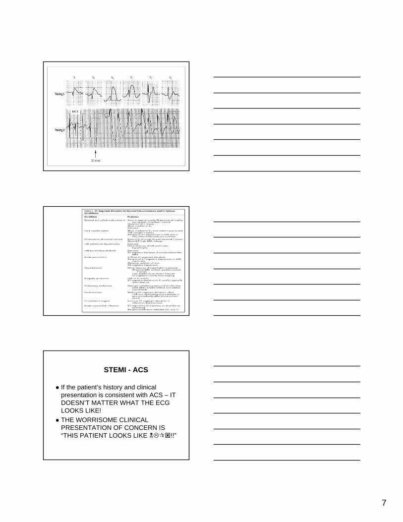
7
STEMI - ACS
If the patient’s history and clinical presentation is consistent with ACS – IT DOESN’T MATTER WHAT THE ECG LOOKS LIKE!THE WORRISOME CLINICAL PRESENTATION OF CONCERN IS “THIS PATIENT LOOKS LIKE !!”

8
ST Elevation – Common Causes
Acute myocardial injury patternAcute pericarditisEarly Repolarization / Normal Variant
+ 0.1mV in any leadUp to 0.3mV in young men in early precordial leads
Myocardial aneurysm
ST Elevation – less common causes
• Pulmonary embolism and acute cor pulmonale (usually lead III)
• Cardiac tumor• Acute aortic dissection• After mitral valvuloplasty• Pancreatitis and
gallbladder disease and other catastrophic illness
• Myocarditis
• Septic shock• Anaphylactic reaction• J wave• Hyperkalemia (“dialyzable”
current of injury)• With any marked QRS
widening (e.g., antidepressant drug overdose) or Class IC antiarrhythmic drugs
Causes of Hypothermia

9
Acute Pericarditis
RBBB
45 yo woman with nausea and vomiting
Which of the following is nottrue about RBBB?
A. RBBB occurs in up to 29% of AMIB. The most common cause in children is post
open heart surgery for Tetralogy of Fallot repair
C. Post MI patients with persistent RBBB are at the same risk for cardiac mortality
D. Transient RBBB is a complication of right heart catheterization

10
Differential Dx of Tall R wave in V1
Definition – R/S ratio > 1 in V1 or V2RBBBRVHTrue Posterior MIWPWBrugada’s Abnormality
65 yo Asian man who presents with recurrent syncope. Echo was normal.
RVH
36 yo woman presenting to the ED with acute dyspnea.

11
Brugada Syndrome
Brugada Abnormality is defined as RBBB with ST elevation and occurrence of sudden cardiac death or syncope due to polymorphous VT (1990)Hereditary bundle branch defect Autosomal dominant trait with variable expressionDiagnosis by EPS with procainamidestimulationTX: AICD
79 yo man admitted with CVA
Diffuse (“Global”) T Wave Inversion
Myocardial Ischemia (Evolving MI is focal)CNS eventApical HCMPericarditisMyocarditisTakatsubo’sCardiomyopathy
Cardiac metastasesCarotid endarterectomyCocaine abusePheochromocytomaAcute illness in women

12
84 yo woman with pneumonia
Which of the following answers about LBBB is not true?
A. LBBB can be caused by toxic, inflammatory changes, hyperkalemia, or digitalis toxicity
B. In most patients with LBBB, septal wall motion abnormalities can exist without coronary artery disease
C. In patients presenting to the ED with chest pain and a LBBB the management is observe with frequent ECGs, treat pain and await enzymes
D. The QRS duration is at least 0.12 seconds
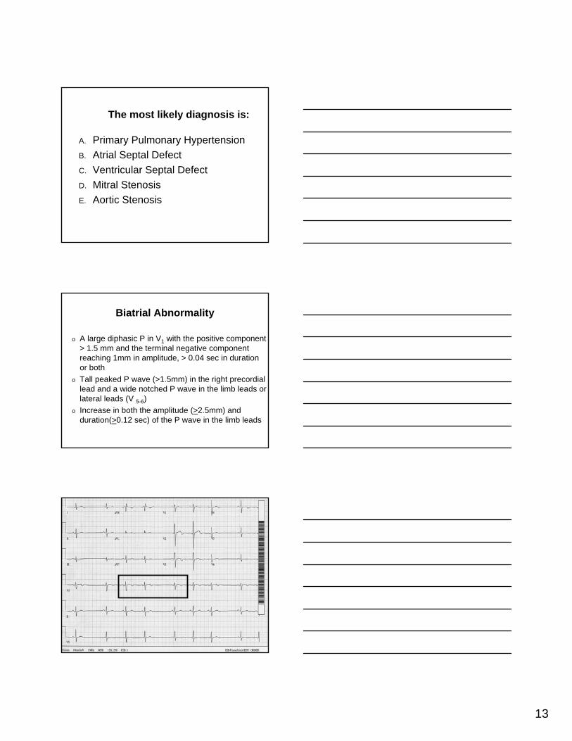
Biatrial Abnormality
32 yo woman from Viet Nam presents with dyspnea on exertion. What is the diagnosis?
13
The most likely diagnosis is:
A. Primary Pulmonary HypertensionB. Atrial Septal DefectC. Ventricular Septal DefectD. Mitral StenosisE. Aortic Stenosis
Biatrial Abnormality
o A large diphasic P in V1 with the positive component > 1.5 mm and the terminal negative component reaching 1mm in amplitude, > 0.04 sec in duration or both
o Tall peaked P wave (>1.5mm) in the right precordial lead and a wide notched P wave in the limb leads or lateral leads (V 5-6)
o Increase in both the amplitude (>2.5mm) and duration(>0.12 sec) of the P wave in the limb leads
Non Conducted PAC’s
14
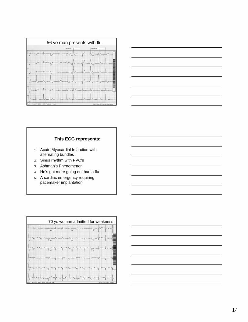
Rate-Related RBBB
56 yo man presents with flu
This ECG represents:
1. Acute Myocardial Infarction with alternating bundles
2. Sinus rhythm with PVC’s3. Ashman’s Phenomenon4. He’s got more going on than a flu5. A cardiac emergency requiring
pacemaker implantation
Giant U waves
70 yo woman admitted for weakness
15
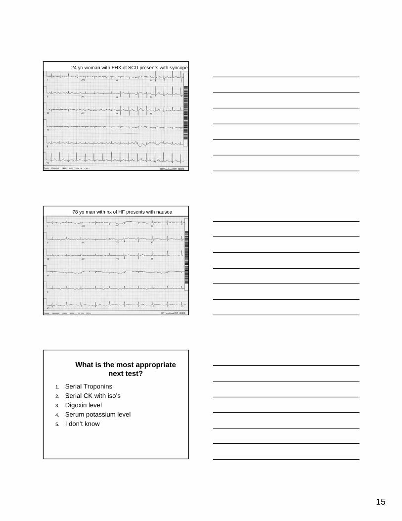
Prolonged QTc = 498 msec
24 yo woman with FHX of SCD presents with syncope
Afib with AV Dissociation
78 yo man with hx of HF presents with nausea
What is the most appropriate next test?
1. Serial Troponins2. Serial CK with iso’s3. Digoxin level4. Serum potassium level5. I don’t know

16
AFib with AV Dissociation
58 58 58 58 58 58 58 58 58
AFib
AFib or AFib/Aflutter or Aflutter?
2nd AVB Mobitz 1
What is the rhythm? What is the treatment?

17
2nd AVB Mobitz 1
What is the rhythm? What is the treatment?
640 640 640p-p
200 240 200pr 240
2nd AVB – Mobitz2
What is the rhythm? What is the treatment?
AVNRT

18
Supraventricular Tachycardias: Which statement is true?
1. If hemodynamically stable, acute management includes Adenosine by slow IV push
2. The majority of SVT is AVNRT3. AVRT does not involve accessory bypass
tracts4. If the patient after adenosine converts from
a narrow complex tachycardia to a wide complex tachycardia, the patient has degenerated to ventricular tachycardia
Slow-fast form of AVNRT
Generation of ECG in Common form of AVNRT

19
Narrow QRS Tachy response to Adenosine
Rx of Hemodynamically stable tachycardia
34 yo woman with idiopathic cardiomyopathy became dizzy and came to the ED.

20
Wide QRS-complex tachycardia
(QRS > 120 ms)
Regular or irregular?
RegularIs QRS identical to that during SR?
If yes, consider:
•SVT and BBB
•Antidromic AVRT
Vagal maneuvers or Adenosine
Previous MI or structural heart disease? If yes, VT is likely
1:1 AV relationship?
Irregular
Atrial Fibrillation
Atrial Flutter/AT with variable conduction and
a) BBB or
b) Antegrade conduction via AP
Yes or unknown No
V rate faster than A rate A rate faster than V rate
VT Atrial Tachycardia
Atrial Flutter
QRS morphology in precordial leads
Next slide
Wide QRS-complex tachycardia I
Wide QRS-complex tachycardia
(QRS > 120 ms)
RegularPrevious MI or structural heart disease? If yes, VT is likely
1:1 AV relationship?
Yes or unknown
QRS morphology in precordial leads
Typical RBBB or LBBB = SVT
Precordial leads VT•Concordant
•No R/S pattern
•Onset of R to nadir > 100 ms
RBBB pattern VT
•qR, Rs or Rr’ in V1
•Frontal plane axis range from +90 degrees to –90 degrees
LBBB pattern VT
•R in V1 > 30 ms
•R to nadir of S in V1 > 60 ms
•qR or qS in V6
Wide QRS-complex tachycardia II
A fib with WPW
21
CHB
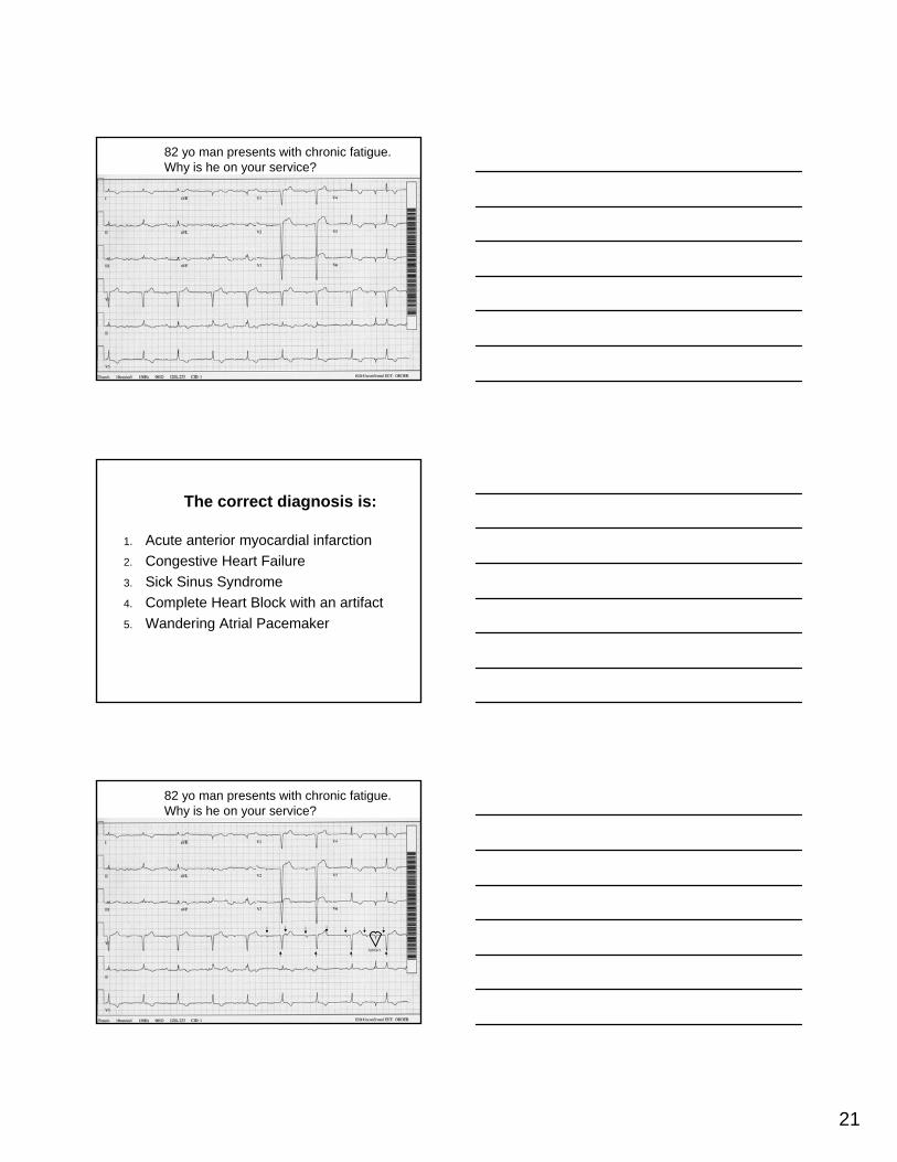
82 yo man presents with chronic fatigue. Why is he on your service?
The correct diagnosis is:
1. Acute anterior myocardial infarction2. Congestive Heart Failure3. Sick Sinus Syndrome4. Complete Heart Block with an artifact5. Wandering Atrial Pacemaker
CHB with Answer
82 yo man presents with chronic fatigue. Why is he on your service?
Artifact
22
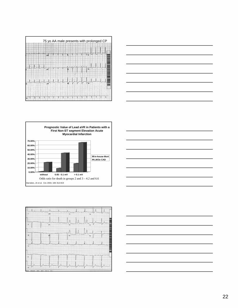
nonSTEMI & aVR
75 yo AA male presents with prolonged CP
Prognostic Value of Lead aVR in Patients with a First Non-ST segment Elevation Acute
Myocardial Infarction
0.00%
10.00%
20.00%
30.00%
40.00%
50.00%
60.00%
70.00%
without 0.05 - 0.1 mV > 0.1 mV
In-house MortLM/3v CAD
Odds ratio for death in groups 2 and 3 – 4.2 and 6.6Barrabes, JA et al. Circ 2003; 108: 814-819
TriFasicular Block

23
First Degree AV Block
Generally benign Bad prognostic sign in:
Bifascicular BlockRBBB + LAFBRBBB + LPFBLBBB
Infectious endocarditis
Q: Had this patient suffered a myocardial infarction?
1. No!2. Yes. Inferior3. Yes. Posterior4. Yes. Septal5. Yes. Localization not possible due to
RBBB
Blocked PAC’s
75 yo man presents with palpitations

24
80 yo man with hx of IMI presents with syncope

25