retrosplenial cortical neurons encode …people.psych.cornell.edu/~dms248/assorted...
TRANSCRIPT
Cerebral Cortex, 2016; 1–11
doi: 10.1093/cercor/bhw192Original Article
O R I G I NA L ART I C L E
Retrosplenial Cortical Neurons Encode NavigationalCues, Trajectories and Reward Locations During GoalDirected NavigationLindsey C. Vedder, Adam M. P. Miller, Marc B. Harrison, and David M. Smith
Department of Psychology, Cornell University, Ithaca, NY 14853, USA
Address correspondence to David M. Smith, Cornell University, Department of Psychology, 211 Uris Hall, Ithaca, NY 14853, USA.Email: [email protected]
AbstractThe retrosplenial cortex (RSC) plays an important role in memory and spatial navigation. It shares functional similaritieswith the hippocampus, including the presence of place fields and lesion-induced impairments in spatial navigation, and theRSC is an important source of visual-spatial input to the hippocampus. Recently, the RSC has been the target of intensescrutiny among investigators of human memory and navigation. fMRI and lesion data suggest an RSC role in the ability touse landmarks to navigate to goal locations. However, no direct neurophysiological evidence of encoding navigational cueshas been reported so the specific RSC contribution to spatial cognition has been uncertain. To examine this, we trained ratson a T-maze task in which the reward location was explicitly cued by a flashing light and we recorded RSC neurons as therats learned. We found that RSC neurons rapidly encoded the light cue. Additionally, RSC neurons encoded the reward andits location, and they showed distinct firing patterns along the left and right trajectories to the goal. These responses mayprovide key information for goal-directed navigation, and the loss of these signals may underlie navigational impairmentsin subjects with RSC damage.
Key words: landmark, learning and memory, visual cue, cingulate cortex
IntroductionThe retrosplenial cortex (RSC) is a key component of the brain’smemory and navigation systems (Vann et al. 2009; Miller et al.2014). Effects of RSC lesions are strikingly similar to the well-known effects of hippocampal lesions, including impairmentsin spatial navigation (Sutherland et al. 1988; Takahashi et al.1997; Harker and Whishaw 2002; Vann and Aggleton 2002), con-textual memory (Keene and Bucci 2008a, 2008b, 2008c, 2009),and episodic memory (Valenstein et al. 1987; Bowers et al.1988). Similar to the hippocampus, RSC neurons exhibit placefields (Cho and Sharp 2001; Smith et al. 2012, but see Alexanderand Nitz 2015) and the firing of RSC neurons is sensitive to thespatial context (Smith et al. 2004). The RSC is an important hubfor visual-spatial information from the dorsal stream into thehippocampus (Kravitz et al. 2011) and inactivation of the RSC
disrupts hippocampal representations (Cooper and Mizumori1999). The RSC also appears to be an important consolidationtarget for hippocampal-dependent memories (Katche et al.2013), especially contextual memories (Keene and Bucci 2008a,2008b, 2008c, 2009; Czajkowski et al. 2014; Tanaka et al. 2014;Sigwald et al. 2015). Consistent with findings from the hippo-campus (Liu et al. 2012; Tonegawa et al. 2015), artificial reacti-vation of the same population of RSC neurons which wasactive during contextual learning can trigger contextual fearmemories (Cowansage et al. 2014).
Despite this growing literature, the specific contribution ofthe RSC to spatial cognition is not known. One hypothesis isthat the RSC plays a critical role in encoding important naviga-tional cues (Auger et al. 2012; Smith et al. 2012; Miller et al.2014). Human subjects with RSC lesions are frequently impaired
© The Author 2016. Published by Oxford University Press. All rights reserved. For Permissions, please e-mail: [email protected]
Cerebral Cortex Advance Access published July 29, 2016 at C
ornell University L
ibrary on August 2, 2016
http://cercor.oxfordjournals.org/D
ownloaded from

in spatial navigation and a striking feature of this deficit is theinability to use landmarks to construct routes to goal locations(Takahashi et al. 1997; Maguire 2001; Ino et al. 2007; Kim et al.2015). Many fMRI studies have suggested an RSC role in naviga-tion (Sugiura et al. 2005; Epstein 2008; Sherrill et al. 2013; Epsteinand Vass 2014), particularly in the use of navigational cues(Wolbers et al. 2004; Epstein et al. 2007). The RSC even appearsto preferentially encode permanent features of the environmentrather than temporary movable objects, which would be lessuseful for navigation (Auger et al. 2012; Auger et al. 2015).However, an unambiguous neurophysiological correlate of navi-gational cue processing has not been observed in RSC neurons.
A previous study from our laboratory provided tentative sup-port for an RSC role in encoding navigational cues (Smith et al.2012). We trained rats on a blocked alternation task, in whichthey approached one location for reward for the first half of thesession and then they switched to a different location for thesecond half. In solving this task, rats invariably employed a“win-stay” strategy: they repeatedly went to the first location aslong as rewards continued to be dispensed there and they onlyswitched to the new location after failing to get a reward at theinitial location. Consistent with the importance of the rewardfor this strategy, a large number of RSC neurons (~40%) select-ively responded to either the left or right reward. It is possiblethat these neurons encoded the reward as a navigational cue(i.e. “return to this location on the next trial”). Alternatively, RSCneurons may have been sensitive to the conjunction of thereward and its spatial location. Similar conjunctive coding is aprominent feature of hippocampal responses (Komorowski et al.2009; McKenzie et al. 2014) and recent findings indicate that RSCneurons are sensitive to conjunctions of movements (left andright turns) and the rat’s location and trajectory within theenvironment (Alexander and Nitz 2015). In order to conclusivelydetermine whether RSC neurons encode navigational cues, wetrained rats on a T-maze task in which the reward location wasexplicitly cued by a flashing light which serves as a beacon thatthe rats must learn to approach. We chose to use a beacon,rather than one or more landmarks, because the light has anunambiguous onset time and we could be confident that therats would attend to this highly salient cue. We recorded RSCneuronal responses to the light cue and other task events aswell as spatial firing patterns throughout learning.
Materials and MethodsSubjects and Surgical Procedures
Subjects were 5 adult male Long Evans rats (Charles RiverLaboratories, Wilmington, MA) weighing 250–300 g upon arrival.
Rats were placed on a 12hr/12 hr light/dark cycle with lights onat 7 am and allowed to acclimate to the vivarium for at least oneweek prior to surgery. Rats were implanted with a custom-builtelectrode microdrive (Macdonald et al. 2011) containing 16 mov-able tetrodes made from twisting together four 17 μm platinum/iridium (90%/10%) wires, platinum plated to an impedance of100–300 kΩ, and arranged in a linear 2 × 8 array that spannedapproximately 4mm along the rostrocaudal axis of the brain.Tetrodes were stereotaxically positioned unilaterally, just abovethe RSC at 2.5–6.5mm posterior to Bregma, 0.5mm lateral, and0.84mm ventral to the cortical surface. Rats were given 7 daysto recover from surgery prior to lowering the tetrodes into theRSC over the course of several days (35–70 μm daily) until adepth of at least 1.0mm was reached (granular area b of theRSC) and a sufficient population of neurons was acquired. Allprocedures complied with the guidelines of the CornellUniversity Animal Care and Use Committee.
Behavioral Training
The behavioral apparatus was a black PVC T-maze (112 cm longstem × 122 cm wide × 68 cm above the floor) that occupied a cir-cular arena enclosed by black curtains. Visual cues of variousshapes, sizes and colors were pinned to the curtains. Prior totraining, rats were food restricted to 85–90% of free-feedingbody weight and acclimated to the maze and chocolate milkrewards (200 μL, Nestle’s Quik) until they consumed 20 rewardswithin 30min, usually 2 sessions.
Prior to the regular training sessions, each rat was given apreliminary training session (pretraining) in order to collectbaseline neural responses to the light cue before learning.During this session, a single light cue, identical to the lightsthat were later used to cue the reward locations (see below),was positioned at the choice point. During this session, thelight was turned on for half of the 20 trials and both rewardcups were always baited. Thus, the light did not signal thereward location and these recordings served as a measure ofbaseline responsivity to the light prior to associative learning.
During regular training sessions, two light cues (each con-structed from side by side LEDs, 5 cm apart that flashed alter-natingly at 3 Hz) were positioned 9 cm above each of the rewardlocations. The timeline for the trials is indicated in Figure 1. Atthe start of each trial, the rat was placed on the stem of themaze facing away from the choice point. As soon as the ratturned around, the experimenter turned on one of the two lightcues to indicate the reward location for that trial. The lightremained on until the rat arrived at the reward location andconsumed the reward, after which the rats were placed on a
Trial Start Light
On
Choice
Point
Reward Light
Off
ITI
~30s ~3s ~1.5s ~1.5s ~3s
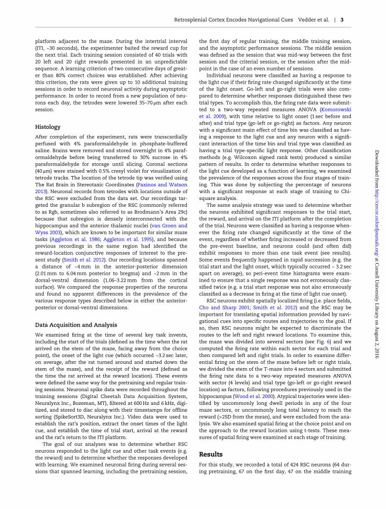
Figure 1. Maze diagram and schematic of the trials. The rats were trained on a T-maze task in which the reward locations (small circles) were indicated by flashing
lights near the rewards (a left-rewarded example is indicated). The schematic timeline indicates key task events, including the trial start, light onset, arrival at the
choice point and the reward. After a ~30 sec ITI, the rats were placed on the maze facing away from the choice point (Trial Start). One of the cue lights was illumi-
nated ~3 sec later when the rat started down the stem of the maze (dashed line on the T-maze diagram). The light remained illuminated throughout the trial, until
after the rat consumed the reward. Approximately 1.5 sec after light onset, the rats reached the choice point and after another ~1.5 sec the rat reached the reward
location. Each of these times varied somewhat from one rat to another and across trials. Analysis of event-related firing of individual neurons compared firing during
the 1 sec before and after the event (grey shaded regions).
2 | Cerebral Cortex
at Cornell U
niversity Library on A
ugust 2, 2016http://cercor.oxfordjournals.org/
Dow
nloaded from
platform adjacent to the maze. During the intertrial interval(ITI, ~30 seconds), the experimenter baited the reward cup forthe next trial. Each training session consisted of 40 trials with20 left and 20 right rewards presented in an unpredictablesequence. A learning criterion of two consecutive days of great-er than 80% correct choices was established. After achievingthis criterion, the rats were given up to 10 additional trainingsessions in order to record neuronal activity during asymptoticperformance. In order to record from a new population of neu-rons each day, the tetrodes were lowered 35–70 μm after eachsession.
Histology
After completion of the experiment, rats were transcardiallyperfused with 4% paraformaldehyde in phosphate-bufferedsaline. Brains were removed and stored overnight in 4% paraf-ormaldehyde before being transferred to 30% sucrose in 4%paraformaldehyde for storage until slicing. Coronal sections(40 μm) were stained with 0.5% cresyl violet for visualization oftetrode tracks. The location of the tetrode tip was verified usingThe Rat Brain in Stereotaxic Coordinates (Paxinos and Watson2013). Neuronal records from tetrodes with locations outside ofthe RSC were excluded from the data set. Our recordings tar-geted the granular b subregion of the RSC (commonly referredto as Rgb, sometimes also referred to as Brodmann’s Area 29c)because that subregion is densely interconnected with thehippocampus and the anterior thalamic nuclei (van Groen andWyss 2003), which are known to be important for similar mazetasks (Aggleton et al. 1986; Aggleton et al. 1995), and becauseprevious recordings in the same region had identified thereward-location conjunctive responses of interest to the pre-sent study (Smith et al. 2012). Our recording locations spanneda distance of ~4mm in the anterior-posterior dimension(2.01mm to 6.04mm posterior to bregma) and ~2mm in thedorsal-ventral dimension (1.06–3.22mm from the corticalsurface). We compared the response properties of the neuronsand found no apparent differences in the prevalence of thevarious response types described below in either the anterior-posterior or dorsal-ventral dimensions.
Data Acquisition and Analysis
We examined firing at the time of several key task invents,including the start of the trials (defined as the time when the ratarrived on the stem of the maze, facing away from the choicepoint), the onset of the light cue (which occurred ~3.2 sec later,on average, after the rat turned around and started down thestem of the maze), and the receipt of the reward (defined asthe time the rat arrived at the reward location). These eventswere defined the same way for the pretraining and regular train-ing sessions. Neuronal spike data were recorded throughout thetraining sessions (Digital Cheetah Data Acquisition System,Neuralynx Inc., Bozeman, MT), filtered at 600Hz and 6 kHz, digi-tized, and stored to disc along with their timestamps for offlinesorting (SpikeSort3D, Neuralynx Inc.). Video data were used toestablish the rat’s position, extract the onset times of the lightcue, and establish the time of trial start, arrival at the rewardand the rat’s return to the ITI platform.
The goal of our analyses was to determine whether RSCneurons responded to the light cue and other task events (e.g.the reward) and to determine whether the responses developedwith learning. We examined neuronal firing during several ses-sions that spanned learning, including the pretraining session,
the first day of regular training, the middle training session,and the asymptotic performance sessions. The middle sessionwas defined as the session that was mid-way between the firstsession and the criterial session, or the session after the mid-point in the case of an even number of sessions.
Individual neurons were classified as having a response tothe light cue if their firing rate changed significantly at the timeof the light onset. Go-left and go-right trials were also com-pared to determine whether responses distinguished these twotrial types. To accomplish this, the firing rate data were submit-ted to a two-way repeated measures ANOVA (Komorowskiet al. 2009), with time relative to light onset (1 sec before andafter) and trial type (go-left or go-right) as factors. Any neuronwith a significant main effect of time bin was classified as hav-ing a response to the light cue and any neuron with a signifi-cant interaction of the time bin and trial type was classified ashaving a trial type-specific light response. Other classificationmethods (e.g. Wilcoxon signed rank tests) produced a similarpattern of results. In order to determine whether responses tothe light cue developed as a function of learning, we examinedthe prevalence of the responses across the four stages of train-ing. This was done by subjecting the percentage of neuronswith a significant response at each stage of training to Chi-square analysis.
The same analysis strategy was used to determine whetherthe neurons exhibited significant responses to the trial start,the reward, and arrival on the ITI platform after the completionof the trial. Neurons were classified as having a response when-ever the firing rate changed significantly at the time of theevent, regardless of whether firing increased or decreased fromthe pre-event baseline, and neurons could (and often did)exhibit responses to more than one task event (see results).Some events frequently happened in rapid succession (e.g. thetrial start and the light onset, which typically occurred ~ 3.2 secapart on average), so peri-event time histograms were exam-ined to ensure that a single response was not erroneously clas-sified twice (e.g. a trial start response was not also erroneouslyclassified as a decrease in firing at the time of light cue onset).
RSC neurons exhibit spatially localized firing (i.e. place fields,Cho and Sharp 2001; Smith et al. 2012) and the RSC may beimportant for translating spatial information provided by navi-gational cues into specific routes and trajectories to the goal. Ifso, then RSC neurons might be expected to discriminate theroutes to the left and right reward locations. To examine this,the maze was divided into several sectors (see Fig. 6) and wecomputed the firing rate within each sector for each trial andthen compared left and right trials. In order to examine differ-ential firing on the stem of the maze before left or right trials,we divided the stem of the T-maze into 4 sectors and submittedthe firing rate data to a two-way repeated measures ANOVAwith sector (4 levels) and trial type (go-left or go-right rewardlocation) as factors, following procedures previously used in thehippocampus (Wood et al. 2000). Atypical trajectories were iden-tified by uncommonly long dwell periods in any of the fourmaze sectors, or uncommonly long total latency to reach thereward (>2SD from the mean), and were excluded from the ana-lysis. We also examined spatial firing at the choice point and onthe approach to the reward location using t-tests. These mea-sures of spatial firing were examined at each stage of training.
ResultsFor this study, we recorded a total of 424 RSC neurons (64 dur-ing pretraining, 67 on the first day, 47 on the middle training
Retrosplenial Cortex Encodes Navigational Cues Vedder et al. | 3
at Cornell U
niversity Library on A
ugust 2, 2016http://cercor.oxfordjournals.org/
Dow
nloaded from
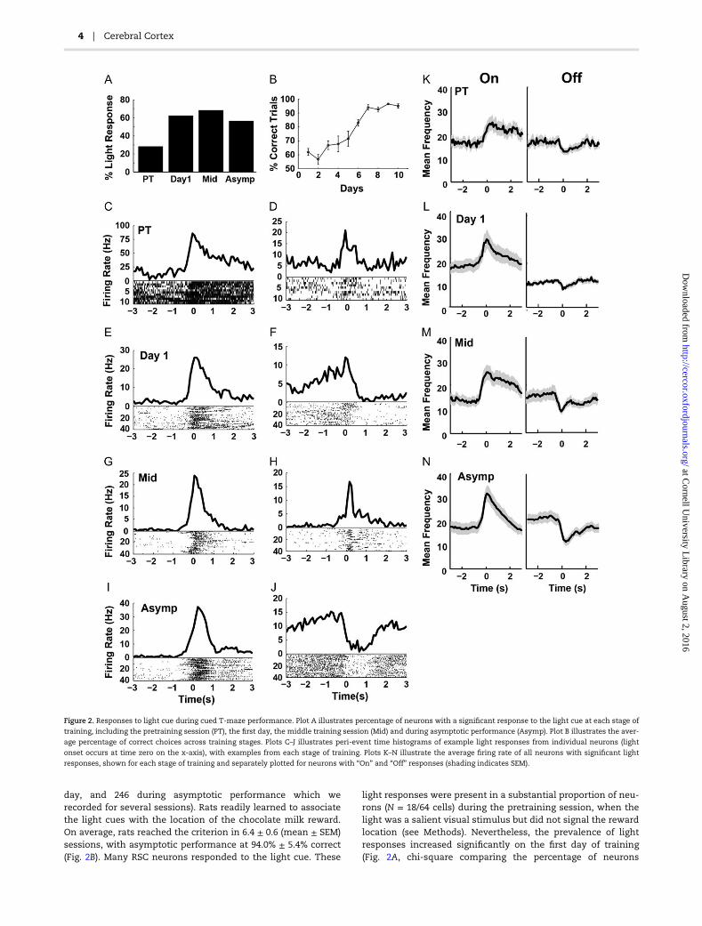
day, and 246 during asymptotic performance which werecorded for several sessions). Rats readily learned to associatethe light cues with the location of the chocolate milk reward.On average, rats reached the criterion in 6.4 ± 0.6 (mean ± SEM)sessions, with asymptotic performance at 94.0% ± 5.4% correct(Fig. 2B). Many RSC neurons responded to the light cue. These
light responses were present in a substantial proportion of neu-rons (N = 18/64 cells) during the pretraining session, when thelight was a salient visual stimulus but did not signal the rewardlocation (see Methods). Nevertheless, the prevalence of lightresponses increased significantly on the first day of training(Fig. 2A, chi-square comparing the percentage of neurons
Figure 2. Responses to light cue during cued T-maze performance. Plot A illustrates percentage of neurons with a significant response to the light cue at each stage of
training, including the pretraining session (PT), the first day, the middle training session (Mid) and during asymptotic performance (Asymp). Plot B illustrates the aver-
age percentage of correct choices across training stages. Plots C–J illustrates peri-event time histograms of example light responses from individual neurons (light
onset occurs at time zero on the x-axis), with examples from each stage of training. Plots K–N illustrate the average firing rate of all neurons with significant light
responses, shown for each stage of training and separately plotted for neurons with “On” and “Off” responses (shading indicates SEM).
4 | Cerebral Cortex
at Cornell U
niversity Library on A
ugust 2, 2016http://cercor.oxfordjournals.org/
Dow
nloaded from
during pretraining and the first day, χ2 = 15.15, P < 0.0001) andremained increased thereafter (χ2 = 22.55, P < 0.0001). The per-centage of neurons with light responses increased significantlyas soon as the light became a reliable indicator of the rewardlocation and, interestingly, long before the rats began to per-form the task reliably. At asymptote, more than half of therecorded neurons (142/246) showed significant responses to thelight cue. Thus, the RSC exhibits large scale encoding of thisnavigational cue.
Light responses took the form of increased firing (“On”responses, 62.6% of light responses, Fig. 2K–N, left) or decreasedfiring (“Off” responses, 37.3% of light responses, Fig. 2K–N, right)at the time of light onset. These “responses” sometimes beganslightly before the onset of the light cue, which is not surprisingsince the rats presumably learned to anticipate that the cuewould be given as soon as they turned around and starteddown the stem of the maze. Anticipatory firing before a predict-able cue has been reported previously in the RSC (Smith et al.2002). Our previous study indicated that RSC reward responseswere remarkably selective for different spatial locations (Smithet al. 2012). In the present study, the two light cues were in dif-ferent locations, so the responses might have been specific toone location or the other. However, only a small minority of theneurons exhibited significantly different responses to the rightand left light cues (5.9% over all training stages) and this per-centage did not change significantly with training (χ2 = 0.93,P = 0.81). Although the light cue was strongly represented inthe RSC, the representation was not sensitive to the location ofthe light.
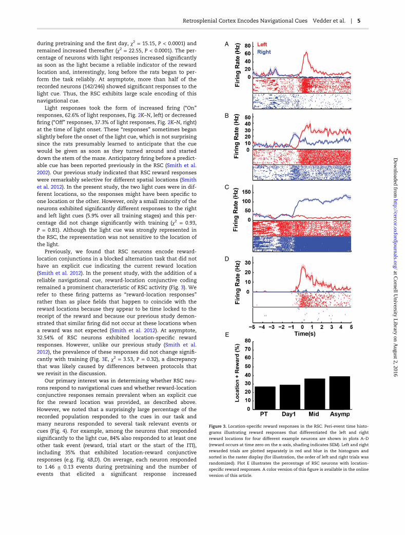
Previously, we found that RSC neurons encode reward-location conjunctions in a blocked alternation task that did nothave an explicit cue indicating the current reward location(Smith et al. 2012). In the present study, with the addition of areliable navigational cue, reward-location conjunctive codingremained a prominent characteristic of RSC activity (Fig. 3). Werefer to these firing patterns as “reward-location responses”rather than as place fields that happen to coincide with thereward locations because they appear to be time locked to thereceipt of the reward and because our previous study demon-strated that similar firing did not occur at these locations whena reward was not expected (Smith et al. 2012). At asymptote,32.54% of RSC neurons exhibited location-specific rewardresponses. However, unlike our previous study (Smith et al.2012), the prevalence of these responses did not change signifi-cantly with training (Fig. 3E, χ2 = 3.53, P = 0.32), a discrepancythat was likely caused by differences between protocols thatwe revisit in the discussion.
Our primary interest was in determining whether RSC neu-rons respond to navigational cues and whether reward-locationconjunctive responses remain prevalent when an explicit cuefor the reward location was provided, as described above.However, we noted that a surprisingly large percentage of therecorded population responded to the cues in our task andmany neurons responded to several task relevant events orcues (Fig. 4). For example, among the neurons that respondedsignificantly to the light cue, 84% also responded to at least oneother task event (reward, trial start or the start of the ITI),including 35% that exhibited location-reward conjunctiveresponses (e.g. Fig. 4B,D). On average, each neuron respondedto 1.46 ± 0.13 events during pretraining and the number ofevents that elicited a significant response increased
Figure 3. Location-specific reward responses in the RSC. Peri-event time histo-
grams illustrating reward responses that differentiated the left and right
reward locations for four different example neurons are shown in plots A–D
(reward occurs at time zero on the x-axis, shading indicates SEM). Left and right
rewarded trials are plotted separately in red and blue in the histogram and
sorted in the raster display (for illustration, the order of left and right trials was
randomized). Plot E illustrates the percentage of RSC neurons with location-
specific reward responses. A color version of this figure is available in the online
version of this article.
Retrosplenial Cortex Encodes Navigational Cues Vedder et al. | 5
at Cornell U
niversity Library on A
ugust 2, 2016http://cercor.oxfordjournals.org/
Dow
nloaded from
significantly to 2.10 ± 0.22 on the first day of training (Fig. 4E,F(3,428) = 7.66, P < 0.0001).
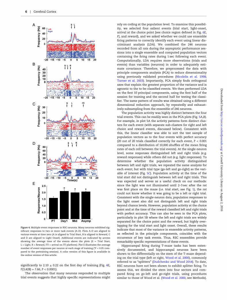
The observation that many neurons responded to multipletrial events suggests that highly specific representations might
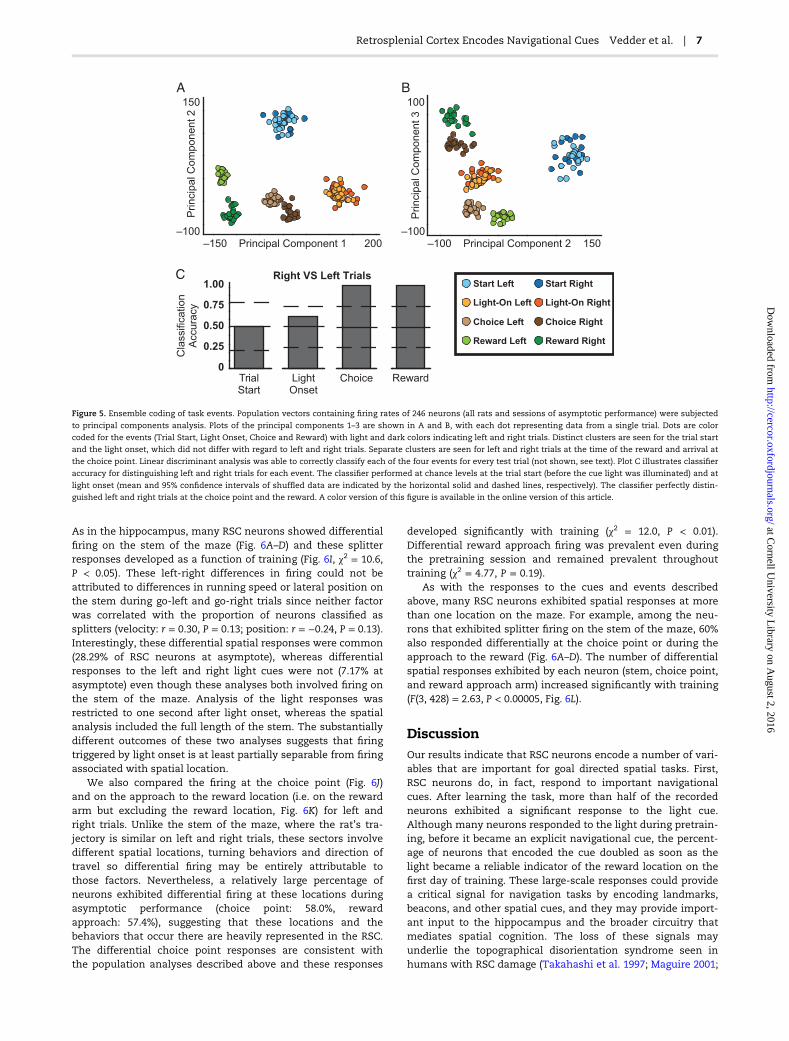
rely on coding at the population level. To examine this possibil-ity, we selected four salient events (trial start, light-onset,arrival at the choice point [see choice region defined in Fig. 6E,F], and reward), and we asked whether we could use ensemblefiring patterns to correctly identify each event using linear dis-criminant analysis (LDA). We combined the 246 neuronsrecorded from all rats during the asymptotic performance ses-sions into a single ensemble and computed population vectorscontaining the firing rates during 1 sec following each event.Computationally, LDA requires more observations (trials andevents) than variables (neurons) in order to adequately esti-mate covariance. Therefore, we preprocessed the data withprinciple components analysis (PCA) to reduce dimensionalityusing previously validated procedures (Nicolelis et al. 1998;Turner et al. 2003). Importantly, PCA simply finds orthogonalaxes that explain the greatest proportion of the variance and isagnostic to the to-be-classified events. We then performed LDAon the first 10 principal components, using the first half of thesession for training and the second half for testing the classi-fier. The same pattern of results was obtained using a differentdimensional reduction approach, by repeatedly and exhaust-ively subsampling from the ensemble of 246 neurons.
The population activity was highly distinct between the fourtrial events. This can be readily seen in the PCA plots (Fig. 5A,B).For example, in plot 5A the activity patterns form distinct clus-ters for each event (with separate sub-clusters for right and leftchoice and reward events, discussed below). Consistent withthis, the linear classifier was able to sort the test sample ofpopulation vectors as to the four events with perfect accuracy(20 out of 20 trials classified correctly for each event, P < 0.001compared to a distribution of 10,000 shuffles of the mean firingrates of each cell between the trial events). At the single-neuronlevel, some responses distinguished left and right trials (e.g.reward responses) while others did not (e.g. light responses). Todetermine whether the population activity distinguishedbetween left and right trials, we repeated the same analysis foreach event, but with trial type (go-left and go-right) as the vari-able of interest (Fig. 5C). Population activity at the time of thetrial start did not distinguish between left and right trials. Thiswas expected and serves as a useful check on our methods:since the light was not illuminated until 2–3 sec after the ratwas first place on the maze (i.e. trial start, see Fig. 1), the ratcould not know whether it was going to be a left or right trial.Consistent with the single-neuron data, population responses tothe light onset also did not distinguish left and right trialsbeyond chance levels. However, population activity at the choicepoint and at the time of the reward classified left and right trialswith perfect accuracy. This can also be seen in the PCA plots,particularly in plot 5B where the left and right trials are widelyseparated for the choice point and the reward, but highly over-lapping for the trial start and light onset. Overall, these resultsindicate that most of the variance in ensemble activity patterns,as reflected in the principle components, coincides with theoccurrence of key task events. Thus, RSC ensembles provideremarkably specific representations of these events.
Hippocampal firing during T-maze tasks has been exten-sively documented, and hippocampal neurons have beenshown to fire differentially on the stem of the T-maze depend-ing on the trial type (left or right, Wood et al. 2000), commonlyreferred to as “splitters” (Dudchenko and Wood 2014). To date,RSC neurons have not been shown to exhibit splitter firing. Toassess this, we divided the stem into four sectors and com-pared firing on go-left and go-right trials, using proceduressimilar to those of Wood et al. (Wood et al. 2000, see Methods).
Figure 4. Multiple event responses in RSC neurons. Many neurons exhibited sig-
nificant responses to two or more task events (A–D). Plots A–D are aligned to
various events at time zero (A is aligned to Trial Start, B is aligned to Reward, C
and D are aligned to Light Onset). Additional events are indicated by arrows
showing the average time of the events above the plots (S = Trial Start,
L = Light, R = Reward, ITI = arrival on ITI platform). Plot E illustrates the average
number of event responses per neuron at each stage of training (*P < 0.05 com-
pared to the pretraining session). A color version of this figure is available in
the online version of this article.
6 | Cerebral Cortex
at Cornell U
niversity Library on A
ugust 2, 2016http://cercor.oxfordjournals.org/
Dow
nloaded from
As in the hippocampus, many RSC neurons showed differentialfiring on the stem of the maze (Fig. 6A–D) and these splitterresponses developed as a function of training (Fig. 6I, χ2 = 10.6,P < 0.05). These left-right differences in firing could not beattributed to differences in running speed or lateral position onthe stem during go-left and go-right trials since neither factorwas correlated with the proportion of neurons classified assplitters (velocity: r = 0.30, P = 0.13; position: r = −0.24, P = 0.13).Interestingly, these differential spatial responses were common(28.29% of RSC neurons at asymptote), whereas differentialresponses to the left and right light cues were not (7.17% atasymptote) even though these analyses both involved firing onthe stem of the maze. Analysis of the light responses wasrestricted to one second after light onset, whereas the spatialanalysis included the full length of the stem. The substantiallydifferent outcomes of these two analyses suggests that firingtriggered by light onset is at least partially separable from firingassociated with spatial location.
We also compared the firing at the choice point (Fig. 6J)and on the approach to the reward location (i.e. on the rewardarm but excluding the reward location, Fig. 6K) for left andright trials. Unlike the stem of the maze, where the rat’s tra-jectory is similar on left and right trials, these sectors involvedifferent spatial locations, turning behaviors and direction oftravel so differential firing may be entirely attributable tothose factors. Nevertheless, a relatively large percentage ofneurons exhibited differential firing at these locations duringasymptotic performance (choice point: 58.0%, rewardapproach: 57.4%), suggesting that these locations and thebehaviors that occur there are heavily represented in the RSC.The differential choice point responses are consistent withthe population analyses described above and these responses
developed significantly with training (χ2 = 12.0, P < 0.01).Differential reward approach firing was prevalent even duringthe pretraining session and remained prevalent throughouttraining (χ2 = 4.77, P = 0.19).
As with the responses to the cues and events describedabove, many RSC neurons exhibited spatial responses at morethan one location on the maze. For example, among the neu-rons that exhibited splitter firing on the stem of the maze, 60%also responded differentially at the choice point or during theapproach to the reward (Fig. 6A–D). The number of differentialspatial responses exhibited by each neuron (stem, choice point,and reward approach arm) increased significantly with training(F(3, 428) = 2.63, P < 0.00005, Fig. 6L).
DiscussionOur results indicate that RSC neurons encode a number of vari-ables that are important for goal directed spatial tasks. First,RSC neurons do, in fact, respond to important navigationalcues. After learning the task, more than half of the recordedneurons exhibited a significant response to the light cue.Although many neurons responded to the light during pretrain-ing, before it became an explicit navigational cue, the percent-age of neurons that encoded the cue doubled as soon as thelight became a reliable indicator of the reward location on thefirst day of training. These large-scale responses could providea critical signal for navigation tasks by encoding landmarks,beacons, and other spatial cues, and they may provide import-ant input to the hippocampus and the broader circuitry thatmediates spatial cognition. The loss of these signals mayunderlie the topographical disorientation syndrome seen inhumans with RSC damage (Takahashi et al. 1997; Maguire 2001;
–150 200–100
150
–100 150–100
100
0
1.00
0.50
0.75
0.25
Start Left
Light-On Left
Choice Left
Reward Left
Start Right
Light-On Right
Choice Right
Reward Right
Principal Component 1 Principal Component 2
Princip
al C
om
ponent 2
Princip
al C
om
ponent 3
Cla
ssific
ation
Accura
cy
TrialStart
LightOnset
Choice Reward
Right VS Left Trials
A
C
B
Figure 5. Ensemble coding of task events. Population vectors containing firing rates of 246 neurons (all rats and sessions of asymptotic performance) were subjected
to principal components analysis. Plots of the principal components 1–3 are shown in A and B, with each dot representing data from a single trial. Dots are color
coded for the events (Trial Start, Light Onset, Choice and Reward) with light and dark colors indicating left and right trials. Distinct clusters are seen for the trial start
and the light onset, which did not differ with regard to left and right trials. Separate clusters are seen for left and right trials at the time of the reward and arrival at
the choice point. Linear discriminant analysis was able to correctly classify each of the four events for every test trial (not shown, see text). Plot C illustrates classifier
accuracy for distinguishing left and right trials for each event. The classifier performed at chance levels at the trial start (before the cue light was illuminated) and at
light onset (mean and 95% confidence intervals of shuffled data are indicated by the horizontal solid and dashed lines, respectively). The classifier perfectly distin-
guished left and right trials at the choice point and the reward. A color version of this figure is available in the online version of this article.
Retrosplenial Cortex Encodes Navigational Cues Vedder et al. | 7
at Cornell U
niversity Library on A
ugust 2, 2016http://cercor.oxfordjournals.org/
Dow
nloaded from

Kim et al. 2015) and the spatial impairments seen in rodentswith RSC lesions (Sutherland et al. 1988; Vann and Aggleton2005; Nelson et al. 2015).
RSC neurons also encoded important spatial conjunctions.For example, many neurons exhibited responses that weretightly time-locked to the reward, but only when the rewardwas presented at one of the two goal locations (left or right).Similar conjunctive coding is a well-documented characteristicof hippocampal neurons, which also respond to reward-location conjunctions (Smith and Mizumori 2006) and conjunc-tions of other task relevant cues, such as odor cues andlocations (Komorowski et al. 2009; McKenzie et al. 2014).Previously, we reported reward-location conjunctive responses inthe RSC (Smith et al. 2012). However, because the rats used awin-stay strategy, continually returning to the previous rewardlocation, the reward location was, itself, an important predictivecue (i.e. it signaled the reward location for the next trial) and wesuggested that the RSC responses may have reflected cue encod-ing processes. However, the present results demonstrate thatRSC neurons exhibit conjunctive responses even when thereward location cannot serve as a predictive cue, as in the pre-sent study where the reward locations were randomized fromone trial to the next. This result suggests that conjunctive coding
of important events and the locations where they occur is a gen-eral characteristic of RSC coding, as it is in the hippocampus.
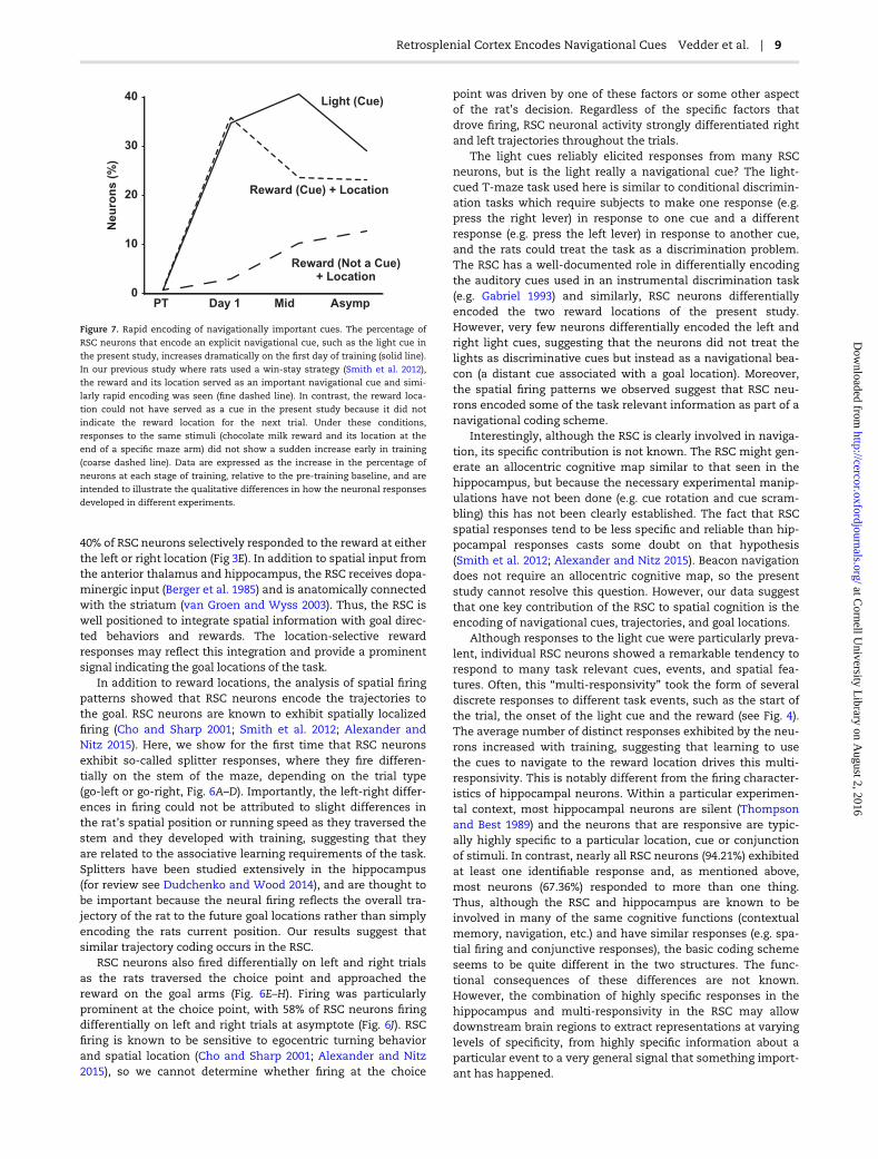
Because we found reward-location conjunctive responses inthe present study, we can compare the development of theseresponses under conditions of differing information value(Fig. 7). In our previous study (Smith et al. 2012), when thereward location served as an important navigational cue,responses developed immediately, just as the light responsesdeveloped in the present study. However, when the rewardlocation could not be used as a navigational cue, the responsesdid not suddenly appear early in learning. This difference isparticularly striking since the stimuli that evoked the response,two drops of chocolate milk delivered to a metal cup at the endof the maze arm, were the same in both experiments. Thiscomparison suggests that whenever a particular stimulus canserve as a navigational cue, regardless of whether it is an expli-cit cue such as the light or a more abstract cue such as thereward location in a win-stay task, it evokes a large and rapidresponse in the RSC. When the same stimuli are not useful asnavigational cues, the RSC may still encode them but not asrapidly or robustly.
Goal locations and trajectories to the goal also appear to beparticularly important for RSC encoding. At asymptote, about
Figure 6. Spatial firing during cued T-maze performance. Examples of neurons that exhibited differential firing on the stem of the maze for go-left and go-right trials
(plots A–D). For each neuron separate firing rate maps are shown for left and right trials, with the sectors of the maze that were analyzed indicated. The average firing
rates in each sector are shown in the line graphs below each plot. RSC neurons also frequently exhibited differential firing on left and right trials at the choice point
(E–F) or on the reward arm as the rat approached the reward (G–H). Mean firing rates for the analyzed maze sections are shown to the left of the firing rate maps. Bar
plots illustrating the percentage of neurons that exhibited differential firing on the stem (I), at the choice point (J) and reward arm (K) are shown for each stage of train-
ing. Plot L illustrates the average number of differential spatial responses per neuron for each stage of training (*P < 0.05 compared to the pretraining session). A color
version of this figure is available in the online version of this article.
8 | Cerebral Cortex
at Cornell U
niversity Library on A
ugust 2, 2016http://cercor.oxfordjournals.org/
Dow
nloaded from
40% of RSC neurons selectively responded to the reward at eitherthe left or right location (Fig 3E). In addition to spatial input fromthe anterior thalamus and hippocampus, the RSC receives dopa-minergic input (Berger et al. 1985) and is anatomically connectedwith the striatum (van Groen and Wyss 2003). Thus, the RSC iswell positioned to integrate spatial information with goal direc-ted behaviors and rewards. The location-selective rewardresponses may reflect this integration and provide a prominentsignal indicating the goal locations of the task.
In addition to reward locations, the analysis of spatial firingpatterns showed that RSC neurons encode the trajectories tothe goal. RSC neurons are known to exhibit spatially localizedfiring (Cho and Sharp 2001; Smith et al. 2012; Alexander andNitz 2015). Here, we show for the first time that RSC neuronsexhibit so-called splitter responses, where they fire differen-tially on the stem of the maze, depending on the trial type(go-left or go-right, Fig. 6A–D). Importantly, the left-right differ-ences in firing could not be attributed to slight differences inthe rat’s spatial position or running speed as they traversed thestem and they developed with training, suggesting that theyare related to the associative learning requirements of the task.Splitters have been studied extensively in the hippocampus(for review see Dudchenko and Wood 2014), and are thought tobe important because the neural firing reflects the overall tra-jectory of the rat to the future goal locations rather than simplyencoding the rats current position. Our results suggest thatsimilar trajectory coding occurs in the RSC.
RSC neurons also fired differentially on left and right trialsas the rats traversed the choice point and approached thereward on the goal arms (Fig. 6E–H). Firing was particularlyprominent at the choice point, with 58% of RSC neurons firingdifferentially on left and right trials at asymptote (Fig. 6J). RSCfiring is known to be sensitive to egocentric turning behaviorand spatial location (Cho and Sharp 2001; Alexander and Nitz2015), so we cannot determine whether firing at the choice
point was driven by one of these factors or some other aspectof the rat’s decision. Regardless of the specific factors thatdrove firing, RSC neuronal activity strongly differentiated rightand left trajectories throughout the trials.
The light cues reliably elicited responses from many RSCneurons, but is the light really a navigational cue? The light-cued T-maze task used here is similar to conditional discrimin-ation tasks which require subjects to make one response (e.g.press the right lever) in response to one cue and a differentresponse (e.g. press the left lever) in response to another cue,and the rats could treat the task as a discrimination problem.The RSC has a well-documented role in differentially encodingthe auditory cues used in an instrumental discrimination task(e.g. Gabriel 1993) and similarly, RSC neurons differentiallyencoded the two reward locations of the present study.However, very few neurons differentially encoded the left andright light cues, suggesting that the neurons did not treat thelights as discriminative cues but instead as a navigational bea-con (a distant cue associated with a goal location). Moreover,the spatial firing patterns we observed suggest that RSC neu-rons encoded some of the task relevant information as part of anavigational coding scheme.
Interestingly, although the RSC is clearly involved in naviga-tion, its specific contribution is not known. The RSC might gen-erate an allocentric cognitive map similar to that seen in thehippocampus, but because the necessary experimental manip-ulations have not been done (e.g. cue rotation and cue scram-bling) this has not been clearly established. The fact that RSCspatial responses tend to be less specific and reliable than hip-pocampal responses casts some doubt on that hypothesis(Smith et al. 2012; Alexander and Nitz 2015). Beacon navigationdoes not require an allocentric cognitive map, so the presentstudy cannot resolve this question. However, our data suggestthat one key contribution of the RSC to spatial cognition is theencoding of navigational cues, trajectories, and goal locations.
Although responses to the light cue were particularly preva-lent, individual RSC neurons showed a remarkable tendency torespond to many task relevant cues, events, and spatial fea-tures. Often, this “multi-responsivity” took the form of severaldiscrete responses to different task events, such as the start ofthe trial, the onset of the light cue and the reward (see Fig. 4).The average number of distinct responses exhibited by the neu-rons increased with training, suggesting that learning to usethe cues to navigate to the reward location drives this multi-responsivity. This is notably different from the firing character-istics of hippocampal neurons. Within a particular experimen-tal context, most hippocampal neurons are silent (Thompsonand Best 1989) and the neurons that are responsive are typic-ally highly specific to a particular location, cue or conjunctionof stimuli. In contrast, nearly all RSC neurons (94.21%) exhibitedat least one identifiable response and, as mentioned above,most neurons (67.36%) responded to more than one thing.Thus, although the RSC and hippocampus are known to beinvolved in many of the same cognitive functions (contextualmemory, navigation, etc.) and have similar responses (e.g. spa-tial firing and conjunctive responses), the basic coding schemeseems to be quite different in the two structures. The func-tional consequences of these differences are not known.However, the combination of highly specific responses in thehippocampus and multi-responsivity in the RSC may allowdownstream brain regions to extract representations at varyinglevels of specificity, from highly specific information about aparticular event to a very general signal that something import-ant has happened.
0
10
20
30
40
PT Day 1 Mid Asymp
Neu
ron
s (
%)
Light (Cue)
Reward (Cue) + Location
Reward (Not a Cue)+ Location
Figure 7. Rapid encoding of navigationally important cues. The percentage of
RSC neurons that encode an explicit navigational cue, such as the light cue in
the present study, increases dramatically on the first day of training (solid line).
In our previous study where rats used a win-stay strategy (Smith et al. 2012),
the reward and its location served as an important navigational cue and simi-
larly rapid encoding was seen (fine dashed line). In contrast, the reward loca-
tion could not have served as a cue in the present study because it did not
indicate the reward location for the next trial. Under these conditions,
responses to the same stimuli (chocolate milk reward and its location at the
end of a specific maze arm) did not show a sudden increase early in training
(coarse dashed line). Data are expressed as the increase in the percentage of
neurons at each stage of training, relative to the pre-training baseline, and are
intended to illustrate the qualitative differences in how the neuronal responses
developed in different experiments.
Retrosplenial Cortex Encodes Navigational Cues Vedder et al. | 9
at Cornell U
niversity Library on A
ugust 2, 2016http://cercor.oxfordjournals.org/
Dow
nloaded from

Another apparent advantage of the multi-responsivity ofRSC neurons is that they produce a remarkably accurateensemble code for task events. Our principle components ana-lysis revealed that population activity states during a givenevent (e.g. light onset) were highly consistent from one trial tothe next, but quite distinct from one event to another (e.g. lightonset compared to reward, Fig. 5A). As a result, a given activitystate could easily be classified as to whether it indicated trialstart, light onset, choice behavior or the reward. Consistentwith the data of individual neurons, the principle componentsand classifier did not distinguish between left and right trialsfor the trial start and light onset but easily distinguished themfor the choice point and reward locations. Thus, RSC outputcould readily inform other brain regions, such as the hippo-campus, anterior thalamus, striatum, anterior cingulate cortex,and prelimbic cortex (van Groen and Wyss 2003) about theoccurrence of salient events.
Our results provide a basis for understanding the fMRI acti-vation seen in human subjects performing spatial tasks(Sugiura et al. 2005; Epstein et al. 2007; Epstein 2008; Sherrillet al. 2013; Epstein and Vass 2014) and the well-documentedspatial deficits seen in patients with RSC damage (Takahashiet al. 1997; Maguire 2001; Kim et al. 2015). However, there isample evidence that the RSC role is not limited to spatial cod-ing (for review see Miller et al. 2014). The RSC is critical for anumber of learning tasks that do not have an obvious naviga-tional component (Keene and Bucci 2008a, 2008b, 2008c, 2009;Nelson et al. 2014; Robinson et al. 2014) and RSC neuronsrespond to cues such as auditory tones used in non-spatial dis-crimination tasks (Gabriel 1993). Consistent with its intercon-nections with frontal regions such as the anterior cingulate andprelimbic cortex, the RSC has recently been implicated in cogni-tive control using a rodent analogue of the Stroop task (Nelsonet al. 2014). Additionally, the RSC is critically involved in con-textual memory processes (Keene and Bucci 2008a, 2008b,2008c, 2009), suggesting that the RSC may encode these non-spatial items and events in conjunction with the context wherethey occur (Bar and Aminoff 2003). Consistent with this idea,RSC neurons show reversed responses to preferred and non-preferred auditory discrimination cues depending on the con-text where they were presented (Smith et al. 2002) and artificialreactivation of RSC neurons triggers the retrieval of a previ-ously learned contextual fear memory (Cowansage et al. 2014).Thus, although the present results suggest a specific role of theRSC in encoding navigationally significant cues, this may beone facet of a broader RSC role in encoding the conjunction ofimportant cues and events and the context in which theyoccur.
FundingThis work was supported by the National Institute of MentalHealth grant number MH083809 awarded to D. Smith.
NotesThank you to William Mau for assisting in the collection ofthese data. Conflict of Interest: None declared.
ReferencesAggleton JP, Hunt PR, Rawlins JN. 1986. The effects of hippo-
campal lesions upon spatial and non-spatial tests of work-ing memory. Behav Brain Res. 19:133–146.
Aggleton JP, Neave N, Nagle S, Hunt PR. 1995. A comparison ofthe effects of anterior thalamic, mamillary body and fornixlesions on reinforced spatial alternation. Behav Brain Res.68:91–101.
Alexander AS, Nitz DA. 2015. Retrosplenial cortex maps theconjunction of internal and external spaces. Nat Neurosci.18:1143–1151.
Auger SD, Mullally SL, Maguire EA. 2012. Retrosplenial cortexcodes for permanent landmarks. PloS One. 7:e43620.
Auger SD, Zeidman P, Maguire EA. 2015. A central role for theretrosplenial cortex in de novo environmental learning.Elife. 4:1–26.
Bar M, Aminoff E. 2003. Cortical analysis of visual context.Neuron. 38:347–358.
Berger B, Verney C, Alvarez C, Vigny A, Helle KB. 1985. Newdopaminergic terminal fields in the motor, visual (area 18b)and retrosplenial cortex in the young and adult rat.Immunocytochemical and catecholamine histochemicalanalyses. Neuroscience. 15:983–998.
Bowers D, Verfaellie M, Valenstein E, Heilman KM. 1988.Impaired acquisition of temporal information in retrosple-nial amnesia. Brain Cogn. 8:47–66.
Cho J, Sharp PE. 2001. Head direction, place, and movement cor-relates for cells in the rat retrosplenial cortex. BehavNeurosci. 115:3–25.
Cooper BG, Mizumori SJ. 1999. Retrosplenial cortex inactivationselectively impairs navigation in darkness. Neuroreport.10:625–630.
Cowansage KK, Shuman T, Dillingham BC, Chang A, GolshaniP, Mayford M. 2014. Direct reactivation of a coherent neocor-tical memory of context. Neuron. 84:432–441.
Czajkowski R, Jayaprakash B, Wiltgen B, Rogerson T, Guzman-Karlsson MC, Barth AL, Trachtenberg JT, Silva AJ. 2014.Encoding and storage of spatial information in the retrosplenialcortex. Proc Natl Acad Sci. 111:8661–8666.
Dudchenko PA, Wood ER. 2014. Splitter cells: hippocampalplace cells whose firing is modulated by where the animal isgoing or where it has been. In: Derdikman D, Knierim JJ,editors. Space,time and memory in the hippocampal forma-tion. Vienna: Springer Vienna. p. 253–272.
Epstein RA. 2008. Parahippocampal and retrosplenial contribu-tions to human spatial navigation. Trends Cogn Sci.12:388–396.
Epstein RA, Parker WE, Feiler AM. 2007. Where am I now?Distinct roles for parahippocampal and retrosplenial corti-ces in place recognition. J Neurosci. 27:6141–6149.
Epstein RA, Vass LK. 2014. Neural systems for landmark-basedwayfinding in humans. Philos Trans R Soc Lond B Biol Sci.369:20120533.
Gabriel M. 1993. Discriminative avoidance learning: A modelsystem. In: Vogt BA, Gabriel M, editors. Neurobiology of cin-gulate cortex and limbic thalamus. Boston: Birkhauser.p. 478–523.
Harker KT, Whishaw IQ. 2002. Impaired spatial performance inrats with retrosplenial lesions: importance of the spatialproblem and the rat strain in identifying lesion effects in aswimming pool. J Neurosci. 22:1155–1164.
Ino T, Doi T, Hirose S, Kimura T, Ito J, Fukuyama H. 2007.Directional disorientation following left retrosplenial hem-orrhage: a case report with fmri studies. Cortex. 43:248–254.
Katche C, Dorman G, Gonzalez C, Kramar CP, Slipczuk L,Rossato JI, Cammarota M, Medina JH. 2013. On the role ofretrosplenial cortex in long-lasting memory storage.Hippocampus. 23:295–302.
10 | Cerebral Cortex
at Cornell U
niversity Library on A
ugust 2, 2016http://cercor.oxfordjournals.org/
Dow
nloaded from

Keene CS, Bucci DJ. 2008a. Contributions of the retrosplenialand posterior parietal cortices to cue-specific and contextualfear conditioning. Behav Neurosci. 122:89–97.
Keene CS, Bucci DJ. 2008b. Involvement of the retrosplenial cor-tex in processing multiple conditioned stimuli. BehavNeurosci. 122:651–658.
Keene CS, Bucci DJ. 2008c. Neurotoxic lesions of retrosplenialcortex disrupt signaled and unsignaled contextual fear con-ditioning. Behav Neurosci. 122:1070–1077.
Keene CS, Bucci DJ. 2009. Damage to the retrosplenial cortexproduces specific impairments in spatial working memory.Neurobiol Learn Mem. 91:408–414.
Kim JG, Aminoff EM, Kastner S, Behrmann M. 2015. A neuralbasis for developmental topographic disorientation.J Neurosci. 35:12954–12969.
Komorowski RW, Manns JR, Eichenbaum H. 2009. Robust con-junctive item-place coding by hippocampal neurons paral-lels learning what happens where. J Neurosci. 29:9918–9929.
Kravitz DJ, Saleem KS, Baker CI, Mishkin M. 2011. A new neuralframework for visuospatial processing. Nat Rev Neurosci12:217–230.
Liu X, Ramirez S, Pang PT, Puryear CB, Govindarajan A,Deisseroth K, Tonegawa S. 2012. Optogenetic stimulation ofa hippocampal engram activates fear memory recall.Nature. 484:381–385.
Macdonald CJ, Lepage KQ, Eden UT, Eichenbaum H. 2011.Hippocampal “time cells” bridge the gap in memory for dis-contiguous events. Neuron. 71:737–749.
Maguire EA. 2001. The retrosplenial contribution to humannavigation: a review of lesion and neuroimaging findings.Scand J Psychol. 42:225–238.
McKenzie S, Frank AJ, Kinsky NR, Porter B, Riviere PD,Eichenbaum H. 2014. Hippocampal representation of relatedand opposing memories develop within distinct, hierarchic-ally organized neural schemas. Neuron. 83:202–215.
Miller AMP, Vedder LC, Law LM, Smith DM. 2014. Cues, context,and long-term memory: the role of the retrosplenial cortexin spatial cognition. Front Hum Neurosci. 8:586.
Nelson AJ, Hindley EL, Haddon JE, Vann SD, Aggleton JP. 2014. Anovel role for the rat retrosplenial cortex in cognitive con-trol. Learn Mem. 21:90–97.
Nelson AJ, Powell AL, Holmes JD, Vann SD, Aggleton JP. 2015.What does spatial alternation tell us about retrosplenial cor-tex function? Front Behav Neurosci. 9:126.
Nicolelis MA, Ghazanfar AA, Stambaugh CR, Oliveira LM,Laubach M, Chapin JK, Nelson RJ, Kaas JH. 1998.Simultaneous encoding of tactile information by three pri-mate cortical areas. Nat Neurosci. 1(7):621–630.
Paxinos G, Watson C. 2013. The rat brain in stereotaxic coordi-nates. San Diego: Academic Press.
Robinson S, Todd TP, Pasternak AR, Luikart BW, Skelton PD,Urban DJ, Bucci DJ. 2014. Chemogenetic silencing of neuronsin retrosplenial cortex disrupts sensory preconditioning.J Neurosci. 34:10982–10988.
Sherrill KR, Erdem UM, Ross RS, Brown TI, Hasselmo ME, SternCE. 2013. Hippocampus and retrosplenial cortex combinepath integration signals for successful navigation.J Neurosci. 33:19304–19313.
Sigwald E, Genoud M, Giachero M, de Olmos S, Molina V,Lorenzo A. 2015. Selective neuronal degeneration in the
retrosplenial cortex impairs the recall of contextual fearmemory. Brain Struct Funct. 221:1861–1875.
Smith DM, Barredo J, Mizumori SJ. 2012. Complimentary rolesof the hippocampus and retrosplenial cortex in behavioralcontext discrimination. Hippocampus. 22:1121–1133.
Smith DM, Freeman JH Jr, Nicholson D, Gabriel M. 2002. Limbicthalamic lesions, appetitively-motivated discriminationlearning and training-induced neuronal activity in rabbits.J Neurosci. 22(18):8212–8221.
Smith DM, Mizumori SJY. 2006. Learning-related developmentof context-specific neuronal responses to places and events:the hippocampal role in context processing. J Neurosci.26:3154–3163.
Smith DM, Wakeman D, Patel J, Gabriel M. 2004. Fornix lesionsimpair context-related cingulothalamic neuronal patternsand concurrent discrimination learning. Behav Neurosci.118:1225–1239.
Sugiura M, Shah NJ, Zilles K, Fink GR. 2005. Cortical representa-tions of personally familiar objects and places: functionalorganization of the human posterior cingulate cortex. J CognNeurosci. 17:183–198.
Sutherland RJ, Whishaw IQ, Kolb B. 1988. Contributions of cin-gulate cortex to two forms of spatial learning and memory.J Neurosci. 8:1863–1872.
Takahashi N, Kawamura M, Shiota J, Kasahata N, Hirayama K.1997. Pure topographic disorientation due to right retrosple-nial lesion. Neurology. 49:464–469.
Tanaka KZ, Pevzner A, Hamidi AB, Nakazawa Y, Graham J,Wiltgen BJ. 2014. Cortical representations are reinstated bythe hippocampus during memory retrieval. Neuron.84:347–354.
Thompson LT, Best PJ. 1989. Place cells and silent cells in thehippocampus of freely-behaving rats. J Neurosci.9:2382–2390.
Tonegawa S, Liu X, Ramirez S, Redondo R. 2015. Memoryengram cells have come of age. Neuron. 87:918–931.
Turner JA, Anderson KC, Siegel RM. 2003. Cell responses inmacaque superior temporal polysensory area measured bytemporal discriminants. Neural Comput. 15:2067–2090.
Valenstein E, Bowers D, Verfaellie M, Heilman KM, Day A,Watson RT. 1987. Retrosplenial amnesia. Brain.110:1631–1646.
van Groen T, Wyss JM. 2003. Connections of the retrosplenialgranular b cortex in the rat. J Comp Neurol. 463:249–263.
Vann SD, Aggleton JP. 2002. Extensive cytotoxic lesions of therat retrosplenial cortex reveal consistent deficits on tasksthat tax allocentric spatial memory. Behav Neurosci.116:85–94.
Vann SD, Aggleton JP. 2005. Selective dysgranular retrosplenialcortex lesions in rats disrupt allocentric performance of theradial-arm maze task. Behav Neurosci. 119:1682–1686.
Vann SD, Aggleton JP, Maguire EA. 2009. What does the retro-splenial cortex do? Nat Rev Neurosci. 10:792–802.
Wolbers T, Weiller C, Buchel C. 2004. Neural foundations ofemerging route knowledge in complex spatial environ-ments. Brain Res Cogn Brain Res. 21:401–411.
Wood ER, Dudchenko PA, Robitsek RJ, Eichenbaum H. 2000.Hippocampal neurons encode information about differenttypes of memory episodes occurring in the same location.Neuron. 27:623–633.
Retrosplenial Cortex Encodes Navigational Cues Vedder et al. | 11
at Cornell U
niversity Library on A
ugust 2, 2016http://cercor.oxfordjournals.org/
Dow
nloaded from