retinal degeneration 6 (rd6): a new mouse model for...
TRANSCRIPT
Retinal Degeneration 6 (rd6): A New Mouse Model forHuman Retinitis Punctata Albescens
Norman L. Hawes,1 Bo Chang,1 Gregory S. Hageman,2 Steven Nusinowitz,3
Patsy M. Nishina,1 Bobbie S. Schneider,2 Richard S. Smith,1 Thomas H. Roderick,1
Muriel T. Davisson,1 and John R. Heckenlively3
PURPOSE. To characterize the genetics and phenotype of a new mouse mutant with retinaldegeneration, rd6, that is associated with extensive, scattered, small white retinal dots seenophthalmoscopically.
METHODS. The phenotype was characterized using ophthalmoscopy, fundus photography, electro-retinography, light microscopy, immunocytochemistry, and electron microscopy. Genetic charac-terization and linkage analysis studies were performed using standard methods.
RESULTS. The inheritance pattern of rd6 is autosomal recessive. Linkage analysis mapped rd6 tomouse Chromosome 9 approximately 24 cM from the centromere, suggesting that the humanhomolog may be on chromosome 11q23. Ophthalmoscopic examination of mice homozygous forrd6 revealed discrete subretinal spots oriented in a regular pattern across the retina. The retinalspots appeared by 8 to 10 weeks of age and persisted through advanced stages of retinaldegeneration. Histologic examination revealed large cells in the subretinal space, typically juxta-posed to the retinal pigment epithelium. The white dots seen on fundus examination correspondedboth in distribution and size to these large cells. By 3 months of age, the cells were filled withmembranous profiles, lipofuscin-like material, and pigment. These cells reacted strongly with anantibody directed against a mouse macrophage-associated antigen. Photoreceptor cells progres-sively degenerated with age, and an abnormal electroretinogram was initially detected between 1and 2 months of age.
CONCLUSIONS. The fundi of mice homozygous for rd6 exhibit phenotypic similarities to the humanflecked retinal disorder retinitis punctata albescens. Thus, rd6/rd6 mice may be a model forunderstanding the etiology of this or similar disorders. The relationship between the aberrantsubretinal cells and the concomitant photoreceptor degeneration remains to be established. (InvestOphthalmol Vis Sci. 2000;41:3149–3157)
Several human retinal disorders are characterized by scat-tered white retinal dots or lesions. Such inherited retinaldisorders were originally classified by Krill1 as “flecked
retina diseases.” The main disorders that he and others havedescribed within this category are retinitis punctata albescens(RPA), fundus albipunctatus (FA), Doyne’s familial maculopa-thy, and fundus flavimaculatus, now more commonly referredto as Stargardt’s disease.1,2 Our understanding of the fundus
appearance of Stargardt’s disease was substantially advancedby a histopathologic study showing that the flecks are aggre-gates of swollen retinal pigment epithelial (RPE) cells.3 Theretinal dots seen in human RPA and FA are similar funduscopi-cally; however, the human retinal histopathology of these twoconditions is unknown. RPA is a progressive disorder thatcauses visual loss similar to that observed in patients withretinitis pigmentosa, whereas FA is a form of congenital sta-tionary night blindness, which tends to be more stable overtime.4
Animal models with spontaneous retinal degenerationhave been used for many years to provide potential insight intothe etiologies of retinal degenerations and tissue to study dis-ease progression and pathology. Many of these animal modelshave come from screening mice from genetically independentmouse strains and stocks at The Jackson Laboratory (TJL) byindirect ophthalmoscopy and electroretinography (ERG).5–8
We have recently identified a new retinal degeneration muta-tion, retinal degeneration 6 (rd6), which is associated withdistinctive white dots or flecking on the retina. This articlepresents the genetic analysis of rd6, showing that it is a newautosomal recessive mutation that maps to mouse Chromo-some 9, and phenotypic characterization of the clinical andmorphologic features of the rd6/rd6 mutant mouse.
From 1The Jackson Laboratory, Bar Harbor, Maine; the 2Depart-ment of Ophthalmology and Visual Sciences, The University of IowaCenter for Macular Degeneration, Iowa City; and the 3Jules Stein EyeInstitute, Harbor–UCLA Medical Center, Torrance, California.
Supported by National Eye Institute Grants EY07758 (JRH, MTD),EY06463 (GSH), EY11515 (GSH), and EY11996 (PMN); Cancer CenterCore Grant CA34196 from the National Cancer Institute (The JacksonLaboratory); a grant from The Foundation Fighting Blindness; a SeniorScientist Award (JRH); a Lew R. Wasserman Merit Award (GSH) fromResearch to Prevent Blindness; and an unrestricted grant to The Uni-versity of Iowa from Research to Prevent Blindness.
Submitted for publication February 7, 2000; revised April 14,2000; accepted April 26, 2000.
Commercial relationships policy: N.Corresponding author: John R. Heckenlively, 100 Stein Plaza,
UCLA Medical Center, Los Angeles, CA [email protected]
Investigative Ophthalmology & Visual Science, September 2000, Vol. 41, No. 10Copyright © Association for Research in Vision and Ophthalmology 3149
Downloaded From: https://iovs.arvojournals.org/pdfaccess.ashx?url=/data/journals/iovs/932909/ on 07/29/2018

METHODS
Animals
The mice in this study were bred and maintained in standard-ized conditions in the Research Animal Facility at TJL. Theywere maintained on NIH31 6% fat chow and acidified water,with a 14-hour light/10-hour dark cycle in conventional facili-ties that are monitored regularly to maintain a pathogen-freeenvironment. All experiments were approved by the Institu-tional Animal Care and Use Committee and conducted in ac-cordance with the ARVO Statement for the Use of Animals inOphthalmic and Vision Research.
Origin
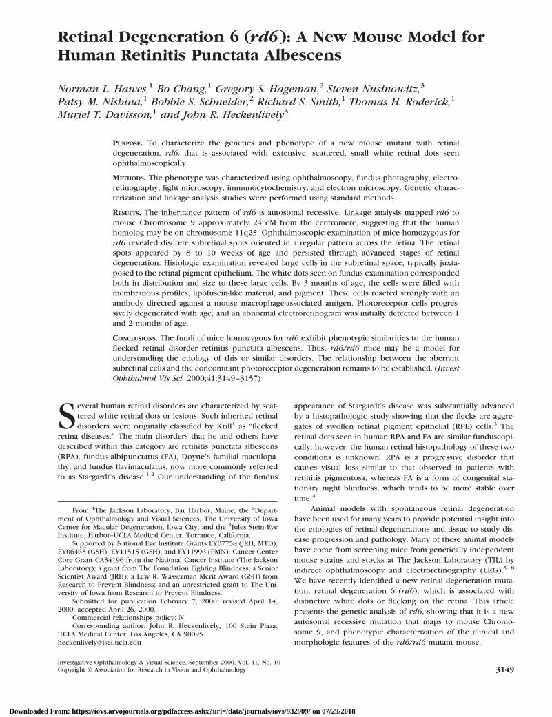
rd6 was discovered in the inbred mouse strain C3HfB/GaCas1b, hereafter referred to as C3HfB. This strain was derivedfrom offspring of irradiated (C3Hx101) F1 male mice crossedto the Oak Ridge National Laboratory “multiple recessive teststock”; the Cas1b mutation was subsequently backcrossed toC3HfB, a C3H subline fostered (f) on C57BL/6 (B). Although allC3H strains are fixed for Pdebrd1, a mutation in the betasubunit of cGMP phosphodiesterase,9 this particular strain ex-hibited an unusual granular retinal appearance compared withother strains homozygous for Pdebrd1. To test for the possibil-ity of a new mutation different from Pdebrd1, mice from thisstrain were mated to C57BL/6J mice, which have a normalretina. The F1 progeny, which did not show ocular abnormal-ities, were backcrossed to the parental strain C3HfB. The back-cross offspring could be separated into 3 groups: Group 1,normal; group 2, Pdebrd1 homozygotes with characteristicpatches of pigment deposits and large diffuse depigmentedregions observable by 3 to 4 weeks of age; and group 3, micewith small, discrete dots present throughout the fundus (Fig.1). On examination by indirect ophthalmoscopy, the dots gavethe retina a granular appearance, a phenotype first observedbetween 8 and 10 weeks of age. Mice from group 3 wereintercrossed to fix the mutation in a stock, free of Pdebrd1 butexhibiting the new retinal phenotype. Subsequently, the rd6stock has been maintained by repeated backcrossing toC57BL/6J to make a congenic inbred strain. Phenotypic char-acterization was performed on mice from the mixed back-ground stock and the incipient B6 congenic. Major aspects of
the phenotype do not appear to differ on the two geneticbackgrounds.
Clinical Retinal Evaluation
All mice in the characterization studies and linkage crosses hadpupils dilated with 1% atropine ophthalmic drops and wereevaluated by indirect ophthalmoscopy with a 78 diopter lens.Signs of retinal degeneration, such as vessel attenuation, alter-ations in the RPE, and presence or absence of retinal dots werenoted. Fundus photographs were taken with a Kowa Genesissmall animal fundus camera (Torrance, CA).10
Electroretinography
After at least 2 hours of dark-adaptation, mice were anesthe-tized with an intraperitoneal injection of normal saline solutioncontaining ketamine (15 mg/gm) and xylazine (7 mg/gm bodywt). Electroretinograms (ERGs) were recorded from the cor-neal surface of one eye after pupil dilation (1% atropine sulfate)using a gold loop electrode referenced to a gold wire in themouth. A needle electrode placed in the tail served as ground.A drop of methylcellulose (2.5%) was placed on the cornealsurface to ensure electrical contract and to maintain cornealintegrity. Body temperature was maintained at a constant tem-perature of 38°C using a heated water pad.
All stimuli were presented in a Ganzfeld dome (LKC Tech-nologies, Gaithersburg, MD) whose interior surface waspainted with a highly reflective white matte paint (No. 6080;Eastman Kodak, Rochester, NY). Stimuli were generated with aGrass Photic Stimulator (model PS33 Plus; Grass Instruments,Worcester, MA) affixed to the outside of the dome at 90° to theviewing porthole. Rod-dominated responses were recorded toshort-wavelength (lmax 5 470 nm; Wratten 47A filter) flashesof light over a 4.0-log unit range of intensities (0.3 log unitsteps) up to the maximum allowable by the photic stimulator.Cone-dominated responses were obtained with white flashes(0.3 steps) on the rod-saturating background after 10 minutesof exposure to the background light to allow complete lightadaptation.
Responses were amplified (Grass CP511 AC amplifier,310,000; 3 dB down at 2 and 10,000 Hz) and digitized using anI/O board (model PCI-1200; National Instruments, Austin, TX)in a personal computer. Signal processing was performed with
FIGURE 1. (A, B) Fundus photo-graphs of normal control C57BL/6Jmouse (A) and a 4-month-old homozy-gous rd6 mouse (B) demonstratingmultiple subretinal retinal spots. Thewhite spots are first detectable oph-thalmoscopically at approximately 8to 10 weeks. They are uniform in size,being slightly larger in diameter than50% of the largest retinal vessels, andevenly distributed throughout theretina.
3150 Hawes et al. IOVS, September 2000, Vol. 41, No. 10
Downloaded From: https://iovs.arvojournals.org/pdfaccess.ashx?url=/data/journals/iovs/932909/ on 07/29/2018

custom software (LabWindows/CVI; National Instruments).Signals were sampled every 0.8 msec over a response windowof 200 msec. For each stimulus condition, responses werecomputer-averaged with up to 50 records averaged for theweakest signals. A signal rejection window could be adjustedonline to eliminate electrical artifacts.
Histology and Electron Microscopy
A total of 40 mice (2 at each time point), ranging in age from1 week to 29 months, were studied histologically. The eyes forlight microscopy were immediately removed and immersed incold fixative (1% paraformaldehyde, 2% glutaraldehyde, and0.1 M cacodylate buffer). Eyes were left in fixative for 24 hours,after which time they were transferred to cold 0.1 M cacody-late buffer solution for an additional 24 hours. Samples wereembedded in methacrylate historesin, and sections were cutand stained with hematoxylin and eosin (H&E). The eyes forultrastructural studies were fixed in one-half strength Kar-
novsky fixative (2% formaldehyde and 2.5% glutaraldehyde in100 mM cacodylate buffer, pH 7.4, containing 0.025% CaCl2)for at least 24 hours. Eyes were cut into small wedges (;1 32 mm), rinsed (3 3 10 min) in 100 mM cacodylate buffer (pH7.4), and post-fixed with 1% osmium tetroxide for 2 hours.Tissues from all four quadrants of each eye were subsequentlyprocessed for transmission electron microscopy.11
Immunocytochemistry
Eyecups were fixed in 4.0% paraformaldehyde in 100 mMcacodylate buffer, pH 7.4, for 2 to 4 hours. Eyes were rinsed in100 mM sodium cacodylate buffer (3 3 10 minutes), infiltrated,and embedded in acrylamide (Boehringer–Mannheim, India-napolis, IN) and optimal cutting temperature (OCT) compound(Miles, Elkhart, NY),12 snap-frozen in liquid nitrogen, sectionedto a thickness of 6 to 8 mm on a cryostat, and mounted onSuperfrost Plus slides (Fisher Scientific, Pittsburgh, PA). Un-fixed eyecups were embedded directly in OCT, without acryl-
FIGURE 2. Stardardized ERGs showing rod- and cone-mediated ERGs across different ages. (A) Representative cone-mediated ERGs for a range ofintensities from 20.19 to 1.13 log photopic trolands in a normal adult C57BL/6J mouse (left) and homozygous rd6 mice ranging in age from 10to 70 weeks (left to right). (B) Representative rod-mediated responses obtained in the dark-adapted state, using a range of intensity settings from23.0 to 1.42 log scotopic trolands, and short-wavelength flashes of light.
IOVS, September 2000, Vol. 41, No. 10 Retinal Degeneration 6 3151
Downloaded From: https://iovs.arvojournals.org/pdfaccess.ashx?url=/data/journals/iovs/932909/ on 07/29/2018

amide infiltration or embedment, and sectioned. Sections thatbisected the optic nerve in temporonasal orientation wereprepared from each eyecup. The sections were blocked in 10mM sodium phosphate, pH 7.4, containing 0.85% NaCl, 1 mMeach of calcium chloride and magnesium chloride, and 1mg/ml globulin-free bovine serum albumin (PBS/M/C/BSA).Sections were then rinsed and incubated in MOMA-2 (Chemi-con, Temecula, CA) antibody for 1 hour.13 Slides were rinsedin PBS/M/C (2 3 10 minutes) and subsequently incubated for30 minutes in fluorescein isothiocyanate (FITC)–conjugatedsecondary antibody. Sections were then washed in PBS/M/C(2 3 10 minutes), and placed on coverslips. For negativecontrols, sections were exposed to PBS/M/C/BSA containingno primary antibody or 1% (vol/vol) normal mouse serum,followed by secondary antibody. Unlabeled, adjacent sectionswere examined at 450 to 550 nm excitation (Olympus BP545filter set; Melville, NY) to detect the presence of autofluores-cence that is characteristic of RPE cells at this wavelength.
Gene Mapping
To determine the chromosomal location of the rd6 gene wemated C3HfB/Ga-Cas1b mice to C57BL/6J mice. The F1 mice,which exhibited no retinal abnormalities, were intercrossedfor the initial linkage study. In a second cross, similar F1 micewere backcrossed to C3HfB/Ga-Cas1b mice. In both crosses, allthe Pdebrd1/Pdebrd1 mice were discarded at 1 month of age.Tail DNA was isolated as previously reported.14 For PCR (poly-merase chain reaction) amplification, 25 ng DNA was used in a10-ml volume containing 50 mM KCl, 10 mM Tris–Cl, pH 8.3,2.5 mM MgCl2, 0.2 mM oligonucleotides, 200 mM dNTP, and0.02 U AmpliTaq DNA polymerase. The reactions, which were
FIGURE 3. Composite showing histologic progression of rd6 retinal degeneration. The upper half left panel depicts a normal retina, RPE, andchoroid from a 2-year-old control, followed by histologic retinal sections from rd6/rd6 mutants at 1, 3, 5, 7, 11, and 29 months (magnification,3400). The lower half shows the aberrant RPE-like cells in the subretinal space at 3 weeks and at 1, 3, 5, 7, and 11 months, and their absence at 29 months(magnification, 3900). Eyes from two to four mice were examined at each time point. There is progressive loss of the photoreceptors over approximately16 months. The aberrant cells are initially unpigmented at 3 weeks as seen in the left panel and become pigmented and enlarged over time. Vacuolesand open spaces in the subretinal space are observed surrounding these aberrant cells.
FIGURE 4. Fluorescence light micrographs depicting immunoreactiv-ity of MOMA-2 antibody to the neural retina (A) and RPE (B) of a4-month-old rd6/rd6 mouse. The two retinal layers became separatedduring tissue processing. Distinct cells located within the subretinalspace react strongly with this antibody. Reactivity is positive based onnegative controls (not depicted). MOMA-2 antibody is not detected inany other region of the RPE or retina, but some reactive cells wereobserved in the choroidal stroma (not shown).
Downloaded From: https://iovs.arvojournals.org/pdfaccess.ashx?url=/data/journals/iovs/932909/ on 07/29/2018

initially denatured for 2 minutes at 95°C, were subjected to 49cycles of 20 seconds at 94°C, 20 seconds at 50°C, 30 secondsat 72°C, and then a final 7-minute extension at 72°C. PCRproducts were separated by electrophoresis on 4% MetaPhor(FMC, Rockland, ME) agarose gels and visualized under UVlight after staining with ethidium bromide. Initially a genomescan of microsatellite (Mit) DNA markers was carried out onpooled DNA samples.15 After detection of linkage on Chromo-some 9, the microsatellite markers D9Mit2, D9Mit54,D9Mit171, D9Mit191, D9Mit197, D9Mit227, D9Mit247, andD9Mit286 were scored on individual DNA samples.
RESULTS
Clinical Phenotype
Mice homozygous for rd6 have small, evenly spaced white dotsthroughout their retinas (Fig. 1B). These small white dotsbecome apparent on ophthalmoscopic examination by 8weeks of age in male and female mice. They are easily visiblethrough about 7 months of age after which they persist but aremore difficult to distinguish. Homozygous rd6 mice begin toshow clinical signs of retinal degeneration at 7 months of age.The fundus develops a mildly pigmented granular and mottledappearance by 15 months; individual spots can still be seen,although less frequently, in mice up to about 2 years of age.Retinal vessels are pale and attenuated by 7 months of age andare not detectable by ophthalmoscopy by 22 months.
ERG Phenotype
ERGs of eyes from homozygous rd6 mice show a slow progres-sive retinal dysfunction of both rods and cones over a 70-weekperiod, beginning at about 1 month of age (data not shown)and being extinguished by 70 weeks of age (Fig. 2). The rodand cone systems are affected by the degenerative process anddiminish at a similar rate.
Histologic Phenotype
Histologic examination revealed progressive changes in theretina and RPE (Fig. 3). By 1 month of age, the distal portionsof the outer segments were mildly distorted, a finding con-firmed by electron microscopy (see below). In addition, by 3weeks of age, a few small cells characterized by an eosinophiliccytoplasm lacking pigment granules were identified within thesubretinal space, juxtaposed to the RPE. From 1 to 2 months ofage, there was a progressive increase in the number of aberrantcells in the subretinal space, and many were characterized bythe presence of pigment granules similar to those in the RPE.The photoreceptor cell outer segments were reduced slightlyin length, with a decrease in the number of photoreceptorcells. Between 3 to 5 and 6 to 11 months of age, there wasprogressive loss of photoreceptor cells in the outer nuclearlayer (ONL), shortening and disorientation of the outer seg-ments, and persistence of the abnormal pigmented cells in thesubretinal space. Overall degeneration appeared to stabilizetemporarily between 3 and 6 months (Fig. 3).
From 15 months of age, the photoreceptor outer segmentswere totally absent, and in some areas, the ONL could not bepositively identified. The large pigmented cells in the subreti-nal space persisted through 22 months of age but could nolonger be found after that time (up to 29 months). Between 4and 11 months, many of these subretinal aberrant cells doubledor tripled in size compared with their size at 2 months of age.
The other characteristic feature of progressive retinal degen-eration was the decrease in thickness of the ONL. In the nearlynormal 1-week-old mice, the ONL was 12 to 14 nuclei thick. By1 month, the ONL decreased to a 7- to 8-nuclei thickness; by 1year of age to 2 to 4 nuclei layers; and by 2 years was only 0 to1 nuclei (Fig. 3, last panel). Higher magnifications of atypicallarge aberrant cells in the subretinal space are shown in Figure3, bottom panels.
Immunocytochemical Analyses
To better characterize the nature of the unusual cells withinthe subretinal space, sections from eyes of 6-month-old rd6/rd6 mice were exposed to various antibodies and lectins. Onlyone monoclonal antibody, MOMA-2, directed against an intra-cellular antigen associated with mouse macrophages andmonocytes,16 reacted strongly with these cells (Fig. 4). TheMOMA-2 antibody did not react with the RPE or retina. Reac-tive choroidal cells, assumed to be monocytes/macrophages,confirmed the specificity of the reaction. The aberrant subreti-nal cells are autofluorescent under 450 to 550 nm excitation.
Electron Microscopy
Ultrastructural studies were performed on eyes derived frommice at 2, 4, and 8 months of age (Figs. 5, 6, and 7). Pyknoticphotoreceptor cell bodies increase in number over the firstyear of life. Approximately four rows of photoreceptor nucleiwere observed at 8 months of age (Fig. 5). Outer segmentsbecome disoriented and disrupted, but the depth of the sub-retinal space does not decrease appreciably over the first 8months (Figs. 5A through 5C). In the subretinal space, distinctaberrant cells are seen that are regularly spaced at intervals thatcorrespond to the dots observed funduscopically (Figs. 5A, 5C,6A through 6C, and 7B). Furthermore, their diameters average35 mm (range, 22–42 mm) in mice between 2 and 8 months ofage. This size is similar to that of the dots observed ophthal-mologically; the average size of the white dots is slightlygreater than 50% of the diameter of the large retinal vessels,which is approximately 60 to 80 mm. Higher magnifications ofthese subretinal space–associated cells in 2-, 4-, and 8-month-old rd6/rd6 mice show that they possess cytoplasmic inclu-sions that are indistinguishable from the pigment granules andlipofuscin in adjacent RPE cells and phagosome-like vesicles(Figs. 6A through 6D). In addition, distinct regions of mem-branous material and vacuoles can be observed in both RPEcells and the cells within the subretinal space (Figs. 7A and 7B).This material is not typically observed in the RPE of controlmice.
Genetic Analysis
rd6 segregated as an autosomal recessive mutation in thegenetic crosses carried out to isolate it on the C57BL/6J geneticbackground. These data were confirmed by the (C3HfB-rd6/rd6 x C57BL/6J[B6])F1 x C3HfB-rd6/rd6 backcross that pro-duced 22 rd6/rd6 and 23 wild-type progeny. Linkage to Chro-mosome 9 was initially detected by genome analysis of pooledDNA samples from 21 progeny (42 meioses) from the C3HfB-rd6/rd6 x B6 intercross described in the Methods section.These 42 meioses showed 1/42 recombinant chromosomewith D9Mit227 and 2/42 with D9Mit171. An additional 45mice from the backcross gave order and recombination per-centages of D9Mit227 2 2/87 5 2.3 6 1.6 2 rd6 2 2/87 52.3 6 1.6 2 D9Mit171 (Fig. 8). The allele sizes of markers
IOVS, September 2000, Vol. 41, No. 10 Retinal Degeneration 6 3153
Downloaded From: https://iovs.arvojournals.org/pdfaccess.ashx?url=/data/journals/iovs/932909/ on 07/29/2018

surrounding the mutation are like C3H, suggesting that themutation arose in a stretch of C3H DNA either from the orig-inally irradiated C3HX101 F1 hybrid mouse or from the C3HfBstrain to which Cas1b was backcrossed. C3HfB alleles in thisregion of Chromosome 9 are identical with strain C3H. Thehomologous region in humans is chromosome 11q22-23.
DISCUSSION
Histologic and physiological evidence indicate that homozy-gous rd6 mice develop an early onset, but slow, retinal degen-eration. Alterations in the photoreceptor layer can be seenshortly after the retina develops; the outer segments shortenand soon show significant disorganization. Photoreceptor cellsdegenerate from the normal 10 to 12 cell–layer thickness to 1to 3 cell layers by 12 months. Functional changes, as measuredby ERG, occur concomitantly with the histologic changes;early rod and cone dysfunction is coincident with initial pho-toreceptor and ONL cell loss, and the ERG is extinguished by70 weeks.
Unique funduscopic features are observed in rd6/rd6mice in association with the retinal degeneration. By 2 monthsof age there are discrete dots distributed in a regular fashionthroughout the retina; these are easily observed by indirect
ophthalmoscopy. Histologic analysis showed a correlation ofthe retinal spots with large, aberrant cells that lie within thesubretinal space. These cells are relatively small in the firstmonth of life and then enlarge and become pigmented by thesecond month. Many of these cells reach a size of approxi-mately 35 to 40 mm in diameter (or about one half the diameterof the retinal vessels, see the Results section) by 8 to 10 weeksof age when the retinal dots are first observed. The appearanceof pigment similar to that seen in the RPE, the presence ofvesicles and membranous profiles at the electron microscopiclevel, and the strong reaction to the monocyte/macrophage-specific MOMA-2 monoclonal antibody suggest that these maybe phagocytic cells that have invaded the subretinal space. Wehave observed histologically similar cells in other mouse mu-tants with retinal degenerations (authors’ unpublished data).The large aberrant cells in the subretinal space are consistentwith the small white dots seen by ophthalmoscopy based ontheir relative size and distribution and on the lack of any otherhistologic finding.
It is likely that a number of genes will be found to beassociated with disorders in which retinal spots and flecks areassociated with retinal degeneration. For example, FA, whichgives rise to retinal spots, has been found to be associated with11-cis retinol dehydrogenase mutations.17 The 11-cis retinol
FIGURE 5. Transmission electron mi-crographs depicting the outer retinaof the homozygous rd6 mouse. (Athrough C) RPE (labeled PE) and cho-roid (CH) shown in the top panel(magnification, 32110); (D throughF) the corresponding ONL (magnifica-tion, 32400) shown in the bottompanel, from 2-month-old (A, D),4-month-old (B, E), and 8-month-old(C, F) rd6 homozygous mice. Photo-receptor degeneration, as evidencedby pyknotic photoreceptor cell bod-ies (asterisks) in the ONL, occursslowly over the first year of life. Cellsthroughout this layer degenerate suchthat approximately 4 rows remain by8 months. Concomitantly, the outersegments become disoriented, al-though the depth of the subretinalspace, between the RPE and outerlimiting membrane (OLM), does notdecrease appreciably between 2 and 8months. Most significantly, distinctcells (arrows; A, C) are observedwithin the subretinal space at theseages. The cells average 35 mm indiameter at these ages. They are reg-ularly spaced at intervals that corre-spond to the whitish-yellow dots ob-served ophthalmoscopically. Notethat no other morphologically detect-able structures are observed thatmight account for the spots.
3154 Hawes et al. IOVS, September 2000, Vol. 41, No. 10
Downloaded From: https://iovs.arvojournals.org/pdfaccess.ashx?url=/data/journals/iovs/932909/ on 07/29/2018

dehydrogenase (Rdh1) gene has not yet been mapped in themouse; its location in human chromosome 12q13-q14 predictsits mouse homolog would map to mouse Chromosomes 10 or15.18 In addition, genes have been identified for other fleckedretinal diseases, for example, Stargardt’s disease,19 and Doyne’sfamilial drusenosis.20 The homologs of the genes mutated inStargardt’s disease (ABCR) and Doyne’s (EFEMP1) also havenot yet been mapped in the mouse. Their locations in humanchromosomes 1p13 and 2p16, respectively, predict that theirhomologs will map to mouse Chromosomes 3 and 17. All threegenes can be examined as potential candidates for rd6; how-ever, the Chromosome 9 location of rd6 suggests that it may
identify yet another mutant gene associated with flecked retinadisorders. However, the retinal phenotype in homozygous rd6mice resembles most the changes seen clinically in humanRPA. RPA is a rare, progressive, recessively inherited diseasethat is characterized by whitish-yellow spots radiating out fromthe posterior pole with no macular involvement. ERG re-sponses are reduced in early stages of RPA and eventuallybecome nonrecordable as the disease progresses.4 The RPAphenotype in humans is genetically heterogeneous because ithas recently been shown to be caused by mutations in bothcellular retinaldehyde-binding protein21 and peripherin.22 Be-cause the mouse cellular retinaldehyde-binding protein gene
FIGURE 6. Higher magnification elec-tron micrographs of the RPE (PE) andphotoreceptor outer segment (OS) in-terface from 2-month-old (A; magnifi-cation, 36415), 4-month-old (B; mag-nification, 34187), and 8-month-old(C, magnification, 34260; and D,magnification, 33580) rd6/rd6 mice.The cells located within the subretinalspace are round to somewhat flat-tened and are characterized by cyto-plasmic inclusions that are indistin-guishable from pigment granules andlipofuscin in the PE. In addition, theyoften contain electron-lucent vacu-oles and whorls of membranous ma-terial. Note that the PE from the8-month-old mouse shown in (C) and(D), but not in the younger mice, con-tains material that is indistinguishablefrom that observed in the subretinalcells. This is shown clearly in Figure 7.
FIGURE 7. Electron micrographs de-picting membranous whorls and vacu-oles in the cells within the subretinalspace (asterisk; A, magnification,34200) and associated RPE cells (as-terisk; B, magnification, 35260) of an8-month-old rd6/rd6 mouse.
IOVS, September 2000, Vol. 41, No. 10 Retinal Degeneration 6 3155
Downloaded From: https://iovs.arvojournals.org/pdfaccess.ashx?url=/data/journals/iovs/932909/ on 07/29/2018

maps to mouse Chromosome 1 and because the retinal-relatedperipherin gene maps to mouse Chromosome 17,18 the rd6mutation is likely to identify yet another gene involved inflecked retina diseases. Further study of this new retinaldegeneration model mouse may give valuable clues aboutthese human disorders characterized by white dots in theretina.
Mouse mutants with retinal disorders provide excellentmodels for human retinal diseases. From a clinical point ofview, the mouse retina has an ophthalmoscopic and histologicappearance very similar to those of the human retina. The mainfunduscopic difference is that mice have no fovea. However,the mouse retina has a cone system that can be easily measuredby ERG and detected histologically. Genetically, it is estimatedthat there is at least 95% conservation of sequence betweenessential coding regions in the human and mouse genomes.This means that human homologs of the mutant genes thatcause inherited disorders in the mouse are likely to be involvedin similar human disorders. Homology maps that allow com-parison of mouse chromosomes to their corresponding humansegments are well developed; and when a mutation is foundin mouse, the human gene location can frequently be pre-dicted.23
A number of ocular disorders, vitelliform macular degen-eration 2 (VMD2), neovascular inflammatory vitreoretinopathy(ADNIV), Usher Syndrome 1B, and familial exudative vitreoreti-nopathy (FEVR), have been mapped to human chromosome11q, which has homology to mouse Chromosome 9.22 How-ever, none of these ocular diseases appears to map unambig-uously to the region of human chromosome 11q, which isdirectly homologous with the rd6 region of mouse Chromo-some 9, neither is their phenotype similar. Whether rd6 is anortholog of a previously described human ocular disease orwhether it identifies a new retinal degenerative disorder locusin this region, which contains a cluster of genes essential fornormal eye function, will be answered by the identification ofthe gene.
Acknowledgments
The authors thank Cindy S. Avery, Ron E. Hurd, Priscilla Jewett, HeidiHoopes Nienhaus, and Lisa Thayer for technical assistance; Jennifer
Smith for graphic services; and Robert Mullins for assistance with theimmunocytochemical analyses.
References
1. Krill AE. Flecked retina diseases. In: Krill AE, Archer DS, eds.Hereditary Retinal and Choroidal Dystrophies Vol. II. New York:Harper and Row; 1977:739–824.
2. Aaberg TM. Stargardt’s disease and fundus flavimaculatus: evalua-tion of morphologic progression and intrafamilial co-existence.Trans Am Ophthalmol Soc. 1986;84:453–487.
3. Eagle RC, Lucier AC, Bernardino VB Jr, Yanoff M. Retinal pigmentabnormalities in fundus flavimaculatus: a light and electron micro-scope study. Ophthalmology. 1980;87:1189–1200.
4. Ellis DS, Heckenlively JR. Retinitis punctata albescens: fundusappearance and functional abnormalities. Retina. 1983;3:27–31.
5. Chang B, Heckenlively JR, Hawes NL, Roderick TH. New mouseprimary retinal degeneration (rd-3). Genomics. 1993;16:45– 49.
6. Heckenlively JR, Chang B, Erway L, et al. Mouse model for Ushersyndrome: linkage mapping suggests homology to Usher type Ireported at human chromosome 11p15. Proc Natl Acad Sci USA.1995;92:11100–11104.
7. Chang B, Bronson RT, Hawes NL, et al. Retinal degeneration inmotor neuron degeneration: a mouse model of ceroid lipofuscino-sis. Invest Ophthalmol Vis Sci. 1994;35:1071–1076.
8. Heckenlively JR, Winston JV, Roderick TH. Screening for mouseretinal degenerations, I: correlation of indirect ophthalmoscopy,electroretinograms, and histology. Doc Ophthalmol. 1989;71:229–239.
9. Pittler SJ, Baehr W. Identification of a nonsense mutation in therod photoreceptor cGMP phosphodiesterase B-subunit of the rdmouse. Proc Natl Acad Sci USA. 1991;88:8322– 8326.
10. Hawes NL, Smith RS, Chang B, Davisson M, Heckenlively JR, JohnSWM. Mouse fundus photography and angiography: a catalogue ofnormal and mutant phenotypes. Mol Vis. 1999;5:22–29.
11. Lazarus H, Sly W, Kyle J, Hageman G. Photoreceptor degenerationand altered distribution of interphotoreceptor matrix proteogly-cans in the mucopolysaccharidosis VII mouse. Exp Eye Res. 1993;56:531–541.
12. Johnson L, Blanks J. Application of acrylamide as an embeddingmedium in studies in lectin and antibody binding in the vertebrateretina. Curr Eye Res. 1984;3:969–974.
13. Hageman G, Mullins R, Russell S, Johnson L, Anderson D. Vitro-nectin is a constituent of ocular drusen and the vitronectin gene isexpressed in human retinal pigmented epithelial cells. FASEB J.1999;13:477–484.
14. Buffone GJ. Isolation of DNA from biological specimens withoutextraction with phenol. Clin Chem. 1985;31:164–165.
FIGURE 8. (A) Eighty-seven meioses from an intercross and a backcross between C3HfB-rd6/rd6 andC57BL/6J were phenotyped and genotyped. Linkage to several markers on mouse Chromosome 9 wasobserved. The columns of squares represent haplotypes (open boxes, C57BL/6J allele; filled boxes, C3HfBallele). The numbers of chromosomes with each haplotype are indicated below each column. (B) Geneticmap of Chromosome 9 in the rd6 region showing the closest markers and the region of human homology.
3156 Hawes et al. IOVS, September 2000, Vol. 41, No. 10
Downloaded From: https://iovs.arvojournals.org/pdfaccess.ashx?url=/data/journals/iovs/932909/ on 07/29/2018

15. Taylor BA, Navin A, Phillips SJ. PCR-amplification of simple se-quence repeat variants from pooled DNA samples for rapidlymapping new mutations of the mouse. Genomics. 1996;21:626–632.
16. Kraal G, Rep M, Janse M. Macrophages in T and B cell compart-ments and other tissue macrophages recognized by monoclonalantibody MOMA-2: an immunohistochemical study. Scand J Im-munol. 1987;26:653–661.
17. Yamamoto H, Simon A, Eriksson U, Harris E, Berson EL, Dryja TP.Mutations in the gene encoding 11-cis retinal dehydrogenase causedelayed dark adaptation and fundus albipunctatus. Nat Genet.1999;22:188–191.
18. Mouse Genome Database (MGD), Mouse Genome InformaticsProject, The Jackson Laboratory, Bar Harbor, Maine. WorldWide Web (URL: http://www.informatics.jax.org). October,1999.
19. Allikmets R, Shroyer NF, Singh N, et al. Mutation of the Stargardtdisease gene (ABCR) in age-related macular degeneration. Science.1997;277:1805–1807.
20. Stone EM, Lotery AJ, Munier FL, et al. A single EFEMP1 mutationassociated with both Malattia Leventinese and Doyne honeycombretinal dystrophy. Nat Genet. 1999;22:199–202.
21. Morimura H, Berson EL, Dryja TP. Recessive mutations in theRLBP1 gene encoding cellular retinaldehyde-binding protein in aform of retinitis punctata albescens. Invest Ophthalmol Vis Sci.1999;40:1000–1004.
22. Kajiwara K, Sandberg MA, Berson EL, Dryja TP. A null mutation inthe human peripherin/RDS gene in a family with autosomal dom-inant retinitis punctata albescens. Nat Genet. 1993;3:208–212.
23. Online Mendelian Inheritance in Man (OMIM). URL: http://www3.ncbi.nlm.nih.gov/Omim/.
IOVS, September 2000, Vol. 41, No. 10 Retinal Degeneration 6 3157
Downloaded From: https://iovs.arvojournals.org/pdfaccess.ashx?url=/data/journals/iovs/932909/ on 07/29/2018