resveratrol does not protect from ischemia-induced...
TRANSCRIPT
Resveratrol Does Not Protect from Ischemia-Induced Acute Kidney Injury in anin Vivo Rat Model
Bienholz, Anja; Mae Pang, Rahel; Guberina, Hana; Rauen, Ursula; Witzke, Oliver;Wilde, Benjamin; Petrat, Frank; Feldkamp, Thorsten; Kribben, Andreas
This text is provided by DuEPublico, the central repository of the University Duisburg-Essen.
This version of the e-publication may differ from a potential published print or online version.
DOI: http://dx.doi.org/10.1159/000485606
URN: urn:nbn:de:hbz:464-20180202-105239-0
Link: https://duepublico.uni-duisburg-essen.de:443/servlets/DocumentServlet?id=45343
License:
This work may be used under a Creative Commons Attribution-NonCommercial-NoDerivatives4.0 International license.
Source: Kidney Blood Press Res, 2017;42:1090–1103; published online: December 05, 2017

Kidney Blood Press Res 2017;42:1090-1103DOI: 10.1159/000485606Published online: December 04, 2017
© 2017 The Author(s). Published by S. Karger AG, Baselwww.karger.com/kbr 1090
Bienholz et al.: Resveratrol in Ischemic AKI
Original Paper
Accepted: November 23, 2017
This article is licensed under the Creative Commons Attribution-NonCommercial-NoDerivatives 4.0 Interna-tional License (CC BY-NC-ND) (http://www.karger.com/Services/OpenAccessLicense). Usage and distribution for commercial purposes as well as any distribution of modified material requires written permission.
DOI: 10.1159/000485606Published online: December 04, 2017
© 2017 The Author(s) Published by S. Karger AG, Baselwww.karger.com/kbr
Resveratrol Does Not Protect from Ischemia-Induced Acute Kidney Injury in an in Vivo Rat ModelAnja Bienholza Rahel Mae Panga Hana Guberinab Ursula Rauenc Oliver Witzkeb Benjamin Wildea Frank Petratc Thorsten Feldkampd Andreas Kribbena
aDepartment of Nephrology, University Hospital Essen, University Duisburg-Essen, Essen, bDepartment of Infectious Diseases, University Duisburg-Essen, Essen, cInstitute of Physiological Chemistry, University Duisburg-Essen, Essen, dDepartment of Nephrology and Hypertension, Christian-Albrechts-University, Kiel, Germany
Key WordsAcute kidney injury • Ischemia and reperfusion injury • Blood pressure • Resveratrol
AbstractBackground/Aims: The natural polyphenol resveratrol (RSV) has been shown to ameliorate ischemia/reperfusion (I/R)-induced damage. Therefore, a rat model of I/R-induced AKI equipped with intensive monitoring was utilized to examine direct renal protection by RSV in vivo. Methods: AKI was induced by bilateral renal clamping (45 min) followed by reperfusion (3 h). Solvent-free RSV was continuously infused intravenously (0.056 and 0.28 mg/kg) in a total volume of 7 ml/kg/h starting from 30 min before renal clamping. At a mean arterial blood pressure below 70 mmHg for more than 5 min, bolus injections of 0.5 ml 0.9% NaCl solution were administered repetitively (max. 5 ml/kg/h). Results: No differences could be found between normoxic control groups with/without RSV. Bilateral renal clamping and subsequent reperfusion caused a progressive rise in creatinine, cystatin C, and CK, a decrease in cellular ATP content and diuresis. Infusion of RSV increased sirtuin 1 expression after ischemia/reperfusion and was associated with decreased blood pressure during ischemia and early reperfusion accompanied by an increased requirement of bolus injections as well as with increased expression of TNFα. Conclusion: RSV did not exert protective effects on I/R-induced AKI in the present short-term in vivo rat model. The lack of protection is potentially connected to aggravation of blood pressure instability.
Dr. med. Anja Bienholz Klinik für Nephrologie, Universitätsklinikum Essen, Universität Duisburg-EssenHufelandstr. 55, Essen (Germany)Tel. +49 201-723 6551, E-Mail [email protected]
© 2017 The Author(s)Published by S. Karger AG, Basel

Kidney Blood Press Res 2017;42:1090-1103DOI: 10.1159/000485606Published online: December 04, 2017
© 2017 The Author(s). Published by S. Karger AG, Baselwww.karger.com/kbr 1091
Bienholz et al.: Resveratrol in Ischemic AKI
Introduction
Acute kidney injury (AKI) is directly associated with increased mortality and is despite full laboratory and clinical recovery an independent risk factor for the development of chronic kidney disease [1, 2]. Damage induced by shortage of nutrition and oxygen supply (ischemia) followed by its restoration (reperfusion) is the main cause of AKI in developed countries with open heart surgery and sepsis being the prime triggers [3]. Therapy is limited to supportive care with no causal treatment being available.
The natural polyphenol resveratrol (trans-3, 5,4`-trihydroxystilbene) (RSV), a component of wine and grapes, has exerted protective effects via a broad variety of anti-inflammatory, immunomodulatory, anti-apoptotic, chemoprotective, antidiabetic and antioxidative properties in different models of damage on cellular, organ and whole animal level [4-10]. In addition, RSV has been shown to ameliorate ischemia/reperfusion (I/R) -induced damage in intestine [11], liver [12], brain [13], skeletal muscle [14], and heart [15, 16]. Proposed protective mechanisms included modulation of enzymes involved in redox activity and production of reactive oxygen species, inhibition of the inducible nitric oxide synthase-NO pathway and cytokine expression. RSV acts as an activator of sirtuin 1. Sirtuin 1 is involved in pathways increasing lifespan in models of calorie restriction [17] and during ischemic preconditioning [18, 19]. Therefore, protective effects through activation of these pathways rather than through direct ones are also anticipated.
Data on protective potential of RSV in I/R-induced AKI are sparse. Despite promising beneficial effects of RSV the poor bioavailability of RSV remains a major concern. RSV has been shown to lower systolic blood pressure in humans [20] and had adverse effects on blood pressure during reperfusion after severe intestinal ischemia in rats [11]. Hemodynamics is a vulnerable point in I/R-induced AKI [21, 22]. Therefore, in the present study a rat model of I/R-induced AKI equipped by intensive monitoring of vital parameters was utilized to examine whether intravenous application of solvent-free RSV provides direct renal protection in vivo.
Materials and Methods
Chemicals/MaterialsResveratrol and hematoxylin were obtained from Sigma-Aldrich (Steinheim, Germany). Formalin
solution (4.5% and 10%, buffered) was from Abbott (Wiesbaden, Germany), isoflurane (Forene) was from AbbVie (Wiesbaden, Germany), ketamine 10% was from Ceva (Düsseldorf, Germany), lidocaine (Xylocaine 1%) from AstraZeneca (Wedel, Germany), Ringer´s solution from Fresenius Kabi (Bad Homburg, Germany), 0.9% NaCl solution and sterile water (Aqua Ecotainer) were from Braun (Melsungen, Germany), paraffin (Paraplast Tissue Embedding Medium REF 501006) was from McCormick Scientific (St. Louis, MO), medical oxygen from Air Liquide (Düsseldorf, Germany) and heparin-sodium 25000 from Ratiopharm GmbH (Ulm, Germany). Triton X-100, zinc chloride, ammonium sulfate and magnesium sulfate were purchased from AppliChem (Darmstadt, Germany), Tris from Serva Electrophoresis (Heidelberg, Germany), Dulbecco´s Phosphate Buffered Saline (DPBS) from Invitrogen (Darmstadt, Germany) and EDTA from Merck (Darmstadt, Germany). Syringe pumps (Perfusor-Secura FT) were from Braun (Melsungen, Germany), protex catheters (0.58 mm i.d., 0.96 mm o.d.) from Smith Medical International (Hythe, U.K.), 4-0 Vicryl sutures from Resorba Medical GmbH (Nürnberg, Germany), 2-ml syringes (Pico50) from Radiometer Medical (Brønhøj, Denmark), safe-lock tubes (2 ml) from Eppendorf (Hamburg, Germany) and 15 ml polypropylene tubes from BD Biosciences (Heidelberg, Germany).
AnimalsMale Sprague Dawley rats (400-470 g) were obtained from Charles River (Sulzfeld, Germany). Animals
were kept for at least one week prior to the experiments in the central animal unit of the University Hospital Essen under standardized conditions of temperature (22 ± 1°C), humidity (55 ± 5%) and 12h/12h light/dark cycles with free access to food (sniff-Spezialdiäten, Soest, Germany) and water, animals were not fasted

Kidney Blood Press Res 2017;42:1090-1103DOI: 10.1159/000485606Published online: December 04, 2017
© 2017 The Author(s). Published by S. Karger AG, Baselwww.karger.com/kbr 1092
Bienholz et al.: Resveratrol in Ischemic AKI
prior to the experimental procedures. All animals received human care according to the standards of the Federation of European Laboratory Animal Science Association (FELASA). The experimental protocol has been approved based on the German animal protection act by the state office for nature, environment and consumer protection (LANUV Recklinghausen, AZ 84-02.04.2011.A369). This article does not contain any studies with human participants performed by any of the authors.
Anesthesia, analgesia and surgical procedureAnesthesia with isoflurane and analgesia with ketamine was performed as described previously [11,
21, 22]. A skin-deep incision was made along the thigh of the right hind limb after application of lidocaine (5 mg/kg s.c.). Portex catheters were placed in the exposed right femoral artery and vein as well as a in the urinary bladder and fixed with 4-0 Vicryl ligatures. Following this procedure, a median abdominal laparotomy was performed along the Linea alba. After an acclimation period of 30 min infusion of resveratrol or sham was started. After an additional 30 min the intestine was carefully evacuated from the abdominal cavity and both kidneys were localized. The vascular pedicle of each kidney was mobilized. In animals undergoing clamping both renal pedicles were occluded for 45 min using atraumatic mini-bulldogs (Aesculap, Tuttlingen, Germany). Clamping was verified visually by change in renal color. The intestine was replaced into the abdominal cavity and covered with moistened compresses and aluminum foil to minimize evaporation and cooling. Subsequent to the ischemic period of 45 min the microvascular clamps were removed and kidneys thus reperfused. At the end of the reperfusion period of 180 min the right kidney was removed. A catheter was placed in the abdominal aorta and the left kidney was perfused at 100 mmHg with 40 ml isotonic NaCl solution containing 1500 I.U. heparin before being resected. Animals remained anesthetized during the whole experiment and were sacrificed by cardiac incision under deep isoflurane anesthesia.
Study groupsThe animal study was performed with eight rats per group. The following experimental groups were
compared:Group 1: normoxic control group, sham operation, 0.9% NaCl;Group 2: I/R control group, 45 min renal clamping, 0.9% NaCl;Group 3: normoxic resveratrol control group, sham operation, 0.28 mg/kg resveratrol (≈ 0.07 mg/
kg/h);Group 4: I/R resveratrolhigh group, 45 min renal clamping, 0.28 mg/kg/h resveratrol (≈ 0.07 mg/kg/h);Group 5: I/R resveratrollow group, 45 min renal clamping, 0.056 mg/kg/h resveratrol (≈ 0.013 mg/
kg/h).Resveratrol for groups 3-5 was administered continuously in a total volume of 7 ml/kg/h starting 30
min before renal clamping until the end of the reperfusion period. Dosages were derived from the finding that amounts higher than 1.0 mg per 100 ml sterile NaCl 0.9% solution were not readily soluble at room temperature. Identical concentrations of intravenous resveratrol were used in previous experiments by Petrat and de Groot in severe intestinal I/R injury [11].
Resveratrol solutions were based on resveratrol freshly dissolved in sterile NaCl 0.9% and pH was adjusted to 7.35 with NaOH. Resveratrol solutions were filtered through a bacteria-tight filter (Minisart 0.2 µm, Sartorius, Göttingen, Germany) and infused with a syringe pump into the right femoral vein.
The normoxic control group of rats undergoing all surgical procedures except clamping of the renal pedicles and the ischemic control group received only 0.9% NaCl solution at a rate of 7 ml/kg/h during the experimental period.
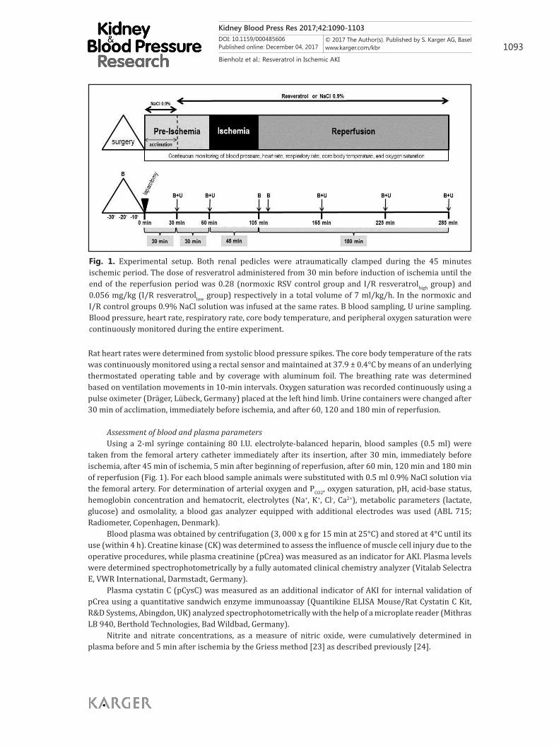
Monitoring of vital parametersVital parameters were assessed every 10 min starting with the beginning of the surgical procedure
(Fig. 1). Systolic, diastolic and mean arterial blood pressures were continuously recorded via the femoral artery catheter that was connected to a pressure transducer (MX 960; Medex Medical, Rossendale, UK). An infusion bag containing Ringer´s solution delivered 3 ml/h to keep the catheter functional. At a mean arterial blood pressure below 70 mmHg for more than 5 min, bolus injections of 0.5 ml 0.9% NaCl solution were administered repetitively through the right femoral artery catheter up to a maximum volume of 5 ml/kg/h.
Kidney Blood Press Res 2017;42:1090-1103DOI: 10.1159/000485606Published online: December 04, 2017
© 2017 The Author(s). Published by S. Karger AG, Baselwww.karger.com/kbr 1093
Bienholz et al.: Resveratrol in Ischemic AKI
Rat heart rates were determined from systolic blood pressure spikes. The core body temperature of the rats was continuously monitored using a rectal sensor and maintained at 37.9 ± 0.4°C by means of an underlying thermostated operating table and by coverage with aluminum foil. The breathing rate was determined based on ventilation movements in 10-min intervals. Oxygen saturation was recorded continuously using a pulse oximeter (Dräger, Lübeck, Germany) placed at the left hind limb. Urine containers were changed after 30 min of acclimation, immediately before ischemia, and after 60, 120 and 180 min of reperfusion.
Assessment of blood and plasma parametersUsing a 2-ml syringe containing 80 I.U. electrolyte-balanced heparin, blood samples (0.5 ml) were
taken from the femoral artery catheter immediately after its insertion, after 30 min, immediately before ischemia, after 45 min of ischemia, 5 min after beginning of reperfusion, after 60 min, 120 min and 180 min of reperfusion (Fig. 1). For each blood sample animals were substituted with 0.5 ml 0.9% NaCl solution via the femoral artery. For determination of arterial oxygen and PCO2, oxygen saturation, pH, acid-base status, hemoglobin concentration and hematocrit, electrolytes (Na+, K+, Cl-, Ca2+), metabolic parameters (lactate, glucose) and osmolality, a blood gas analyzer equipped with additional electrodes was used (ABL 715; Radiometer, Copenhagen, Denmark).
Blood plasma was obtained by centrifugation (3, 000 x g for 15 min at 25°C) and stored at 4°C until its use (within 4 h). Creatine kinase (CK) was determined to assess the influence of muscle cell injury due to the operative procedures, while plasma creatinine (pCrea) was measured as an indicator for AKI. Plasma levels were determined spectrophotometrically by a fully automated clinical chemistry analyzer (Vitalab Selectra E, VWR International, Darmstadt, Germany).
Plasma cystatin C (pCysC) was measured as an additional indicator of AKI for internal validation of pCrea using a quantitative sandwich enzyme immunoassay (Quantikine ELISA Mouse/Rat Cystatin C Kit, R&D Systems, Abingdon, UK) analyzed spectrophotometrically with the help of a microplate reader (Mithras LB 940, Berthold Technologies, Bad Wildbad, Germany).
Nitrite and nitrate concentrations, as a measure of nitric oxide, were cumulatively determined in plasma before and 5 min after ischemia by the Griess method [23] as described previously [24].
Fig. 1. Experimental setup. Both renal pedicles were atraumatically clamped during the 45 minutes ischemic period. The dose of resveratrol administered from 30 min before induction of ischemia until the end of the reperfusion period was 0.28 (normoxic RSV control group and I/R resveratrolhigh group) and 0.056 mg/kg (I/R resveratrollow group) respectively in a total volume of 7 ml/kg/h. In the normoxic and I/R control groups 0.9% NaCl solution was infused at the same rates. B blood sampling, U urine sampling. Blood pressure, heart rate, respiratory rate, core body temperature, and peripheral oxygen saturation were continuously monitored during the entire experiment.

Kidney Blood Press Res 2017;42:1090-1103DOI: 10.1159/000485606Published online: December 04, 2017
© 2017 The Author(s). Published by S. Karger AG, Baselwww.karger.com/kbr 1094
Bienholz et al.: Resveratrol in Ischemic AKI
Determination of tissue ATPThe caudal poles of both kidneys were cut off at the end of reperfusion, immediately before kidney
removal, stored in safe-lock tubes (2 ml) containing 1 M perchloric acid in an extracellular buffer for deproteinization as described previously [25], were mixed vigorously and immediately frozen in liquid nitrogen. Samples were kept frozen at -80°C. Tissue ATP content was measured using a luciferase-driven bioluminescence assay (ATP Bioluminescence Assay Kit CLS II, Roche, Mannheim, Germany). After thawing, samples were diluted in buffer containing 100 mM Tris and 4 mM EDTA (pH 7.75) and mixed immediately with luciferase reagent. Light emission was detected at 550 nm by a luminometer (Berthold Detection Systems, Pforzheim, Germany). The tissue protein content was determined according to Lowry [26].
Histopathological evaluation of the ischemia-reperfusion injury of the kidneyFor histological examination the left blood-free kidney was sliced in half and fixed in formalin (10%,
neutral buffered) for 24-48 h. Thereafter, it was embedded in paraffin and cut on a rotary microtome in serial sections of 2 µm thickness. Tissue sections were mounted on slides and stained with hematoxylin-eosin. Histopathological changes were evaluated in a blinded fashion based on the following criteria: 1. Blood content in glomeruli; 2. shrunken or swollen glomeruli; 3. loss of brush border; 4. shrunken or swollen tubular epithelial cells; 5. intraluminal casts and 6. indicators of cell death (loss of nuclei).
Gene expressionA quarter of the left, blood-free kidney was stored in RNA-stabilizer (Qiagen, Hilden, Germany)
at -80°C immediately after the end of the experimental procedure. Total RNA was extracted after tissue homogenization and disruption (TissueLyser and Qiashredder; Qiagen) using RNeasy Mini Kit (Qiagen) according to the manufacturer´s instructions. Thereafter, total RNA was converted to cDNA using the Qiagen QuantiTect Reverse Transcription Kit. Total cDNA was used for real-time RT-PCR quantification with TaqMan Fast Universal PCR Master Mix (Applied Biosystems by Thermo Fisher Scientific, Oberhausen, Germany) on a real-time PCR system (StepOnePlus, Applied Biosystems by Fisher Scientific). Primers for SIRT1, IL-6, TNFα and the housekeeping gene β-actin were purchased from Applied Biosystems by Thermo Fisher Scientific (TaqMan Primer). Real-time PCR data were analyzed using 2-∆∆CT method as described previously [27].
StatisticsExperiments were performed with eight animals per experimental group. Biochemical assays were
run in duplicate unless stated otherwise. Data are expressed as mean value ± standard error of the mean. Comparisons among multiple groups were performed using analysis of variances either for nonrecurring or for repeated measures (analysis over time) or Kruskal-Wallis test followed by Tukey´s, Bonferroni or Dunns post-hoc analysis. A p < 0.05 was considered significant.
Results
Effects of resveratrol on blood pressure and other vital parametersThe mean arterial blood pressure (MAP) of all animal groups was around 76 mmHg at
the start of the experimental procedure (Fig. 2). In the normoxic control group MAP remained stable during the entire experiment. No differences between normoxic control group animals receiving NaCl solution and normoxic control group animals receiving resveratrol (0.28 mg/kg) could be detected (Fig. 2a). Renal clamping as performed in the I/R control group moderately and continuously decreased MAP, but alterations did not reach statistical significance in comparison to the normoxic control group. Significant differences in MAP were observed between animals undergoing I/R and receiving continuous infusions of resveratrol in comparison to the I/R control group during ischemia and early reperfusion (p<0.05; Fig. 2b). These changes in blood pressure were reflected by an increased requirement of bolus injections during ischemia (Fig. 3).
Heart rates, breathing rates and peripheral oxygen saturations did not change during

Kidney Blood Press Res 2017;42:1090-1103DOI: 10.1159/000485606Published online: December 04, 2017
© 2017 The Author(s). Published by S. Karger AG, Baselwww.karger.com/kbr 1095
Bienholz et al.: Resveratrol in Ischemic AKI
Fig. 2. Mean arterial blood pressure. Mean arterial blood pressure was recorded via a catheter placed in the femoral artery. Resveratrol was administered at 0.28 (normoxic RSV control group and I/R resveratrolhigh group) and 0.056 mg/kg (I/R resveratrollow group) respectively in a total volume of 7 ml/kg/h. In the normoxic and I/R control groups 0.9% NaCl solution was infused at the same rates. Dark grey-shaded area indicates time of renal ischemia induced by atraumatic clamping of both renal pedicles. Values are means ± SEM of eight rats per group. For other experimental details see Fig. 1. (A) Mean arterial blood pressure of normoxic control group and RSV control group. (B) Mean arterial blood pressure of normoxic control group, I/R control group and both I/R resveratrol groups. Significant differences between the I/R control group and both I/R resveratrol
groups were observed during ischemia and early reperfusion (p<0.05).
Fig. 3. Number of bolus injections used to help support blood pressure during renal ischemia. Rats of the I/R control group and I/R RSVhigh and I/R RSVlow groups were subjected to clamping of both renal vascular pedicles for 45 minutes followed by three hours of reperfusion as diagrammed in Fig. 1. Besides a continuous intravenous infusion rate of 7 ml/kg/h, bolus injections of 0.5 ml 0.9% NaCl were administered repetitively up to a maximum volume of 5 ml/kg/h at a mean arterial blood pressure below 70 mmHg for more than five minutes. Values are means ± SEM of eight rats per group. 1 Bolus injection = 0.5 ml 0.9% NaCl. *p<0.05 vs. I/R control group; ##p<0.01 and ###p<0.001 vs. normoxic control group.
B
A

Kidney Blood Press Res 2017;42:1090-1103DOI: 10.1159/000485606Published online: December 04, 2017
© 2017 The Author(s). Published by S. Karger AG, Baselwww.karger.com/kbr 1096
Bienholz et al.: Resveratrol in Ischemic AKI
Normoxic Control Group I/R Control Group Normoxic RSV Control Group I/R RSVhigh Group I/R RSVlow Group
Baseline Values End Values
pH 7.34 ± 0.01 7.35 ± 0.02 7.25 ± 0.01### 7.36 ± 0.02 7.30 ± 0.02** 7.29 ± 0.01##
HCO3-, mmol/l 21.5 ± 0.3 22.2 ± 0.5 18.4 ± 0.4### 22.7 ± 0.5 18.9 ± 0.2### 16.7 ± 2.2###
Base excess, mmol/l -3.4 ± 0.4 -2.6 ± 0.6 -7.0 ± 0.5### -2.1 ± 0.7 -6.8 ± 0.3### -6.9 ± 0.5###
pO2, mmHg 401 ± 13 447 ± 13 419 ± 23 399 ± 31 454 ± 8 452 ± 14
pCO2, mmHg 42 ± 2 42 ± 2 46 ± 2 42 ± 3 38 ± 2** 40 ± 1
Na+, mmol/l 139.8 ± 0.9 139.0 ± 0.5 140.0 ± 0.4 139.3 ± 0.3 139.9 ± 0.5 139.4 ± 0.7
Cl-, mmol/l 114.0 ± 1.4 112.5 ± 1.0 119.0 ± 1.3### 113.6 ± 0.9 118.0 ± 0.8### 116.8 ± 1.3#
K+, mmol/l 5.0 ± 0.1 4.4 ± 0.1+ 6.2 ± 0.1### 4.6 ± 0.1 6.3 ± 0.2### 6.3 ± 0.2###
Ca2+, mmol/l 1.5 ± 0.01 1.4 ± 0.01+++ 1.4 ± 0.02 1.4 ± 0.03 1.4 ± 0.03 1.4 ± 0.02
the course of the experiment and were neither affected by I/R nor by resveratrol.
Effects of resveratrol on nitrite and nitrate concentrationsCumulative nitrite and nitrate concentration
was low and within the normal physiological range in all groups. No differences were detected between groups neither before nor after ischemia. In addition, there were no significant changes in any group between both time points of measurement (Fig. 4).
Effects of resveratrol on parameters of blood gas analysis, electrolytes, metabolic parameters, osmolality, hemoglobin, and hematocritIn the normoxic control group K+ and Ca2+
levels decreased significantly during the course of the experiment, but stayed well within the physiological range (Table 1).
Compared with the values obtained from the last blood sampling of the normoxic control group animals, in the I/R control group clamping of both renal pedicles and subsequent reperfusion resulted in significant changes in pH, bicarbonate, base excess, Cl-, and K+ (Table 1). Resveratrol treatment of sham group animals had no significant effect on any of the parameters studied.
Fig. 4. Nitrite and nitrate concentration. Cumulative nitrite and nitrate concentrations were assessed in plasma obtained (A) before and (B) 5 min after ischemia. Values are means ± SEM of 5-8 rats per group.
Table 1. Measurements of blood pH, bicarbonate, base excess, pCO2, pO2 and electrolytes. Values are means ± SE; n = 8 per group. I/R: ischemia/reperfusion; RSV: resveratrol. Baseline values were obtained from the normoxic control group after the acclimation period; these values were not significantly different from the baseline values of the other experimental groups. The end values were assessed from the last blood sampling drawn after 3 h of reperfusion. For experimental details, see Fig. 1. +p<0.05 and +++p<0.01 vs. baseline values; #p<0.05, ##p<0.01 and p<0.001 vs. normoxic control group; **p<0.01 vs. I/R control group

Kidney Blood Press Res 2017;42:1090-1103DOI: 10.1159/000485606Published online: December 04, 2017
© 2017 The Author(s). Published by S. Karger AG, Baselwww.karger.com/kbr 1097
Bienholz et al.: Resveratrol in Ischemic AKI
In animals undergoing I/R and receiving the higher dosage of resveratrol pH was significantly less acidotic than in the I/R control group directly following renal clamping and at the end of the reperfusion period presumably due to a lowered pCO2 which did not go along with an increased breathing rate.
Glucose and lactate levels, osmolality, hemoglobin concentrations and hematocrit remained stable during the entire experiment and were neither affected by I/R nor by resveratrol (data not shown).
Effects of resveratrol on urine outputIn the normoxic control group urine output increased during the course of the experiment
(Fig. 5). No differences between normoxic control group animals receiving NaCl solution and normoxic control group animals receiving resveratrol (0.28 mg/kg) could be detected. Bilateral renal clamping caused a highly significant reduction in urine output compared to the normoxic control group, which did not recover during reperfusion and was not altered
Fig. 5. Urine output. Urine was collected via a bladder catheter and measured during indicated time intervals. Rats received continuous intravenous infusions at a volume of 7 ml/kg/h during the entire experimental procedure starting 60 min before induction of renal ischemia. Ischemia was induced by atraumatic clamping of both renal pedicles for 45 min followed by 3 h of reperfusion. For experimental details see Fig. 1. Values are means ± SEM of eight rats per group. +p<0.05 and +++p<0.001 vs. baseline value of normoxic control group; ###p<0.001 vs. normoxic control group.
Baseline Values Normoxic Control Group I/R Control Group Normoxic RSV Control Group I/R RSVhigh Group I/R RSVlow Group
CK, U/l 354 ± 62
Pre-ischemia 296 ± 51 169 ± 16 208 ± 18 226 ± 25 235 ± 56
End 276 ± 49 661 ± 88### 226 ± 31 839 ± 188### 828 ± 102###
pCrea, mg/dl 0.48 ± 0.06
Pre-ischemia 0.48 ± 0.05 0.48 ± 0.05 0.56 ± 0.05 0.43 ± 0.06 0.59 ± 0.04
Post-ischemia 0.44 ± 0.06 0.68 ± 0.06# 0.54 ± 0.04 0.66 ± 0.06# 0.75 ± 0.05###
End 0.43 ± 0.02 1.24 ± 0.08### 0.54 ± 0.07 1.28 ± 0.06### 1.38 ± 0.06###
pCysC, mg/l 0.93 ± 0.05
Post-ischemia 0.88 ± 0.06 2.99 ± 0.17### 0.84 ± 0.05 2.69 ± 0.25### 2.56 ± 0.16###
End 0.86 ± 0.03 5.49 ± 0.30### 0.77 ± 0.04 5.07 ± 0.38### 4.76 ± 0.20###**
ATP, nmol/mg protein 8.1 ± 0.6 2.8 ± 0.2## 7.9 ± 0.9 3.0 ± 0.2## 3.2 ± 0.1##
Table 2. Effects of RSV on CK, pCrea, pCysC and ATP. Values are means ± SE; n = 8 per group. I/R: ischemia/reperfusion; RSV: resveratrol; CK: creatine kinase; pCrea: plasma creatinine; pCysC: plasma cystatin C. Baseline values were obtained from the normoxic control group drawn after the acclimation period. These values were not significantly different from the baseline values of the other experimental groups. The other values were assessed at the time points indicated. #p<0.05, ##p<0.01 and ###p<0.001 vs. normoxic control group; **p<0.01 vs. I/R control group

Kidney Blood Press Res 2017;42:1090-1103DOI: 10.1159/000485606Published online: December 04, 2017
© 2017 The Author(s). Published by S. Karger AG, Baselwww.karger.com/kbr 1098
Bienholz et al.: Resveratrol in Ischemic AKI
by continuous infusion of resveratrol.
Effects of resveratrol on markers of organ injury and tissue ATP contentIn the normoxic control group CK, pCrea and pCysC levels remained stable during the
entire experimental procedure (Tab 2). No differences between normoxic control group animals receiving NaCl solution and normoxic control group animals receiving resveratrol (0.28 mg/kg) could be detected regarding these parameters.
In the I/R control group CK levels were significantly elevated as of 2 h reperfusion compared to the normoxic control group. Continuous infusion of resveratrol during I/R did not alter CK levels.
In the I/R control group pCrea and pCysC levels were significantly increased at the end of ischemia. Continuous infusion of resveratrol in animals undergoing I/R did not alter pCrea levels. In the I/R resveratrollow group pCysC levels were significantly lower at the end of 3 h of reperfusion compared to the I/R control group.
Tissue ATP content measured after 3 h or reperfusion was no different in normoxic control group animals receiving NaCl solution and normoxic control group animals receiving resveratrol (0.28 mg/kg) (Tab. 2). In the I/R control group tissue ATP content was significantly lower than in the normoxic control group. No alterations by continuous infusion of resveratrol could be detected.
Effects of resveratrol on histopathological changesNormal kidney histology was revealed by light-microscopy (hematoxylin-eosin
staining) in the normoxic control group. Exposition to I/R resulted in loss of brush border, increased numbers of shrunken glomeruli and tubular epithelial cells as well as in formation of intraluminal casts. Continuous infusion of resveratrol at any concentration to animals undergoing I/R did not improve histopathological changes.
Effects of resveratrol on gene expressionRenal expression of SIRT1 was neither altered in the I/R control group nor in normoxic
control group animals receiving resveratrol (Fig. 6). SIRT1 expression was significantly
Fig. 6. Sirtuin1 expression. Real-time PCR data for Sirtuin1 expression in the kidney at the end of the experimental procedure was analysed using 2-∆∆CT. Each value was normalized for the mean of the normoxic control group. Values and mean are given for 6-8 rats per group. *p<0.05 vs. I/R control group; #p<0.05 and ##p<0.01 vs. normoxic control group.

Kidney Blood Press Res 2017;42:1090-1103DOI: 10.1159/000485606Published online: December 04, 2017
© 2017 The Author(s). Published by S. Karger AG, Baselwww.karger.com/kbr 1099
Bienholz et al.: Resveratrol in Ischemic AKI
increased in the kidney in animals undergoing I/R and receiving resveratrol at both concentrations compared to the normoxic control group as well as to the I/R control group.
Renal expression of IL-6 was dramatically increased in all animals following renal clamping (Fig. 7A). Infusion of resveratrol to animals of the normoxic control group did not alter IL-6 expression. TNFα expression in the kidney was neither changed in the I/R control group nor in animals of normoxic control group receiving resveratrol compared to the normoxic control group (Fig. 7B). Application of I/R plus resveratrol significantly increased renal TNFα expression compared to the normoxic control group as well as to the I/R control group (resveratrolhigh only).
Discussion
Continuous intravenous infusion of resveratrol did not provide protection from renal injury in a rat model of I/R-induced AKI (45 min ischemia, 180 min reperfusion), but aggravates blood pressure instability. Renal protection by mostly intraperitoneally or orally administered resveratrol has been described in various models of acute kidney injury associated with cisplatin, sepsis or hemorrhagic shock [28-31] and of I/R [32, 33]. Oral or intravenous application of resveratrol, at similar or lower concentrations, prior to I/R attenuated renal injury observed after 24 h of reperfusion by antioxidative action and preservation of NO bioactivity. Orally administered resveratrol is well-absorbed, but undergoes a high first-pass effect resulting in a biological availability of <1% [34]. Degradation takes mainly place during resorption [35]. Degradation is far less expected during continuous intravenous application and in addition resveratrol cannot be excreted by the kidney during bilateral renal clamping. Although, resveratrol proved to induce the sirtuin pathway in animals undergoing I/R-induced AKI, protection from ischemia- induced
Fig. 7. IL-6 and TNFα expression. Real-time PCR data for IL-6 (A) and TNFα (B) expression in the kidney at the end of the experimental procedure was analysed using 2-∆∆CT. Each value was normalized for the mean of the normoxic control group. Values and mean are given for 6-8 rats per group. **p<0.01 vs. I/R control group; ##p<0.01 and ###p<0.001 vs. normoxic control group.

Kidney Blood Press Res 2017;42:1090-1103DOI: 10.1159/000485606Published online: December 04, 2017
© 2017 The Author(s). Published by S. Karger AG, Baselwww.karger.com/kbr 1100
Bienholz et al.: Resveratrol in Ischemic AKI
injury was not observed in our experiments.Destabilisation of blood pressure associated with bilateral (and not unilateral) renal
ischemia has been described before [21, 22, 36], although to our knowledge studies performing close continuous arterial blood pressure monitoring especially during the clamping period have not been performed by other groups. The mechanisms involved in induction of hypotension during bilateral renal clamping remain unclear, but it is conceivable that this effect is related to interference by the clamping itself with sympathetic nerves surrounding the renal artery as decreasing blood pressure is observed immediately following mechanical manipulation. This effect has also been described in humans, in which bilateral nephrectomy is an unfavorable, but effective treatment for uncontrolled hypertension [37]. Blood pressure lowering effects after nephrectomy in humans are heterogeneous and shown an early and late response phase [38] which has been ascribed not only to interruption of the renin-angiotensin-aldosterone system, but also to decreased sympathetic activity. Infusion of resveratrol during normoxia or late reperfusion had no immediate effect on MAP, but lowered MAP during ischemia and early reperfusion to an extent exceeding that of ischemia only. Infusion of resveratrol did not result in increased levels of nitric oxide which potentially could have been responsible for direct reduction of blood pressure. Nitric oxide release had been hypothesized as part of the protective pathway after resveratrol application [32]. But, the combination of I/R plus resveratrol was found to be associated with increased expression of TNFα after 3 h of reperfusion, a proinflammatory cytokine being involved in blood pressure regulation [39]. Increased TNFα expression during this early phase therefore offers a potential explanation for increased blood pressure instability associated with resveratrol treatment. Aggravation of hypotension by resveratrol has also been described before. Although intravenous resveratrol proved beneficial in a model of severe low-flow intestinal ischemia, adverse effects on blood pressure during reperfusion subsequent to superior mesenteric artery occlusion were documented despite administration of additional intravenous 0.9% NaCl solution [11]. In a mouse model of sepsis-induced acute kidney injury high doses of intraperitoneal resveratrol (100 mg/kg) were without obvious signs of toxicity in sham mice, but rapidly killed mice subjected to cecal ligation and puncture [29]. Protective effects of lower doses of resveratrol (10 and 30 mg/kg) were among others ascribed to an improvement of renal microcirculation. It is conceivable that resveratrol increases (micro-) capillary perfusion with detrimental effects on blood pressure in situations of hemodynamic instability such as septic shock or bilateral renal ischemia as studied here. Detailed mechanisms of the observed effects remain eventually unclear.
As described previously [11, 21, 22] intravenous bolus injections of NaCl 0.9% were used to compensate for hypotension. Due to the low MAP in the context of bilateral renal clamping animals of both I/R RSV groups received numerous bolus injections during ischemia. Application of NaCl 0.9% has been identified as potentially critical. Negative effects of NaCl 0.9% are ascribed to its unphysiologically high chloride content. The high chloride content is associated with an increased risk of hyperchloraemic metabolic acidosis [40-43] and animal studies have suggested that it is the critical determinant for changes in renal blood flow, mediated primarily by effects on afferent and intrarenal arterial vessels [44-47]. Chloride levels of all I/R groups were significantly elevated, but of questionable physiological implication. However, pH was lowest in the I/R control group, which revealed high chloride levels in the absence of a significant increase in bolus injections. Considering the only slight changes in chloride levels and the improved pH in the RSV groups compared to the I/R control group, the absence of protective effects cannot be ascribed to an increased application of NaCl 0.9% in the RSV groups.
In summary, resveratrol does not to prevent the development of AKI possibly due to its negative effects on the already strained blood pressure during bilateral renal clamping. Resveratrol might still exhibit its beneficial effects during recovery by reduction of oxidative stress [29, 48] and suppression of inflammation at later phases than analyzed in our short term model [30, 48]. The reduction in pCysC might point towards that, but a reperfusion

Kidney Blood Press Res 2017;42:1090-1103DOI: 10.1159/000485606Published online: December 04, 2017
© 2017 The Author(s). Published by S. Karger AG, Baselwww.karger.com/kbr 1101
Bienholz et al.: Resveratrol in Ischemic AKI
time aiming to answer this question is far beyond the scope of the current study.In conclusion, in vivo studies on I/R-induced AKI testing substrates with protective
effects observed in other models of organ injury should include biomonitoring parameters to reveal confounding systemic effects. Those models thereby closer depict clinical situations and reveal issues which may otherwise simply be included in the results of unfavorable outcome.
Disclosure Statement
The authors declare no conflicts of interest.
Acknowledgements
The authors thank Tanja Hinkeldein, Simone Leyting and Barbara Nilewski-Kühl for their excellent technical assistance and Sven Benson for his help in statistical analysis. This study was supported by the Dr. Werner Jackstädt-Foundation.
AB received a research grant from the Dr. Werner Jackstädt-Foundation. TF was supported by a Junior Excellence Research Group grant by the Dr. Werner Jackstädt-Foundation. Funding bodies had no role in the design of the study and collection, analysis, and interpretation of date and in writing the manuscript.
References
1 Rodrigue F, Bruetto R, Torres U, Otaviano A, Zanetta D, Burdmann E: Incidence and mortality of acute kidney injury after myocardial infarction: a comparison between KDIGO and RIFLE criteria. PLoS One 2013;8:e69998.
2 Singbartl K, Kellum J: AKI in the ICU: definition, epidemiology, risk stratification, and outcomes. Kidney Int 2012;81:819-825.
3 Bellomo R, Kellum J, Ronco C: Acute kidney injury. Lancet 2012;380:756-766.4 Rieder S, Nagarkatti P, Nagarkatti M: Multiple anti-inflammatory pathways triggered by resveratrol lead to
amelioration of staphylococcal enterotoxin B-induced lung injury. Br J Pharmacol 2012;167:1244-1258.5 Sha H, Ma Q, Jha R, Wang Z: Resveratrol ameliorates lung injury via inhibition of apoptosis in rats with
servere acute pancreatitis. Exp Lung Res 2009;35:344-358.6 Cigremis Y, Akgoz M, Ozen H, Karaman M, Kart A, Gecer M, Atalan G: Resveratrol ameliorates cisplatin-
induced oxidative injury in New Zealand rabbits. Can J Physiol Pharmacol 2015;93:727-735.7 Hall E, Vaishnav R, Mustafa A: Antioxidant therapies for traumatic brain injury. Neurotherapeutics
2010;7:51-61.8 Huang J, Huang S, Deng J, Chang C, Day Y, Hung L: Insulin and resveratrol act synergistically, preventing
cardiac dysfunction in diabetes, but the advantage of resveratrol in diabetics with acute heart attack is antagonized by insulin. Free Radic Biol Med 2010;49:1710-1721.
9 Kim D, Jung Y, Lee J, Lee A, Kang K, Lee S, Park S, Han M, Lee S, Ramkumar K, Sung M, Kim W: SIRT1 activation by resveratrol ameliorates cisplatin-induced renal injury through deacetylation of p53. Am J Physiol Renal Physiol 2011;301:F427-435.
10 Moridi H, Karimi J, Sheikh N, Goodarzi M, Saidijam M, Yadegarazari R, Khazaei M, Khodadadi I, Tavilani H, Piri H, Asadi S, Zarei S, Rezaei A: Resveratrol-dependent down-regulation of receptor for advanced glycation end-products and oxidative stress in kidney of rats with diabetes. Int J Endocrinol Metab 2015;13:e23542.
11 Petrat F, deGroot H: Protection against severe interstinal ischemia/reperfusion injury in rats by intravenous resveratrol. J Surg Res 2011;167:e145-155.

Kidney Blood Press Res 2017;42:1090-1103DOI: 10.1159/000485606Published online: December 04, 2017
© 2017 The Author(s). Published by S. Karger AG, Baselwww.karger.com/kbr 1102
Bienholz et al.: Resveratrol in Ischemic AKI
12 Nivet-Antoine V, Cottart C, Lemaréchal H, Vamy M, Margaill I, Beaudeux J, Bonnefont-Rousselot D, Borderie D: trans-Resveratrol downregulates Txnip overexpression occurring during liver ischemia-reperfusion. Biochimie 2010;92:1766-1771.
13 Shin J, Lee H, Lim Y, Koh Y, Choi J, Park E: Therapeutic effects of resveratrol during acute periods following experimental ischemic stroke. J Neuroimmunol 2010;227:93-100.
14 Ikizler M, Ovali C, Dernek S, Erkasap N, Sevin B, Kaygisiz Z, Kural T: Protective effects of resveratrol in ischemia-reperfusion injury of skeletal muscle: A clinically relevant animal model for lower extremity ischemia. Chin J Physiol 2006;49:204-209.
15 Dernek S, Ikizler M, Erkasap N, Ergun B, Koken T, Yilmaz K, Sevin B, Kaygisiz Z, Kural T: Cardioprotection with resveratrol pretreatment: improved beneficial effects over standard treatment in rat hearts after global ischemia. Scand Cardiovasc J 2004;38:245-254.
16 Wang S, Qian Y, Gong D, Zhang Y, Fan Y: Resveratrol attenentuates acute hypoxic injury in cardiomyocytes: correlation with inhibition of iNOS-NO signaling pathway. Eur J Pharm Sci 2011;44:416-421.
17 Cohen H, Miller C, Bitterman K, Wall N, Hekking B, Kessler B, Howitz K, Gorospe M, de Cabo R, Sinclair D: Calorie restriction promotes mammalian cell survival by inducing the SIRT1 deacetylase. Science 2004;305:390-392.
18 Das S, Das D: Resveratrol: A therapeutic promise for cardiovascular diseases. Recent Pat Cardivasc Drug Discov 2007;2:133-138.
19 Della-Morte D, Dave K, DeFazio R, Bao Y, Raval A, Perez-Pinzon M: Resveratrol pretreatment protects rat brain from cerebral ischemic damage via a sirtuin 1-uncoupling protein 2 pathway. Neuroscience 2009;159:993-1002.
20 Liu Y, Ma W, Zhang P, He S, Huang D: Effect of resveratrol on blood pressure: a meta-analysis of randomized controlled trials. Clin Nutr 2015;34:27-34.
21 Bienholz A, Petrat F, Wenzel P, Ickerott P, Weinberg J, Witzke O, Kribben A, deGroot H, Feldkamp T: Adverse effects of α-ketoglutarate/malate in a rat model of acute kidney injury. Am J Physiol Renal Physiol 2012;303:F56-63.
22 Bienholz A, Reis J, Sanli P, de Groot H, Petrat F, Guberina H, Wilde B, Witzke O, Saner F, Kribben A, Weinberg J, Feldkamp T: Citrate shows protective effects on cardiovascular and renal function in ischemia-induced acute kidney injury. BMC Nephrol 2017;18:130.
23 Green L, Wagner D, Glogowski J: Analysis of nitrate, nitrite, and [15N]nitrate in biological fluids. Anal Biochem 1982;126:131-138.
24 Hamburger T, Broecker-Preuss M, Hartmann M, Schade F, DeGroot H, Petrat F: Effects of glycine, pyruvate, resveratrol, and nitrite on tissue injury and cytokine response in endotoxemic rats. J Surg Res 2013;183:e7-e21.
25 Feldkamp T, Weinberg J, Hörbelt M, vonKropff C, Witzke O, Nürnberger J, Kribben A: Evidence for involvement of nonesterified fatty acid-induced protonophoric uncoupling during mitochondrial dysfunction caused by hypoxia and reoxygenation. Nephrol Dial Transplant 2009;24:43-51.
26 Lowry O, Rosebrough N, Farr A, Randall R: Protein measurement with the Folin phenol reagent. J Biol Chem 1951;193:265-275.
27 Livak K, Schmittgen T: Analysis of relative gene expression data using real-time quantitative PCR and the 2-∆∆CT method. Methods 2001;25:402-408.
28 Hao Q, Xiao X, Zhen J, Feng J, Song C, Jiang B, Hu Z: Resveratrol attenuates acute kidney injury by inhibiting death receptor-mediated apoptotic pathways in a cisplatin-induced rat model. Mol Med Rep 2016;14:3683-3689.
29 Holthoff J, Wang Z, Seely K, Gokden N, Mayeux P: Resveratrol improves renal microcirculation, protects the tubular epithelium, and prolongs survival in a mouse model of sepsis-induced acute kidney injury. Kidney Int 2012;81:370-378.
30 Chen L, Yang S, Zumbrun E, Guan H, Nagarkatti P, Nagarkatti M: Resveratrol attenuates lipopolysaccharide-induced acute kidney injruy by suppressing inflammation driven by macrophages. Mol Nutr Food Res 2015;59:853-864.
31 Wang H, Guan Y, Karamercan M, Ye L, Bhatti T, Becker L, Baur J, Sims C: Resveratrol rescues kidney mitochondrial function following hemorrhagic shock. Shock 2015;44:173-180.

Kidney Blood Press Res 2017;42:1090-1103DOI: 10.1159/000485606Published online: December 04, 2017
© 2017 The Author(s). Published by S. Karger AG, Baselwww.karger.com/kbr 1103
Bienholz et al.: Resveratrol in Ischemic AKI
32 Chander V, Chopra K: Protective effect of nitric oxide pathway in resveratrol renal ischemia-reperfusion injury in rats. Arch Med Res 2006;37:19-26.
33 Giovannini L, Migliori M, Longoni B, Das D, Bertelli A, Panichi V, Filippi C, Bertelli A: Resveratrol, a polyphenol found in wine, reduces ischemia reperfusion injury in rat kidneys. J Cardiovasc Pharmacol 2001;37:262-270.
34 Walle T, Hsieh F, DeLegge M, Oatis J, Walle U: High absorption but very low bioavailability of oral resveratrol in humans. Drug Metab Dispos 2004;32:1377-1382.
35 Kapetanovic I, Muzzio M, Huang Z, Thompson T, McCormick D: Pharmacokinetics, oral bioavailability, and metabolic profile of resveratrol and its dimethylether analog, pterostilbene, in rats. Cancer Chemother Pharmacol 2011;68:593-601.
36 Mitaka C, Si M, Tulafu M, Yu Q, Uchida T, Abe S, Kitagawa M, Ikeda S, Eishi Y, Tomita M: Effects of atrial natriuretic peptide on inter-organ crosstalk among the kidney, lung, and heart in a rat model of renal ischemia-reperfusion injury. Intensive Care Med Exp 2014;2:28.
37 Knehtl M, Bevc S, Hojs R, Hlebic G, Ekart R: Bilateral nephrectomy for uncontrolled hypertension in hemodialysis patient: a forgotten option? Nephrol Ther 2014;10:528-531.
38 Schwarz E, Heintz B, Stefanidis I, vom Dahl J, Sieberth H: The heterogeneous and delayed course of blood pressure normalization in hypertensive patients after bilateral nephrectomy with and without subsequent renal transplantation. Ren Fail 2000;22:591-604.
39 Ramseyer V, Garvin J: Tumor necrosis factor-α: regulation of renal function and blood pressure. Am J Physiol Renal Physiol 2013;304:F1231-1242.
40 Reid F, Lobo D, Williams R, Rowlands B, Allison S: (Ab)normal saline and physiological Hartmann’s solution: a randomized double-blind crossover study. Clin Sci (Lond) 2003;104:17-24.
41 Lobo D, Stanga Z, Aloysius M, Wicks C, Nunes Q, Ingram K, Risch L, Allison S: Effect of volume loading with 1 liter intravenous infusions of 0.9% saline, 4% succinylated gelatin (Gelofusine) and 6% hydroxyethyl starch (Voluven) on blood volume and endocrine responses: a randomized, three-way crossover study in healthy volunteers. Crit Care Med 2010;38:464-470.
42 Williams E, Hildebrand K, McCormick S, Bedel M: The effect of intravenous lactated Ringer’s solution versus 0.9% sodium chloride solution on serum osmolality in human volunteers. Anesth Analg 1999;88:999-1003.
43 Kellum JB, R, Kramer D, Pinsky M: Etiology of metabolic acidosis during saline resuscitation in endotoxemia. Shock 1998;9:364-368.
44 Wilcox C: Regulation of renal blood flow by plasma chloride. J Clin Invest 1983;71:726-735.45 Nashat F, Tappin J, Wilcox C: The renal blood flow and the glomerular filtration rate of anaesthetized dogs
during acut changes in plasma sodium concentration. J Physiol 1976;256:731-745.46 Hansen P, Jensen B, Skott O: Chloride regulates afferent ateriolar contraction in response to depolarization.
Hypertension 1998;32:1066-1070.47 Bullivant E, Wilcox C, Welch W: Intrarenal vasoconstriction during hyperchloremia: role of thromboxane.
Am J Physiol 1989;256:F152-157.48 Khader A, Yang W, Kuncewitch M, Prince J, Marambaud P, Nicastro J, Coppa G, Wang P: Novel resveratrol
analogues attenuate renal ischemic injury in rats. J Surg Res 2015;193:807-815.