restoration of missing data in limited angle tomography
TRANSCRIPT
Restoration of Missing Data in Limited AngleTomography Based on Consistency Conditions
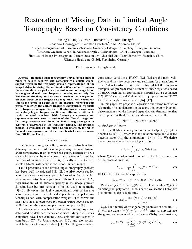
Yixing Huang∗, Oliver Taubmann∗†, Xiaolin Huang∗‡,Joachim Hornegger∗†, Guenter Lauritsch¶ and Andreas Maier∗†
∗Pattern Recognition Lab, Friedrich-Alexander-University Erlangen-Nuremberg, Erlangen, Germany†Erlangen Graduate School in Advanced Optical Technologies (SAOT), Erlangen, Germany
‡Institute of Image Processing and Pattern Recognition, Shanghai Jiao Tong University, Shanghai, China¶Siemens Healthcare GmbH, Forchheim, Germany
Email: [email protected]
Abstract—In limited angle tomography, only a limited angularrange of data is acquired and consequently a double wedge-shaped region in the frequency domain representation of theimaged object is missing. Hence, streak artifacts occur. To restorethe missing data, we perform a regression and an image fusionin sinogram domain and frequency domain, respectively. Wefirst convert the sinogram restoration problem into a regressionproblem based on the Helgason-Ludwig consistency conditions.Due to the severe ill-posedness of the problem, regression onlypartially recovers the correct frequency components, especiallylower frequency components, and will introduce erroneous ones,particularly higher frequencies. Bilateral filtering is utilized toretain the most prominent high frequency components andsuppress erroneous ones. A fusion of the filtered image andthe image reconstructed from the limited angle sinogram isperformed afterwards in the frequency domain. The proposedmethod is evaluated on the Shepp-Logan phantom, for whichthe root-mean-square error of the reconstructed image decreasesfrom 310 HU to 136 HU.
I. INTRODUCTION
In computed tomography (CT), image reconstruction fromdata acquired in an insufficient angular range is called limitedangle tomography. It arises when the gantry rotation of a CTsystem is restricted by other system parts or external obstacles.Because of missing data, artifacts, typically in the form ofstreak artifacts, will occur in the reconstructed images.
The ill-posedness of the limited angle tomography problemhas been well investigated [1], [2]. Iterative reconstructionalgorithms can incorporate prior information. In particular,iterative reconstruction algorithms with total variation (TV)regularization, which exploits sparsity in the image gradientdomain, have become popular in limited angle tomography[3]–[6]. However, the high computational cost of iterativealgorithms restrains their clinical applications. Deep learningtechniques can learn compensation weights [7] to correct themass loss in a filtered back-projection (FBP) reconstructionwhile keeping the same computational complexity [8].
An alternative approach is to restore the missing projectiondata based on data consistency conditions. Many consistencyconditions have been explored, e.g., epipolar consistency incone-beam CT [9], John’s equation [10], and the polyno-mial behavior of truncated data [11]. The Helgason-Ludwig
consistency conditions (HLCC) [12], [13] are the most well-known and they are necessary and sufficient for a transform tobe a Radon transform [14]. Louis reformulated the sinogramextrapolation problem into a system of linear equations basedon HLCC such that an approximate sinogram can be estimated[15]. Willsky et al. and Kudo et al. also proposed to use HLCCfor limited angle reconstruction [16], [17].
In this paper, we propose a regression and fusion method torestore the missing data for limited angle tomography. Numeri-cal experiments on the Shepp-Logan phantom demonstrate thatthe proposed method can reduce streak artifacts well.
II. METHOD AND MATERIALS
A. Background theory
The parallel-beam sinogram of a 2-D object f(x, y) isdenoted by p(s, θ), where θ is the rotation angle and s is thedetector index with the assumption −1 ≤ s ≤ 1. We definethe nth order moment curve of p(s, θ) as,
an(θ) =
∫ 1
−1p(s, θ)Tn(s)ds, (1)
where Tn(s) is a polynomial of order n. The Fourier transformof the moment curve is,
bn,m =1
2π
∫ 2π
0
an(θ)e−imθdθ. (2)
HLCC [12], [13] can be expressed as,
bn,m = 0, |m| > n or n+m is odd. (3)
Restoring p(s, θ) from an(θ) is feasible only when Tn(s) isan orthogonal polynomial. In this paper, we use the Chebyshevpolynomial of the second kind,
Un(s) =sin((n+ 1) arccos(s))√
1− s2. (4)
Un(s) is a family of orthogonal polynomials at domain [-1,1] with the weight W (s) = (1−s2)1/2. Thus, an approximatesinogram can be restored by the inverse Chebyshev transform,
pnr (s, θ) =2
π
nr∑n=0
an(θ)(W (s) · Un(s)), (5)

where nr stands for the number of orders used.The Fourier transform of W (s) · Un(s) is computed as,
F(W (s) · Un(s))(w) = π(J ′n+2(iw)− J ′n(iw)), (6)
where F is the Fourier transform operator and J ′n(z) is themodified Bessel function of order n [18]. Because J ′n(z)rapidly tends to zero when |z| becomes less than n, W (s) ·Un(s) can be regarded as a high-pass filter with an cut-off frequency denoted by wc,n. Therefore, only a circulararea with radius wc,nr
is restored completely in the Fourierrepresentation of the imaged object if we use pnr
(s, θ) forreconstruction, since the missing polynomials of orders higherthan nr only contribute to the frequency range above wc,nr .
B. Regression method for sinogram restoration
We denote the projection angles as θ = [θ0, θ1, ..., θN−1]>
where 0 ≤ θk < θmax, k = 0, 1, ..., N − 1, θmax isthe maximum scanned angle, and N is the total numberof acquired projections. We further denote the missing an-gular range as Φ = π − θmax. Then, N samples onthe moment curve of order n are available, denoted byan(θ) = [an(θ0), an(θ1), an(θ2), ..., an(θN−1)]>. We seek torestore the complete 180◦ sinogram at the angles θcomp =[0, 1, ...,K − 1] · ∆θ from the acquired samples where K =bπ/∆θc and ∆θ is the angular step.
Due to HLCC, Eq. (2) can also be represented as thefollowing trigonometric Fourier series,
an(θ) = cn,0 +
n∑m=1
(cn,m cos(mθ) + dn,m sin(mθ)), (7)
where cn,0 = bn,0 and (cn,m−dn,mi)/2 = bn,m. Accordingly,cn,m = 0 and dn,m = 0 when n+m is odd. That is, when nis even, m can be 0, 2, 4, ..., n− 2, n. Thus, an(θ) has n+ 1unknown coefficients denoted by,
βn,e = [cn,0, cn,2, dn,2, cn,4, dn,4, . . . , cn,n, dn,n]>. (8)
As a result, we get the linear regression problem,
[1, cos(2θ), sin(2θ), cos(4θ), sin(4θ), . . . ,cos(nθ), sin(nθ)]βn,e = an(θ),
(9)
where cos(·) and sin(·) are element-wise operators.When n is odd, we get a similar regression problem with
again n+ 1 unknown parameters,
[cos(θ), sin(θ), cos(3θ), sin(3θ), . . . ,cos(nθ), sin(nθ)]βn,o = an(θ),
(10)
where
βn,o = [cn,1, dn,1, cn,3, dn,3, . . . , cn,n, dn,n]>. (11)
For each case, the regression problem can be written as,
Xn(θ)βn = an(θ). (12)
With an estimate βn of parameters βn, the complete nthmoment curve an(θcomp) is obtained as
an(θcomp) = Xn(θcomp)βn. (13)
30 60 90 120 150 180missing angle Φ
50
100
150
200
250
300
350
orde
r n
0
5
10
15
20
infinity
Fig. 1. Condition numbers of Xn(θ) as a function of the order and themissing angular range. The condition numbers are logarithmized as log10(κn)and the lines are contours with step size 2.
Then the complete sinogram can be restored using Eq. (5) andthe object can be reconstructed.
The conversion of the sinogram restoration problem into theregression problem in Eq. (12) has the following benefits:
(i) θ can be a partial angular range and it does not have tobe uniformly distributed.
(ii) We can investigate its ill-posedness conveniently bycomputing the condition number of matrix Xn(θ). An exam-ple is displayed in Fig. 1. It indicates that when the order n orthe missing angular range Φ increases, the condition numberincreases drastically. When n ≥ N = (180◦ − Φ)/∆θ (theupper right triangle), Xn(θ) with size N × (n+ 1) becomesunderdetermined. Therefore, the regression problem (Eq. (12))for sinogram restoration is ill-posed.
(iii) Various existing algorithms are available to solve ill-posed regression problems. Here we use Lasso regression [19],
βn = arg min1
2||Xn(θ)βn − an(θ)||+ τn||βn||1, (14)
where τn is a regularization coefficient. It can be solved bythe iterative soft-thresholding algorithm [20].
C. Image fusion in frequency domain
The proposed regression method is applied to restore thecomplete sinogram pnr (s, θ). The image reconstructed frompnr
(s, θ) is denoted by fHLCC(x, y). Since the condition num-ber of Xn(θ) increases drastically when n increases, onlycertain orders of the moment curves are estimated correctly.Let nc denote the highest order that is still estimated correctly.Then, the frequency components of fHLCC(x, y) are onlycorrect inside a circular area with radius wc,nc
. Consequently,regression errors in the restored moment curves from ordernc + 1 to nr will introduce artifacts.
To obtain only the most prominent and reliable high fre-quency components associated with sharp edges and suppresserroneous smaller ones, a strong bilateral filter (BF) [21] isapplied to fHLCC. The filtered image is denoted by fBF.
We denote the image reconstructed from the limited anglesinogram by flimited(x, y) and its 2-D Fourier transform in

polar coordinates by Flimited(w, θ). The central slice theoremreveals that a double wedge region is missing, i.e.,
Flimited(w, θ)|θmax≤θ<π,−∞<w<∞ = 0. (15)
We only want to use the information contained in fBF tofill in these unobserved regions. For this purpose, we designa double wedge-shaped mask M(w, θ) in frequency domainwhere values outside the double wedge zero region are 1. Thebinary mask M(w, θ) is smoothed by a Gaussian filter to geta smooth transition at its boundary. Then the following imagefusion (Fig. 2) is performed,
Ffused = Flimited ·M + FBF · (1−M), (16)
where FBF is the 2-D Fourier transform of fBF and theoperators are to be understood element-wise. The fused imageffused can be obtained from Ffused afterwards.
=
Flimited
+
FBF Ffused
Fig. 2. Illustration of the fusion in frequency domain. The black and blueareas are the missing and measured frequency components respectively. Thegreen frequency components are estimated by HLCC and the bilateral filterwhere the faded green area might be estimated incorrectly.
D. Experimental setup
To evaluate the performance of our proposed method, asimulation experiment is conducted with the standard high-contrast pixelized Shepp-Logan phantom (Fig. 4(a)). Its imagesize is 512× 512 with an isotropic pixel size of 0.4 mm. Thelinear attenuation coefficients are between [-1000, 3000] HU.
The limited angle sinogram is simulated with a parallel-beam trajectory using a ray-driven method with a sampling rateof 7.5/mm. No noise is simulated. The total scanned angularrange is 160◦ and the angular step is 0.5◦. The number ofthe equal-space detector pixels ND is 1537 and the detectorelement size is 0.2 mm.
Empirically, we choose nr = 720 to restore the sinogram.For each order n, the soft-threshold τn = 0.001 · (1−n/1000)
is used and the iteration stops when ||βl+1
n − βl
n||2/||βl
n||2 <10−4 where β
l
n are the estimated parameters at l-th iteration.The images are reconstructed using FBP with the Ram-Lak
kernel. The bilateral filter is characterized by the geometricspread σc = 30 and the photometric spread σs = 0.05defined on an N = 40 × 40 neighborhood [21]. The binarymask M(w, θ) is smoothed by a Gaussian filter with a cutofffrequency at 0.4 Nyquist frequency. The experimental setup isimplemented in CONRAD [22].
0 50 100 150 200 250 300 350angle θ
-3
-2
-1
0
1
2
3
4
an(θ
)
ground truth curvemeasured samplesestimated curve
Fig. 3. Plot of curve a100(θcomp) as an example to illustrate the regression.The area in the two red lines is the missing part and the angular range[180◦, 360◦) is displayed based on p(s, θ) = p(−s, θ + π).
III. RESULTS AND DISCUSSION
The curve of a100(θcomp) is plotted in Fig. 3(a) as anexample to illustrate the regression problem. It shows that theestimated moment curve has some deviations to the groundtruth moment curve a100(θcomp), indicating the existence ofregression errors.
The reconstructed images and their absolute differencesfrom the ground truth are shown in Fig. 4. Comparing fHLCCwith flimited, large streak artifacts are reduced and the shape ofthe outer boundary is reconstructed better in fHLCC. However,it suffers from artifacts caused by regression errors, whichappear as small streaks. Fig. 4(g) reveals that the edge areasin fHLCC also have large errors. This results from the missinghigh frequency components due to the low number of nr.Figs. 4(d) demonstrates that the bilateral filter can removeartifacts and partially recover the high contrast edges. Withthe image fusion, streak artifacts are reduced in ffused whileavoiding the introduction of new artifacts due to regression.
IV. CONCLUSION
In this paper, we propose a regression and fusion methodfor limited angle tomography to restore missing data. Thelimited angle sinogram restoration problem is converted intoa regression problem based on HLCC. A strong bilateral filteris used to preserve prominent sharp edges and reduce artifactscaused by the regression. Afterwards, a fusion in the frequencydomain utilizes the restored frequency components to fill themissing double wedge region. With our proposed method,streak artifacts can be reduced in the final fused image.
Disclaimer: The concepts and information presented inthis paper are based on research and are not commerciallyavailable.
REFERENCES
[1] M. E. Davison, “The ill-conditioned nature of the limited angle tomog-raphy problem,” SIAM Journal on Applied Mathematics, vol. 43, no. 2,pp. 428–448, 1983.

(a) fGT (b) flimited (c) fHLCC (d) fBF (e) ffused
(f) difflimited (g) diffHLCC (h) diffBF (i) difffused
Fig. 4. Reconstructions of the Shepp-Logan phantom and their absolute difference from the ground truth. The root-mean-square errors (RMSEs) for flimited,fHLCC, fBF, and ffused are 310 HU, 189 HU, 178 HU, and 136 HU, respectively. Window: [-1400, 3400] HU and [-1000, 1000] HU for the top row and themiddle row images, respectively.
[2] A. K. Louis, “Incomplete data problems in X-ray computerized tomog-raphy: I. singular value decomposition of the limited angle transform,”Numerische Mathematik, vol. 48, no. 3, pp. 251–262, 1986.
[3] E. Y. Sidky and X. Pan, “Image reconstruction in circular cone-beamcomputed tomography by constrained, total-variation minimization,”Physics in Medicine and Biology, vol. 53, no. 17, p. 4777, 2008.
[4] L. Ritschl, F. Bergner, C. Fleischmann, and M. Kachelrieß, “Improvedtotal variation-based CT image reconstruction applied to clinical data,”Physics in Medicine and Biology, vol. 56, no. 6, p. 1545, 2011.
[5] Z. Chen, X. Jin, L. Li, and G. Wang, “A limited-angle CT reconstructionmethod based on anisotropic TV minimization,” Physics in Medicine andBiology, vol. 58, no. 7, p. 2119, 2013.
[6] Y. Huang, O. Taubmann, X. Huang, V. Haase, G. Lauritsch, andA. Maier, “A New Scale Space Total Variation Algorithm for LimitedAngle Tomography,” in CT-Meeting 2016 Proceedings (The 4th Interna-tional Meeting on Image Formation in X-Ray Computed Tomography),M. Kachelrieß, Ed., 2016, pp. 149–152.
[7] C. Riess, M. Berger, H. Wu, M. Manhart, R. Fahrig, and A. Maier, “TVor not TV? That is the Question,” in Fully 3D Image Reconstruction inRadiology and Nuclear Medicine, 2013, pp. 341–344.
[8] T. Wurfl, F. C. Ghesu, V. Christlein, and A. Maier, “Deep LearningComputed Tomography,” in Medical Image Computing and Computer-Assisted Intervention MICCAI 2016, Springer, Ed., vol. 3, 2016, pp.432–440.
[9] A. Aichert, M. Berger, J. Wang, N. Maass, A. Doerfler, J. Hornegger,and A. Maier, “Epipolar consistency in transmission imaging,” IEEETransactions on Medical Imaging, vol. 34, no. 10, pp. 1–15, 2015.
[10] S. K. Patch, “Consistency conditions upon 3D CT data and the waveequation,” Physics in Medicine and Biology, vol. 47, no. 15, p. 2637,2002.
[11] R. Clackdoyle and L. Desbat, “Data consistency conditions for truncatedfanbeam and parallel projections,” Medical Physics, vol. 42, no. 2, pp.831–845, 2015.
[12] S. Helgason, “The radon transform on euclidean spaces, compact two-point homogeneous spaces and grassmann manifolds,” Acta Mathemat-ica, vol. 113, no. 1, pp. 153–180, 1965.
[13] D. Ludwig, “The radon transform on euclidean space,” Communicationson Pure and Applied Mathematics, vol. 19, no. 1, pp. 49–81, 1966.
[14] S. R. Deans, The Radon transform and some of its applications. CourierCorporation, 2007.
[15] A. K. Louis, “Approximation of the radon transform from samples inlimited range,” in Mathematical Aspects of Computerized Tomography.Springer, 1981, pp. 127–139.
[16] A. S. Willsky and J. L. Prince, “Constrained sinogram restoration forlimited-angle tomography,” Optical Engineering, vol. 29, no. 5, pp. 535–544, 1990.
[17] H. Kudo and T. Saito, “Sinogram recovery with the method of convexprojections for limited-data reconstruction in computed tomography,”Journal of the Optical Society of America A, vol. 8, no. 7, pp. 1148–1160, 1991.
[18] G. B. Arfken and H. J. Weber, Mathematical methods for physicistsinternational student edition. Academic press, 2005.
[19] R. Tibshirani, “Regression shrinkage and selection via the lasso,”Journal of the Royal Statistical Society. Series B (Methodological), pp.267–288, 1996.
[20] I. Daubechies, M. Defrise, and C. De Mol, “An iterative thresholdingalgorithm for linear inverse problems with a sparsity constraint,” Com-munications on pure and applied mathematics, vol. 57, no. 11, pp. 1413–1457, 2004.
[21] C. Tomasi and R. Manduchi, “Bilateral filtering for gray and colorimages,” in Computer Vision, 1998. Sixth International Conference on.IEEE, 1998, pp. 839–846.
[22] A. Maier, H. Hofmann, M. Berger, P. Fischer, C. Schwemmer, H. Wu,K. Muller, J. Hornegger, J. Choi, C. Riess, A. Keil, and R. Fahrig,“CONRAD - a software framework for cone-beam imaging in radiol-ogy,” Medical Physics, vol. 40, no. 11, p. 111914, 2013.