responses of hair cells to statocyst rotation
TRANSCRIPT
Responses of Hair Cells to Statocyst Rotation
D A N I E L L. A L K O N
From the Section on Neural Systems, Laboratory of Biophysics, and the Laboratory of Neuro- physiology, National Institute of Neurological and Communicative Disorders and Stroke, National Institutes of Health, Bethesda, Maryland 20014. The present address of Dr. Alkon and of the Section on Neural Systems is the Marine Biological Laboratory, Woods Hole, Massachusetts 02543.
ABSTRACT A new technique is described for stimulating hair cells of the Hermissenda statocyst. The preparation and recording apparatus can be rotated at up to 78 rpm while recording intracellular potentials. Hair cells in front of the centrifugal force vector depolarize in response to rotation. Hair cells in back of the centrifugal force vector hyperpolarize in response to rotation. Mech- anisms by which the hair cell generator potential might arise are examined.
I N T R O D U C T I O N
Molluscan hair cells have previously been shown to be sensitive to mechanical stimulation as determined by intracellular recording (Alkon and Bak, 1973). The hair cells of the nudibranch mollusc Hermissenda crassicornis respond with increasing depolarization and firing to increasing displacements of the stato- cyst. These displacements were effected by a piezoelectric device and were monitored by stroboscopic photography. Depolarizing responses were ob- served to arise from an increase in conductance and tended to approach re- versal at approximately 40 mV above the resting potential (Alkon and Bak, 1973).
Although the piezoelectric technique appeared to be adequately controlled, there remained a need for a stimulus with greater similarity to those encoun- tered naturally by the animal. Behavioral and extracellular observations (Wolff, 1975; Alkon, 1973 b) as well as a preliminary intracellular study (Wiederhold, 1974) suggested the efficacy of a gravitational stimulus. Further- more, Hermissenda have recently been shown to respond with a stereotypic behavior to vigorous rotation (Alkon, 1974). Finally, a rotational stimulus seemed of particular interest in light of the recent demonstration of changes in the synaptic interaction of hair cells and photoreceptors after repeated pair- ing of a visual stimulus with rotation (Alkon, 1975 a, b).
In the present study a new technique of stimulating the hair cells of Hermis- senda is introduced. This consists of rotating the isolated circumesophageal nervous system at speeds up to 78 rpm while recording intracellular potentials from the statocyst hair cells. I t will be shown that hair cells located on the
THE JOURNAL OF GENERAL PHYSIOLOGY " VOLUME 66,1975 " pages507--530 507
on December 25, 2018jgp.rupress.org Downloaded from http://doi.org/10.1085/jgp.66.4.507Published Online: 1 October, 1975 | Supp Info:

5 0 8 T H E J O U R N A L O F G E N E R A L P H Y S I O L O G Y " V O L U M E 66 • 1 9 7 5
s ta tocyst so as to be in f ront of the centr i fugal force vec tor depolar ize and increase firing in response to rotat ion. H a i r cells located on the s ta tocyst so as to be in back of the centr i fugal force vec tor hyperpo la r i ze and decrease firing
in response to rotat ion. T h e depolar iz ing response to ro ta t ion, like the de- polar iz ing response to s ta tocyst d i sp l acemen t appea r s to arise, a t least in par t , f r o m an increase in conduc t ance to an ion wi th an equ i l ib r ium potent ia l posit ive wi th respect to the rest ing level. Ev idence will also be presented which indicates t ha t the depola r iz ing response arises f rom m a n y t rans ien t depola r iz - ing waves which p r e s u m a b l y reflect the in te rac t ion of hairs wi th the s ta to- conia.
M E T H O D S
Preparation
Hermissenda were provided by Dr. Rimmon Fay of the Pacific Bio-Marine Supply Co. (Venice, Calif.) and Mr. Michael Morris of the Peninsula Marine Biological Supply Co. (Monterrey, Calif.). Animals were maintained in an "Instant Ocean" aquarium at 13°C. The circumesophageal nervous system of Hermissenda was dissected and iso- lated as previously described (el. Alkon and Fuortes, 1972; Alkon, 1972 a). The stato- cysts (70-100 ~m in diameter) are located symmetrically at the junction of the pedal and cerebropleural ganglia.
I t has been previously reported that the transparent statoeyst of Herrnissenda is com- posed of 12 to 13 hair cells 40-50/~m in diameter and 5-10 ~m thick (Alkon and Bak, 1973). The hair cells in Herrnissenda, unlike vertebrate hair cells, have axons which
join together along the anterior border of the statocyst to form the static nerve. The static nerve leaves the statocyst anterodorsally, and at this point is 7-15 #m in diam- eter. After traveling a distance of 25-40 ~m it enters the ipsilateral cerebropleural ganglion approximately 10 ~m from the optic nerve's point of entry. All intracellular recordings were made from the hair cells' somata within the statocyst.
Suspended in the cyst fluid (statolymph) are a cluster of numerous refractile par- ticles (3-10 #m in diameter) known as statoconia. The statocyst with its statoconia can be clearly visualized through a conventional binocular microscope or a dissecting microscope by placing the isolated circumesophageal nervous system, immersed in a few drops of seawater, in a depression slide. Visualization is also excellent for prepara- tions mounted as described below for recording during rotation.
Mounting of Preparation
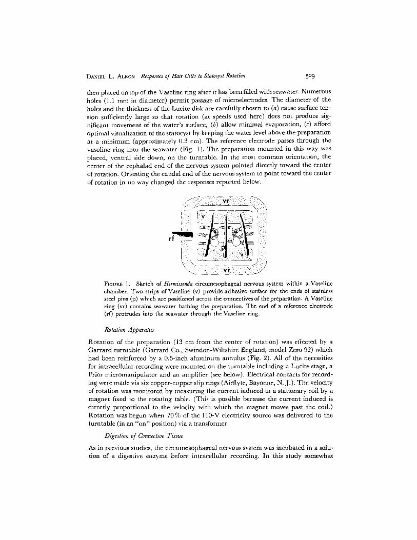
The isolated eircumesophageal nervous system is placed, immersed in a few drops of seawater, on a conventional microscope glass slide. Two strips of Vaseline, 1 cm long are placed immediately above and below the preparation. Stainless steel pins are then laid across the connectives of the nervous system. The pins are in direct contact with surface of the glass slide and are held this way because the ends of each pin are im- bedded in one of the Vaseline strips. A ring of Vaseline (approximately 0.4 em high and 3 cm in diameter) is then spread around the preparation pinned as described be- tween the two Vaseline strips. A Lucite disk, 0.2 mm thick and 3.5 cm in diameter, is
DANIEL L. ALKON Responses of Hair Cells to Statocyst Rotation 509
then placed on top of the Vaseline ring after it has been filled with seawater. Numerous holes (1.1 mm in diameter) permit passage of mieroeleetrodes. The diameter of the holes and the thickness of the Lucite disk are carefully chosen to (a) cause surface ten- sion sufficiently large so that rotation (at speeds used here) does not produce sig- nificant movement of the water's surface, (b) allow minimal evaporation, (c) afford optimal visualization of the statocyst by keeping the water level above the preparation at a minimum (approximately 0.3 cm). The reference electrode passes through the vasoline ring into the seawater (Fig. 1). The preparation mounted in this way was placed, ventral side down, on the turntable. In the most common orientation, the center of the cephalad end of the nervous system pointed directly toward the center of rotation. Orienting the caudal end of the nervous system to point toward the center of rotation in no way changed the responses reported below.
,:::i:::' ~ ~.. i ";'" ' : : " ' : ' ~
; ::. : ~
FIGURE l. Sketch of Hermissenda circumesophageal nervous system within a Vaseline chamber. Two strips of Vaseline (v) provide adhesive surface for the ends of stainless steel pins (p) which are positioned across the connectives of the preparation. A Vaseline ring (vr) contains seawater bathing the preparation. The end of a reference electrode (rf) protrudes into the seawater through the Vaseline ring.
Rotation Apparatus
Rotation of the preparation (13 cm from the center of rotation) was effected by a Garrard turntable (Garrard Co., Swindon-Wiltshire England, model Zero 92) which had been reinforced by a 0.5-ineh aluminum annulus (Fig. 2). All of the necessities for intracellular recording were mounted on the turntable including a Lucite stage, a Prior mieromanipulator and an amplifier (see below). Electrical contacts for record- ing were made via six copper-copper slip rings (Airflyte, Bayonne, N. J.). The velocity of rotation was monitored by measuring the current induced in a stationary coil by a magnet fixed to the rotating table. (This is possible because the current induced is directly proportional to the velocity with which the magnet moves past the coil.) Rotation was begun when 70 % of the l l0-V electricity source was delivered to the turntable (in an "on" position) via a transformer.
Digestion of Connective Tissue
As in previous studies, the circumesophageal nervous system was incubated in a solu- tion of a digestive enzyme before intracellular recording. In this study somewhat

51o T H E J O U R N A L O F G E N E R A L P H Y S I O L O G Y • V O L U M E 66 • 1975
FIOURE 2. Photograph of modified Garrard turntable and recording apparatus. (f) Farraday cage. (p) pedestal for preparation. (a) amplifier. (s) slip rings.
milder treatments were used: (a) 7-10 min of incubation in protease (CB, Calbiochem, San Diego, Calif.) solution (0.3-0.5 mg/cma). (b) 15 rain of incubation in collagenase (type 1, Sigma Chemical Co., St. Louis, Mo.) solution (0.2-0.5 mg/cma). From lot lo lot of the enzyme, optimal conditions were found to be more variable for protease than for collagenase, although greater ease of penetration was usually provided by protease.
Amplifier
Intraeellular signals were amplified by a high input impedance amplifier (impedance converter) with input capacity neutralization. This amplifier, as pictured in Fig. 2, was located on the turntable itself. Its characteristics are similar to those of the original design by Bak (1958). It utilizes, however, a battery-powered solid-state operational amplifier (no. 1402, Teledyne Philbriek, Dedham, Mass.). Capacitance neutralization was also accomplished by an operational amplifier (no. 40 1, Analog Devices, Inc., Norwood, Mass.) which was powered remotely by an Analog Devices power supply.

DANIEL L. ALKON Responses of Hair Cells to Statocyst Rotation 5II
Intracellular Recording
Intracellular recordings were made with glass micropipettes filled with 4 M potassium acetate (resistances of 60-100 M~2). The electrode was connected via a silver wire to the input stage of the high impedance amplifier. The reference electrode was a chloride/silver wire. A Wheatstone bridge circuit was used to pass current through the recording electrode. Current was monitored by recording the potential drop across a 10-Mf~ resistor in series with the electrode. All experiments were performed at room temperature (approximately 22°C).
R E S U L T S
Movement of Statoconia
SPONTANEOUS Previous observations revealed that the statoconia are constantly moving (Alkon and Bak, 1973). Videotapes of these movements demonstrate a wide range of movement frequencies, from occasional coarse movements of individual statoconia to very frequent rapid movements com- plete in less than 30 ms. Although the individual statoconia show much independence of motion, the cluster of statoconia, appearing as a sphere, often makes movements of its own, i.e. the entire cluster of crystals can rotate.
A few of the numerous hairs which project into the statocyst from the inner surface of the hair cells (Alkon and Bak, 1973) can occasionally be visualized using light microscope techniques (_> × 1,000). These hairs also can be seen constantly experiencing a wide range of movement frequencies. It is reason- able to suppose then, that the hairs are responsible for the cluster formation of the statoconia and that the hairs impart some of their own movement to the statoconia.
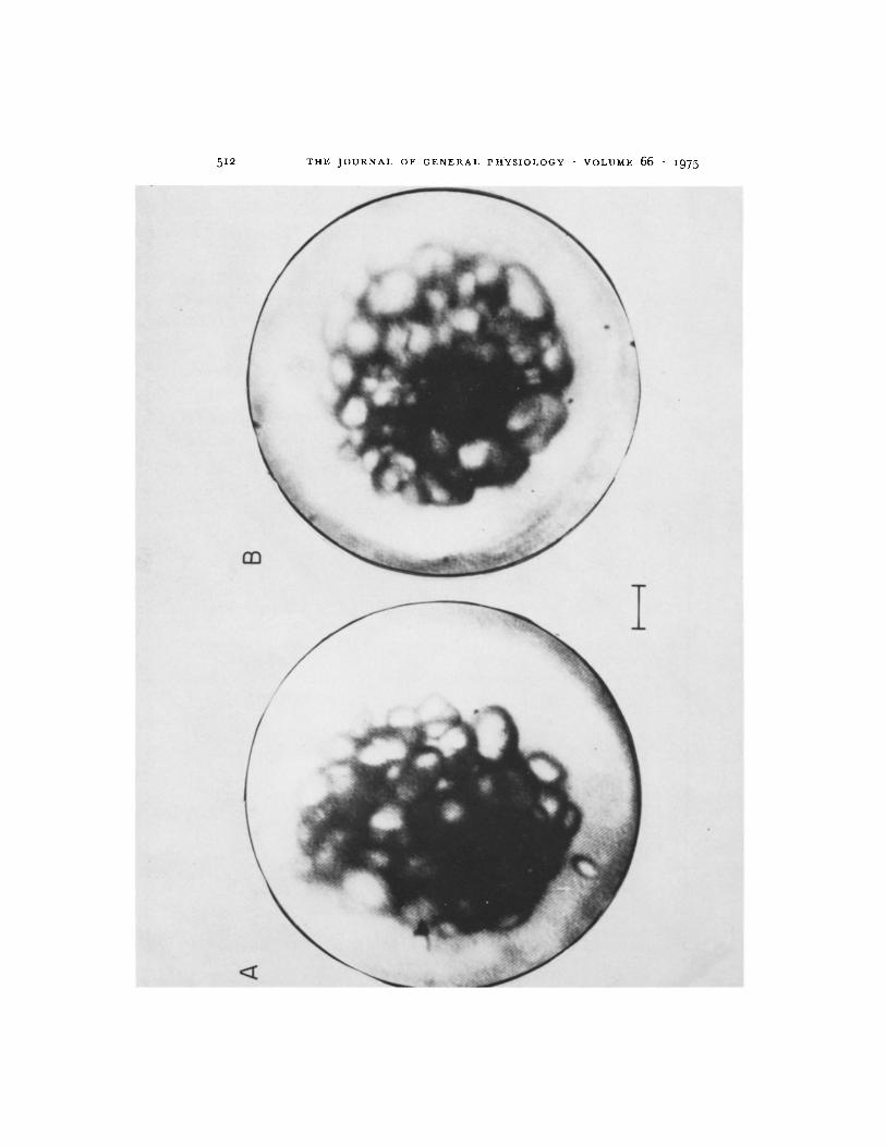
RESPONSE TO TILTING It has been reported that tilting of the statocyst causes the statoconia cluster to fall within the statocyst in the direction of the tilt (Alkon and Bak, 1973). This is demonstrated in Fig. 3. A small increase in the space between the cluster and the statocyst wall (on the side opposite to the direction of tilt) is apparent. The shape of the cluster also becomes less spheri- cal. The space between the cluster and the statocyst wall on the side in the direction of tilt is maintained, diminished only slightly by the tilting. These observations suggest that in response to a change in direction of gravity with respect to the cyst the cluster shape is distorted and somewhat compressed. In addition, the cluster now exerts more force (by its own weight) on the hairs of these cells in the direction of the tilt.
Spontaneous Activity of Hair Cells
Previous recordings from hair cells indicated that hair cells show a wide range of spontaneous firing (0-200 impulses/rain). In those hair cells which fired infrequently or not at all, it was possible to observe a rather large base-line
5 1 2 T H E J O U R N A L O F G E N E R A L P H Y S I O L O G Y • V O L U M E 66 • 1 9 7 5

DANIEL L. ALKON Responses of Hair Cells to Star, cyst Rotation 513
"noise" which was never observed for any other neural cell in Hermissenda
(Alkon and Bak, 1973). I t was suggested that this noise could arise from: (a) synaptic input to the hair cells (e.g. from the visual system, Alkon, 1973 b), (b) the constant interaction of the statoconia with the hairs, (c) spontaneous activity of an excitable focus (within the hair cell itself) which is subthreshold for firing hair cell impulses.
In the present study, collagenase, as well as protease in lower concentra- tions and for much shorter incubation periods than previously used, was used for penetration of hair cells. With this type of enzyme treatment the statocyst remained completely intact for several hours. With the enzyme treatment used in previous studies cell swelling was clearly apparent after 2 h and usually resulted in collapse of the statocyst shortly thereafter. In addition, hair cells treated with enzyme as described for the present study usually did not require steady hyperpolarization after penetration to reduce what is probably largely injury discharge. Generally with the use of the milder enzyme pretreatment described here the spontaneous firing of hair cells was reduced, spike ampli- tudes were larger and the base-line noise of hair cells was seen more clearly and more frequently to be comprised of depolarizing local potentials and distinct inhibitory potentials.
Tha t some of the base-line noise is not synaptic is illustrated in Fig. 4. This record was obtained from a hair cell whose axon had been cut before the re- cording was made. All of the evidence available to date (Alkon, 1973 b,
10 mV I 1.0 s
FmuR~. 4. Base-line activity of hair cell with cut axon. The axon was cut (approxi- mately 40 ~m from the cell soma) before intracellular recording. Because synapses are made distal to the cell soma, the activity pictured is nonsynaptic in origin.
FICURE 3. Dorsal view of Hermissenda statocyst. (A) Viewing microscope and prepara- tion are tilted 30 ° (to left in photograph) from the earth's gravitational vector. (B) Untilted preparation. Statoconia tend to fall in the direction of ti!ting, i.e. away from the right side of the statocyst. One statoconium (bottom A) approaches the inner surface of the hair cells, presumably falling between the hairs which support the cluster of statoconia, preventing them from coming any nearer to the statocyst wail. Calibration: 10/~m.

514 T I l E J O U R N A L O F G E N E R A L P H Y S I O L O G Y • V O L U M E 66 - 1975
Detwiler and Alkon, 1973), indicates that there is no synaptic input to hair cells at or near their somata. It is reasonable to assume, therefore, that a hair cell whose axon has been cut (betbre its point of entry into the cerebropleural ganglion) no longer sends or receives synaptic signals. The base-line noise of hair cells isolated from synaptic input can even be somewhat greater than that of intact hair cells. This may partially be due to irritation of the cells caused by the axon lesions.
With mild enzyme pretreatment, in approximately 50~o of cells penetrated, large discrete inhibitory potentials were observed (cf. Figs. 14, 16). These were considered synaptic since their frequency was greatly reduced or abolished by steps of light presented to the Hermissenda eyes (Alkon, in preparation), sug- gesting that these inhibitory potentials arise from ipsilateral optic ganglion cells (see Alkon, 1973 a, b). Presumably these inhibitory potentials arise from a conductance increase to an ion with an equilibrium potential negative with respect to the resting level since these potentials are diminished and eventually abolished by hyperpolarizing the hair cell with steady negative currents (Figs. 15, 16).
In hair cells with infrequent spontaneous impulses (<20 /min) distinct subthreshold local potentials were also often observed (Figs. 11, 13). Records from other hair cells showed frequent spontaneous impulses ( > 2 0 / m i n ) but less discernible hyperpolarizing waves or discrete local potentials (Fig. 7). Different types of hair cell base-line activity presumably would arise from hair cells located differently in the statocyst with respect to the earth's gravitational force. Injury associated with cell penetration could also account for differences in the resting potential of hair cells and thus, their base-line activity.
Subthreshold local potentials were elicited by weak depolarizing currents in a previous report (Alkon and Bak, 1973). In the present study this observation was repeated and in addition it was possible to observe a considerable range in the magnitude of those local potentials produced by depolarizing current pulses (Fig. 5). These potentials occurred less frequently if the depolarizing pulse was repetitively injected; i.e. they were subject to fatigue (Fig. 5).
From the above observations, then, the base-line noise of the hair cells arises from: (a) local potentials which are a property of the hair cell membrane itself and which are a function of the hair cell membrane potential (subthresh- old local potentials might also arise from firing of electrically coupled hair cells with spike thresholds lower than that of the penetrated hair cell [see Detwiler
FIGURE 5. Responses of hair cell to injection of depolarizing current pulses. Monitor deflections (lower traces in each record) are proprotional to current injected, the maxi- mum (top record) being 0.25 nA. Subthreshold local potentials are elicited by current pulses. Their frequency is significantly reduced when the pulse is repetitively injected (rate: 30/min) as in the two sets of double records (lower record in each set after five repetitions).

\
I I ! r
lO mv
L_ o.is
5x5

5z6 T H E J O U E N A L O F G E N E R A L P H Y S I O L O G Y • V O L U M E 6 6 • z 9 7 5
and Alkon, 1973]), (b) hyperpolar iz ing waves, synaptic in origin, arising f rom the visual pa thway. S t imula t ion of the hair cells by the cons tant movements of the s tatoconia as well as by o ther unknown synaptic inputs could, of course, set the m e m b r a n e potent ia l and thus affect the occur rence and magn i tude of the local potentials and the hyperpola r iz ing waves.
Responses of Hair Cells to &atocyst Rotation
Visualizat ion of the statocyst in the isolated c i rcumesophagea l nervous system was usually f rom its dorsal aspect. Those hai r cells were pene t ra t ed wi th cell bodies a t the equa to r of the cyst visualized as just described. Those hai r cells pene t ra ted toward the center of the ro ta t ing table will be descr ibed as being " i n back of the centr ifugal force vec tor . " Those hair cells pene t ra t ed on tha t half of the equator ia l c i rcumference away f rom the center of the ro ta t ing table will be described as being " in f ront of the centr i fugal force vec to r . " ryhe visualizat ion never being perfect, some al lowance must be m ad e for the hai r cell pene t ra ted not always being exact ly on the equator ia l c i rcumference.
A B
r r F I T I
_ _ 5 m V k ~ -
0.5s
¢ D
I
FIGURE 6. Depolarizing response of hair cell to rotation. Hair cell is located in fl-ont of the centrifugal force vector. Rotation produces depolarization, occurrence of local potentials (B) and finally firing of the hair cell (C). A hyperpolarizing wave is apparent in D. Amplitude of monitor signal as well as the inverse of the interval between monitor signals is proportional to the angular velocity of the turntable. (Here, lines have been substituted for the monitor signals.) Note the infrequent base-line activity. Occasional hyperpolarizing waves followed by an impulse occurred spontaneously.

I!I II1!! l ' twl , , f" , . : III ii!II ]~::
I ' : ~ ' IIJ o i i ; ~ ~ , :
I Jl !:' , :
b
! i ! ' ~ . / '
|T~r!l!!~,.!!! :,::. : i iillJ ::
" I l I ' T " ~ v ' - . ~ r - v ~ - - - , . . ' ~ - V , , , = p , , . , V , t , ~ I ,
~ ' , . . , / k . . . / k . . . / k . . . / ~
[ I I , * . . . . . . .
!pl J!! !: i
I0 mV I
1.0 :
FIGURE 7. Depolarizing response of hair cell to rotation. Hair cell is located in front of the centrifugal force vector. Rotation produces depolarization and a marked increase in firing. Ampli tude of minitor signal as well as the inverse of the intervals between monitor signals are proportional to the angular velocity of the turntable. Note the frequent occurrence of spontaneous impulses (top record). Base-line activity: 72 im- pulses/min.
517

518 T H E J O U R N A L O F G E N E R A L P H Y S I O L O G Y • V O L U M E 66 - i975
Hair cells in front of the centrifugal force vector always depolarized and increased their firing in response to rotation. This was true of hair cells regard- less of their base-line activity (Figs. 6, 7). The depolarization and increased firing always followed the onset of rotation after a short delay (Fig. 8). Simi-
Begin Rotation t/
I
If°it .2 0
12 o
x2
8 o
O z
~5
/ N i \
/ \
d s ~
/
' b
~--.q i
I I L I [
4 8 12 16 20 lIME (s)
': I 0 1
o_ co
1.5-' g
$ 2.0-
>
uJ
\\
25_
, \ " 30
\
'o- -o,
"q /,.o I "~- -c- --o- -o'
1 I I
24 28 32 36
- 1 0
- 0 8
E v
06 ~ w -
<[
g 04-----
g
02
FmUR~ 8. Relation of hair cell firing to angular velocity. Points of lowest plot are determined by the number of spikes in 2-s intervals as a function of time. Middle and upper plots are respectively determined by the amplitude of the monitor signal and the intervals between monitor signals as a function of time. The hair cell is located in front of the centrifugal force vector. Rotation produces a marked increase in firing. Ampli tude of monitor signal as well as the inverse of the intervals between monitor signals is proportional to the angular velocity of the turntable. Note the delay between onset of increased firing and the onset of rotation.
larly, the response reached its maximum a short time after the maximum rotating velocity (and thus the maximum centrifugal force) was achieved. Very frequently after the deceleration of the table was completed the hair cell activity (and membrane potential) briefly fell to a lower than resting level and then returned to the previous base-line impulse activity and membrane poten- tial (Fig. 8). If the rotation at maximum velocity was maintained, the hair cell impulse activity as well as membrane potential reached a transient maximum followed by a new steady state of impulse activity and potential, still sub-

DANIEL L. ALKON Responses of Hair Cells to Statocyst Rotation 519
stantially elevated above the resting level (Fig. 9). For different ceils, a wide range of responses to the smaller centrifugal forces was observed. In general, for cells with little spontaneous firing 0.5 g was the threshold stimulus (Fig. 9). Other hair cells, however, responded to as little as 0.25 g (Fig. 10).
16 Begin Begin
Acceleration Deceleration
12 l ,,m 78 rpm
8
I ~ f / I I I [ I I i i -, o , 8 2o 2, 2B 32 , : ,'8 5'2 5; 6; TIME (s)
FIGURE 9. Transient and steady-state phases of the depolarizing response of hair cells to rotation. Points are determined by the number of spikes in 2-s intervals as a function of time. The hair cell is located in front of the centrifugal force vector. Rotation at 78 rpm produces a marked increase in firing. Angular velocity reaches its maximum within 8 s. The maximum velocity is maintained for 44 s before deceleration begins. The decrease in the level of firing during the steady-state phase parallels a comparable decrease in membrane potential.
16 ~ 8 8 4 g
Begin 10 12 Rotation
0 ' ~ : ' ./ ~, / 0.2 0 . 6 1 . 0
~ , j ! ~, MAX. ACCELERATION (g)
\k.2, , I , I , I , I I I I
-4 4 8 12 16 20 24 28 TIME (s)
FIGURE 10. Relation of hair cell depolarizing response to maximum acceleration. For each plot deceleration of the turntable began immediately after the attainment of different maximum accelerations (0.244, 0.436, 0.721, 0.884 g). Points are determined by the number of spikes in 2-s intervals as a function of time. The hair cell is located in front of the centrifugal force vector. Inset: Peak firing frequency as a function of the maximum acceleration achieved.

B
l I , , , ,
I
~ - , o e"
T y , J ~
5m Lm 0.2 s
FIGURE I 1. Hyperpolarizing response of hair cell to rotation. Hair cell is located in back of the centrifugal force vector. Rotation produces a slight hyperpolarization and a cessation of impulse activity. (A) (top to bottom) Depolarizing local potentials de- crease in amplitude and frequency as a maximum acceleration is achieved. (B) (top to bottom) Depolarizing local potentials increase in amplitude and frequency as turn- table comes to rest. Ampli tude of monitor signal as well as the inverse of the intervals between monitor signals are proportional to the angular velocity of the turntable. Base- line activity of hair cell: 120 impulses/rain.
52o

DANIEL L. ALKON Responses of Hair Cells to Statocyst Rotation 52I
Hair cells in back of the centrifugal force vector usually hyperpolar ized (slightly) and markedly decreased their firing in response to rotation. Again, this was true of hair cells regardless of their base-line activity (Fig. 11, 13). Al though impulse activity often ceased entirely dur ing rotat ion of hair ceils in back of the centrifugal force vector, subthreshold, depolarizing local poten- tials, were often apparen t (Fig. 11). Slight hyperpolar izat ion of hair cells in back of the centrifugal force vector could reduce or remove spontaneous im- pulses and reveal spontaneously occurring depolarizing local potentials, thus approximate ly dupl icat ing the effect of rotat ion (cf. Figs. 11, 12). Fur the r hyperpolar izat ion could abolish these depolarizing local potentials, also simu- lat ing the effect of rotat ion (Fig. 12). Thus, rotat ion of hair cells in back of the
5 mV[__ 0.2 s
FIGURE 12. Comparison of hyperpolarizing response of hair cell to rotation to hyper- polarization produced by current injection. (A) Hair cell of Fig. 11 was hyperpolarized by 5 mV with injection of 0.05 nA of steady negative current. (B) Rotation produces a hyperpolarization and a cessation of local potentials. (C) Local potentials return after rotation stops. (D) Injection of an additional 0.025 nA in hair cell also causes cessation of local potentials. Dashed lines indicate level of membrane potential in A and C. Amplitude of minitor signal in C as well as the inverse of the intervals between moni- tor signals are proportional to the angular velocity of the turntable.
centrifugal force vector produces an effect not obviously different from tha t produced by simple hyperpolar izat ion of the hair cell.
In other hair cells ( toward the center of rotation) in which base-line firing was only occasional, depolarizing local potentials were apparen t wi thout rotation. For these cells, rota t ion of the statocyst often produced sufficient hyperpolar izat ion to abolish the local potentials which were a part of the base-line activity (Fig. 13).
Table I summarizes the n u mb e r of hyperpolar iz ing and depolarizing re-

522 THE JOURNAL OF GENERAL PHYSIOLOGY - VOLUME 66 • i975
A ~ B 5 rnvL~ - s ~
%,-- %,- -V~--
FIGURE 13. H y p e r p o l a r i z i n g response of ha i r cell to ro ta t ion . H a i r cell is loca ted in
back of the cen t r i fuga l force vector. R o t a t i o n p r o d u c e s a h y p e r p o l a r i z a t i o n a n d a cessa-
t ion of local potent ia ls . Note i n f r e q u e n t o c c u r r e n c e of s p o n t a n e o u s impu l se s in A a n d C,
b u t obv ious va r i a t i on in m a g n i t u d e of local potent ia ls . A m p l i t u d e of m o n i t o r s ignal
as well as t h e inverse of the in te rva ls be tween m o n i t o r s ignals a re p r o p o r t i o n a l to the
a n g u l a r veloci ty of the t u rn t ab l e . Base- l ine ac t iv i ty of ha i r cell: 15 i m p u l s e s / m i n .
T A B L E I
H A I R C E L L R E S P O N S E S T O S T A T O C Y S T R O T A T I O N *
Depolarizing Hyperpolarizi ng response with response with
increased firing decreased firing Unresponsive
H a i r ceils in front:~ of force vec to r
H a i r cells in back:~ of force vec tor
37 0 2
2 11 1
* T o t a l n u m b e r of cel ls: 53, to ta l n u m b e r of s t a tocys t s : 38, to ta l n u m b e r of a n i m a l s : 30. Not cons ide r ing u n r e s p o n s i v e cells, a c h i - s q u a r e test for a two-by - two tab le showed t h a t the
two k inds of responses o c c u r r e d for the two loci in a d i s t r i b u t i o n w h i c h was s ign i f ican t ly d i f fe ren t (P < o.ooi).
sponses encountered on the half-circumference toward and away from the center of the rotation table. Although there are exceptions (which could be due to the imperfection of electrode placement) the general rule was followed that hair cells nearer the center of rotation hyperpolarize, and hair cells away from the center of rotation depolarize in response to centrifugal force.
Effect of Current on Depolarizing Responses to Rotation
For many hair cells the current (input)-voltage (induced) relation is approxi- mately linear for a substantial range of potentials negative with respect to the resting membrane potential (Alkon and Bak, 1973). With steady hyperpolari- zations effecting negative changes in hair cell membrane potential, the rota- tional stimulus was repeated a number of times for many of the hair cells studied. For most of the hair cells studied after stimulating at different levels of membrane potential, it was possible to reproduce the initial response of the hair cell at its resting membrane potential level. Records from those hair cells which did not give reproducible responses to rotation were discarded.
Steady hyperpolarization of hair cells always reduced the base-line noise. As already mentioned (Figs. 14, 15, 16), the prominent inhibitory waves seen

DANIEL L. ALKON
A
I
Responses of Hair Cells to Statocyst Rotation 523
B
I
I I0 rnV i
I.Os
~ . . . ~ ,x...~ ~_.t '._./k.d k_/k_d ~ I I t h i
FICURE 14. Effect of negative current injection on depolarizing response to rotation. Upper records: (A) Hair cell spontaneous activity (15 impulses/min). (B) Depolarizing response of hair cell to rotation. Depolarization is slight although increase in impulse activity with rotation is clear. Lower records: (A) Hair cell is hyperpolarized 11 mV with minus 0.10 nA current injection. (B) Depolarizing response of hyperpolarized hair cell to rotation. Dashed lines in upper and lower records indicate level of resting mem- brane potential. Note the large increase in the absolute magnitude of the depolarizing response to rotation when the hair cell is hyperpolarized. Amplitude of monitor signal (at lower gain in upper records) as well as the inverse of the intervals between monitor signals are proportional to the angular velocity of the turntable.
clearly in some cells were diminished and eventually abolished by hyper- polarization. The depolarizing local potentials which occurred spontaneously decreased in magnitude and, with sufficient hyperpolarization, in frequency (Figs. 11, 12).
Injection of small negative currents (_<0.125 nA) causing DC potential changes of 7-15 mV invariably produced an increase in absolute magnitude of the depolarizing response to rotation (Figs. 14, 16). For these small steady changes in hair cell potential, resistance clearly decreased (and thus conduct- ance increased) during rotation (Table II). For additional steady changes in hair cell potential (using currents >0.125 nA), resistance showed a small increase during rotation (Table II). With most cells it was possible to sub- stantially reduce or remove the depolarizing response to rotation with suffi-

5 2 4 T H E J O U R N A L O F G E N E R A L P H Y S I O L O G Y • V O L U M E 66 . i 9 7 5
c i e n t s t e a d y h y p e r p o l a r i z a t i o n (F ig . 15). T h e o r i g i n a l r e s p o n s e r e t u r n e d w h e n
t h e h a i r c e l l w a s n o l o n g e r h y p e r p o l a r i z e d .
W i t h h y p e r p o l a r i z a t i o n o f t h e h a i r ce l l , c l e a r d e p o l a r i z i n g w a v e s w e r e o b -
s e r v e d t o b e p a r t o f t h e d e p o l a r i z i n g r e s p o n s e to r o t a t i o n (F igs . 14, 16). T h e s e
d e p o l a r i z i n g w a v e s c o u l d , i f o f s u f f i c i e n t m a g n i t u d e , t r i g g e r t h e a f o r e m e n -
t i o n e d d e p o l a r i z i n g l o c a l p o t e n t i a l s (cf. F ig . 16).
T A B L E II
H A I R CELL C O N D U C T A N C E (g) AT REST AND D U R I N G R O T A T I O N *
W i t h ro ta t ion W i t h ro ta t ion
1' l" AI(I" -- I') ,~V1 gl(AI/AVI) AV2 g~(AI/AV~)
nA nA nA mV mhos X 10 -6 mV mhos X 10 -6
For currents (I) ___ 0.125 nA
0 --0.1 --0.1 --11.5 0.0087 --2.5 0.040 0 --0.07 --0.07 --15 0.0047 --11.5 0.006
~0 --0.063 --0.063 --7 0.0090 --4.2 0.015 §0 --0.125 --0.125 --12 0.0104 --3.5 0.0357
Means 0.008311 0.02411
SD 4- 0.0027 SD 4- 0,014
For currents (I) > 0.125 nA
--0.175 --0.288 --0.113 --8.5 --0.148 --0.32 --0.175 --17.5
~--0.138 --0.275 --0.138 -- 16.0 §--0.125 --0.175 --0.050 - -4 .0
0.0132 -- 14.5 0.0078 0.0099 --17,25 0,0101 0.0086 --22,0 0.00625 0.0125 - -6 ,0 0,0084
Means 0.011 0.00815
SD 4- 0.00188 SD 4- 0.00165
* Each value of V(mV) was obtained by averaging four or more maximal potent ial values (excluding discrete local potentials and spikes) in a 10-s sample of the hair cell recording. AV1 was then obtained by substract ing the average potent ial value for one level of current inject ion from the average potent ial value at a second level. AV~ was obta ined in a similar man- ner for potent ia l values during statoeyst rotat ion. ~: § Indica te lines of data taken from the same cell, Other lines of data were taken from different cells. All da ta on any one line were from the same cell. [I Using a t test for paired observations, these means are significantly different (0.005 < P < 0.0125).
I0 mV
'l.Os
FIGURE 15. Effect of addi t ional negative current injection on depolarizing response to rotation. Hai r cell of Fig. 14 is hyperpolar ized 16 m V with minus 0.15-nA cur ren t injection. Rota t ion causes little or no depolarizing response. Ampl i tude of moni tor signal as well as the inverse of the intervals between moni tor signals are propor t ional to the angular velocity of the turntable .

DANIEL L. ALKON Responses of Hair Cells to Statocyst Rotation 525
0.0
' , I
"V" W"-
:.o;Ik . . . . . . . . . . . . . . . . . . . . . . . . . . . . . . . . . . . . . .
w V
10 mV L___
0.2 s
-0.2 nA
_ - A _ _ . .
I i
"W "V'-
FIGURE 16. Effect of negative current injection on depolarizing response to rotation. Records on left are of the hair cell at different levels of membrane potential (resting level in the absence of current indicated by dashed lines). Records on right show the effect of rotation on the hair cell when hyperpolarized to different levels of membrane potential. Hyperpolarization clearly reduces the magnitude of the spontaneously occurring hyperpolarizing waves and increases the absolute magnitude of the depolarizing response to rotation. In the lower right record a local potential is triggered by a depolarizing wave which was caused by rotation. Ampli tude of monitor signal as well as the inverse of the intervals between monitor signals are proportional to the angular velocity of the turntable.

526 T H E J O U R N A L O F G E N E R A L P H Y S I O L O G Y • V O L U M E 6 6 • i 9 7 5
Some hair cells which appeared to be quiet at their resting level showed an especially large depolarizing response and increase in occurrence of hyper- polarizing and depolarizing waves (Fig. 6). These hair cells were presumably already somewhat hyperpolarized because of their position relative to the earth's gravitational force.
DISCUSSION
The base-line activity in hair cells was shown to arise at least in part from synaptic hyperpolarizing waves and subthreshold depolarizing local poten- tials. In response to rotation, hair cells in front of the centrifugal force vector depolarize and increase their firing, whereas hair cells in back of the vector hyperpolarize and decrease their firing.
Artifactual Stimulation
A number of observations argue against the possibility that the technique used in this study causes an artifactual stimulation of the hair cells: (a) Only hair cells respond as described to rotation. No other cells in the circumesophageal nervous system of Hermissenda respond similarly. Type A and type B photo- receptors which receive synaptic input from hair cells give their own unique responses to statocyst rotation (Alkon, in preparation). Records from many other neural cells in the circumesophageal system show no response to rota- tion. (b) No potential changes are recorded when rotating the preparation with the electrode immediately outside of the hair cells. (c) The differences in responses which are dependent on the location of the hair ceils on the stato- cyst with respect to the center of rotation can only be explained physiologically since all recording conditions and rotation conditions are the same regardless of the electrode's position on the statocyst. (d) The depolarizing response in- creases with steady injection of small negative currents. With larger negative current injections, the response is diminished and ultimately abolished. These effects of steady hyperpolarizing currents most likely are physiological. An artifactual response in all probability would not be membrane-potential dependent, particularly within the linear range of the hair ~ells' current-volt- age characteristics. (e) The depolarizing response of hair cells to rotation was seen to consist of an initial transient phase followed by a smaller steady-state phase. An artifactual response would be expected to closely follow in one phase, the gradual at tainment of maximal rotational velocity.
Mechanism of Hair Cell Responses to Rotation : Depolarizing Response
The results reported in this study suggest that the following chain of events leads to the depolarizing response of hair cells to rotation. The centrifugal force produced by rotation, acting as demonstrated for the gravitational force of the earth, causes the statoconia to push against the hairs of those statocyst

DANIEL L. ALKON Responses of Hair Cells to Statocyst Rotation 527
cells located away from the center of rotation. The force of the statoconia bends the hairs which in turn causes the hair cell to depolarize. Depolarizing waves as a part of the depolarization produced by bending the hairs trigger subthreshold depolarizing local potentials. Local potentials of sufficient mag- nitude trigger impulses and thus results in an increase in firing of the hair cell.
The depolarization of the hair cells, believed to be caused by the bending of the sensory hairs, appears to arise, at least in part, from an increase in con- ductance to an ion(s) with a positive equilibrium potential. This was sug- gested by the effect of small negative current injections on the depolarizing response to rotation. The depolarizing response elicited by a displacement (or vibration) stimulus also was increased by steady hyperpolarizations (Alkon and Bak, 1973).
The decrease in the depolarizing response to rotation caused by larger negative current injections was not observed for the response to a displace- ment stimulus. (Responses to weak displacement stimuli, however, were not examined at different levels of hair cell membrane potential.) The reduction of the depolarizing response to rotation caused by larger (>0 . 125 nA) current injections raises the possibility that a membrane-potential-dependent con- ductance increase is responsible for the hair cells' response. Alternatively such a finding could be explained were the depolarizing response to require a second change in conductance, namely of an ion with a negative equilibrium potential. Finally, a parallel might be drawn between hair cell depolarizing responses to rotation and the generator potentials of Hermissenda photorecep- tors. A depolarizing generator response in dark-adapted cells (Alkon and Fuortes, 1972) and a hyperpolarizing response, observed in light-adapted cells, were identified for these photoreceptors. The hyperpolarizing response, studied in photoreceptors with cut axons, and thus isolated from synaptic in- put, was found to arise from an increase in conductance to an ion with a nega- tive equilibrium potential. I The second phase of the depolarizing generator potentials were seen to increase with steady hyperpolarizations of the photo- receptor (indicating increased conductance to an ion with a positive equi- l ibrium potential) until a critical level was reached, usually with large nega- tive current injections causing potential shifts as great as 50 mV. With the photoreceptor hyperpolarized below this critical level the photoreceptor re- sponse was dramatically reduced in absolute magnitude. This suggested, within the depolarizing response to light, the existence of two separate de- polarizing components. Thus, three separate conductances could underlie the photoreceptor response to light: one for each of the two depolarizing com- ponents and a third for the hyperpolarizing response. Such additional com- ponents, then, although much smaller than those of the photoreceptor's re- sponse, might also exist for the hair cell generator potential.
1 Alkon, D. L. 1972. Unpublished observations.

528 THE JOURNAL OF GENERAL PHYSIOLOGY • VOLUME 66 • 1975
Attenuation of Hair Cell Transducer and~or Synaptic Events
One advantage provided by a second depolarizing component in the hair cell generator potential (see above) could be amplification of a signal which is otherwise attenuated by the cable properties of the hair. Thus, if the primary transduction process occurs on the hairs, the potential reaching the spike focus and the synaptic sites (as well as that reaching the recording electrode) might be very small unless this potential is amplified, e.g. by triggering a second much greater signal. These second triggered signals might be the depolarizing waves (cf. Figs. 14 and 16) observed within the depolarizing response to rotation.
Although the actual distance of the synaptic sites, like the distance of the hairs, from the recording electrode is probably small, the attenuation of synap- tic signals arising on the terminal branches and hairs of the hair cell may be great. This would account for the difficulty encountered here and in previous studies in reversing the inhibitory synaptic potentials (cf. Figs. 14 and 16) recorded in hair cells.
Hyperpolarizing Response
The centrifugal force produced by rotation will cause the statoconia to move away from and thus exert less force against the hairs of those cells on that half of the statocyst toward the center of rotation. With less force against the hairs of these cells, they would be expected to become less depolarized. The mem- brane potential thus falls below levels critical for eliciting depolarizing local potentials of sufficient magnitude (cf. Figs. 11, 12) to trigger impulses. This sequence of events was seen to be not clearly distinguishable from the results of simple negative current injection into the hair cells (Fig. 12). It might be asked why weak negative currents, in hair cells without rotation (Figs. 12 D; 14 A, bottom; 16, left), do not increase and /or make clearer the depolarizing waves which are only seen in the responses of these hair cells, in front of the force vector, during rotation (Figs. 5 B, C; 14 B, bottom; 16 right). This could be explained if these depolarizing waves only occur when a certain minimum force, such as that generated during rotation, is exerted by the statoconia on the hairs. Small steady forces might, nevertheless, be exerted by the statoconia on the hairs of the equatorial statocyst cells. Whether or not the depolarization caused by these small steady forces brings the membrane po- tential to a level at which local potentials of various magnitudes, or even impulse activity, occur would depend on the particular membrane properties of each hair cell as well as the tonic synaptic input it receives from other cells within the statocysts and /or the visual system.
The inhibitory interactions identified between hair cells of the same stato- cyst (Detwiler and Alkon, 1973) could serve to enhance the differences ob- served for hair cells located on opposite sides of the statocyst. Thus, a hair cell
DANIEL L. ALKON Responses of Hair Cells to Statocyst Rotation 529
in front of the centrifugal force vector depolarizes and increases its firing in response to rotation, thereby increasing its inhibition of hair cells located on the opposite side of the statocyst. This inhibition, then,, would add to the hyperpolarization of a hair cell expected to hyperpolarize, in any case, as its own response to statocyst rotation.
S U M M A R Y
(a) A new technique is described for stimulating hair cells of the Hermis- senda statocyst. The preparation and recording apparatus can be rotated at up to 78 rpm while recording intracellular potentials.
(b) Hair cells on the equator of the statocyst away from the center of rota- tion depolarize and increase their firing in response to rotation. This de- polarizing response has a transient and a smaller steady-state phase.
(c) Hair cells on the equator of the statocyst toward the center of rotation hyperpolarize and decrease their firing in response to rotation.
(d) Base-line activity of hair cells, at least in part, consists of depolarizing local potentials and synaptic hyperpolarizing waves.
(e) Injection of small negative currents produces a sizable increase in the absolute magnitude of the depolarizing response to rotation.
(f) Injection of larger negative currents reduces and ultimately abolishes the depolarizing response to rotation.
(g) Injection of negative currents has approximately the same effect on hair cells as does rotation when the hair cell is located toward the center of rota- tion (i.e. in back of the centrifugal force vector).
(h) The results reported in this study suggest that the following chain of events leads to the depolarizing response of hair cells to rotation-. The cen- trifugal force produced by rotation causes the statoconia to push against the hairs of those statocyst cells located away from the center of rotation. The force of the statoconia bends the hairs which in turn causes, at least in part, an increase in conductance to an ion with a positive equilibrium potential. This increase in conductance, in turn, causes steady depolarization and depolariz- ing waves which can trigger subthreshold depolarizing local potentials. Local potentials of sufficient magnitude trigger impulses and this results in an in- crease in firing of the hair cell.
I am grateful to Dr. W. J. Adelman, Jr . for his helpful discussion and suggestions. Without A. F. Bak's engineering assistance this work would not have been possible.
Received for publication 1 February 1975.
R E F E R E N C E S
ALKON, D. L. 1973 a. Neural organizat ion of a molluscan visual system. J. Gen. Physiol. 61:444. ALKON, D. L. 1973 b. Intersensory interactions in Hermissenda. J. Gen. Physiol. 62:185. ALKON, D. L. 1974. Associative t ra in ing of Hermissenda. J. Gen, Physiol. 6,t:70.

5 3 ° T H E J O U R N A L O F G E N E R A L P H Y S I O L O G Y " V O L U M E 66 • 1 9 7 5
ALKON, D. L. 1975 a. Neural correlates of associative training in Hermissenda. J . Gen. Physiol. 65:46--56.
ALKON, D. L. 1975 b. A dual synaptic effect on hair cells in Hermissenda. J. Gen. Physiol. 65:385. ALKON, D. L., and A. BAK. 1973. Hair cell generator potentials. J. Gen. Physiol. 61:619. ALKON, D. L., and M. G. F. FUORTES. 1972. Responses of photoreceptors in Hermissenda. J. Gen.
Physiol. 60:1. BAK, A. F. 1958. A unity gain cathode follower. Electroencephalogr. Clin. Neurophysiol. 10 (No. 4):
745. DETWIL~R, P. B., and D. L. ALtCON. 1973. Hair cell interactions in the statocyst of Hermissenda.
J . Gen. Physiol. 62:618. WmDERI~OLD, M. L. 1974. Aplysia statocyst receptor cell3: intraceUular responses to physiologic
stimuli. Brain Res. 78:490. WOLFF, H. G. 1975. Statocysts and geotactic behavior in gastropod molluscs. Fortschr. Zool.
23:63.