response of the ventral pallidal/mediodorsal thalamic system to antipsychotic drug administration:...
TRANSCRIPT
N
EUROPSYCHOPHARMACOLOGY
1998
–
VOL
.
18
,
NO
.
5
© 1998 American College of NeuropsychopharmacologyPublished by Elsevier Science Inc. 0893-133X/98/$19.00655 Avenue of the Americas, New York, NY 10010 PII S0893-133X(97)00165-6
Response of the Ventral Pallidal/Mediodorsal Thalamic System to Antipsychotic Drug Administration: Involvement of thePrefrontal Cortex
Antonieta Lavin, Ph.D., and Anthony A. Grace, Ph.D.
Intracellular recordings were obtained from rat ventral pallidal (VP) and mediodorsal thalamic (MD) cells
in vivo
and the effects of antipsychotic drugs on their basal and evoked electrophysiological characteristics were assessed. Administration of either haloperidol or clozapine caused a significant decrease in the average firing rate, accompanied by a hyperpolarization of the membrane potential in the VP cells recorded. However, neither drug induced a substantial change in the other basic membrane properties of the MD cells or VP cells tested. In addition, in 50% of the MD cells tested, both antipsychotic drugs caused a change in spike discharge from an oscillatory pattern to a tonic discharge mode. In rats that had
received ibotenic acid lesions of the prefrontal cortex (PFCtx) 4–8 weeks prior to recording, cells in the VP exhibited similar changes in firing frequency in response to haloperidol administration as those in the intact rats. However, in contrast to the intact rats, MD cells recorded from rats with PFCtx lesions exhibited a significant increase in firing rate after haloperidol administration. The results from this study suggest that the prefrontal cortex plays a role in modulating the response of the thalamus to antipsychotic drugs.
[Neuropsychopharmacology 18:352–363, 1998]
© 1998 American College of Neuropsychopharmacology.Published by Elsevier Science Inc.
KEY
WORDS
:
Mediodorsal thalamic nucleus; Ventral Pallidum; Haloperidol; Clozapine; Schizophrenia; Prefrontal cortical lesions
The ventral pallidum (VP) is a ventral extension of theglobus pallidus, and together with its projection to themediodorsal thalamic nucleus (MD) are integral partsof the limbic system within the basal ganglia. The VP isthe major synaptic target of nucleus accumbens neu-rons, and in return influences higher cortical structuresvia its projections to the MD (Cornwall and Phillipson1988; Gröenewegen 1988; Gröenewegen et al. 1993;
Haber et al. 1985; Lavin and Grace 1994; Young et al.1984; Zahm and Heimer 1987) and the reticular thalamicnucleus (RTN) (Cornwall et al. 1990; Jones 1985; Lavinand Grace 1994), thus forming a loop that encompassesthe prefrontal cortex–nucleus accumbens–VP–thalamus–cortex. The MD thalamic nucleus has attracted a greatdeal of interest due to its proposed role in the integrationof information flow between the limbic system and cen-ters of higher cognitive function. The MD nucleus hasalso been an area of interest due to its potential involve-ment in schizophrenia, both in its role as a major corti-cal relay of the limbic system and because of its inter-connections with the prefrontal cortex (Divac et al. 1978;Goldman-Rakic et al. 1984; Goldman-Rakic and Porrino1985; Markowitsch 1982; O’Donnell et al. 1997).
Models of cortical function suggest that the thalamo-cortical system, in conjunction with the RTN, serves as agate for cortical input or cortico-cortical integration. Re-
From the Departments of Neuroscience and Psychiatry, Center forNeuroscience, University of Pittsburgh, Pittsburgh, Pennsylvania.
Address correspondence to: Dr. Antonieta Lavin, Department ofNeuroscience, 446 Crawford Hall, University of Pittsburgh, Pitts-burgh, PA 15260.
Received March 25, 1997; revised July 20, 1997; accepted August7, 1997.

N
EUROPSYCHOPHARMACOLOGY
1998
–
VOL
.
18
,
NO
.
5
VP/MD System and Antipsychotic Drugs
353
sults of anatomical (Cornwall et al. 1990; Jones 1985;O’Donnell et al. 1997) and electrophysiological (Lavinand Grace 1994) studies suggest that the limbic systemoutput to the thalamus is positioned to exert a strong in-fluence on prefrontal cortical activity via its projection tothe MD and the RTN. Therefore, information arisingfrom cortical and limbic sites is integrated within theventral striatum under the modulatory influence ofdopamine, which then exerts regulatory control overMD and RTN activity via the ventral striatal projectionto the VP and the medial segment of the globus pallidus(Gröenewegen 1988; Young et al. 1984). Like the MD, theVP itself is thought to play a significant role in the cogni-tive and motor skills necessary to perform reward-medi-ated behaviors (Grace 1995; Mogenson and Yang 1991;Richardson and DeLong 1991; Wilson and Rolls 1990)and represents the main output of the limbic system.
Because of this potential relationship between corti-cal-subcortical interactions and schizophrenia, manystudies have examined the mode of action of antipsy-chotic drugs by testing their effects on basal ganglianeurons. However, comparatively less effort has beenexpended to examine how classical and atypical anti-psychotic drug treatment affects the output sites of thelimbic system and their subsequent impact on thalamo-cortical activity. For example, Deutch et al. (1995) foundthat clozapine administration increases the number ofcells expressing Fos-like immunoreactivity and theamount of Fos protein in the paraventricular thalamicnucleus, but D
2
-like or D
1
-like antagonists did not in-duce Fos in this nucleus. Furthermore, extracellular re-cordings of VP neuron activity revealed that systemicadministration of haloperidol produced a suppression intheir firing rate (Napier et al. 1991a). In this article, weexamined the spontaneous and evoked activity of neu-rons that comprise the output sites of the limbic system(i.e., the VP and the MD thalamic nucleus) in responseto acute administration of two drugs with distinct phar-macological profiles that are known to have potenttherapeutic actions in schizophrenia: the classical anti-psychotic drug haloperidol and the atypical antipsy-chotic drug clozapine (Chouinard and Annable 1976;Gerlach et al. 1974, 1975; Racagni et al. 1980). The im-pact of lesions of the prefrontal cortex on these re-sponses was also examined as a way of evaluatingwhether the hypofrontality that is reported to bepresent in schizophrenics (Ingvar and Franzen 1974;Pettegrew et al. 1989, 1991; Williamson et al. 1991;Weinberger et al. 1986) may alter the effects of antipsy-chotic drugs when compared to the intact system.
METHODS
All animals were handled in accordance with the proce-dures outlined in the
Guide for the Care and Use of Labora-
tory Animals
published by the USPHS, and the specificprotocols used were approved by the University ofPittsburgh Institutional Animal Care and Use Commit-tee. Most of the methods used have been described pre-viously (Lavin and Grace 1994).
In Vivo Intracellular Recording andDrug Administration
Sprague-Dawley albino male rats were anesthetized withchloral hydrate (400 mg/kg IP) and fixed in a stereotaxicapparatus (Narishige model SN-2). In experiments usinganimals with cortical lesions, the recordings were per-formed 4–6 weeks after the lesion. Supplemental doses ofchloral hydrate and test drugs were administered IV viaa lateral tail vein cannula. The scalp was exposed andholes drilled in the skull overlying the VP (coordinates:AP
5
2
0.4mm; L
5
2
1.9 mm; V
5
7.5–8.5 mm), the MDthalamic nucleus (coordinates: AP
5
2
3.4 mm; L
5
2
0.5mm; V
5
5.2–5.6 mm), and the prefrontal cortex (coordi-nates: AP
5
2.7 mm; L
5
2
1.6 mm; V
5
2.5 mm, accord-ing to Paxinos and Watson 1986). Body temperature wasmaintained at 37
6
0.5
8
C by a thermostatically controlledheating pad. Intracellular recordings were performed us-ing electrodes pulled from 1.0 mm O.D. Omegadot glasstubing (WPI) using a Flaming-Brown P-80/PC electrodepuller and filled with either: (1) 3 M K
1
acetate (resis-tance
5
60–120 M
V
), or (2) 10% Lucifer yellow in 0.1 MLiCl (resistance
5
100–250 M
V
).All electrophysiological recordings were digitized us-
ing a NeuroData Neurocorder and stored on VHS video-cassettes for subsequent off-line analysis with a custom-designed data analysis program (Neuroscope). Inputresistance, time constant, firing pattern, and evoked re-sponses from VP or MD stimulation were examined foreach cell recorded. Electrical stimulation of the VP or MDnuclei was performed using bipolar concentric electrodes(Kopf, model SNE-10). Single pulses (0.3 ms duration, 0.1to 1.5 mA) were delivered at approximately 1 Hz. Spon-taneous and evoked responses were sampled from singlecells before and after drug administration. The input re-sistance was determined by injecting a series of hyperpo-larizing current pulses of increasing amplitude into theneuron and measuring the resultant changes in mem-brane potential. The drugs used were: haloperidol (0.5mg/kg) (McNeil Pharmaceutical), or clozapine (20 mg/kg, dissolved in 1N HCl, pH
5
4.5) (RBI). The final vol-ume was adjusted using isotonic saline. Only haloperidolwas tested in the animals with prefrontal cortical lesions.In the cases in which the effects of drugs were examined,only one cell per animal was recorded.
Prefrontal Cortical Lesions
Lesions of neurons intrinsic to the PFCtx were producedin Sprague-Dawley (Zivic-Miller) albino male rats weigh-

354
A. Lavin and A.A. Grace N
EUROPSYCHOPHARMACOLOGY
1998
–
VOL
.
18
,
NO
.
5
ing 200–250 g. The rats were anesthetized with Equithesin(0.3 ml/0.1 kg) followed by local bilateral injection of theexcitotoxin ibotenic acid (5
m
g/0.5
m
l/side) into the pre-frontal cortex (coordinates: AP
1
3.2; L
6
0.7; V 3.2 mm,according to Paxinos and Watson 1986) using a glassmicropipette (75–100
m
m tip diameter). After the mi-cropipette had remained in position for at least 5 min,ibotenic acid was introduced by pressure ejection over a2.5-min period. The micropipette was then left in placefor an additional 5 min before withdrawal. Followingthe lesions, animals were allowed to recover for 4–6weeks before recording, with food and water availablead libitum. Rats were housed 2 per cage on a 12-h light-dark cycle. The animals did not exhibit any substantialbehavioral abnormalities following recovery from thelesion and were observed to consume food and waternormally. The lesions did not encompass the entirePFCtx, but instead were limited to a region of the cortexthat included the medial prefrontal cortex, dorsal ante-
rior cingulate cortex and prelimbic cortex, as illustratedin Figure 1.
Histology
In a subset of experiments, the location of the cells wasverified by staining the cells after recording via intracel-lular injection of the fluorescent dye Lucifer yellow(Stewart 1978). Upon stable penetration of VP or MDneurons, the dye was injected by applying 1–2 nA con-tinuous hyperpolarizing current through the electrode,which was interrupted by 12-ms, 2-nA depolarizingcurrent pulses delivered at 7 Hz to prevent blocking ofthe electrode (Grace and Llinás 1985). At the end of theexperiment, the animal was killed by administering anoverdose of chloral hydrate and was perfused transcar-dially with saline followed by 10% formalin. The brainwas removed and placed overnight in 15% sucrose solu-tion at 4
8
C. Slices of 75–120
m
m thickness were cut in the
Figure 1. Extent of lesion induced by ibotenic acid injection into the prefrontal cortex. Diagrams modified from Paxinosand Watson atlas (1986) show the extent of the lesion. Ibotenic acid was infused bilaterally by pressure ejection into the pre-frontal cortex of rats 4–8 weeks before recording. The area of the lesion encompassed the cingulate cortex areas 1 and 3 andthe infralimbic cortex (hatched blocks). The AP coordinate listed in each diagram reflects the distance in mm anterior toBregma.

N
EUROPSYCHOPHARMACOLOGY
1998
–
VOL
.
18
,
NO
.
5
VP/MD System and Antipsychotic Drugs
355
coronal plane using a freezing microtome and collectedin phosphate buffer (1.0 M, pH 7.4). The slices were thenwashed with 100% DMSO for 2
3
20 min. The Luciferyellow–labeled cells exhibiting fluorescence were ob-served using a Leitz fluorescence microscope fittedwith a custom-made filter cube (Omega Optical; excita-tion bandpass
5
370–490 nm, low pass filter
5
510 nm,dichromatic mirror
5
500 nm, 45
8
). In the cases inwhich the cells recorded were not stained with Luciferyellow, slices cut in an identical manner were trans-ferred to phosphate buffer and then stained with cresylviolet to aid in the localization of the electrode tracks.To facilitate localization of the electrode tracks instained tissue, the electrode was “jiggled” after comple-tion of the recording to increase the diameter of thetrack sufficient for visual confirmation of the record-ing site.
Data Analysis
A two-way ANOVA with mixed design was used with“lesion” as the between-subjects factor and “drug” asthe within subject (repeated) factor. Planned compari-sons of the effects of lesion or drug were made with inde-pendent or paired
t
-tests, respectively. The effects ofdrugs on cell firing rate are expressed as percent of con-trol to compensate for the variability in baseline firingrate among individual neurons. Therefore, in experi-ments using repeated measures of drug effects on singlecells, changes of
.
35% were considered to be biologi-cally significant. All data are presented as mean
6
SEM.Only data collected from cells that exhibited stable in-tracellular penetrations were used, with stability de-fined as exhibiting a membrane potential of at least
2
50 mV, action potentials with amplitudes of 50 mV orgreater, and cell impalements maintained for at least15 min to allow sufficient time for data collection. Themembrane potential histograms were generated using aminimum of 260 samples for each histogram. This num-ber of samples represents a minimum of 5 min sam-pling during control and during drug conditions.
RESULTS
Stable intracellular recordings were obtained in vivofrom a total of 79 neurons. Of these neurons, 45 cellswere recorded in 20 intact animals and 34 cells were re-corded in 21 rats with bilateral prefrontal cortical le-sions (PFCtx lesion). The cells were divided as follows:
n
5
28 VP cells in intact animals (haloperidol
5
10, cloz-apine
5
12);
n
5
17 MD cells in intact animals (haloperi-dol
5
7, clozapine
5
6);
n
5
16 VP cells in animals withPFCtx lesions (haloperidol
5
9);
n
5
18 MD cells in ani-mals with PFCtx lesion (haloperidol
n
5
5). The effect ofclozapine was not assessed in PFCtx-lesioned animals.
The responses to drug administration were examinedon one cell per animal in a repeated measures designwith one dose of a single drug tested for each cell. Inaddition, eight of the cells were stained by intracellularinjection of Lucifer yellow to confirm their location asfollows: in animals with PFCtx lesions, two of the cellswere located in the VP, and three cells were located inthe MD nucleus; in intact animals three cells were local-ized to the MD nucleus. All of the MD cells stained withLucifer yellow exhibited six to 10 primary dendritesand a spindle or stellate shape, with the somata rangingbetween 16 and 26
m
m in diameter. The cells stainedand recorded in rats with lesions did not differ in grossmorphological characteristics from the cells stained inintact animals. The MD neurons could be reliably iden-tified by their stereotaxic location and the short-latencyipsps (1.3
6
0.6 ms) that were evoked in response to VPstimulation; this served as identification criteria for sub-sequent recordings using KAc-filled electrodes. As wedescribed previously (Lavin and Grace 1996), neuronsin the VP can be classified as Type A, B, or C, accordingto the action potential waveform and firing pattern.Briefly, Type A cells are characterized by short durationaction potentials (1–1.5 ms) that do not exhibit a sub-stantial afterhyperpolarization (AHP), the Type B cellsexhibit longer duration action potentials (2–3 ms) and apronounced AHP, and cells classified as Type C firespikes grouped in couplets or in bursts. In the VP of le-sioned rats, two cells were stained and exhibited theType A spike pattern. These cells had small polygonal-shaped somata measuring 23.6
6
11.8 and 28.3
6
9.1
m
macross their short and long axes, respectively, and ex-hibited four thick primary dendrites that extended fromtheir somata without branching. Overall, 15 cells wereclassified as Type A, nine were Type B, and four wereType C. The anatomical location of the recording siteswas also assessed by reconstruction of the electrodetracks in cresyl violet–stained tissue slices.
Ventral Pallidal Neurons
Electrophysiological Properties of VP Neurons.
The ba-sic properties of the VP cells recorded in intact and le-sioned rats are listed in Table 1. The VP cells recordedfrom animals with PFCtx lesions (
n
5
16) did not dis-play significant differences in their basic physiologywhen compared with the VP cells recorded in intact rats(Table 1). Possibly due to the small number of VP cellsrecorded in lesioned rats and the variability in baselinerates, the firing rate of VP cells recorded in PFCtx-lesioned rats was not significantly different from con-trols, although there was a trend towards lower firingfrequencies (ANOVA
p
,
.11). In the intact animals,17.8% of the cells (5/28) were quiescent, whereas in thePFCtx-lesioned animals 50% (8/16) of the cells recordedwere found to be in the silent state.

356
A. Lavin and A.A. Grace N
EUROPSYCHOPHARMACOLOGY
1998
–
VOL
.
18
,
NO
.
5
Effects of Haloperidol Administration.
The primary re-sponse of VP cells recorded in intact animals to acute ad-ministration of haloperidol (0.5 mg/kg, IV) was a statisti-cally significant decrease in the firing rate (
p
,
.05,
t
5
2.21) and a significant hyperpolarization of the mem-brane potential (
p
,
.009,
t
5
23.0) (Table 2). The re-sponse to acute haloperidol administration was also ex-amined in nine cells in the VP following PFCtx lesions.Haloperidol administration again produced a statisti-cally significant decrease in firing rate (by 64%; p , .05,t 5 2.2), accompanied by a trend toward more hyperpo-larized membrane potentials (by 4.8 6 3.0 mV; p , .06,t 5 1.4). The cells also exhibited an increase in averageinput resistance by 48%, although the magnitude of thechanges in input resistance did not reach statistical sig-nificance (Table 2). Approximately 28% of the VP cellsrecorded in intact animals also exhibited a slow (,1Hz)oscillatory pattern of spike discharge. In these cells,spikes were found to ride on a wave of depolarizedmembrane potential (4.8 6 1.5 mV amplitude, 58.0 6151.2 ms duration). This pattern was not disrupted byantipsychotic administration. However, after the anti-psychotic application there was a shift in the distribu-tion of membrane potentials, with the hyperpolarizedphase reaching more negative membrane potential val-ues (Figure 2). Thus, the membrane potential histogramsrevealed that, after antipsychotic drug administrationthese cells spent more time in the hyperpolarized state.No changes in latency or amplitude of the MD-evokedepsps were found after drugs or PFCtx lesions.
Effects of Clozapine Administration. The response ofVP cells to clozapine administration was examined inintact animals. The primary response to this drug was asignificant decrease in the mean firing rate (p , .05, t 52.21) of the cells recorded (n 5 12), which shifted from a
control rate of 1.3 6 0.8 Hz to an average of 0.3 6 0.2 Hzafter clozapine administration. It should be noted thatthis decrease was significant despite the fact that, due tothe variability in basal firing rates, baseline pre-drugfiring rate of the cells tested in this group were lowerthan those recorded during the same period in the halo-peridol group (Table 2).
Mediodorsal Thalamic Neurons
Electrophysiological Properties of MD Thalamic Neu-rons. In the MD of intact animals 10/17 cells (58.8%)were found to be in a quiescent state; on the other hand,in animals with PFCtx lesions 8/18 (44.4%) were foundto be nonfiring. In animals that had received PFCtx le-sions, the average firing frequency of neurons in theMD thalamic nucleus was significantly higher thanthose recorded in intact rats (p , .05, t 5 21.1 Table 1).With respect to oscillatory activity, neither the fre-quency of oscillations (5.1 6 0.2 Hz in lesion animals vs.5.4 6 0.3 Hz in intact animals) nor the proportion ofMD cells that were found in the oscillatory state (40% inlesion animals vs. 38.0% in intact animals) were differ-ent in the lesioned rats.
Effect of Haloperidol Administration. All of the MDneurons tested responded to VP stimulation with evokedipsps. Seven MD cells in intact rats were tested for theirresponse to haloperidol administration. Haloperidoladministration caused a change in the firing pattern of3/5 MD cells, with neurons changing from an oscilla-tory discharge pattern to a tonic firing mode, accompa-nied by a trend toward higher firing frequencies (in-crease by 22%; Table 3, Figure 3).
The effect of haloperidol was studied on five MD cellsrecorded in rats with PFCtx lesions. As in the control
Table 1. Comparison of the Basic Physiological Characteristics of VP and MD Cells Recorded in Intact and PFCtx-Lesioned Animals
VP IntactVP PFCtx
Lesion % Change MD IntactMD PFCtx
Lesion % Change
Membrane Potential (mV) 63.8 6 1.7(n 5 28)
66.9 6 0.9(n 5 16)
23.7 6 1.4 63.4 6 2.3(n 5 17)
62.7 6 1.5(n 5 18)
1.1 6 1.9
Input resistance (MV) 48.4 6 22.8(n 5 20)
55.9 6 29.3(n 5 13)
115.4 6 26 34.1 6 22.1(n 5 14)
59.2 6 12.2(n 5 5)
173.6 6 17.1
Firing rate (Hz) 5.3 6 1.0(n 5 18)
3.9 6 1.8(n 5 8)
226.4 6 1.4 7.2 6 0.5(n 5 7)
10.5 6 5.3(n 5 10)
145.8 6 2.9
Type A (%) 53.5(15/28)
62.5(10/16)
212.4
Type B (%) 32.1(9/28)
12.5(2/16)
270.7
Type C (%) 14.2(4/28)
18.7(3/16)
21.5
% of cells with slow oscillations (,1 Hz)
21.4(6/28)
18.7(3/16)
234.3

NEUROPSYCHOPHARMACOLOGY 1998–VOL. 18, NO. 5 VP/MD System and Antipsychotic Drugs 357
sample, haloperidol did not elicit significant changes inthe input resistance, membrane potential or in the pro-portion of MD cells that exhibited oscillations followinghaloperidol. Furthermore, unlike that observed in intact
animals, haloperidol caused a change in the activitystate of 2/5 cells, which shifted from a non-oscillatorymode to an oscillatory pattern (Table 3). Neither halo-peridol nor PFCtx lesions produced changes in the am-
Figure 2. Representative example of the response of a VP neuron recorded in an intact rat to haloperidol administration.(A) Prior to haloperidol administration, this cell was tonically discharging action potentials in a regular pattern. Acute injec-tion of haloperidol (0.5 mg/kg IV, arrow) caused an 11 mV hyperpolarization of the membrane and an attenuation of sponta-neous spike discharge. (B1) Membrane potential histograms of the same cell shown in (A) reveal the bimodal distribution ofmembrane potentials characteristic of cells exhibiting spontaneous oscillatory activity. (B2) The membrane potential histo-gram constructed from the same VP neuron shown in (B1), 2 min following haloperidol administration shows a shift towardmore hyperpolarized values and also an increase in the proportion of time that the cell membrane is in the hyperpolarizedstate. The abscissa represents the membrane potential in mV, the ordinate represents the % of total time/bin, calculated bytaking the number of events/bin 3 bin width (0.1 ms) divided by the total time of sampling.
Table 2. Effects of Antipsychotic Drug Administration on VP Cells Recorded in Intact Animals, and in Animals with PFCtx Lesions
Intact PFCtx Lesion
CTRLHAL
(0.5 mg/kg) % Change CTRLCLOZ
(20 mg/kg) % Change CTRLHAL
(0.5 mg/kg) % Change
Membrane potential (mV)
63.2 6 2.2(n 5 10)
70.7 6 2.9a
(n 5 10)211.6 6 2.5 69.7 6 5.1
(n 5 12)71.8 6 7.2
(n 5 12)23.0 6 6.1 65.7 6 1.0
(n 5 9)72.0 6 0.5
(n 5 9)27.9 6 0.7
Input resistance (MV)
71.9 6 4.5(n 5 10)
70.8 6 12.2(n 5 10)
21.5 6 8.3 70.7 6 8.6(n 5 10)
70.9 6 8.3(n 5 10)
10.28 6 8.5 77.9 6 2.9(n 5 5)
115.2 6 11.5(n 5 5)
147.8 6 7.2
Firing frequency (Hz)
5.9 6 0.5(n 5 10)
1.0 6 0.1b
(n 5 10)283 6 0.3 1.3 6 0.8
(n 5 8)0.3 6 0.2b
(n 5 8)276.9 6 0.5 4.2 6 2.1
(n 5 7)1.5 6 0.1b
(n 5 7)264.2 6 1.1
CTRL 5 Control; HAL 5 Haloperidol; CLOZ 5 Clozapine.ap , .009, t 5 23.07.bp , .05, t 5 2.21.Bold 5 statistical significant difference p , .05 or .35% change in response to drug administration (repeated measures design).

358 A. Lavin and A.A. Grace NEUROPSYCHOPHARMACOLOGY 1998–VOL. 18, NO. 5
Table 3. Effects of Antipsychotic Drug Administration on MD Cells Recorded in Intact Animals, and in Animals with Prefrontal Cortical Lesions
Intact
CTRLHAL
(0.5 mg/kg) % Change CTRLCLOZ
(20 mg/kg) % Change
Membrane potential (mV)
63.6 6 2.5(n 5 7)
60.9 6 4.7(n 5 7)
13.7 6 3.6 65.1 6 5.3(n 5 6)
63.6 6 3.3(n 5 6)
22.3 6 4.3
Input resistance (MV)
47.1 6 9.6(n 5 6)
40.2 6 7.2(n 5 6)
214.6 6 8.4 42.1 6 2.4(n 5 3)
43.3 6 1.0(n 5 3)
12.8 6 1.7
Firing frequency (Hz)
6.7 6 0.9(n 5 7)
8.2 6 7.0(n 5 7)
122.3 6 3.9 8.6 6 6.0(n 5 3)
15.9 6 12.4(n 5 3)
184.8 6 9.2
Change in state OSC→TONIC 3/5
OSC→TONIC 3/7
VP-evoked IPSP amplitude (mV)
7.3 6 0.7 4.9 6 0.8 232.8 6 7.1 5.9 6 2.2 4.3 6 0.65 227.1 6 35.2
PFCtx Lesions
CTRLHAL
(0.5 mg/kg) % Change
Membrane potential (mV)
63.8 6 3.6(n 5 5)
65.3 6 4.9(n 5 5)
22.3 6 4.2
Input resistance (MV)
62.2 6 6.2(n 5 5)
58.0 6 8.3(n 5 5)
26.7 6 7.2
Firing frequency (Hz)
5.3 6 2.0(n 5 5)
10.3 6 3.6a
(n 5 5)194.34 6 8.4
Change in state Non-OSC→OSC2/5
VP-evoked IPSP amplitude (mV)
7.5 6 1.3 6.7 6 0.85 210.6 6 17.3
ap , .05.Bold 5 statistically significant difference p , .05 or .35% in response to drug administration (repeated measures design).
plitude of VP stimulation-evoked ipsps recorded in MDcells in lesioned animals (pre-haloperidol: 7.5 6 1.3 mV,haloperidol: 6.7 6 0.85 mV).
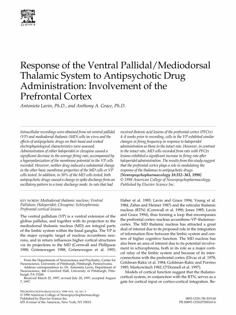
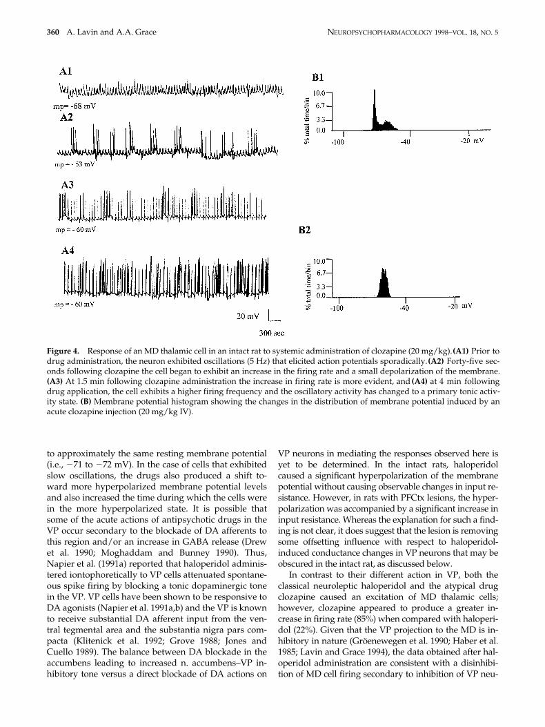
Effect of Clozapine. The response to the acute admin-istration of clozapine was examined in six MD cells re-corded in control animals. Clozapine produced an in-crease in firing frequency (85%) in the MD cells tested(Table 3 and Figure 4). In three out of seven cases thedrug changed the mode of discharge from an oscilla-tory pattern to one of tonic firing. No changes in inputresistance or resting membrane potential were ob-served after clozapine; however, the amplitude of theipsps evoked by VP stimulation exhibited a trend to-ward a decrease.
DISCUSSION
Antipsychotic drugs exert a broad array of actions onneurons within the basal ganglia; as a result, it has been
difficult to ascertain which of the actions are related tothe therapeutic response of schizophrenic patients. In aninitial approach to this complex problem, we have fo-cused on an examination of antipsychotic drug action onthe primary common output pathway of the limbic sys-tem: the ventral pallidal-mediodorsal thalamic pathway.
Impact of Prefrontal Cortical Lesions on VP-MD Cell Physiology
Studies into the potential sites of pathology in theschizophrenic brain have failed to yield consistent re-sults, with alterations described with respect to sulcalsize, ventricular enlargement, and neuronal disarray inthe hippocampus and prefrontal cortex (Benes et al.1991; Bogerts et al. 1985; Conrad et al. 1991; Roberts andBruton 1990; Shelton et al. 1988). However, one charac-teristic that has been described repeatedly in the schizo-phrenic is the presence of hypofrontality—i.e., a de-crease in either the activity or in the task-specificactivation of the prefrontal cortex in patients with

NEUROPSYCHOPHARMACOLOGY 1998–VOL. 18, NO. 5 VP/MD System and Antipsychotic Drugs 359
schizophrenia (Ingvar and Franzen 1974; Pettegrew etal. 1989; 1991; Weinberger 1988; Williamson et al. 1991).Therefore, in an attempt to assess the relevance of pre-frontal cortical function to the response to antipsychoticdrugs, we performed cell-specific lesions of the prefron-tal cortex in one set of rats approximately 1 month priorto recording and evaluation of drug action.
Following prefrontal cortical lesions, only minorchanges in the basal activity of limbic system neuronswere noted. Neurons in the VP showed a tendency tofire more slowly (26% decrease) with even smallerchanges in their other physiological properties such asinput resistance or resting membrane potential. This ef-fect could be due to the loss of direct excitatory PFCtxinput to the VP (Sesack et al. 1989) or via an effect in thePFCtx–nucleus accumbens–VP circuit, since at least aportion of the n. accumbens input to the VP is also exci-tatory (Chrobak and Napier 1993; Lavin and Grace1996; Napier et al. 1995). Neurons in the MD of prefron-tally lesioned rats showed a more marked (46%) in-crease in baseline firing rate, however this change didnot reach statistical significance. It is not clear whyremoval of excitatory PFCtx drive to MD cells (Gröe-newegen 1988) did not result in a decrease in MD cell
activity. One possibility is that, as a result of decreasedPFCtx input to the limbic system, compensatory pro-cesses acting via a decrease in VP-mediated MD inhibi-tion were introduced. This would be consistent with theincrease in firing rate combined with an increase in theaverage input resistance in the MD occurring through aremoval of inhibitory inputs secondary to decreased VPafferent activity.
Response of VP and MD Cells to Administration of Antipsychotic Drugs
In this study, we examined the actions of both the clas-sical neuroleptic drug haloperidol and the atypicalantipsychotic drug clozapine. Although these classes ofdrugs vary in several dimensions with regard to theirreceptor binding specificity and propensity for induc-ing extrapyramidal side effects, the one feature thatthese drugs have in common is their ability to elicit atherapeutic response in schizophrenic patients. Consis-tent with this common therapeutic action, cells in theVP exhibited a significant decrease in firing rate in re-sponse to both drugs; in both cases drug administrationcaused a hyperpolarization sufficient to bring the cells
Figure 3. Typical example of the activation of a nonfiring MD thalamic cell by systemic administration of halperidol in anintact animal. (A1) This MD thalamic cell was quiescent prior to drug administration. (A2) Two and a half minutes followinghaloperidol administration (0.5 mg/kg, IV) the cell exhibited a small depolarization of the membrane potential, and (A3) by4.5 min the cell displayed a tonic spike discharge pattern and a change to a non-oscillatory membrane potential activitystate. Overall, this cell exhibited an 11 mV depolarization of the membrane from pre-drug levels. (B) Membrane potential his-tograms showing the changes in firing states produced by the systemic administration of haloperidol (0.5 mg/kg, IV) to aMD cell (B1 5 control period, B2 5 after haloperidol).
360 A. Lavin and A.A. Grace NEUROPSYCHOPHARMACOLOGY 1998–VOL. 18, NO. 5
to approximately the same resting membrane potential(i.e., 271 to 272 mV). In the case of cells that exhibitedslow oscillations, the drugs also produced a shift to-ward more hyperpolarized membrane potential levelsand also increased the time during which the cells werein the more hyperpolarized state. It is possible thatsome of the acute actions of antipsychotic drugs in theVP occur secondary to the blockade of DA afferents tothis region and/or an increase in GABA release (Drewet al. 1990; Moghaddam and Bunney 1990). Thus,Napier et al. (1991a) reported that haloperidol adminis-tered iontophoretically to VP cells attenuated spontane-ous spike firing by blocking a tonic dopaminergic tonein the VP. VP cells have been shown to be responsive toDA agonists (Napier et al. 1991a,b) and the VP is knownto receive substantial DA afferent input from the ven-tral tegmental area and the substantia nigra pars com-pacta (Klitenick et al. 1992; Grove 1988; Jones andCuello 1989). The balance between DA blockade in theaccumbens leading to increased n. accumbens–VP in-hibitory tone versus a direct blockade of DA actions on
VP neurons in mediating the responses observed here isyet to be determined. In the intact rats, haloperidolcaused a significant hyperpolarization of the membranepotential without causing observable changes in input re-sistance. However, in rats with PFCtx lesions, the hyper-polarization was accompanied by a significant increase ininput resistance. Whereas the explanation for such a find-ing is not clear, it does suggest that the lesion is removingsome offsetting influence with respect to haloperidol-induced conductance changes in VP neurons that may beobscured in the intact rat, as discussed below.
In contrast to their different action in VP, both theclassical neuroleptic haloperidol and the atypical drugclozapine caused an excitation of MD thalamic cells;however, clozapine appeared to produce a greater in-crease in firing rate (85%) when compared with haloperi-dol (22%). Given that the VP projection to the MD is in-hibitory in nature (Gröenewegen et al. 1990; Haber et al.1985; Lavin and Grace 1994), the data obtained after hal-operidol administration are consistent with a disinhibi-tion of MD cell firing secondary to inhibition of VP neu-
Figure 4. Response of an MD thalamic cell in an intact rat to systemic administration of clozapine (20 mg/kg). (A1) Prior todrug administration, the neuron exhibited oscillations (5 Hz) that elicited action potentials sporadically. (A2) Forty-five sec-onds following clozapine the cell began to exhibit an increase in the firing rate and a small depolarization of the membrane.(A3) At 1.5 min following clozapine administration the increase in firing rate is more evident, and (A4) at 4 min followingdrug application, the cell exhibits a higher firing frequency and the oscillatory activity has changed to a primary tonic activ-ity state. (B) Membrane potential histogram showing the changes in the distribution of membrane potential induced by anacute clozapine injection (20 mg/kg IV).

NEUROPSYCHOPHARMACOLOGY 1998–VOL. 18, NO. 5 VP/MD System and Antipsychotic Drugs 361
ronal afferent discharge. However, it was not clear whyhaloperidol and clozapine exhibited equivalent levels ofinhibition of VP neurons, whereas clozapine caused amuch greater elevation in the firing rate of MD neurons.Although the reason for this is not apparent, one possi-bility is that it is related to the broad pharmacologicalprofile of clozapine and its actions at other receptor sites,such as serotoninergic, muscarinic, and adrenergic re-ceptors. In this context, it is interesting to note that PFCtxlesions can augment the responses of MD cells to halo-peridol to an extent in which it is equivalent to clozapinewithout affecting the response of VP neurons. It shouldbe noted, however, that since these recordings were ob-tained in anesthetized animals, and chloral hydrate isknown to attenuate some of the responses of neurons toantipsychotic drugs (Bunney et al. 1973), the effects ofthese drugs may be more robust in awake preparations.
Another effect observed in MD cells in response toboth haloperidol and clozapine administration was ashift in activity from an oscillatory to a tonically dis-charging state. Studies have shown that the reticularnucleus of the thalamus has a central role in regulatingprimary thalamic cell activity patterns (Steriade and De-schênes 1984). Furthermore, our work has shown thatprojections from the VP innervates both the MD and thereticular thalamic nucleus (RTN) (O’Donnell et al 1997)and that stimulation of the VP both inhibits the MD di-rectly as well as disinhibits dorsal thalamic nuclei via anaction in the RTN (Lavin and Grace 1994). Therefore, it ispossible that inhibition of neurons in the VP by antipsy-chotic drugs may affect MD cell firing directly as well asmodulating its activity pattern via the RTN.
Conclusions and Functional Extensions
The augmented response of MD cells to haloperidol ad-ministration in rats with lesions of the PFCtx may reflectdifferences in the acute response to these drugs as ob-served in normal versus schizophrenic patients. Severalstudies have shown that schizophrenics exhibit hypo-frontality, or a decrease in basal activity and/or task-specific activation of the PFCtx when compared withcontrols (Chabrol et al. 1986; Farkas et al. 1984; Ingvarand Franzen 1974; Pettegrew et al. 1989; 1991; Wein-berger 1987). Drawing from our observations, it is pos-sible that the augmented effect produced by haloperi-dol administration in rats with PFCtx lesions is aconsequence of the diminished activity in the prefrontalcortical efferent pathways. Since this does not occur inconcert with an augmented effect on the VP, one possi-bility is that it involves an alteration in the PFCtx–MDloop. For example, if one proposes that the MD–PFCtxloop functions to provide a stabilizing influence in thissystem, one possible explanation is that the intactPFCtx–MD connection partially offsets the influence of
haloperidol acting via the nucleus accumbens–VP–MDcircuit. This would be consistent with models of corti-cal/subcortical DA interactions, in which PFCtx DAand subcortical DA are regulated in opposite directions.In this context, one could suggest that the potent effectsof clozapine in the MD as well as in the schizophrenicpatients are at least partially due to its ability to circum-vent this stabilizing influence. Furthermore, the findingthat the responses of MD cells to haloperidol in the le-sioned rats are similar in magnitude to the effects ofclozapine in intact animals may relate to the greatersedative actions of haloperidol in a system that has notbeen compromised—i.e., the non-schizophrenic individ-ual (Berger and Waldhorn 1995; Kornetsky and Mirsky1966; King and Henry 1992).
ACKNOWLEDGMENTS
The authors thank McNeil Laboratories for their generous giftof haloperidol, and Mr. Brian Lowry for providing the com-puter programs for data analysis. We also thank Dr. H. Moorefor her helpful comments and advice on the statistical analy-sis. This work was supported by USPHS MH 01055, MH45156, and a fellowship from the National Alliance for Re-search on Schizophrenia and Depression (AL).
REFERENCES
Benes FM, McSparren J, Bird ED, SanGiovanni JP, VincentSL (1991): Deficits in small interneurons in prefrontaland cingulate cortices of schizophrenic and schizoaffec-tive patients. Arch Gen Psychiatry 48:996–1001
Berger I, Waldhorn RE (1995): Analgesia, sedation and paral-ysis in the intensive care unit. American Family Physi-cian 51:166–172
Bogerts B, Meertz E, Schonfeldt-Bausch R (1985): Basal gan-glia and limbic system pathology in schizophrenia. Amorphometric study of brain volume and shrinkage.Arch Gen Psychiatry 42:784–791
Bunney BS, Walters JR Roth RH, Aghajanian GK (1973)Dopaminergic neurons: Effect of antipsychotic drugsand amphetamine on single cell activity. J PharmacolExp Ther 185:561–571
Chabrol H, Guell A, Bes A, Moron P (1986): Cerebral flow inschizophrenic adolescents. Am J Psychiatry 143:130
Chrobak JJ, Napier TC (1993): Opioid and GABA modula-tion of accumbens-evoked ventral pallidal activity. JNeural Transm 93:123–124
Chouinard G, Annable L (1976) Clozapine in the treatmentof newly admitted schizophrenic patients. A pilot study.Clin Pharmacol 16:289–297
Conrad AJ, Abebe T, Austin R, Forsythe S, Scheibel A (1991):Hippocampal pyramidal cell disarray in schizophrenia asa bilateral phenomenon. Arch Gen Psychiatry 48:413–417
Cornwall J, Cooper JD, Phillipson OT (1990): Projections tothe rostral reticular thalamic nucleus in the rat. ExpBrain Res 80:157–171

362 A. Lavin and A.A. Grace NEUROPSYCHOPHARMACOLOGY 1998–VOL. 18, NO. 5
Cornwall J, Phillipson OT (1988): Afferent connections of thedorsal thalamus of the rat as shown by retrograde lectintransport. I. The mediodorsal nucleus. Neuroscience24:1035–1049
Deutch AY, Öngür D, Duman RS (1995): Antipsychotic drugsinduced Fos protein in the thalamic paraventricularnucleus: a novel locus of antipsychotic drug action.Neuroscience 66:337–346
Divac I, Kosmal A, Björklund A, Lindvall O (1978): Subcorti-cal projections to the prefrontal cortex in the rat asrevealed by the horseradish peroxidase technique. Neu-roscience 3:785–796
Drew KL, O’Connor WT, Kehr J, Ungerstedt U (1990):Regional specific effects of clozapine and haloperidol onGABA and dopamine release in rat basal ganglia. Eur JPharmacol 187:385–397
Farkas T, Wolf AP, Jaeger J, Brodie JD, Christman DR,Fowler JS (1984): Regional brain glucose metabolism inchronic schizophrenia. Arch Gen Psychiatry 41:293–300
Gerlach J, Koppelhus P, Helweg E, Monrad A (1974): Cloza-pine and haloperidol in a single-blind crossover trial:Therapeutic and biochemical aspects in the treatment ofschizophrenia. Acta Psychiatr Scand 50:410–424
Gerlach J, Thorsen K, Fog R (1975): Extrapyramidal reactionsand amine metabolites in cerebrospinal fluid duringhaloperidol and clozapine treatment of schizophrenicpatients. Psychopharmacologia 40:341–350
Goldman-Rakic PS, Porrino LJ (1985): The primate medio-dorsal (MD) nucleus and its projection to the frontallobe. J Comp Neurol 242:535–560
Goldman-Rakic PS, Selemon LD, Schwartz ML (1984): Dualpathways connecting the dorsolateral prefrontal cortexwith the hippocampal formation and parahippocampalcortex in the Rhesus monkey. Neuroscience 12:719–743
Grace AA, Llinás R (1985): Dehydration-induced morpho-logical artifacts in intracellularly stained neurons: cir-cumvention using rapid DMSO clearing. Neuroscience16:461–475
Grace AA (1995): The tonic/phasic model of dopamine sys-tem regulation: its relevance for understanding howstimulant abuse can alter basal ganglia function. DrugAlcohol Depend 37:111–129
Gröenewegen HJ (1988): Organization of the afferent con-nections of the mediodorsal thalamic nucleus in the rat,related to the mediodorsal-prefrontal topography. Neu-roscience 24:379–432
Gröenewegen HJ, Berendse HW, Wolters JG, Lohman AHM(1990): The anatomical relationship of the prefrontal cor-tex with the striatopallidal system, the thalamus and theamygdala: evidence for a parallel organization. InUylings HBM, Van Eden CG, De Bruin JPC, Corner MA,Fenestra MGP (eds), Progress in Brain Research. ThePrefrontal Cortex: Its Structure, Function and Pathol-ogy, vol. 85. Amsterdam, Elsevier, pp 95–118
Gröenewegen HJ, Berendse HW, Haber SN (1993): Organi-zation of the output of the ventral striatopallidal systemin the rat: Ventral pallidal efferents. Neuroscience 57:113–142
Grove EA (1988): Neural associations of the substantiainnominata in the rat: Afferent connections. J CompNeurol 277:315–346
Haber SN, Gröenewegen HJ, Grove EA, Nauta WJH (1985):Efferent connections of the ventral pallidum: Evidenceof a dual striato pallidofugal pathway. J Comp Neurol235:322–335
Ingvar DH, Franzen G (1974): Abnormalities of cerebralblood flow distribution in patients with chronic schizo-phrenia. Acta Psychiatr Scand 50:425–462
Jones EG (1985): The thalamus. In Emson PC (ed), ChemicalNeuroanatomy. New York, Raven, pp 257–293
Jones BE, Cuello AC (1989) Afferents to the basal forebraincholinergic cell area from pontomesencephalic-cate-cholamine, serotonin and acetylcholine-neurons. Neu-roscience 31:37–61
King DJ, Henry G (1992): The effects of neuroleptics on cog-nitive and psychomotor function: A preliminary studyin healthy volunteers. Br J Psychiatry 160:647–653
Klitenick MA, Deutch AY, Churchill L, Kalivas PW (1992):Topography and functional role of dopaminergic pro-jections from the ventral mesencephalic tegmentum tothe ventral pallidum. Neuroscience 50:371–386
Kornetsky C, Mirsky AF (1966): On certain psychopharma-cological differences between schizophrenics and nor-mal persons. Psychopharmacologia 8:309–318
Lavin A, Grace AA (1996): Physiological properties of ratventral pallidal neurons recorded intracellularly in vivo.J Neurophysiol 75:1432–1443
Lavin A, Grace AA (1994): Modulation of dorsal thalamiccell activity by the ventral pallidum: Its role in the regu-lation of thalamocortical activity by the basal ganglia.Synapse 18:104–127
Markowitsch HJ (1982): Thalamic mediodorsal nucleus andmemory: A critical evaluation of studies in animals andman. Neurosci Behav Rev 6:351–380
Mogenson GJ, Yang CR (1991): Contribution of basal fore-brain to limbic-motor integration and the mediation ofmotivation to action. In Napier TC, Kalivas P, Hanin I(eds), The Basal Forebrain: Anatomy to Function. NewYork, Plenum, pp 267–290
Moghaddam B, Bunney BS (1990): Acute effect of typical andatypical antipsychotic drugs on the release of dopaminefrom the prefrontal cortex, nucleus accumbens, and stri-atum of the rat: An in vivo microdialysis study. J Neuro-chem 54:1755–1760
Napier TC, Simson PE, Givens BS (1991a): Dopamine elec-trophysiology of ventral pallidal/substantia innominataneurons: Comparison with the dorsal globus pallidus. JPharmacol Exp Ther 258:249–262
Napier TC, Muench MB, Maslowski RJ, Battagllia G (1991b):Is dopamine a neurotransmitter within the ventral palli-dum/substantia innominata? The basal forebrain: Anat-omy to function. In Napier TC, Kalivas P, Hanin I (eds),The Basal Forebrain: Anatomy to Function. New York,Plenum Press, pp 183–195
Napier TC, Mitrovic I, Churchill L, Klitenick MA, KalivasPW (1995): Substance P in the projection from thenucleus accumbens to the ventral pallidum: Anatomy,electrophysiology and behavior. Neuroscience 69:59–70
O’Donnell P, Lavin A, Enquist LW, Grace AA, Card JP(1997): Interconnected parallel circuits between ratnucleus accumbens and thalamus revealed by retro-

NEUROPSYCHOPHARMACOLOGY 1998–VOL. 18, NO. 5 VP/MD System and Antipsychotic Drugs 363
grade transynaptic transport of pseudo rabies virus. JNeurosci 17:2143–2167
Paxinos G, Watson C (1986): The Rat Brain in StereotaxicCoordinates. San Diego, CA, Academic, pp 1–83
Pettegrew JW, Keshavan M, Panchalingam K, Strychor S,Kaplan DB, Tretta M, Allen M (1991): Alteration in brainhigh-energy phosphate and membrane phospholipidmetabolism in first-episode, drug naive schizophrenics.Arch Gen Psychiatry 48:563–568
Pettegrew JW, Keshavan M, Panchalingam K (1989): 31PNMR studies in schizophrenia. Biol Psychiatry 25:15A
Racagni G, Bruno F, Bugatti A, Parenti M, Apud JA, SantiniV, Carenzi A, Gropetti A, Cattabeni F (1980): Behavioraland biochemical correlates after haloperidol and cloza-pine long-term treatment. Adv Biochem Psychopharma-col 24:45–51
Richardson RT, DeLong MR (1991): Electrophysiologicalstudies of the function of the nucleus basalis in pri-mates. In Napier TC, Kalivas P, Hamin I (eds), The BasalForebrain: Anatomy to Function. New York, Plenum,pp 232–252
Roberts GW, Bruton CJ (1990): Notes from the graveyard:Neuropathology and schizophrenia. Neuropathol ApplNeurobiol 16:3–16
Sesack SR, Deutch AY, Roth RH, Bunney BS (1989): Topo-graphical organization of the efferent projections of themedial prefrontal cortex in the rat: An anterogradetract-tracting study with Phaseolus vulgaris Leucoagglu-tinin. J Comp Neurol 290:213–242
Shelton RC, Karson CN, Doran AR, Pickar D, Bigelow LB,Weinberger DR (1988): Cerebral structural pathology in
schizophrenia: evidence for a selective prefrontal corti-cal defect. Am J Psychiatry 145:154–163
Steriade M, Deschênes M (1984): The thalamus as a neuronaloscillator. Brain Res Rev 8:1–63
Stewart WW (1978): Functional connections between cells asrevealed by dye-coupling with a highly fluorescentnaphthalimide tracer. Cell 13:741–759
Weinberger DR, Berman KF, Zec RF (1986): Physiologicaldysfunction of dorsolateral prefrontal cortex in schizo-phrenia. Arch Gen Psychiatry 43:114–124
Weinberger DR (1987): Implications of normal brain devel-opment for the pathogenesis of schizophrenia. ArchGen Psychiatry 44:660–669
Weinberger DR (1988): Schizophrenia and the frontal lobe.Trends Neurosci 11:367–370
Williamson P, Drost D, Stanley J, Carr T, Morrison S, Mers-key H (1991): Localized phosphorus 31 magnetic reso-nance spectroscopy in chronic schizophrenic patientsand normal controls. Arch Gen Psychiatry 48:578
Wilson FAW, Rolls ET (1990): Neuronal responses to rein-forcement in the primate basal forebrain. Brain Res 509:213–231
Young III WS, Alheid FG, Heimer L (1984): The ventral pal-lidal projection to the mediodorsal thalamus: A studywith fluorescent retrograde tracers and immunohisto-fluorescence. J Neurosci 4:1626–1638
Zahm DS, Heimer L (1987): The ventral striatopallidal pro-jection. III. Striatal cells of the olfactory tubercle estab-lish direct synaptic contact with ventral pallidal cellsprojecting to the mediodorsal thalamus. Brain Res404:327–331