respiratory toxicity of multi-wall carbon nanotubes toxicity of multi-wall carbon nanotubes julie...
TRANSCRIPT
www.elsevier.com/locate/ytaap
Toxicology and Applied Pharma
Respiratory toxicity of multi-wall carbon nanotubes
Julie Mullera, Francois Huauxa, Nicolas Moreaub, Pierre Missona, Jean-Francois Heiliera,
Monique Delosc, Mohammed Arrasa, Antonio Fonsecab, Janos B. Nagyb, Dominique Lisona,TaIndustrial Toxicology and Occupational Medicine Unit, Universite Catholique de Louvain, Clos Chapelle-aux-Champs, 30.54; 1200 Brussels, Belgium
bLaboratory of Nuclear Magnetic Resonance, Facultes Universitaires Notre-Dame de la Paix, Namur, BelgiumcLaboratory of Pathology, University Hospital of Mont-Godinne, Yvoir, Belgium
Received 14 September 2004; accepted 5 January 2005
Available online 5 March 2005
Abstract
Carbon nanotubes focus the attention of many scientists because of their huge potential of industrial applications, but there is a paucity of
information on the toxicological properties of this material. The aim of this experimental study was to characterize the biological reactivity of
purified multi-wall carbon nanotubes in the rat lung and in vitro.
Multi-wall carbon nanotubes (CNT) or ground CNT were administered intratracheally (0.5, 2 or 5 mg) to Sprague–Dawley rats and we
estimated lung persistence, inflammation and fibrosis biochemically and histologically. CNT and ground CNT were still present in the lung
after 60 days (80% and 40% of the lowest dose) and both induced inflammatory and fibrotic reactions. At 2 months, pulmonary lesions
induced by CNT were characterized by the formation of collagen-rich granulomas protruding in the bronchial lumen, in association with
alveolitis in the surrounding tissues. These lesions were caused by the accumulation of large CNT agglomerates in the airways. Ground CNT
were better dispersed in the lung parenchyma and also induced inflammatory and fibrotic responses. Both CNT and ground CNT stimulated
the production of TNF-a in the lung of treated animals. In vitro, ground CNT induced the overproduction of TNF-a by macrophages. These
results suggest that carbon nanotubes are potentially toxic to humans and that strict industrial hygiene measures should to be taken to limit
exposure during their manipulation.
D 2005 Elsevier Inc. All rights reserved.
Keywords: Carbon nanotubes; Lung toxicity; Inflammation; Fibrosis
Introduction
Carbon nanotubes are a new form of crystalline carbon
currently attracting intense research efforts because of their
unique properties that make them suitable, as such or after
modification, for many industrial developments such as in
high strength materials, electronics or biomedical applica-
tions (Martin and Kohli, 2003). The global market for
carbon nanotubes was estimated at $12 million for 2002 and
is expected to grow up to $700 million by 2005 (Carbon
Nanotubes—Worldwide Status and Outlook: Applications,
Applied Industries, Production, R&D and Commercial
Implications, 2002). There is, however, a disquieting lack
0041-008X/$ - see front matter D 2005 Elsevier Inc. All rights reserved.
doi:10.1016/j.taap.2005.01.008
T Corresponding author. Fax: +32 2 764 32 28.
E-mail address: [email protected] (D. Lison).
of information about the possible human health and
environmental impacts of manufactured carbon nanotubes
and other nanomaterials. The disaster associated with the
use of asbestos fibers in the past highlights the importance
of identifying rapidly the potential hazards of new materials
(Mossman et al., 1990). The extreme aspect ratio of
individual carbon nanotubes suggests toxic properties
similar to those observed with other fibrous particles
(Maynard et al., 2004). Additional concerns come from
studies revealing that particles with nanoscopic dimension
are markedly more toxic than larger sized particles
(Oberdorster, 2001).
The existing information on the lung toxicity of carbon
nanotubes is limited and remains inconclusive. In a short
report, it has been found that non-purified carbon nanotubes
did not induce significant signs of lung toxicity 4 weeks
after intratracheal (i.t.) administration in guinea pigs
cology 207 (2005) 221–231

J. Muller et al. / Toxicology and Applied Pharmacology 207 (2005) 221–231222
(Huczko et al., 2001). Lam and colleagues observed in mice
that a single i.t. treatment with single-wall carbon nanotubes
induced persistent epithelioid granulomas and interstitial
inflammation; the severity of the lesions was dose depend-
ent (Lam et al., 2004). In another study, single-wall carbon
nanotubes soot administered in rats produced transient
inflammation and cell injury and resulted in the formation
of multifocal granulomas centered around nanotubes,
similar to a foreign body reaction. The toxicological
relevance of these observations was, however, questioned
because of the absence of a dose response and because the
formation of granulomas was suspected to be the conse-
quence of instilling a bolus of agglomerated nanotubes
(nanoropes) (Warheit et al., 2004). A major difficulty
reported by these investigators was that carbon nanotubes,
because of their electrostatic nature, tend to form large
agglomerates that are far beyond the range of respirable
particles and therefore difficult to manipulate and administer
to experimental animals. In order to circumvent these
difficulties, we compared the lung response to multi-wall
carbon nanotubes that had been ground or not.
The particles were administered intratracheally (0.5, 2 or
5 mg) to Sprague–Dawley rats. First, we determined
whether carbon nanotubes are biopersistent in the lung.
We characterized the lung inflammatory and fibrotic
responses, and finally we examined the expression of
TNF-a, a pro-inflammatory and pro-fibrotic cytokine, in
vivo and in vitro. The response to nanotubes was calibrated
with two reference particles (carbon black or asbestos).
Materials and methods
Animals. Female Sprague–Dawley rats weighing 200–250
g were purchased from Charles River Laboratories (St
Germain-sur-l’Arbresle, France). The animals were kept in a
conventional animal facility and housed in positive-pressure
air-conditioned units (25 8C, 50% relative humidity) on a
12:12-h light/dark cycle. The experimental protocol has
been approved by the local ethical committee for animals in
research.
Particle preparation and characterization. All carbon
nanotubes were provided by the laboratory of Nuclear
Magnetic Resonance at the Facultes universitaires Notre-
Dame de la Paix in Namur. Multi-wall carbon nanotubes
(15 carbon layers on average) were synthesized by the
decomposition of ethylene on an alumina support doped
with a cobalt–iron catalyst mixture (Willems et al., 2000)
and purified by subsequent treatment with NaOH. A
fraction of multi-wall carbon nanotubes was ground in an
oscillatory agate ball mill (Pulverisette 0, Fritsch), with a
vertical vibration of 1 mm applied during 6 h. Asbestos
(Rhodesian Chrysotile bAQ; Asb) and ultrafine carbon
black (ENSACO 250; CB) were obtained, respectively,
from the Union Internationale contre le Cancer (Geneva,
Switzerland) and ERACHEM Comilog S.A. (Willebroek,
Belgium). CB had a specific surface area of 66.8 m2/g,
an ash content of 0.01%, volatile matter 0.11% and a
density of 174 kg/m3. The fiber length and width of
UICC Rhodesian chrysotile A is, respectively, 2.4; 2.3
and 0.17; 1.8 Am (geometrical mean; standard deviation)
(Kohyama et al., 1996).
Nanotube characterization. The morphological character-
ization of nanotubes (length, diameter) was done by low-
resolution transmission electron microscopy (TEM), using a
TECNAI 10 (Philips) microscope.
The carbon content of the samples was determined by
thermal analysis on a STA-409 PC analyzer (Netzsch),
under dry air flow (50 ml/min). Temperature range was from
30 to 1400 8C, at a heating rate of 10 8C/min. The residual
mass was recorded at 1400 8C. The Co content of the
nanotubes was determined by proton-induced X-ray emis-
sion (PIXE) with a proton beam of 2 MeV from a Tandetron
accelerator.
Specific surface areas were measured at �196 8C by
nitrogen adsorption–desorption (Brunauer Emmet Teller
method or BET) on an ASAP 2000 (Micrometrics). Prior
to measurements, the samples were outgassed under vacuum
at 320 8C. Oxidized forms of carbon on the surface of
nanotubes were probed by X-ray photoelectron spectro-
scopy (SSX-100, Surface Science Instrument) (Ka = 1.486
keV).
Particle instillation. After heat inactivation of any possible
trace of endotoxin (200 8C, 2 h), the preparations were
suspended and sonicated in sterile 0.9% saline containing
1% of Tween 80. The suspensions were then injected
directly into the lungs by i.t. instillation (Lasfargues et al.,
1992). All instillations (500 Al/rat) were performed after
surgical opening of the neck on animals anesthetized with a
mix of Ketalar, 6 mg/rat (Warner-Lambert, Zaventem,
Belgium), and Rompun, 0.8 mg/rat (Bayer A6, Leverkusen,
Germany), given intraperitoneally.
General experimental design. Intact multi-wall carbon
nanotubes (CNT) and ground CNT were administered
intratracheally (0.5, 2 or 5 mg/animal) to Sprague–Dawley
rats and we estimated pulmonary persistence, inflammation
and fibrosis at different time points (Table 1). The lung
response to nanotubes was calibrated with a single dose (2
mg/rat) of two reference particles sharing with carbon
nanotubes nature (CB) or aspect properties (Asb). The
inflammatory response was assessed at days 3 and 15 by
measuring several parameters in bronchoalveolar lavage
(Table 1). The fibrotic response was assessed biochemically
(soluble collagen and hydroxyproline) and histopathologi-
cally at day 60. To assess the biopersistence of carbon
nanotubes the lowest dose of CNT and ground CNT (0.5
mg) was selected because (1) doses of 0.5–2 mg have been
recommended in the literature to perform biopersistence
Table 1
General experimental design (in vivo studies)
Day 0 1 h Days 3 and 15 Day 28 Day 60
Biopersistence IT administration of NaCl 0.9%;
0.5 mg CNT; 0.5 mg ground CNT
Co-measurement – Co-measurement Co-measurement
Inflammation IT administration of NaCl 0.9%;
2 mg Asb; 2 mg CB; 0.5 or 2 mg CNT;
0.5 or 2 mg ground CNT
– LDH activity;
total proteins;
cellular parameters;
TNF-a protein
– –
Fibrosis IT administration of NaCl 0.9%; 2 mg Asb;
2 mg CB; 0.5, 2 or 5 mg CNT;
0.5, 2 or 5 mg ground CNT
– – – Hydroxyproline;
type I soluble collagen;
histology; TNF-a protein
J. Muller et al. / Toxicology and Applied Pharmacology 207 (2005) 221–231 223
studies (Muhle and Belmann, 1995) and (2) to minimize the
possible influence of inflammation. The biopersistence was
assessed up to 2 months after administration to allow a
direct comparison with the evaluation of the fibrotic
response (2 months). The expression of TNF-a was
measured in vivo during the inflammatory (day 3) and
fibrotic (day 60) phases of the lung response.
To further characterize the biological response to CNT,
we performed in vitro assays on peritoneal macrophages
which have been intensively used to estimate and rank the
toxicity of inhaled (Lison and Lauwerys, 1990; Miller,
1978). Macrophages were exposed to CB, Asb, intact and
ground CNT (20, 50 and 100 Ag/ml) and cytotoxicity was
assessed by measuring LDH release. TNF-a production was
assessed at the transcript and protein levels at 6 and 24 h,
respectively.
Determination of nanotube biopersistence. At selected
time intervals after nanotube administration (Table 1), whole
lungs were excised and mineralized in acid (14N HNO3:
12N HCl 65:35; both Merck, Darmstad, Germany) in a high
pressure microwave (Multiwave, Anton Paar GmbH, Graz,
Austria). The amount of nanotubes was then determined by
measuring the cobalt content on an atomic absorption
spectrometer (Spectra AA 300, Varian Zeeman Inc., Palo
Alto, CA, USA) equipped with a graphite furnace atomizer
and a Zeeman system for correction of non-specific
absorbance.
Bronchoalveolar lavage (BAL) and whole lung
homogenates. At the indicated time intervals after particle
treatment, animals were sacrificed with sodium pentobarbi-
tal (60 mg/rat ip) and a BAL was performed by cannulating
the trachea and perfusing the lungs with a volume of 10 ml
saline. The recovered BAL fluid (BALF) was centrifuged
(250 � g, 10 min, 4 8C) and the cell-free supernatant used
for biochemical measurements. The cell pellets were
resuspended in saline and then used to determine total and
differential cell numbers. Leukocyte differentials were
performed on cytocentrifuge preparations fixed in methanol
and stained with Diff Quick (Dave NV/SA, Brussels,
Belgium).
The perfused lungs were then excised and placed into a
Falcon tube (Becton-Dickinson) chilled on ice to which 18
ml of phosphate-buffered saline (PBS) were added. The
content of each tube was then homogenized using an Ultra-
Turrax T25 homogenizer (Janke and Kunkel, Brussels,
Belgium) for 1 min. The tubes were then centrifuged at
4 8C, 1000 � g for 10 min, and supernatants were kept
frozen at �80 8C until use.
Biochemical assays. TNF-a concentrations in BALF were
measured by ELISA (detection limit 7 pg/ml) according to
the manufacturer’s instructions (Pharmingen, BD Bioscien-
ces, San Diego, CA, USA). LDH activity was measured by
following the reduction of NAD+ at 340 nm (Technicon RA
systems, Bayer Diagnostics Domont, France). Total protein
concentration was estimated spectrophotometrically at 600
nm after complexation with molybdate pyrogallol red
(Watanabe et al., 1986).
Collagen assays. Collagen deposition was estimated by
measuring hydroxyproline and soluble type I collagen
contents in lung homogenates. Hydroxyproline was assessed
by high-performance liquid chromatography analysis in lung
homogenates hydrolyzed in 6N HCl overnight at 110 8C(Biondi et al., 1997). Soluble type I collagen contents were
measured in the supernatant of the homogenates with a
standardized direct ELISA. Samples and standards were
diluted in PBS and coated directly in Nunc-immuno ELISA
plates (MaxiSorp) for overnight at 4 8C. After blocking with
BSA, polyclonal anti-mouse type I collagen Ab (1:200
times; Biodesign, Saco, ME) were then added and incubated
2 h at room temperature. Polyclonal HRP-conjugated goat
anti-rabbit Ig Ab (1:1000; BD Biosciences) was used to
measure the fixation of primary Abs. Purified mouse type I
collagen obtained from Novotec (Saint-Martin-la-Garenne,
France) was used as standard to calibrate each assay. The
detection limit of this ELISA is 40 ng/ml.
Histology. In a separate set of animals, the lungs were
excised 2 months after administration and fixed in Bouin
solution (Merck-Belgolabo, Belgium). Paraffin-embedded
sections were stained with hematoxylin and eosin (H&E) or
Masson’s trichrome for light microscopic examination.
Peritoneal macrophage culture. Peritoneal macrophages
were harvested from rats injected intraperitoneally 3 days
J. Muller et al. / Toxicology and Applied Pharmacology 207 (2005) 221–231224
before with 5 ml of casein 6% in saline. After hypertonic
lysis of the erythrocytes, the cells were pooled, counted and
dispersed in Dulbecco’s modified Eagle’s medium (DMEM,
Invitrogen) supplemented with 10% fetal bovine serum
(FBS), l-glutamine (2 mM) and antibiotics (penicillin 50 U/
ml and streptomycin 50 Ag/ml). Cells were plated in 24-well
culture plates at a density of 106 macrophages/well. Cultures
were incubated overnight at 37 8C in a humidified incubator
in an atmosphere of 5% CO2. On the second day of culture,
the cells were washed with 1 ml PBS and exposed for 6 and
24 h in DMEM supplemented with FBS (0.5%), l-
glutamine (2 mM) and antibiotics (penicillin 50 U/ml and
streptomycin 50 Ag/ml) containing different dilutions of the
tested compound.
RNA extraction and quantification. Total RNA from
adherent cells was isolated with the RNeasy mini-kits
(Qiagen, Hilden, Germany) following the manufacturer’s
protocols. RNA (1 Ag) was reverse-transcribed using
Superscript RNase H� reverse transcriptase (Invitrogen)
with 350 pM random hexamers (Eurogentec, Seraing,
Belgium) in a final volume of 25 Al. Resulting cDNA was
then diluted 25-fold in sterile distilled water and used as a
template in subsequent real-time polymerase chain reactions
(PCR). Sequences of interest were amplified using the
following forward primers (Invitrogen): 5V-GAGTACAA-CCTTCTTGCAGCTCC-3V (h-actin), 5V-ATGGGCTCCCT-CTCATCAGT-3V(TNF-a); and reverse primers: 5V-TTGTCQGACGACGAGCGC-3V(h-actin), 5V-ACTCCAGCTGCTCQCTCTGCT-3V (TNF-a). h-actin was used as endogenous
reference housekeeping gene. The quantification of mRNA
expression was performed on an ABI 7000 (Applied
Biosystems, Foster City, CA) in the following conditions:
2 min 50 8C, 10 min 95 8C, (15 s 95 8C, 1 min 60 8C for) �40 cycles. Six serial 1:10 dilutions of a standard of cDNA
(asbestos-stimulated macrophages) were used to calibrate the
assay in each reaction. Standard and samples (5 Al) wereamplified with 300 nM primers using SYBR green PCR
master mix (Applied Biosystems) in a total volume of 25 Al.
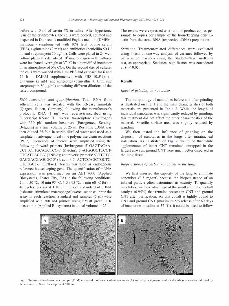
Fig. 1. Transmission electron microscopic (TEM) images of multi-wall carbon nan
the arrows (B). Scale bars represent 500 nm.
The results were expressed as a ratio of product copies per
sample to copies per sample of the housekeeping gene h-actin from the same RNA (respective cDNA) preparation.
Statistics. Treatment-related differences were evaluated
using t tests or one-way analysis of variance followed by
pairwise comparisons using the Student–Newman–Keuls
test, as appropriate. Statistical significance was considered
at P b 0.05.
Results
Effect of grinding on nanotubes
The morphology of nanotubes before and after grinding
is illustrated on Fig. 1 and the main characteristics of both
materials are presented in Table 2. While the length of
individual nanotubes was significantly reduced by grinding,
this treatment did not affect the other characteristics of the
material. Specific surface area was slightly reduced by
grinding.
We then tested the influence of grinding on the
dispersion of nanotubes in the lungs after intratracheal
instillation. As illustrated on Fig. 2, we found that while
agglomerates of intact CNT remained entrapped in the
largest airways, ground CNT were much better dispersed in
the lung tissue.
Biopersistence of carbon nanotubes in the lung
We first assessed the capacity of the lung to eliminate
nanotubes (0.5 mg/rat) because the biopersistence of an
inhaled particle often determines its toxicity. To quantify
nanotubes, we took advantage of the small amount of cobalt
catalyst (0.95%) that remains present in CNT and ground
CNT after purification. As this cobalt is tightly bound to
CNT and ground CNT (maximum 5% release after 60 days
of incubation in saline at 37 8C), it could be used to follow
otubes (A) and of typical ground multi-wall carbon nanotubes indicated by

Table 2
Characteristics of individual carbon nanotubes (CNT) ground during 6 h or
not
CNT Ground CNT
Length (Am) 5.9 F 0.05 0.7 F 0.07
Average inner diameter (nm) 5.2 F 1.5 5.1 F 2.1
Average outer diameter (nm) 9.7 F 2.1 11.3 F 3.9
Specific surface area (m2/g) 378 F 20 307 F 15
Oxidized forms (atomic %) 13.7 F 0.7 13.1 F 0.7
Carbon content (%) 97.8 F 0.2 98.0 F 0.2
J. Muller et al. / Toxicology and Applied Pharmacology 207 (2005) 221–231 225
the persistence of carbon nanotubes in the lungs. Rats were
sacrificed at day 0, 28 or 60 after administration of CNT or
ground CNT and cobalt contents measured in whole lung
homogenates as an index of nanotube biopersistence. The
data presented in Table 3 indicate that CNT were not or
slowly eliminated from the lung (81.2% of CNT recovered
after 60 days) whereas ground CNT were cleared more
rapidly, especially during the second month (36% after 60
days). Thus, we conclude that CNT persist in the lung and
their length appears to modulate clearance kinetics.
Pulmonary inflammation in response to carbon nanotubes
We then examined whether nanotubes could cause
pulmonary inflammation. Three days after administration,
bronchoalveolar lavage (BAL) fluid was obtained from rats
administered with CNT (0.5 or 2 mg/rat), ground CNT (0.5
or 2 mg/rat), asbestos (Asb, 2 mg/rat), carbon black (CB, 2
mg/rat) or a saline solution (NaCl, 0.9%, controls). BAL
fluid LDH activity, a marker of cell toxicity, was signifi-
cantly increased after Asb, but not after CB. Administration
of ground CNT and CNT induced a dose-dependent increase
in LDH release which was more marked with the ground
material (Fig. 3A). The protein concentration in BAL fluid,
which reflects alveolo–capillar permeability and/or alveoli-
tis, was also increased after administration of ground CNT
and CNT (Fig. 3B). Accumulation of granulocytes in the
lung was also evident after treatment with the different
particles. CNT and ground CNT induced the accumulation
of both neutrophils and eosinophils (Figs. 3C and D).
Similar data were observed 15 days after particle admin-
istration (data not shown).
Altogether, we concluded that nanotubes induced an
inflammatory response which was to some extent more
marked with ground CNT.
Pulmonary fibrosis in response to carbon nanotubes
Next, we examined whether these nanotubes could induce
lung fibrosis. Collagen deposition was first assessed quanti-
tatively by measuring lung hydroxyproline (OH-proline) and
soluble collagen I contents 60 days after particle admin-
istration. While OH-proline levels were significantly and
dose-dependently increased after CNT, only the highest dose
of ground CNT (5 mg/rat) induced a significant elevation of
OH-proline levels (Fig. 4A). As expected, Asb treatment was
also accompanied by exaggerated OH-proline accumulation
and no similar increase was noted after CB (Fig. 4A). Asb,
CNT as well as ground CNT induced a significant increase of
the type I collagen lung levels in comparison with the control
rats. No similar increase was noted after administration of CB
(Fig. 4B).
Both measurements therefore indicated that the fibrotic
response to nanotubes was dose dependent. Of note, the
intensity of the fibrotic response induced by 5 mg of ground
CNT was equivalent to that induced by 2 mg of CNT.
Sixty days after particles treatment, the morphological
characteristics of the lesions induced by CNT, ground CNT,
Asb or CB (2 mg/rats) were also evaluated by standard
hematoxylin–eosin and Masson trichrome staining (Figs. 5A
and B). This histopathological study revealed also the
presence of collagen-rich granulomas in the bronchi of
animals instilled with CNT, which blocked partially or
completely the bronchial lumen. These granulomas were re-
epithelialized, organized around CNT material and formed
of multinuclear giant cells as well as macrophages and other
mononuclear inflammatory cells. Histological analysis
clearly showed that ground CNT were better dispersed in
the parenchyma and induced granulomas in the interstitial
tissue. These granulomas were localized in the alveolar
spaces or the interstitium and consisted of macrophages
laden with particles, multinuclear giant cells and some
inflammatory cells. As expected, the administration of Asb
induced a parenchymal thickening with fibrosis and
alveolitis. Occasionally, obstructive lesions were localized
in bronchioles. Those injuries consisted of lymphocytes,
fibroblasts, collagen deposition and sometimes of multi-
nuclear giant cells. In CB-treated rats, we observed a simple
accumulation of particles in alveolar macrophages without
significant change of the alveolar architecture. Altogether,
we concluded that nanotubes were able to induce a fibrotic
response.
TNF-a in response to carbon nanotubes (in vivo)
Because TNF-a is a key mediator in inflammation and
fibrosis, we then assessed TNF-a production in the lung of
experimental rats. During the acute inflammatory reaction
(day 3), the BAL levels of TNF-a were significantly
increased after instillation of Asb, CB, CNT (2 mg) or
ground CNT (0.5 and 2 mg) (Fig. 6A). At the fibrotic stage
(day 60), this increased expression persisted only in the
groups of rats treated with Asb or ground CNT (0.5 and 2
mg) (Fig. 6B).
In vitro studies
Finally, we examined the effects induced by carbon
nanotubes on peritoneal macrophages used as models to
study the biological reactivity of mineral dust exposure
(Miller, 1978).
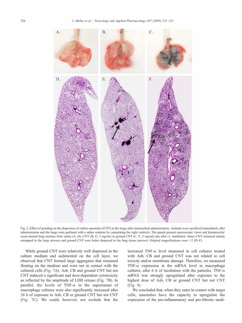
Fig. 2. Effect of grinding on the dispersion of carbon nanotubes (CNT) in the lungs after intratracheal administration. Animals were sacrificed immediately after
administration and the lungs were perfused with a saline solution by cannulating the right ventricle. The panels present macroscopic views and hematoxylin/
eosin-stained lung sections from saline (A, D), CNT (B, E; 2 mg/rat) or ground CNT (C, F; 2 mg/rat) rats after i.t. instillation. Intact CNT remained mainly
entrapped in the large airways and ground CNT were better dispersed in the lung tissue (arrows). Original magnifications were �2 (D–F).
J. Muller et al. / Toxicology and Applied Pharmacology 207 (2005) 221–231226
While ground CNT were relatively well dispersed in the
culture medium and sedimented on the cell layer, we
observed that CNT formed large aggregates that remained
floating on the medium and were not in contact with the
cultured cells (Fig. 7A). Asb, CB and ground CNT but not
CNT induced a significant and dose-dependent cytotoxicity
as reflected by the amplitude of LDH release (Fig. 7B). In
parallel, the levels of TNF-a in the supernatant of
macrophage cultures were also significantly increased after
24 h of exposure to Asb, CB or ground CNT but not CNT
(Fig. 7C). We could, however, not exclude that the
increased TNF-a level measured in cell cultures treated
with Asb, CB and ground CNT was not related to cell
toxicity and/or membrane damage. Therefore, we measured
TNF-a expression at the mRNA level in macrophage
cultures, after 6 h of incubation with the particles. TNF-a
mRNA was strongly upregulated after exposure to the
highest dose of Asb, CB or ground CNT but not CNT
(Fig. 8).
We concluded that, when they enter in contact with target
cells, nanotubes have the capacity to upregulate the
expression of the pro-inflammatory and pro-fibrotic medi-

Table 3
Pulmonary biopersistence of carbon nanotubes (CNT) ground during 6 h or
not
Single i.t. dose Time after particle administration
Day 0 Day 28 Day 60
NaCl 0.9% NDa NDa NDa
0.4 F 0.1b 0.3 F 0.1b 0.4 F 0.1b
0.5 mg CNT (78.4% F 15.3)c (81.2% F 26.4)c
0.5 F 0.1b 0.4 F 0.1b 0.2 F 0.1b
0.5 mg ground CNT (78.4% F 12.4)c (36.0% F 13.2)c
a ND = not detected.b Amount of CNT recovered in the lung of rats (mg, means F SD, n = 5)
based on AAS measurement of cobalt (see Materials and methods).c Relative to day 0.
J. Muller et al. / Toxicology and Applied Pharmacology 207 (2005) 221–231 227
ator TNF-a. The apparent absence of effect of CNT on
TNF-a expression in vitro is most probably related to their
low availability to the test system.
Fig. 4. Instillation of carbon nanotubes (CNT or ground CNT) induced a
fibrotic response in the rat lung. The graphs represent hydroxyproline levels
(A) and type I collagen content (B) in the lung tissue 60 days after
intratracheal instillation of CNT (0.5, 2 or 5 mg/rat), ground CNT (0.5, 2 or
5 mg/rat), asbestos (Asb, 2 mg/rat), carbon black (CB, 2 mg/rat) or saline
(NaCl, 0.9%). Each bar represents the mean F SEM of 4–6 observations.
*P b 0.05; **P b 0.01, ***P b 0.001 denote significant differences
between mean values measured in the indicated group compared to saline,
as analyzed by the Student–Newman–Student–Newman–Keuls multiple
comparison test.
Discussion
In this study, we show that when they reach the lungs
multi-wall carbon nanotubes are not rapidly eliminated and
have the potential to cause inflammatory and fibrotic
reactions. The pro-inflammatory and pro-fibrotic mediator
TNF-a is upregulated in response to carbon nanotubes in
vivo and in vitro.
Intratracheal instillation is a useful procedure for the
evaluation of the respiratory toxicity of particles (hazard
assessment), especially when, as in the case of carbon
Fig. 3. Instillation of carbon nanotubes (CNT or ground CNT) induced an inflamm
intratracheal instillation with CNT (0.5 or 2 mg/rat), ground CNT (0.5 or 2 mg/r
0.9%). Levels of lactate dehydrogenase (LDH, A), total proteins (B), number of ne
lavage fluid (BALF) 3 days after particle treatment. Each bar represents the mean
significant differences between mean values measured in the indicated group c
comparison test.
nanotubes, the amount of test material is too limited for the
generation of an atmosphere at adequate concentrations and
for a sufficient duration. It yields qualitatively similar results
atory response in the rat lung. Sprague–Dawley rats were administered by
at), asbestos (Asb, 2 mg/rat), carbon black (CB, 2 mg/rat) or saline (NaCl,
utrophils (C) as well as eosinophils (D) were determined in bronchoalveolar
F SEM of 4–6 observations. *P b 0.05; **P b 0.01, ***P b 0.001 denote
ompared to saline, as analyzed by the Student–Newman–Keuls multiple
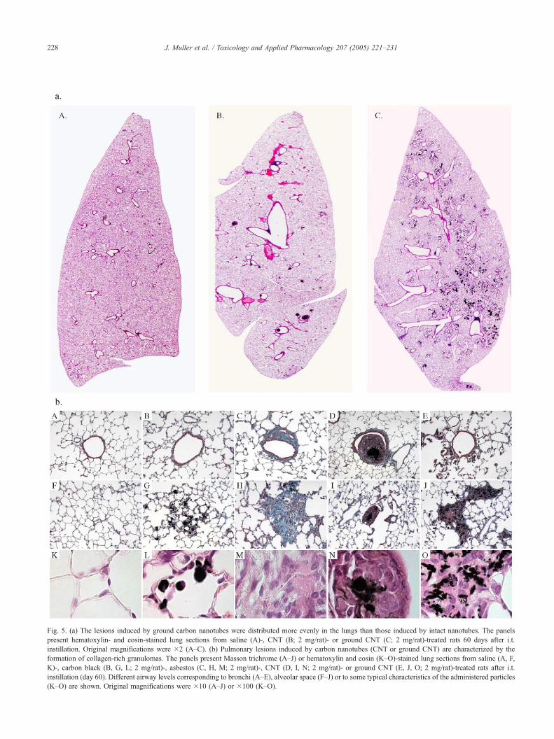
Fig. 5. (a) The lesions induced by ground carbon nanotubes were distributed more evenly in the lungs than those induced by intact nanotubes. The panels
present hematoxylin- and eosin-stained lung sections from saline (A)-, CNT (B; 2 mg/rat)- or ground CNT (C; 2 mg/rat)-treated rats 60 days after i.t.
instillation. Original magnifications were �2 (A–C). (b) Pulmonary lesions induced by carbon nanotubes (CNT or ground CNT) are characterized by the
formation of collagen-rich granulomas. The panels present Masson trichrome (A–J) or hematoxylin and eosin (K–O)-stained lung sections from saline (A, F,
K)-, carbon black (B, G, L; 2 mg/rat)-, asbestos (C, H, M; 2 mg/rat)-, CNT (D, I, N; 2 mg/rat)- or ground CNT (E, J, O; 2 mg/rat)-treated rats after i.t.
instillation (day 60). Different airway levels corresponding to bronchi (A–E), alveolar space (F–J) or to some typical characteristics of the administered particles
(K–O) are shown. Original magnifications were �10 (A–J) or �100 (K–O).
J. Muller et al. / Toxicology and Applied Pharmacology 207 (2005) 221–231228
Fig. 6. Instillation of carbon nanotubes (CNT or ground CNT) induced an
increase of TNF-a in BAL fluid. Sprague–Dawley rats were administered
by intratracheal instillation with CNT (0.5 or 2 mg/rat), ground CNT (0.5 or
2 mg/rat), asbestos (Asb, 2 mg/rat), carbon black (CB, 2 mg/rat) or saline
(NaCl, 0.9%). Level of TNF-a was measured in BAL fluid 3 days (A) or 60
days (B) after particle treatment. Each bar represents the mean F SEM of
4–6 observations. *P b 0.05; **P b 0.01; ***P b 0.001 denote significant
differences between mean values measured in the indicated group compared
to saline, as analyzed by the Student–Newman–Keuls multiple comparison
test.
Fig. 7. Treatment with carbon nanotubes (CNT or ground CNT) induced
different effects in vitro. Here we show the typical dispersion of CNT (A1)
or ground CNT (A2) in peritoneal macrophages cultures. 24 h after CNT
(20, 50 or 100 Ag/well), ground CNT (20, 50 or 100 Ag/well), asbestos(Asb; 20, 50 or 100 Ag/well), carbon black (CB; 20, 50 or 100 Ag/well) orculture medium (control), we assessed the level of TNF-a (B) and of lactate
dehydrogenase (LDH, C) in the supernatant of macrophage cultures. Total
LDH content was determined in parallel after cell disruption in 0.1% Triton
X-100. Each bar represents the mean F SEM of 3 observations. **P b
0.01, ***P b 0.001 denote significant differences between mean values
measured in the indicated group compared to saline, as analyzed by the
Student–Newman–Keuls multiple comparison test.
J. Muller et al. / Toxicology and Applied Pharmacology 207 (2005) 221–231 229
than those obtained by inhalation for a variety of biologic
endpoints such as pulmonary inflammation and fibrosis in
rats (Driscoll et al., 2000). However, intratracheal admin-
istration has several limitations, in particular because the
administration of a single bolus of often entangled material
may produce artifactual granulomatous lesions in the
airways (Drew et al., 1987). Thus, previous investigators
who examined the toxicity of carbon nanotubes faced
difficulties to adequately deliver the test material to the
lung, mainly because these particles tended to form
aggregates that remained entrapped in the airways. They
were therefore perplex about the significance of their
observations (non-uniform distribution of the lesions, no
dose response, absence of surrounding inflammation) and
questioned the toxicological relevance of their findings
(Warheit et al., 2004). In the present study, we also found
that the administration of intact multi-wall carbon nanotubes
(CNT) induces the formation of bronchial granulomas
developing around focal aggregates of CNT (Fig. 5B);
these granulomas were very similar to those reported
previously in the airways of rats (Warheit et al., 2004) and
in the airways and interstitium of mice (Lam et al., 2004).
Ground CNT were much better dispersed in the vehicle,
which allowed a better distribution of the particles in the
lungs and avoided the formation of the large intraluminal
granulomas observed with CNT (Fig. 5B). Ground CNT
induced the formation of lung inflammation and fibrosis
and the lesions had a more uniform distribution, followed a
dose–effect relationship and persisted for at least 2 months.
Since the nature of carbon nanotubes was not significantly
affected by grinding, our data support the idea that carbon
nanotubes are intrinsically toxic to the lung.
Industrial applications of carbon nanotubes include a
number of instances in which this material is included in
polymers or other matrices. For these applications, a perfect
dispersion of the particles is essential and ground nanotubes
are used to this end (Liu et al., 2004; Pierard et al., 2001).
It is likely that in the near future, the industrial use of
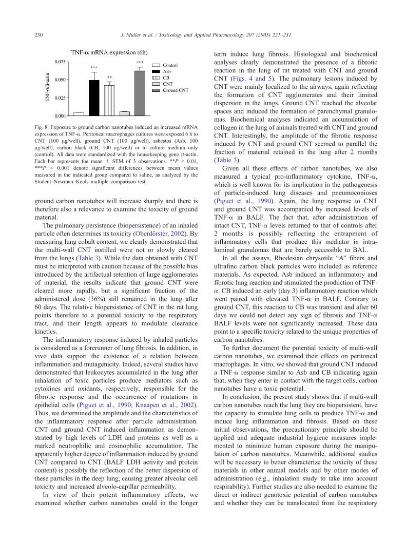
Fig. 8. Exposure to ground carbon nanotubes induced an increased mRNA
expression of TNF-a. Peritoneal macrophages cultures were exposed 6 h to
CNT (100 Ag/well), ground CNT (100 Ag/well), asbestos (Asb, 100
Ag/well), carbon black (CB, 100 Ag/well) or to culture medium only
(control). All data were standardized with the housekeeping gene h-actin.Each bar represents the mean F SEM of 3 observations. **P b 0.01,
***P b 0.001 denote significant differences between mean values
measured in the indicated group compared to saline, as analyzed by the
Student–Newman–Keuls multiple comparison test.
J. Muller et al. / Toxicology and Applied Pharmacology 207 (2005) 221–231230
ground carbon nanotubes will increase sharply and there is
therefore also a relevance to examine the toxicity of ground
material.
The pulmonary persistence (biopersistence) of an inhaled
particle often determines its toxicity (Oberdorster, 2002). By
measuring lung cobalt content, we clearly demonstrated that
the multi-wall CNT instilled were not or slowly cleared
from the lungs (Table 3). While the data obtained with CNT
must be interpreted with caution because of the possible bias
introduced by the artifactual retention of large agglomerates
of material, the results indicate that ground CNT were
cleared more rapidly, but a significant fraction of the
administered dose (36%) still remained in the lung after
60 days. The relative biopersistence of CNT in the rat lung
points therefore to a potential toxicity to the respiratory
tract, and their length appears to modulate clearance
kinetics.
The inflammatory response induced by inhaled particles
is considered as a forerunner of lung fibrosis. In addition, in
vivo data support the existence of a relation between
inflammation and mutagenicity. Indeed, several studies have
demonstrated that leukocytes accumulated in the lung after
inhalation of toxic particles produce mediators such as
cytokines and oxidants, respectively, responsible for the
fibrotic response and the occurrence of mutations in
epithelial cells (Piguet et al., 1990; Knaapen et al., 2002).
Thus, we determined the amplitude and the characteristics of
the inflammatory response after particle administration.
CNT and ground CNT induced inflammation as demon-
strated by high levels of LDH and proteins as well as a
marked neutrophilic and eosinophilic accumulation. The
apparently higher degree of inflammation induced by ground
CNT compared to CNT (BALF LDH activity and protein
content) is possibly the reflection of the better dispersion of
these particles in the deep lung, causing greater alveolar cell
toxicity and increased alveolo-capillar permeability.
In view of their potent inflammatory effects, we
examined whether carbon nanotubes could in the longer
term induce lung fibrosis. Histological and biochemical
analyses clearly demonstrated the presence of a fibrotic
reaction in the lung of rat treated with CNT and ground
CNT (Figs. 4 and 5). The pulmonary lesions induced by
CNT were mainly localized to the airways, again reflecting
the formation of CNT agglomerates and their limited
dispersion in the lungs. Ground CNT reached the alveolar
spaces and induced the formation of parenchymal granulo-
mas. Biochemical analyses indicated an accumulation of
collagen in the lung of animals treated with CNT and ground
CNT. Interestingly, the amplitude of the fibrotic response
induced by CNT and ground CNT seemed to parallel the
fraction of material retained in the lung after 2 months
(Table 3).
Given all these effects of carbon nanotubes, we also
measured a typical pro-inflammatory cytokine, TNF-a,
which is well known for its implication in the pathogenesis
of particle-induced lung diseases and pneumoconioses
(Piguet et al., 1990). Again, the lung response to CNT
and ground CNT was accompanied by increased levels of
TNF-a in BALF. The fact that, after administration of
intact CNT, TNF-a levels returned to that of controls after
2 months is possibly reflecting the entrapment of
inflammatory cells that produce this mediator in intra-
luminal granulomas that are barely accessible to BAL.
In all the assays, Rhodesian chrysotile bAQ fibers and
ultrafine carbon black particles were included as reference
materials. As expected, Asb induced an inflammatory and
fibrotic lung reaction and stimulated the production of TNF-
a. CB induced an early (day 3) inflammatory reaction which
went paired with elevated TNF-a in BALF. Contrary to
ground CNT, this reaction to CB was transient and after 60
days we could not detect any sign of fibrosis and TNF-a
BALF levels were not significantly increased. These data
point to a specific toxicity related to the unique properties of
carbon nanotubes.
To further document the potential toxicity of multi-wall
carbon nanotubes, we examined their effects on peritoneal
macrophages. In vitro, we showed that ground CNT induced
a TNF-a response similar to Asb and CB indicating again
that, when they enter in contact with the target cells, carbon
nanotubes have a toxic potential.
In conclusion, the present study shows that if multi-wall
carbon nanotubes reach the lung they are biopersistent, have
the capacity to stimulate lung cells to produce TNF-a and
induce lung inflammation and fibrosis. Based on these
initial observations, the precautionary principle should be
applied and adequate industrial hygiene measures imple-
mented to minimize human exposure during the manipu-
lation of carbon nanotubes. Meanwhile, additional studies
will be necessary to better characterize the toxicity of these
materials in other animal models and by other modes of
administration (e.g., inhalation study to take into account
respirability). Further studies are also needed to examine the
direct or indirect genotoxic potential of carbon nanotubes
and whether they can be translocated from the respiratory

J. Muller et al. / Toxicology and Applied Pharmacology 207 (2005) 221–231 231
system to other organs. Comparative studies of different
types of carbon nanotubes (single and multi-wall, function-
alized, produced by metal catalysis or plasma, etc.) will also
contribute to decipher the mechanisms that may govern the
toxicity of these materials.
Acknowledgments
This work was supported in part by the Actions de
Recherche concertees, Communaute francaise de Belgique-
Direction de la Recherche scientifique and by the Region
wallonne, BINANOCO project. F.H. is Research assistant
with the Fonds national de la Recherche scientifique
(FNRS), Belgium. We thank Dr N. Probst (Erachem
Comilog) for the gift of carbon black. The authors also
acknowledge the excellent technical assistance of Youssof
Yakoub, Claudine Gathy, Stephane Lagasse and Johan
Casters.
References
Biondi, P.A., Chiesa, L.M., Storelli, M.R., Renon, P., 1997. A new
procedure for the specific high-performance liquid chromatographic
determination of hydroxyproline. J. Chromatogr. Sci. 35, 509–512.
Carbon Nanotubes—Worldwide Status and Outlook: Applications, Applied
Industries, Production, R&D and Commercial Implications, 2002.
http://www.researchandmarkets.com/reports/7730/.
Drew, R.T., Kuschner, M., Bernstein, D.M., 1987. The chronic effects of
exposure of rats to sized glass fibres. Ann. Occup. Hyg. 31, 711–729.
Driscoll, K.E., Costa, D.L., Hatch, G., Henderson, R., Oberdorster, G.,
Salem, H., Schlesinger, R.B., 2000. Intratracheal instillation as an
exposure technique for the evaluation of respiratory tract toxicity: uses
and limitations. Toxicol. Sci. 55, 24–35.
Huczko, A., Lange, H., Clko, E., Grubek-Jaworska, H., Droszcz, P., 2001.
Physiological testing of carbon nanotubes: are they asbestos-like?
Fuller. Sci. Technol. 9, 251–254.
Knaapen, A.M., Albrecht, C., Becker, A., Hohr, D., Winzer, A., Haenen,
G.R., Borm, P.J., Schins, R.P., 2002. DNA damage in lung epithelial
cells isolated from rats exposed to quartz: role of surface reactivity and
neutrophilic inflammation. Carcinogenesis 23, 1111–1120.
Kohyama, N., Shinohara, Y., Suzuki, Y., 1996. Mineral phases and some
reexamined characteristics of the international union against cancer
standard asbestos samples. Am. J. Ind. Med. 30 (5), 515–528 (Nov.).
Lam, C., James, J.T., McCluskey, R., Hunter, R.L., 2004. Pulmonary
toxicity of single-wall carbon nanotubes in mice 7 and 90 days after
intratracheal instillation. Toxicol. Sci. 77, 126–134.
Lasfargues, G., Lison, D., Maldague, P., Lauwerys, R., 1992. Comparative
study of the acute lung toxicity of pure cobalt powder and cobalt-
tungsten carbide mixture in rat. Toxicol. Appl. Pharmacol. 112, 41–50.
Lison, D., Lauwerys, R., 1990. In vitro cytotoxic effects of cobalt-
containing dusts on mouse peritoneal and rat alveolar macrophages.
Environ. Res. 52 (2), 187–198 (Aug.).
Liu, C.H., Huang, H., Wu, Y., Fan, S.S., 2004. Thermal conductivity
improvement of silicone elastomer with carbon nanotube loading. Appl.
Phys. Lett. 84 (21), 4248–4250.
Martin, C.R., Kohli, P., 2003. The emerging field of nanotube biotechnol-
ogy. Nat. Rev., Drug Discov. 2, 29–37.
Maynard, A.D., Baron, P.A., Foley, M., Shvedova, A.A., Kisin, E.R.,
Castranova, V., 2004. Exposure to carbon nanotube material: aerosol
release during the handling of unrefined single-walled carbon nanotube
material. J. Toxicol. Environ. Health, Part A 67, 87–107.
Miller, K., 1978. The effects of asbestos on macrophages. CRC Crit. Rev.
Toxicol. 5 (4), 319–354 (Sep.).
Mossman, B.T., Bignon, J., Corn, M., Seaton, A., Gee, J.B., 1990.
Asbestos: scientific developments and implications for public policy.
Science 247, 294–301.
Muhle, H., Belmann, B., 1995. Biopersistence of man-made vitreous fibres.
Ann. Occup. Hyg. 39 (5), 655–660 (Oct.).
Oberdfrster, G., 2001. Pulmonary effects of inhaled ultrafine particles. Int.
Arch. Occup. Environ. Health 74, 1–8.
Oberdfrster, G., 2002. Toxicokinetics and effects of fibrous and nonfibrous
particles. Inhalation Toxicol. 14, 29–56.
Pierard, N., Fonseca, A., Konya, Z., Willems, I., Van Tendeloo, G., Nagy,
J.B., 2001. Production of short carbon nanotubes with open tips by ball
milling. Chem. Phys. Lett. 335, 1–8.
Piguet, P.F., Collart, M.A., Grau, G.E., Sappino, A.P., Vassalli, P., 1990.
Requirement of tumour necrosis factor for development of silica-
induced pulmonary fibrosis. Nature 344, 245–247.
Warheit, D., Laurence, B., Reed, K., Roach, D., Reynold, G., Webb, T.,
2004. Comparative pulmonary toxicity assessment of single wall carbon
nanotubes in rats. Toxicol. Sci. 77, 117–125.
Watanabe, N., Kamei, S., Ohkubo, A., Yamanaka, M., Ohsawa, S., Makino,
K., Tokuda, K., 1986. Urinary protein as measured with a pyrogallol
red-molybdate complex, manually and in a Hitachi 726 automated
analyzer. Clin. Chem. 32 (8), 1551–1554.
Willems, I., Konya, Z., Colomer, J.-F., Van Tendeloo, G., Nagajaru, N.,
Fonseca, A., Nagy, J.-B., 2000. Control of the outer diameter of thin
carbon nanotubes synthesised by catalytic decomposition of hydro-
carbons. Chem. Phys. Lett. 317, 71–76.