respiratory therapy training for long-term care nurses ... therapy training fo… · acute...
TRANSCRIPT

10/26/2012
1
October 2012
COURSE OBJECTIVES
1. Refresher on basic anatomy of respiratory system.
2. Common disorders of the respiratory system.
3. Respiratory system assessment.
4. Lungs sounds auscultation.
5. Pulse oximetry.
6. Oxygen delivery systems.
7. Suctioning.
8. Nebulizer treatment.
9. Tracheostomy care.
10. Refresher on commonly used respiratory medications.
Proprietary of Morningside Ministries
ANATOMY OF
THE RESPIRATORY SYSTEM
1. Respiratory tract: upper and lowerA. Organs in the upper respiratory tract include: nares
(nostrils), nasal cavity, pharynx, larynx and trachea.
B. Structures of the lower respiratory tract include: the lower trachea, bronchi, bronchioles, alveoli, lungs, pleural membranes and intercostal muscles.
2. Pleural membranesA. Parietal pleura
B. Visceral pleura

10/26/2012
2
UPPER RESPIRATORY TRACTA
LOWER RESPIRATORY TRACTB
PLEURAL MEMBRANES

10/26/2012
3
BOYLE’S LAW
� The smaller the volume,
the higher the pressure.
� The larger the volume,
the lower the pressure.
GAS EXCHANGE
AGING PROCESS
As a person ages, changes occur to the respiratory system include:� Decreased exchange of oxygen and carbon dioxide, caused by
decreased circulation.
� Increased anterior/posterior diameter of the chest due to
skeletal changes associated with aging (kyphosis).
� Chest wall becomes stiffer and more difficult to move.
� Respiratory muscles may weaken.
� Increased airway resistance.
� Impaired cough mechanism.
� Lungs lose some of their elastic recoil (like a rubber band in the sun).
� Muscles of the larynx and pharynx atrophy.
� Decreased vital capacity, residual volume and functional capacity.

10/26/2012
4
COMMON RESPIRATORY DISORDERS
� Acute bronchitis
� Etiology/Pathophysiology
� Inflammation of the trachea and bronchial tree
� Usually secondary to upper respiratory infection
� Exposure to inhaled irritants
� Clinical manifestations/assessment
� Productive cough; wheezes
� Dyspnea; chest pain
� Low-grade fever
� Malaise; headache
COMMON RESPIRATORY DISORDERS
� Acute bronchitis (continued)
� Medical management/nursing interventions
� Cough suppressants
� Antitussives
� Antipyretics
� Bronchodilators
� Antibiotics
� Vaporizer
� Encourage fluids
COMMON RESPIRATORY DISORDERS
� Adult respiratory distress syndrome
� Etiology/pathophysiology
� Complication of other disease processes
� Direct or indirect pulmonary injury
� Clinical manifestations/assessment
� Respiratory distress
� Tachycardia
� Hypotension
� Decreased urinary output

10/26/2012
5
COMMON RESPIRATORY DISORDERS
� Adult respiratory distress syndrome (continued)
� Medical management/nursing interventions
� Treat cause
� Oxygen
� Corticosteroids
� Diuretics
� Morphine
� Lanoxin
� Antibiotics
COMMON RESPIRATORY DISORDERS
� Asthma
� Etiology/pathophysiology
� Narrowing of the airways due to various stimuli
� Extrinsic or intrinsic factors
� Influenced by secondary factors
� Antigen-antibody reaction
COMMON RESPIRATORY DISORDERS
� Asthma (continued)
� Clinical manifestations/assessment
� Mild asthma
� Dyspnea on exertion
� Wheezing
� Acute asthma attack
� Tachypnea
� Expiratory wheezing; productive cough
� Use of accessory muscles; nasal flaring
� Cyanosis

10/26/2012
6
COMMON RESPIRATORY DISORDERS
� Asthma (continued)
� Medical management/nursing interventions
� Maintenance therapy
� Serevent inhalant, prophylactic
� Corticosteroid inhalant
� Avoid allergens
� Acute or rescue therapy
� Proventil inhalant
� Aminophylline IV (Acute Care, LTAC)
� Corticosteroid and epinephrine (PO or SubQ)
� Oxygen
COMMON RESPIRATORY DISORDERS
Did you know?
That residents with acute severe asthma should have their nebulizers administered via oxygen or they will become hypoxic. If necessary, low-flow oxygen may be administered via nasal cannula to residents while a drug is nebulized with air. This is because it requires high-flow oxygen to nebulize a drug (6-8 liters/minute) and if the resident has chronic respiratory disease he will only require a low-flow of oxygen to stimulate his respiration.
COMMON RESPIRATORY DISORDERS
� Atelectasis
� Etiology/pathophysiology
� Collapse of lung tissue due to occlusion of air to a portion of the lung
� Clinical manifestations/assessment
� Dyspnea; tachypnea
� Pleural friction rub; crackles
� Restlessness
� Elevated temperature
� Decreased breath sounds

10/26/2012
7
COMMON RESPIRATORY DISORDERS
� Atelectasis (continued)
� Medical management/nursing interventions
� Cough and deep-breathe
� Analgesia
� Early ambulation
� Incentive spirometry
� intermittent positive-pressure breathing (IPPB)
� Oxygen
� Chest percussion and postural drainage
� Bronchodilators; antibiotics; mucolytic agents
� Chest tube
COMMON RESPIRATORY DISORDERS
� Chronic Obstructive Pulmonary Disease (COPD)
� Chronic bronchitis
� Etiology/pathophysiology
� Hypertrophy of mucous glands causes hypersecretion and alters cilia function
� Increased airway resistance causes bronchospasm
� Clinical manifestations/assessment
� Productive cough
� Dyspnea
� Use of accessory muscles to breathe
� Wheezing
COMMON RESPIRATORY DISORDERS
� Chronic bronchitis (continued)
� Medical management/nursing interventions
� Bronchodilators
� Mucolytics
� Antibiotics
� Oxygen (low-flow)
� Pursed-lip breathing

10/26/2012
8
COMMON RESPIRATORY DISORDERS
� Chronic Obstructive Pulmonary Disease (COPD)� Emphysema
� Etiology/pathophysiology
� The bronchi, bronchioles, and alveoli become inflamed as a result of chronic irritation
� Air becomes trapped in the alveoli during expiration, causing alveolar distention, rupture, and scar tissue
� Complication
� Cor pulmonale
� Right-sided congestive heart failure due to pulmonary hypertension
COMMON RESPIRATORY DISORDERS
� Emphysema (continued)
� Clinical manifestations/assessment
� Dyspnea on exertion
� Sputum
� Barrel chest
� Chronic weight loss
� Emaciation
� Clubbing of fingers
COMMON RESPIRATORY DISORDERS
� Emphysema (continued)
� Medical management/nursing interventions
� Oxygen (low-flow)
� Chest physiotherapy
� Bronchodilators; corticosteroids; antibiotics; diuretics
� Humidifier
� Pursed-lip breathing
� High-protein, high-calorie diet
10/26/2012
9
COMMON RESPIRATORY DISORDERS
Did you know?
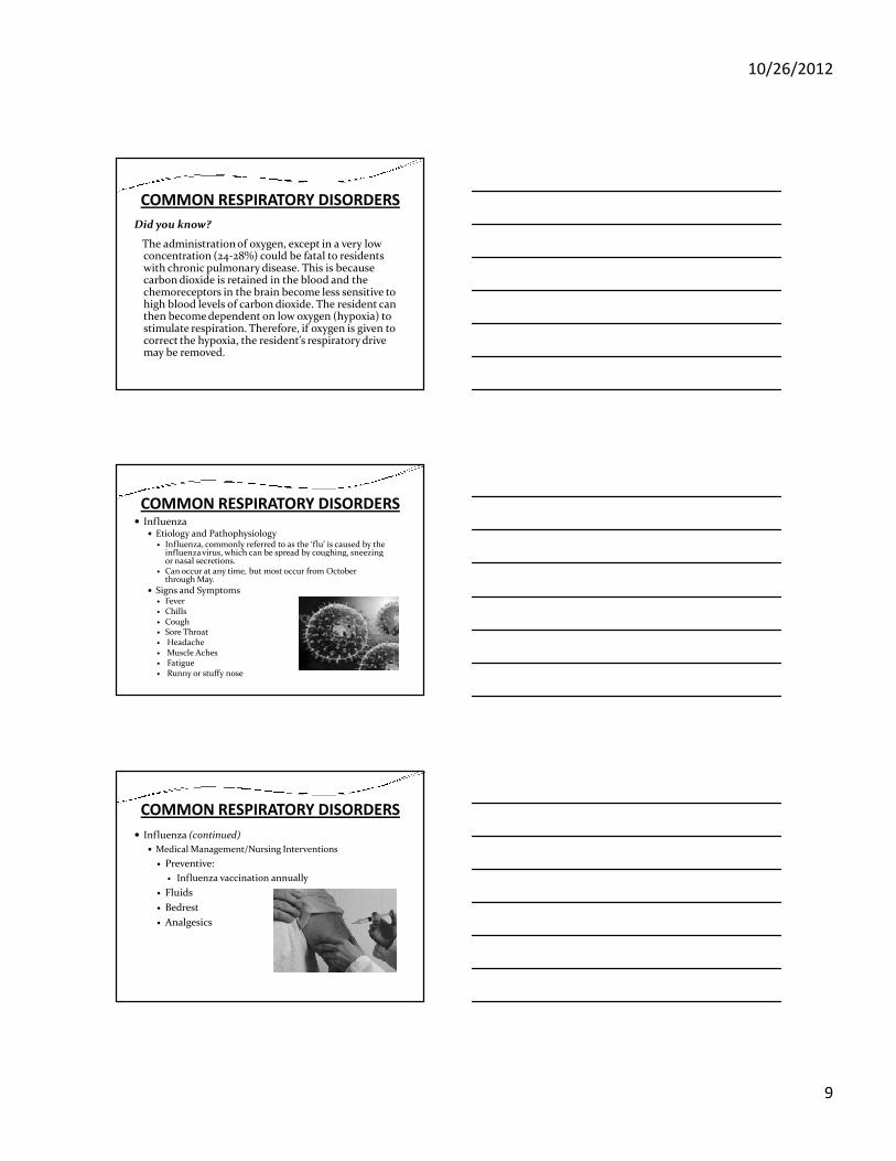
The administration of oxygen, except in a very low concentration (24-28%) could be fatal to residents with chronic pulmonary disease. This is because carbon dioxide is retained in the blood and the chemoreceptors in the brain become less sensitive to high blood levels of carbon dioxide. The resident can then become dependent on low oxygen (hypoxia) to stimulate respiration. Therefore, if oxygen is given to correct the hypoxia, the resident’s respiratory drive may be removed.
COMMON RESPIRATORY DISORDERS� Influenza
� Etiology and Pathophysiology� Influenza, commonly referred to as the ‘flu’ is caused by the
influenza virus, which can be spread by coughing, sneezing or nasal secretions.
� Can occur at any time, but most occur from October through May.
� Signs and Symptoms� Fever� Chills� Cough� Sore Throat� Headache� Muscle Aches� Fatigue� Runny or stuffy nose
COMMON RESPIRATORY DISORDERS
� Influenza (continued)
� Medical Management/Nursing Interventions
� Preventive:
� Influenza vaccination annually
� Fluids
� Bedrest
� Analgesics

10/26/2012
10
COMMON RESPIRATORY DISORDERS
� Legionnaires’ disease
� Etiology/Pathophysiology
� Legionella pneumophila
� Thrives in water reservoirs
� Causes life-threatening pneumonia
� Leads to respiratory failure, renal failure, bacteremic shock, and ultimately death
COMMON RESPIRATORY DISORDERS
� Legionnaires’ disease (continued)
� Clinical manifestations/assessment
� Elevated temperature
� Headache
� Nonproductive cough
� Difficult and rapid respirations
� Crackles or wheezes
� Tachycardia
� Signs of shock
� Hematuria
COMMON RESPIRATORY DISORDERS
� Legionnaires’ disease (continued)
� Medical management/nursing interventions
� Oxygen
� Mechanical ventilation, if necessary
� IV therapy
� Antibiotics
� Antipyretics
� Vasopressors

10/26/2012
11
COMMON RESPIRATORY DISORDERS
� Lung cancer
� Etiology/pathophysiology
� Primary tumor or metastasis
� Small-cell, non-small–cell, squamous cell and large-cell carcinoma
� Clinical manifestations/assessment
� Hemoptysis
� Dyspnea
� Wheezing
� Fever and chills
� Pleural effusion
COMMON RESPIRATORY DISORDERS
� Lung cancer (continued)
� Medical management/nursing interventions
� Surgery
� Most are not diagnosed early enough for curative surgical intervention
� Segmental resection
� Lobectomy
� Pneumonectomy
� Radiation
� Chemotherapy
COMMON RESPIRATORY DISORDERS
� Pleural effusion/empyema
� Etiology/pathophysiology
� Pleural effusion
� Accumulation of fluid in the pleural space
� Empyema—infection
� Clinical manifestations/assessment
� Dyspnea
� Air hunger
� Respiratory distress
� Fever

10/26/2012
12
COMMON RESPIRATORY DISORDERS
� Pleural effusion/empyema (continued)
� Medical management/nursing interventions
� Thoracentesis
� Chest tube with closed water-seal drainage system
� Antibiotics
� Cough and deep-breathe
COMMON RESPIRATORY DISORDERS
� Pleurisy
� Etiology/pathophysiology
� Inflammation of the visceral and parietal pleura
� Bacterial or viral
� Clinical manifestations/assessment
� Sharp inspiratory pain
� Dyspnea
� Cough
� Elevated temperature
� Pleural friction rub
COMMON RESPIRATORY DISORDERS
� Pleurisy (continued)
� Medical management/nursing interventions
� Antibiotics
� Analgesics
� Antipyretics
� Oxygen
� Anesthetic block for intercostal nerves

10/26/2012
13
COMMON RESPIRATORY DISORDERS
� Pneumonia
� Etiology/pathophysiology
� Inflammatory process of the bronchioles and the alveolar spaces due to infection
� Bacteria, viruses, mycoplasma, fungi, and parasites
� Clinical manifestations/assessment
� Productive cough
� Severe chills; elevated temperature
� Increased heart rate and respiratory rate
� Dyspnea
COMMON RESPIRATORY DISORDERS
� Pneumonia (continued)� Medical management/nursing interventions
� Oxygen
� Chest percussion and postural drainage
� Encourage to cough and deep-breathe
� Antibiotics
� Analgesics
� Expectorants
� Bronchodilators
� Humidifier
� Nebulizer treatments
COMMON RESPIRATORY DISORDERS
� Pulmonary edema� Etiology/pathophysiology
� Accumulation of serous fluid in interstitial tissue and alveoli
� Clinical manifestations/assessment
� Dyspnea; cyanosis
� Tachypnea; tachycardia
� Pink or blood-tinged, frothy sputum
� Restlessness; agitation
� Wheezing; crackles
� Decreased urinary output; sudden weight gain

10/26/2012
14
COMMON RESPIRATORY DISORDERS
� Pulmonary edema (continued)
� Medical management/nursing interventions
� Oxygen
� Mechanical ventilation, if necessary (Acute Care, LTAC settings)
� Diuretics
� Narcotic analgesics
� Nipride (Acute Care Hospital only)
� Strict I & O; daily weight
� Low-sodium diet
COMMON RESPIRATORY DISORDERS
� Pulmonary embolus
� Etiology/pathophysiology
� Foreign substance in the pulmonary artery
� Blood clot, fat, air, or amniotic fluid
� Clinical manifestations/assessment
� Sudden, unexplained dyspnea, tachypnea
� Hemoptysis
� Chest pain
� Elevated temperature
� Increased WBCs
COMMON RESPIRATORY DISORDERS
� Pulmonary embolus (continued)
� Medical management/nursing interventions
� Oxygen
� HOB up 30 degrees
� Anticoagulants
� Fibrinolytic agents

10/26/2012
15
COMMON RESPIRATORY DISORDERS
� Tuberculosis
� Etiology/pathophysiology
� Inhalation of tubercle bacillus (Mycobacterium tuberculosis)
� Infection versus active disease
� Presumptive diagnosis
� Mantoux tuberculin skin test
� Chest x-ray
� Acid-fast bacilli smear x 3
� Confirmed diagnosis
� Sputum culture; positive for TB bacilli
COMMON RESPIRATORY DISORDERS
� Tuberculosis (continued)
� Clinical manifestations/assessment
� Fever
� Weight loss; weakness
� Productive cough; hemoptysis
� Chills; night sweats
� Medical management/nursing interventions
� Tuberculosis isolation (acid fast bacilli [AFB])
� Multiple medications to which the organisms are susceptible
RESPIRATORY SYSTEM ASSESSMENT
An assessment of the respiratory resident include:
� Evaluations and routine monitoring of the respiratory system.
� All residents should have a respiratory assessment upon admission, whenever a change occurs in the resident’s respiratory status and as indicated by the resident’s diagnosis and/or medical conditions.

10/26/2012
16
RESPIRATORY SYSTEM ASSESSMENT
Knowledge of the resident’s current and past respiratory history includes:� Prior oxygen usage� Any lab results and/or diagnostics (chest x-ray)� Steroid use� Current medication use� Dietary requirements� Smoking history� Any history of cancer� Risk factors for pulmonary emboli� Sputum production to include color, amount and odor� Sleeping position, such as number of pillows, fan use, etc.
RESPIRATORY SYSTEM ASSESSMENT
LUNGS SOUNDS AUSCULTATION
Anterior Posterior

10/26/2012
17
NORMAL, ABNORMAL,ADVENTITIOUS
PULSE OXIMETRY
� Measuring the amount of oxygen absorbed in thearterial blood will provide an indication of theeffectiveness of the resident’s breathing and/or oxygentherapy.
� This is usually done by using pulse oximetry. A two-sided probe is used to transmit an alternating lightthrough a finger (preferred site), toe or earlobe. Thewavelength of the light that emerges indicates thepercentage of oxyhemoglobin present in thecapillaries.
PULSE OXIMETRY
� Normal values of oxygen saturation are 95-99%.
� Readings of 90-95% are usually a cause for concern; however, a resident’s medical history must be taken into consideration. � For example, a resident with chronic obstructive
pulmonary disease (COPD) may have a baseline oxygen
saturation of 88% and are comfortable at that level.
Did you know?
You should never attach a sensor by using adhesive tape as this has the potential to cause tissue necrosis.

10/26/2012
18
OXYGEN THERAPY
� Oxygen therapy is the administration of oxygen at a concentration greater than that in normal air, with the intent of treating or preventing the symptoms and manifestations of hypoxia.
� A range of tubing, connectors and masks ensure that the most appropriate product or combination of products are utilized for the resident whether the oxygen is delivered from an oxygen tank, cylinder or an oxygen concentrator.
OXYGEN FACTS� Oxygen is a colorless, odorless gas.
� Oxygen does not burn, however it makes fires burn faster and hotter as it acts as a catalyst.
� Avoid using any electrical equipment, such as a hair dryer or electric razor near oxygen tanks.
� Keep flammable materials, such as oil and grease away from oxygen equipment.
� Do not use petroleum-based products on your face and hands.
Did you know?
Oxygen should always be prescribed by a physician or physician extender and include the flow rate, delivery system, duration, for what medical condition/disease and how often to monitor saturations.
OXYGEN DELIVERY DEVICES
Oxygen Cylinders Oxygen Concentrator
� ‘E’ cylinders are the most commonly used in the long-term setting.
� ‘H’ cylinders are used frequently for continuous oxygen when a concentrator is not available.
� Portable cylinders, classified as ‘M’ cylinders are primarily used by private-pay and managed
� care residents when they are admitted from home.
� Oxygen concentrators function by pulling in room air and removes nitrogen, providing a greater percentage of oxygen concentration to the resident.
� Oxygen concentrators come in a variety of sizes and capacities, from 5 to 10 liters/minute.
� Usage and maintenance should be in accordance with manufacturer’s recommendations and
guidelines.

10/26/2012
19
OXYGEN SUPPLIES
1. Nasal cannula
� Most common delivery device used.
� Best used for residents with mild hypoxia of oxygen saturations between 90% an 93% and for long-term use in COPD residents to maintain oxygen saturations between 88% to 92%.
� Tubing varies in length to allow for mobility. In some cases, an extension tubing can be connected.
� Monitor for skin breakdown behind the ears.
� Monitor for drying of nares; if flow is 4 liters/minute or greater, a humidifier should be utilized.
� Flow rates greater than 5 liters/minute should be avoided as this will dry out nasal mucosa and cause nasal irritation.
� Never use a petroleum based jelly in nares; water-based gels should be utilized for nasal irritation.
OXYGEN SUPPLIES
2. Simple Facemask� The simple facemask is made of a clear soft vinyl construction mask
to cover the mouth and nose, a metal frame to ensure a tight fit at the nose.
� The strap on the mask ensures a tight fit around the cheeks and chin, providing maximum resident comfort.
� Holes in the side of the mask allow for air to be drawn into the mask to supplement the oxygen accumulating in the mask itself.
� Best used for residents with moderate hypoxia and oxygen saturation between 85% and 93%.
� Must be used with a concentrator that delivers >5 liters/minute.� Flow rates less than 5 liters/minute will result in unwanted carbon
dioxide (CO2) retention.� Usual order is between 6 – 10 liters/minute for a concentration of 40
- 60% of oxygen.� Do not place a humidifier on this device.� Switch to a nasal cannula for meals.
OXYGEN SUPPLIES3. Non-rebreather Facemask
� The non-rebreather facemask comes with a clear soft vinyl construction mask to cover the mouth and nose, a metal frame to ensure a tight fit at the nose.
� A strap on the mask to ensure a tight fit around the cheeks and chin and an oxygen reservoir bag to store oxygen to allow for much greater oxygen than the standard mask.
� Best used for residents with severe hypoxia and oxygen saturations below 85%.
� Often used in emergency situations.� Do not use a humidifier.� Flow rates less than 8 liters/minute will fail to sufficiently inflate the
oxygen reservoir bag resulting in rebreathing unwanted carbon dioxide and a sensation of suffocation.
� It is essential that the oxygen reservoir bag is fully inflated before applying the facemask to the resident. This can be achieved quickly by placing a finger over the outlet plug of the oxygen reservoir bag.
� Oxygen flow should be over 10 liters/minute for maximum effectiveness.� Liter flow is determined by the resident’s current respiratory needs. For
example, hyperventilation requires increase in liter flow.

10/26/2012
20
OXYGEN SUPPLIES4. Venturi Mask
� The Venturi mask is often referred to as a variable flow oxygen mask. � The mask mixes oxygen with room air based on the color port
attached. � This system provides for the most accurate constant oxygen
percentages of all the masks available on the market. � The larger the ports in the colored connector, the more external air
that is drawn in during a breath, reducing the percentage of oxygen.� Best used when there is concern about carbon dioxide retention.� The only mask that will give a precise oxygen concentration based
on manufacturer liter flow.� Best used when exact oxygen concentration must be utilized when
treating certain disease processes.� Venturi adapter is color coded to indicate percentage of oxygen and
liter flow delivered. � The adaptors allow for an oxygen range between 24% at 4
liters/minute to 50% at 12 liters/minute.� Never humidified.
SUCTIONING
� Suctioning is a removal of secretions through the use of negative pressure utilizing a suction catheter device.
� Suctioning is intended to remove accumulated secretions, blood, vomitus and other foreign material from the trachea that cannot be removed by the resident’s cough.
� The need to maintain a patent airway and remove secretions from the trachea due to the inability to clear secretions and audible and visual evidence of secretions that persist in spite of the resident’s best cough effort.
SUCTIONING
� Hazards:
� Hxypoxia
� Hypotension
� Gagging/vomiting
� Nosocomial infection
� Misdirection of catheter
� Cardiac arrest
• Bleeding disorder• Epiglottis• Laryngospasm
� Contraindications:
� Occluded nasal passages
� Acute head or neck injury
� Irritable airway
� Nasal bleeding
� Respiratory arrest
� Laryngospasm
� Atelectasis
� Increased intracranial pressure
� Bradycardia
� Uncontrolled coughing
� Bronchospasm

10/26/2012
21
SUCTIONING
� The following should be monitored during and following suctioning:� Breath sounds
� Skin color
� Breathing pattern
� Pulse rate and rhythm (regular/irregular)
� Sputum/secretions
� Color
� Consistency
� Amount
� Presence of blood
� Cough
� Pulse oximetry
SUCTIONING
� Effectiveness of suctioning should be reflected by improved breath sounds and by removal of secretions. Suctioning can be completed through multiple access areas:
� Nasal
� Nasopharyngeal
� Oral – catheter or Yankauer
� Tracheostomy
TRACHEOSTOMY CARE
� Reasons for a tracheostomy:
� Maintain a patent airway by means of bypassing upper airway (mouth/throat) obstructions.
� Facilitate removal of secretions .
� Decrease work of breathing and increase volume of air entering lungs by reducing anatomical dead space.
� Prevent aspiration of gastric contents

10/26/2012
22
TRACHEOSTOMY CARE
� Tracheostomy care consists of two parts, suctioning and cleaning of the tracheostomy tube.
� Suctioning removes secretions and fluids from the trachea, bronchi, and throat while stimulating the cough reflex. Tracheal suctioning aids to maintain a patent airway and provide optimal gas exchange of oxygen and carbon dioxide. An indication for suctioning may include:
� increased heart rate
� increased respiratory rate
� decreased oxygen saturation (commonly referred to as O2 sats)
� restlessness
� noisy expirations
� noted mucous in the tracheostomy tube
TRACHEOSTOMY CARE
� Cleaning keeps the cannula path open. Cleaning of the tracheostomy tube includes:
� the removal and cleaning of the inner cannula (in some cases, the inner cannula is disposable and replaced instead of the cleaning process)
� cleaning around the outer cannula
� cleaning of the stoma
NEBULIZER TREATMENTS
� Nebulizers are used to turn prescribed liquid medication into a mist so that it can be inhaled.
� These aerosolized medications are utilized to relieve bronchospasms, mobilize bronchial secretions, administer antibiotics and humidify the respiratory tract.
� Basic guidelines for nebulizer treatments are as follows:
� Require a physician order to include dosage of medication(s), frequency and length of time of treatment.
� Monitor the resident before, during and after each treatment for heart rate, breath sounds, respiratory rate, pulse oximetry and effectiveness of treatment.
� Encouraging/assisting the resident to deep breath and cough after each treatment.

10/26/2012
23
NEBULIZER TREATMENTS
� Monitor the resident for side effects. If he/she experience any of the following, stop the treatment, rest for five to ten minutes. If the sensation goes away, continue with your treatment.
� If physical problems persist, stop the treatment and notify the physician:� Increase in pulse by 20 beats per minute� Palpitations (noticeable heart beat)� Hyperventilation� Dizziness� Shakiness� Nausea� Chest pain� Uncontrollable coughing� Facial redness
RESPIRATORY MEDICATIONS
� Bronchodilators� Albuterol
� Levalbuterol
� Ipratropium Bromide
� Ipratropium Bromide/Albuterol
� Tiotropium Bromide
� Reduce Mucus and Secretions� Acelycysteine (Mucomyst)
� Reduce and Control Inflammation� Fluticasone Proprionate and Salmeterol (Advair)
� Budesonide
� Budesonide/Formoterol Fumarate (Symbicort)
QUESTIONS & ANSWERS

10/26/2012
24
RESOURCES*
� 2013 Lippincott’s Nursing Drug Guide; Lippincott Williams & Wilkins, Wolters Kluwer Company. Rochester, NY. June, 2012.
� Braman SS. Postinfectious cough: ACCP evidence-based clinical practice guidelines. Chest. 2006;129(1):138S-146S.
� Centers for Disease Control and Prevention. Vaccination Information Sheets (VIS).
� Lippincott Manual of Nursing Practice, Ninth Edition; Lippincott Williams & Wilkins, Wolters Kluwer Company. Rochester, NY. 2010
� Littmann Education; 3M Solutions* List is not all inclusive.