respiratory care : the official journal of the american ... · re/piratoryq^re...
TRANSCRIPT

August 1997
Volume 42, Number 8
ISSN 0020-1324-RECACP
J^43'^'' International Respiratory Congress
December 6-9 • New Orleans, Louisiana
A MONTHLY SCIENCE JOURNAL41ST YEAR—ESTABLISHED 1956
Validation of Metabolic Cart for
Measurement of VqA/t
Bronchoalveolar Lavage: A Review
CRCE through the Journal
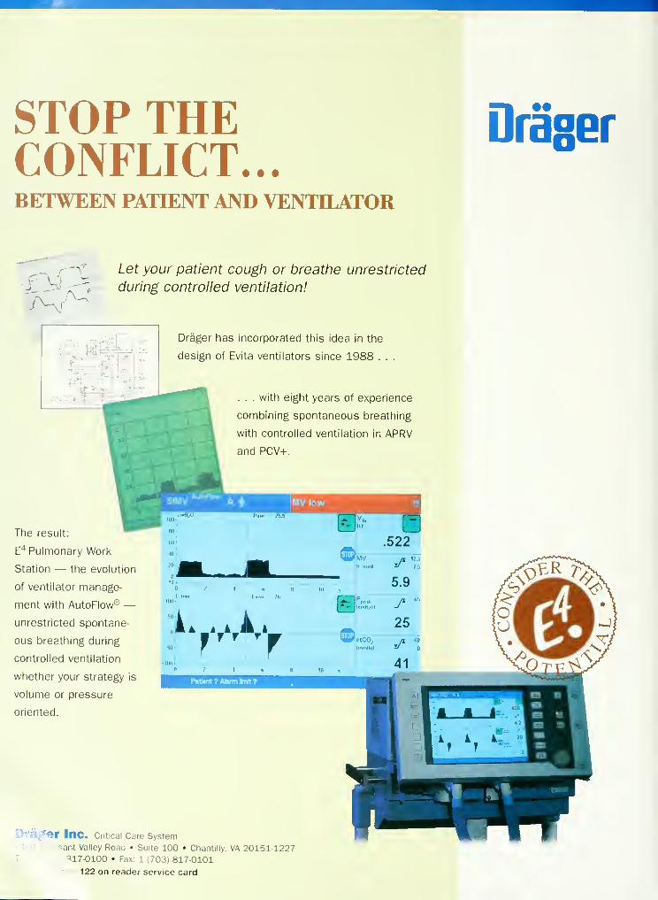
STOP THECONFLICT...BETWEEN PATIENT AND VENTILATOR
Draser
j'^ :\^
Let your patient cough or breathe unrestricted
during controlled ventilation!
Drager has incorporated this idea in the
design of Evita ventilators since 1988 . .
. . . with eight years of experience
combining spontaneous breathing
with controlled ventilation in APRV
and PCV+.
I SIMV'
IVIV lowt'jw ?i.l
The result:'"^
E"* Pulmonary Work
Station — the evolution
of ventilator manage-
ment with AutoFlow® —unrestricted spontane-
ous breathing during
controlled ventilation
whether your strategy is
volume or pressure
oriented.
Te^-
'<ir Inc. Critical Care System
-asant Valley Road • Suite 100 • Chantilly. VA 20151-1227703. 317-0100 • Fax: 1 (703) 817-0101
Oir 122 on reader service card

VpcfateTUe (F^MI^^respiratory care meeting in the worCd
AA^ 43rcfInternationaf^spiratory Congress
of the
American Associationfor ^spiratory Care
(DecemSer 6-9, 1997 (Saturcfay-Tuesday)
Ernest% 'MoriafConvention Center • [Kew Orfeans
CONTINUING EDUCATION - "The AARC meeting is the best place to earn CRCE" stated an attendee at last
year's Congress in San Diego. When asked to elaborate, she added that at the AARC meeting she was "able to
accumulate all the CRCE hours required by her state licensure, from 7 am to 5 pm for four days." At the NewOrleans Congress you, too, can earn as much as 25 hours of continuing education.
ADVANCE PROGRAM - The official advance program for the 1997 Congress will be mailed in early
September. In addition to containing all the events scheduled for New Orleans, the program will include the reg-
istration fonn and instructions for you to make your hotel reservations.
WHAT TO DO IN NEW ORLEANS - The September issue ofAARC Times will contain all kinds of information
about New Orleans—from what to see, to what to do, to where to eat. In the meantime, ifyou want to read about the
"Big Easy," visit the New Orleans Convention & Visitors Bureau's website on the Internet (www.nawlins.com).
PROGRAM HOURS EXPANDED - The AARC Program Committee has decided to begin the educational pro-
grams during the four days of the Congress at 8:30 am instead of 9 am, as in past years. This change is necessitat-
ed to accommodate the large number of programs to the presented in New Orleans. Exhibit hours will be from 1
1
AM to 4 PM on the first three days and from 1 1 am to 3 pm on the last day.
Open Forum - The results of scientific studies by clinicians, managers, and educators will again be presented
during the popular Open Forum sessions at the 1997 Congress. Organized and presented by the journal
Respiratory Care, the Open Forum papers are clustered into Minisymposia—with posters, one-on-one discus-
sions, study implications, and group discussion. The history of the Minisymposia is one of stimulating interac-
tions and lively group discussions. CRCE credits are available for participation in the Open Forum. As each
minisymposium is completed, the abstract posters will be displayed on the Wall of Fame (located in the Exhibit
Hall), for the remaining days of the Congress.
MEDICAL DIRECTORS/RCPs INTERACTION SYMPOSIUM - The premier program at the 1997 Congress,
the New Horizons Symposium, will address the issue of medical directors/RCPs interaction in delivering respira-
tory care. Chaired by Dr. James Stoller of Cleveland, the symposium will discuss the issues of current roles of med-
ical directors; what can be done about the inactive medical director; the RCP/medical director team; and interac-
tion of the two outside the hospital environment. The symposium will end with a panel discussion. This is the 13th
year the AARC presents the New Horizons Symposium.
UNIOUE PHYSIOLOGY COURSE TO BE PRESENTED - For the first time ever, a clinical respiratory physi-
ology mini-course will be presented during the 1 997 Congress. A four-part series ofone-hour daily lectures, the mini-
course is designed to review basic physiology in a plain-speaking, clinically-focused way using case examples.
HEADOUARTERS HOTEL FOR 1997 CONGRESS - The New Orleans Hilton has been designated as the
headquarters hotel for the 1997 Congress. Located beside the banks of the mighty Mississippi River, the Hilton is
considered among the top five hotels in New Orleans.

The South Carolina Society for
Respiratory Care and the AARC present a
Special
Pre-(onvention
kminarTuesday, September 30, 1997
Pawley's Island, South Carolina
MAPPING A smmiDIBECTION fOR SKCiSS IN
THE (HANGING HEALTH
(ARE ENVIRONMENTThe 26th Annual Meeting and Exhibition of the
South Carolina Society for Respiratory Care (SCSRC)will be held at the beautiful Litchfield by the SeaHotel in Pawley's Island on October 1-3, 1997. This
year's convention will once again be preceded by a
special seminar organized in cooperation with the
American Association for Respiratory Care. Theseminar will present timely information on howrespiratory care practitioners and their employerscan change and benefit from today's health care
environment. The Special Seminar is approved for
6 hours of continuing respiratory care education
(CRCE) credit by the AARC.

RE/PIRATORy Q^REA Monthly Science Journal. Established 1956. Official Journal of the American Association for Respiratory Care
Contents ...Editor
Pat Brougher BA RRT
Managing Editor
Ray Masferrer BA RRT
Editorial Board
James K Stoller MD, Chairman
Cleveland Clinic Foundation
Cleveland. Ohio
Richard D Branson RRT
University of Cincinnati
Medical Center
Cincinnati. Ohio
Crystal L Dunlevy EdD RRT
Atlanta, Georgia
Charles G Durbin Jr MDThe University of Virginia
Health Sciences Center
Charlottesville. Virginia
Dean R Hess PhD RRT
Massachusetts General Hospital
Harvard Medical School
Boston. Massachusetts
Neil R Maclntyre Jr MD
Duke University Medical Center
Durham, North Carolina
Shelley C Mishoe PhD RRT
Medical College of Georgia
Augusta, Georgia
Joseph L Rau PhD RRT
Georgia State University
Atlanta. Georgia
August 1997
Volume 42, Number 8
Original Contributions
761 Validation of the Deltatrac Metabolic Cart for
Measurement of Dead-Space-to-Tidal-Voltime Ratio
by John C MacKinnon. Patricia L Houston, and Glenn P
McGuire—Toronto. Ontario. Canada
Reviews, Overviews, & Updates
765 Bronchoalveolar Lavage: A Useful Method for
Diagnosis of Some Pulmonary Disorders
b\ Ali Emad^Sliiraz. Iran
Graphics Corner
791
Letters
796
796
796
Excessive Work of Breathing, Active Exhalation,
and Retardation of Expiratory Flow: What's the
Problem and Where's the Problem?
by Richard D Branson. Robert S Camplicll. and Jay A
Johannigman—Cincinnati. Ohio
Asthma & SCUBA Diving
b\ Lawrence Martin—Cleveland. Ohio
Graphics Corner Makes Waves?
b\ Phil Mercurio—Albuquerque. New Mexico
Airflow Resistance & Zero Flow: An Apparent
Contradiction?
/)\ Robert Chatburn—Cleveland. Ohio
Historical Notes
798 "Nebulizer" from Effective Inhalation Therapy
by Edwin R Levine MD and published by National Cylinder Gas
Co in 1953. pages 150-154. submitted by Teri Nikolai Wilson—
Daxton. Ohio
Continuing Education Examination
800 CRCE through the Journal— 1997
Respiratory Care • August '97 Vol 42 No 8 747

The Wisconsin Society for Respi^toj^^affi^nd ttt^AABCi
WISCONSIN SOCIETY9:00 a.m. - 9:05 a.m.
Program OverviewPatrick J. Dunne, MEd, RRT,
Southwest Medical Emporium,Fullerton CA
9:05 a.m. -9:55 a.m.
Managing the "INs andOUTs" of Managed CarePatrick J. Dunne, MEd, RRTDiscusses the key concepts
underlying ttie managed care
movement and the resultant
changes the process brings to the
traditional health care delivery
paradigm. Explores the trend of
"risk-shifting" and its implications,
as well as the opportunities this
phenomena portends for respiratory
care practitioners in all care settings.
10:00 a.m. - 10:55 a.m.
Managing Acute Care in a
Managed Care EnvironmentKevin L. Shrake, MA, RRl,
FACHE, Memorial Medical
Center, Springfield IL
E.\plains how guidelines andprotocols fit into the managed care
environment. Describes how to
establish a utilization control
program that has gained widespread
acceptance from employees,
physicians, insurance companies,and administrative staff.
11:00 a.m. - 11:55 p.m.
A Guide to Disease andDemand ManagementPatrick J. Dunne, MEd, RRTDefines and discusses the key
attributes of disease and demandmanagement strategies. Focuses onthe application and use of these
concepts and techniques byrespiratory care practitioners.
Provides examples of successful
implementation and the value of
disease and demand management in
fostering change while safeguarding
quality health care services.
1:30 p.m. -2:25 p.m.
Developing CollaborativeRelationships withPhysiciansKevin L. Shrake, MA, RRT,
F.-XCHE
Describes proven skills to
communicate and collaborate withphysicians. Kxplaiiis the basis for the
development of key physicianbehavioral traits and how to
effectively ask for support for keyprograms.
MAPPING A
STRATEGIC
DIRECTION FOR
SUCCESS IN
THE CHANGING
HEALTH CARE
ENVIRONMENT
2:30 p.m. - 3:25 p.m.
Developing a PerformanceMeasurement System forRespiratory Care Services
Patrick J. Dunne, MEd, RRTDiscusses the growing demands in
health care for the collection,
analysis, and use of objective,
quantifiable information aboutprovider performance. Explores
various utilization and clinical
outcomes appropriate for
measurement by providers of
respiratory care services. Reviews
Oryx, the JC.'XHO's new initiative to
integrate the use of performancemeasures into the accreditation
process.
3:30 p.m. - 4:25 p.m.
Creating Opportunitiesthrough Career MarketingKevin I.. Shrake, MA, RRT,
FACHEPresents a systematic plan on howto create career opportunities byeffectively marketing skills andservices to key decision-makers andcolleagues. Outlines how to create
skills inventory, how to identify key
players, and choosing techniques to
prove value.
Conference
Thursday, October 2, 1997
MERRIMAC,WISCONSIN
The Annual I-'all Conference of the
Wisconsin Society for Respiratory Care
will be held at the beautiful Devil's
Head Resort in Merrimac on October
1-2, 1997. This year's conference will
include a special seminar organized in
cooperation with the American
Association for Respiratory Care. The
seminar will present timely
information on how respiratory care
practitioners and their employers can
change and benefit from today's
health care environment. The Special
Seminar is approved for 6 hours of
continuing respiratory care education
(CRCE) credit by the AARC.
Registration Fees(Pre-Registration Deadline: September 20, 1997)
' ^e-ReQistration AAP.C Memirer Nor-N/embef
-911 Conference $100 $125
"^oistration AAFiC Mefr.t::er Non-lvtefT'.bef
. ;--'ce $110 $135
For hotel room reservations and their special
rate for the WSRC Pall Conference, please call
the DcNil's Head Resort direct (1-800-472-6670)
and identify yourself as an attendee. RoomRate: 554 single/ double.
For additional information, please call or write
Al Ludin (414) 334-5533"i,r: Soc.e:v '; ^esp^faic^y Ca^e
P.O. Box 26646.;ivvaukee, Wi 53226

Assistant EditorKris Williams BA
Editorial Assistant
Linda Barcus BBA
Production CoordinatorKaren Singletern BS
Section EditorsRobert R Fluck Jr MS RRT
MS Jastremski MDBlood Gas Corner
Hugh S Mathewson MDDrug Capsule
John O Nilsestuen PhD RRTKen Hargett BS RRT
Roben Harwood MSA RRTGraphics Corner
Richard D Branson RRTRobert S Campbell RRT
Kittredge 's Corner
Charles G Ir\in PhD
Jack Wanger MBA RPFT RRTPFT Corner
Patricia Ann Doorley MS RRTCharles G Durbin Jr MDTest Your Radiologic Skill
Barbara Wilson MEd RRTJon Meliones MD
John Palmisano RRTCardiorespiratoiy Interactions
Consulting EditorsFrank E Biondo BS RRTHoward J Birenbaum MDRobert L Chatbum RRTDonald R Ellon MDRonald B George MDJames M Hurst MD
Robert M Kacmarek PhD RRTMichael McPeck BS RRT
David J Pierson MDJohn Shigeoka MD
Jeffrey J Ward MEd RRT
ProductionDonna Knauf
PublisherSam P Giordano MBA RRT
MarketingDale Gnffiths
Director
Tim GoldsburyDirector ofAdvertising Sales
Beth Binkley
Advertising Assistant
Editorial Office
11030 Abies Lane
Dallas TX 75229
(972) 243-2272
ContentsAugust 1997
Volume 42, Number 8
In This Issue
809750824824822813811
817818
AARC Membership Application
Abstracts from Other Journals
Advertisers Index & Help Lines
Author Index
Calendar of Events
Manuscript Preparation GuideMedWatchNew Products & Services
Notices
RUSPIRATORV Care llSSN 0020-1324, USPS OtSQ-NOl is published monthly by Daedalus Enterprises Inc. at
1 1030 Abies Lane. Dallas TX 75229-4593, for the American Association for Respiratory Care. One volume is pub-
lished per year beginning each January . Subscription rales are $65 per year in the US; S80 in all other countries (for
airmail, add S84).
The contents of the Journal are indexed in Hospital and Health Administration Index. Cumulative Index to Nurs-
ing and Allied Health Literature. Excerpla Medica. and RNdex Library Edition. Abndged versions of RESPIRV-
TORY Care are also published in Italian and Japanese, with peniiission from Daedalus Enterprises Inc.
Periodicals postage paid at Dallas TX and at additional mailing offices. POSTMASTER: Send address changes
to Respi]<.atory Care. Membership Office, Daedalus Enterprises Inc, 1 1030 Abies Lane, Dallas TX 7522y-45«
'
Printed in the United States ofAmerica Copyright ® 1997. by Daedalus Enterprises I:c.
RESPIRATORY CARE • AUGUST '97 VOL 42 NO 8 749

Abstracts Summ^iries of Pertinent Articles in Other JoumaI>
Editorials, Coinmenlaries, and Reviews To Note
Perioperative Cardiac Dysrlijthmias: Diagnosis and Management I Review)—JL Atlee. Anes-
thesiology 1997;86(fi):I.W7-l424.
The Outcomes of \ ery Low Birth Weight Infants: Arc We Aslting the Right Questions? (Com-
inentary)—MCMcCorniick. Pediatrics iyy7;99(6):869-876.
Intrapartum Hypoxic-Ischemic Cerebral Injury and Subsequent Cerebral Palsy: Medicole-
gal Issues ( Review )—JM Ferlnian. Pediatrics 1997;99(6):851-X57.
Downsizing Competence: The Myth of Quality or Let the Barber Do the Surgery (Editorial)
—
MS Braunstein. Chest 1997;) 1 1|6): 1478.
Noninvasive Positive Pressure V entilation for Acute Respiratory Failure: I'nderutilized or < )ver-
raled? RM Jasmer. JM Luce. MA Matthay, Chest 1997;! 1 1(6): 1672- 1678. Seethe related edilo-
rial: Noninvasive Positive Pressure Ventilation: A Positive View in Need ofSupportive Evidence—J/ Rosenberg. RS Goldstein. Chest 1997:1 1 H6):1479-I4H2.
Croup (Review)—GC Geelhoed. Pediatr Pul-
monol 1997:23:370.
the mode of administration will need to be decided
by the attending clinician.
brands and different diameters is mandatory prior
to their utilization.
The management of mild to severe croup has
undergone dramatic changes in the last 5 years,
primarily due to the increased understanding of
the benefits of treating it w ith steroids. Steroids
have been useil in the treatment of croup for many
years but. until recently, their use has remained
controversiiil. luirlier studies were ohen not blinded
or used inappropriate outcome measures, such as
respiratory rate, which have not provcti appropriate.
Two attempts to review the literature in 1980 and
1989 cautiously supported the use of steroids.
Despite these recommendations many practitioners
continued to \ lew croup in most cases as a benign
self-limited condition, and since steroids have
potential side effects, their use was not consid-
ered justified. More recently, however, a number
of developments such as the successful use of the
inhaled steroid budesonide and oral dexametha-
sone have reinforced the argument for using
steroids. Recent work has also shown that both
inhaled and systemic steroids work by I hour and
dramaticall) reduce morbidity and hospitaliza-
tion time. The demonstration that an oral dose of
0. 1 5 ing/kg desaniethasonc is as cffectiv e as kirger
doses has made the use of systemic steroids more
acceptable to many practitioners. All children with
croup severe enough to be admitted to hospital
should receive steroids. Two recent studies ha\ e
shown that steroids also benefited children whopresented to emergency de[)artinents for treatment,
but w hose croup was not considea'd se\ ea- enough
for admission. The type of steroid, the dose, and
Correctly Selecting a Liquid-Filled Nasogas-
tric Infant Feeding Catheter lo Measure Intra-
esophageal Pressure—CCJ Hartford. GO Rogers.
M.1 furner. Pediatr Pulmonol 1997:23:362.
Polyvinyl chloride iPVCl nasogastric feeding
catheters are used clinically to measure intra-
esophageal pressure as an estimate of pleural pres-
sure for calculating lung compliance in infants.
Tlie accuracy of pressure measurement of 4 French
gauge (HG) catheter sizes and 3 brands of liquid-
filled catheter manometer systems (CMS) was
evaluated by determining their resonance-fre-
quency amplitude and phase properties. .All CMSwere undeidaniix'd :uid a'sonated. No CMS e.vhib-
ited a uniform mean frequency response above
1 1 H/. The inaximum respiratory rate (Fr,) within
vv hich CMS could potentially measure dynamic
inlracsophageal pressure within a y/i error limit
was detemiined (F„): the highest mean V„ recorded
reliably in large-diameter catheters was 82
ba>aths/min. Significant CMS tliflerences in accu-
racy existed between catheter FG sizes and be-
tween catheters of similar diameters but differ-
ing brands. Correlation (r) between catheter inner
diameter and CMS Frr was 0.66 across brands.
In conclusion, intraesophagcal PVC liquid-filled
feeding catheters aiie suitable for estimating pleu-
ral pressures in subjects mechanically ventilated
vv ithout sharp inspiratory wavefonns or high res-
piratory rates. Quantitative frequency response
characterization ol ditlerent nasocastric catheter
Retrospecti> e Rev iew of the Effects of rhDNa-se
in Children vvllli Cystic Fibrosis—J Dav les. M-T
Trindade. C Wallis. M Rosenthal. O Craw lord.
A Bush. Pediatr Pulmonol 1997:23:243.
Trials of rhDNase in mixed groups of adults and
children with cystic fibrosis (CF) have demon-
strated improvements in lung function and well-
being. This has led to many pediauic CF patients
receiving regular rhDNase therap>' although their
response to treatment may not be the same as that
seen in adults. We have retrospectively reviewed
the effects ofrhDNa.se during the first yeiir of tlier-
apy in 65 children reeelv ing the dmg ;it 2 teni;iry
referral centers. Outcome measures included
changes in lung function, oxygen saturation, use
of inmivcnous antibiotics, and subjective improve-
ment. Median baseline lung function CJc of pre-
dicted) was 4S'7f for forced expiratory volume in
1 second (FEV I ) and .iX^r for forced vital capac-
ity (FVC). At 3-4 months following initiation of
therapy the group demonstrated median (9.S';i- CI)
increa.ses of 14.2-* (<iy/c CI 7.3: 21.1% ) in FEV,
and 79r (0: 14%) in FVC. Within this wide scal-
ier of responses, one-quarter of children deteri-
orated, but almost .5()7f showed significant im-
provements of > 10%. A similar pattern was seen
at 9 months, with median increases for the group
of 11.1% (0; 18.8%)inFEV|and5.6%(0; 17%)
in FVC. again with approximately one third of
the group deteriorating and one half improving
sisinificantlv . Intravenous antibiotic use decrea-sed
RESPIRATORS Care • August "97 Vol 42 No 8

Abstracts
significantly. Almost all the children (89%),
including those with a tall in lung function,
described subjecti\e improvement. There were
no predictive markers at baseline of a good
response to the drug. However, there was a good
correlation betw een lung function response at .^
months and that at 6. 9. and 12 months. Thus, chil-
dren respond to rhDNase at least as well a.s adults,
and a therapeutic trial is justified in those over
5 years with significantly impaired lung function.
Response is highly variable, making careful indi-
vidual assessment mandatory. Baseline charac-
teristics are not useful in predicting those chil-
dren who will respond well to treatment, but
long-term response to the drug can be predicted
on the basis of spirometric improvement at .^
months. Therefore, this would be a useful time
period for a therapeutic trial.
Hospitalization Patterns in Severe Acute
.\sthma in Children—S Schuh. D Johnson. D
Stephens. S Callahan. G Canny. Pediatr Pulmonol
1997;23;184.
We set out to determine associations betw een hos-
pitalization and disease severity before and 2 hour,
after initiation of a.sthma therapy in the Emergency
Department, and to describe the outcome of pa-
tients admitted and discharged. This is a retro-
spective review of data and charts from a ran-
domized, double blind, placebo-controlled trial
IRCT) of 120 asthmatics 5-17 years of age with
baseline forced expiratory volume in 1 second
(FEV| ) < .'i09r predicted, treated with .^ or 1 or
doses of nebulized ipratropium added to 3
albuterol nebulizations administered o\ er 1 hour.
None of the clinical parameters measured at base-
line were associated with hospitalization. How-
ever, by 2 hours after initiation of therapy, both
the FEV| percent of predicted values C^t pred) and
the total asthma score were associated w ith like-
lihood of hospital admission. Baseline O: satu-
ration < 92^* indicated a longer hospital stay (75..^
± 5 1 hours vs 4.3.0 ± 24.4 hours, p = 0.01 .5 1 and
a later onset of infrequent nebulizations (46.7 ±
35. 1 vs 26.6 ± 17.4 hours, p = 0.006). By 2 hours,
those with a post-treatment FEV] % pred < 30%
and an asthma score > 6 of 9 had a high likelihood
of hospitalization (86 and 80%. respectively, com-
bined probability 100%). whereas FEV 1 % pred
> 60% and total asthma score < 3 were associated
with successful discharge (probability of 92 and
83%. respectively). We conclude that pre-treat-
ment assessments were not associated with hos-
pitalization, while patients with posttreatment
FEVi % pred < 30% and a score > 6 had high like-
lihood of hospitalization.
.Aerosol Delivery to Wheezy Infants: A Com-
parison between a Nebulizer and Two Small
Volume Spacers—JH \\ ildhaber. SG Dcvada-
son. MJ Hayden. E Eber. QA Summers. PN
LeSouef Pediatr Pulmonol 1997:23:212.
Inhalation therapy for wheezy infants with either
a nebulizer or a pressurized metered dose inhaler
(pMDI) through a spacer is common practice. The
aim of our study was to compare aerosol deliv-
en to w heezv infants from a nebulizer and from
a pMDI via 2 small volume spacers. Twenty
wheezy infants (aged 4-12 months) were recruited.
They inhaled salbutamol from a Pari-Baby ' neb-
ulizer, from a detergent-coated Babyhalei'- . and
from a Nebuchamber'' in random order. A filter
was placed between the inhalation systems and
the patients. The amount of salbutamol deposited
on the filter was measured using an ultraviolet
spectrophotometer and was expressed as a per-
centage of the total nebulized or actuated doses.
The mean total iwhiilired dose for the P;in-Baby*
(1,030 ^g) was higher (p < 0.001 ) than the mean
You need it now—not next month,
not next week.
And you
can have it!
The fastest answers to your questions
about products and services advertised in
the current issue of Respiratory Care are
just a "Ciicl< " away when you go online:
http://www.aarc.org/resources/html
r /J
^Oxtf^e^
jlfUifiten.
Helps eliminate tangling of oxygen tubing.
Allows the patient greater mobility with
less worry.
Attaches easily to all types of cannulas and
oxygen tubing.
Lightweight, but durable.
Integral clip for added convenience.
Linjorel Products Inc.
2275-C Freed Watj
Pittsburg, CA 94565
Tel: 510.427.4013
Fax: 510.427.5988
Visit Us on the World Wide Web at^^
Circle 117 on reader service card
RESPIRATORY CARE • AUGUST "97 VOL 42 NO 8 751

Respiratory Care Fashion Appare
To order call the
RC Week Hotline at
(972) 406-4684
or fax your order
24 hours a day to
(972) 484-2720 or
(972)484-6010.
WEEKT-SHIRT
"Sine Qua Non —There is no equal for
respiratory health and
your respiratory care
practitioner." This year's
theme connes to life on
these 100 percent cotton
shirts in two fashion
colors; eggplant and sea
green. Available in
M, L, XL, and XXL.
American Association for Respiratory Care|11030 Abies Ln
, Dallas. TX 75229-4693Texas customers only, olease add 6.25<>i> sales t
(including shipping ch:-(rges). Texas customers tr
are exempt from sales t.-^x must attach aixl
exerrr-tion certificate. Puces subject to eha- ^cwith}' ' notice.

1997RC WEEKSWEATSHIRTThis top-of-the-line,
burgundy, heley-style
sweatshirt features a two-
button placket. RC Weel<
logo is embroidered on
the left chest. A must
have for those cool, fall
evenings. Available in M,
L, XL, and XXL.
1997RC WEEKCAPItem R17$15($20 nonmembers)
This classic cap is dressier than ever before. Features
a low crown, with suede bill and leather strap
closure. A must-have.

Abstracts
aciimied dose from a pMDI for the Babyhalei*
(374 jug) and for the Nebuchamber® (378 /Jg).
Mean drug deposition on the filter was 40.2% ( 1 50
^g) of the total actuated dose for the detergent-
coated Babyhaler® and 40.7';; ( 1 54 /jg) of the total
actuated dose for the Nebuchambei*\ There was
no significant difference in drug deposition on the
filter between the 2 spacers. Mean drug deposi-
tion on the filter was 25.3'7( (26()/jg) of the total
nebulized dose for the Pari-BabV* nebulizer. There
was no weight dependence in drug deposition on
the filter for the 2 spacers, but. drug deposition
increased with the subject's weight for the neb-
ulizer. We have shown that aerosol delivery' to
whee/y infants from a pMDI through small vol-
ume spacers is effective and that a higher per-
centage of the total amount of salbutamol is deliv-
ered than from a nebulizer. Tlie weight dependence
in drug deposition for the nebulizer can be of clin-
ical relevance.
A Prospective Study of the Safety of Tracheal
Extubation I'sing a Pediatric Airway Kxchangc
Catheter for Patients with a Known Difllcult
Airway—EPLoudcmiilk. M Hartmunnsgruber.
DP Stollzfus. PB Langevin. Chest 1W7:111
(6): 1660.
OBJECTIVE: To determine the usefulness of rou-
tinely inserting a hollow airway exchange catheter
(jet styletl prior to tracheal extubation of adull
patients with risk factors for difficult U'acheal intu-
bation. DESIGN: Prospective, 1-year study of 40
consecutive patients undergoing mechanical ven-
tilation who had one or more risk factors for dif-
ficult tracheal reintubation. SETTING: Surgical
ICU of a tertiary university medical center.
INTERVENrriONS: Study patients at risk fordil-
ficult tracheal reintubation were extubated using
a No. 1 1 Cook airway exchange catheter (CAEC ).
Following tracheal extubation. the CAEC was
secured, and humidified oxygen was insufflated
through the cenU"al lumen 1 2 to 8 Uniin) for a min-
imum of 4 hours, during which oxyhemoglobin
saturation (Spo,) and respiratory frequency were
monitored. Stridor or other signs of respiratory
difficulty were also assessed. The CAEC was
removed when it became clinically apparent that
the need for tracheal reintubation was unlikely.
When patients failed to respond to tracheal extu-
bation. the CAEC was used to facilitate reintu-
bation of these difficult airways. RESULTS: Res-
piratory distress necessitating tracheal reintubation
occurred in 3 of 40 patients (S'i ). One patient
failed to respond to tracheal extubation tw icc. None
of the patients developed oxyhemoglobin desat-
uration (SpO; < 90%) before or during tracheal rein-
tubation. All 4 reintubations were accomplished
during the first attempt using the CAEC as a stylet.
The CAEC was kept in the trachea for a inean
duration of 9.4 hours. There were no adverse
events documented. CONCLUSIONS: The No.
1 1 CAEC is a useful and effective tool for giv-
ing patients a trial of extubation .Administration
of oxygen through the CAEC diminishes the
potential for hypoxia while maintaining the abil-
ity to reintubate the U'achea, especially when rein-
tubation might prove challenging. Previous data
suggest that the CAEC is rigid enough to facil-
itate tracheal reintubation in adults; this was con-
firmed in the 3 patients in our study who required
tracheal reintubation. The risk of aspiration, baro-
trauma, or other airw ay trauma during prolonged
placement of the CAEC appears to be low (zero
incidence in 40 patients in this study), and use of
the No. 1 1 CAEC appeared to be safe. Since oxy-
gen can be delivered through the CAEC, it may
provide a means to safely e\ aluate an airway dur-
ing a trial of extubation, ie, a reversible extuba-
tion. Finally, oxygen administration through the
CAEC may obviate the need for face mask or nasal
cannula following tracheal extubation. See ilie
leldled editorial: Airway Exchange Cathetersji>r
Safe Extubation: The Clinical and Scientific
Details That Make the Concept Work—JL Benii-
mof. Chcsl 1997:1 1 U6t:l4K.^-l4fi5.
Ultrasonic Nehulization of .Mhuterol Is NoMore EITective Than Jet Nehulization for the
Treatment of Acute Asthma in Children—AKNiikanishi, BM Lamb, C Foster. BK Rubin. Chest
1997:1 11(6): 1505.
OBJECTIVE: Nebulizer systems used to gener-
ate therapeutic aerosols vary in their ability to
deli\ er medication to the airway. In a recent study
in 17 adults with stable asthma, albuterol given
using an ultrasonic nebulizer ( UN ) appeared to pa>-
duce greater bronchodilatation than the same dose
of albuterol given by ajet nebulizer (JNl. The pur-
pose of this smdy was to detemiine if the UN used
in that snjdy would produce a better bronchodilator
response in children with acute asthma than the
JN system that has been in use at Cardinal Glen-
non Children's Hospital. DESIGN: Randomized,
prospecti\e, unblinded study. SETTING: An urb;in
uni\ersity children's hospital emergency depart-
ment. PARTICIPANTS: One hundred thirteen
children, aged 7 to 16 years, who presented for
treatment of acute mixierately severe asthma com-
pleted this study. INTERVENTIONS: After ran-
domization and exclusion of dropouts. 46 childa'n
received albuterol by UN, and 67 were treated by
JN. MEASUREMENTS: Pulmoinirv function tests
(PFTs) by spirometry, pulse o\imetr\, and symp-
tom score at baseline and at 15 and 30 minutes fol-
lowing a single prescribed treatment. RESULTS:
Pl-T on entry to the study was the same in both
groups (FEV|; p > 0.97). I'he ch;mge in FEV| after
therapy (UN -i- 0.22 1. vs JN + 0.37 L) was sig-
nificant (p < 0.05) and favored JN. There was no
difference in the improvement in pulmonary func-
tion between JN and UN therapy in children with
an initial FEV|/FVC > 75% predicted but when
FI-;V|/1-VC < 75%. the improvement in FEV,
again favored the JN (UN -f 0.2 vs JN -i- 0.47; p
< 0.05). CONCLUSION: For the treatment of
acute exacerbations of asthma in children, there
is no greater bronchodilator response when al-
buterol is administered by a UN than by a JN.
Measurement of Respiratory Resistance in the
Emergency Department: Feasibility in \'oung
Children with Acute .Asthma—F Ducharme.
GM Davis. Chest 19y7;l! 1(6):15I9.
OBJECTIVES: To assess, in acutely ill asthmatic
children, the feasibility of obtaining reproducible
measurements of 2 independent lung function tests,
namely spirometry and respiratory resistance, using
the forced oscillation technique (Rfo). DESIGN/
SETTING: A prospective observational study of
150 previously untrained children, aged 2 to 17
years, treated for acute asthma in a tertiary-care
pediatric emergency department. ME.ASURE-
MENTS: Following a standardized physical exam-
ination. 3 measurements of respiratory resistance
by forced oscillation were attempted at 8 Hz (Rfoj)
and at 16 Hz (Rfoi6). followed by spirometry, all
using the same instrument (Custo Vii R; Custo
Med; Munich. Gennany). RESULTS: On the ini-
tial assessment. 9S (65*^; ) children, aged 2 to 17
years, were able to reprtxiucibly perform the Rfos
measurement. 77 (5 1 % ) were able to reproducibly
perform the Rfoi(, measurement, while only 65
(43%) subjects managed to reliably perform
spirometry. A notable proportion of preschool-
aged children cooperated w ith the Rfog technique:
19% of 3-year-olds. 40% of 4-year-olds, and 83%-
of 5-year-olds. Tlie superior success rate w ith Rfos
as compared with spirometry was seen in all age
groups but was most striking both in preschool-
ers (relative risk |RR1 = 10.5; 95% confidence
interval [CI], 8.0 to 13.8) and in children aged 6
to9 years (RR = 1.28; 95% CL 1.18 to 1.39). Rfo„
values correlated significantly with clinical mark-
ers of asthma severity such as respiratory rate (r
= 0.38) and heart rate (r = 0.23) as well as with
FEV| values (r- = 0.73). CONCLUSIONS: This
study demonstrates the feasibility of obtaining
reproducible measurements of respiratory resis-
tance in a notable proportion of untrained, acutely
ill. astlimatic children. The forced oscillation tech-
nique appears as an attractive alternative to objec-
tively assess lung function in children too young
or too ill to cooperate with spirometry.
Preoperative Spirometry and Laparotomy:
Blowing Away Dollars—LA De Nino. VA1 jwrencc. EC .-Vvcrvt. SO Hilscnbeck. R Dhanda.
CP Page. Chest 1W7:1 1 1(61:15.36.
OBJECTIVE: Increasing evidence indicates that
routine preoperative diagnostic spirometry (pul-
monary function tests |PFTsl) before elective
atxiominal surgery does not predict individual risk
of postoperative pulmonary complications and is
oveioitilizcd. Tliis economic evaluation estimates
potential savings from reduced use of preopera-
tive PFTs. DESIGN: Analyses of ( 1 ) real costs
(resource consumption to perform tests) and (2)
reimbursements (expenditures for charges) by
RESPIRATORY CARE • AUGUST "97 VOL 42 NO 8

Abstracts
third-party payers. SETTING; University-affil-
iated public and Veterans Affairs hospitals.
PATIENTS; .^dults undergoing elective abdom-
inal operations. MEASUREMENTS & RE-
SULTS; Average real cost of PFTs was $19.07
(95Vf confidence interval |CI|, S18.53 to $19.61 ).
based on a time and motion study. Average re-
imbursement expenditure by third-party payers
for PFTs was $85 (range. $33 to $150. <)59c CI.
$68 to $ 103). based on Medicare payment of $52
and a survey of 9 urban U.S. hospitals with a spec-
trum of bed sizes and teaching status. Estimates
Ironi published literature included the following;
( 1 1 iuinual number of major abdominal operations.
3.5 million; and ( 2 ) proportion of PFTs not meet-
ing current guidelines. 39^:;^ (95-* CI. 0.3 1 lo 0.47 ).
Local data were used when estimates were not
available in the literature; ( 1 ) proportion of laparo-
tomies that are elective. 76% (95% CI. 0.73 to
0.79); and (2) frequency of PFTs before laparo-
tomy, 69% (95% CI. 0.54 to 0.84). Estimated
annual national real costs for preoperative PFTs
are $25 million to $45 million. If use of PFTs were
reduced by our estimate for the proportion of PFTs
not meeting current guidelines, potential annual
national cost savings would be $7,925,411 to
$21,406,707. National reimbursement expendi-
tures by third-party payers range from more than
$90 million to more than $235 million. If use were
reduced, potential annual savings in reimburse-
ments would be $29,084,076 to $1 1 1.345.440.
Potential savings to Medicare approach $8 mil-
lion to $20 million annually. CONCLUSION;
Reduced use of PFTs before elective abdominal
surgery could generate substantial savings. Cur-
rent evidence indicates reduced use would not
compromise patients' outcomes.
Detection of Small Airway Dysfunction Using
Specific Airway Conductance—AG Bassiri. RE
Girgis, RL Doyle. J Theodore. Chest 1997;1 1
1
(6); 1533.
OBJECTIVE; To assess the potential utility of spe-
cific airway conductance (sGaw ) in detecting sm;ill
airways dysfunction, the postlung-transplant bron-
chiolitis obliterans syndrome (BOS) was used as
a model of small airways dysfunction. BOS is
defined as an otherwise unexplained 20% reduc-
tion in FEV|. We hypothesized that if sGaw is sen-
sitive to small airways dysfunction, it should
decrease before the decline in FEV|. DESIGN/
METHODS; The pulmonary function test and
sGaw ineasurements of patients who underwent
heart-lung orhilatend lung transplantation between
May 1981 and January 1993 were reviewed.
Patients with and without BOS were identified.
A significant decrease in sGaw was defined as a
20% fall from baseline. RESULTS; Twenty-six
BOS and 15 non-BOS patients had at least 3 sGaw
measurements such that trends could he examined.
Eleven of the 26 BOS patients (42% I had a sig-
nificant decrease in sGaw before a 20% decrease
in FEV|. as compared to 2 of the 15 non-BOS
patients (13%) (p = 0.08). In comparison. 12 of
the 26 BOS patients (46%) and 4 of the 15 non-
BOS patients (27%) had a significant decrease in
forced expiratory flow at 25 to 75% of the forced
lung volume (FEF:5.7s) (p = 0.32), an accepted
test of small airways dysfunction. CONCLUSION;
sGaw tended to decrease before FEVi in BOS.
The trend in sGaw was similar to the trend in
FEF>5-7.'i. We conclude that ( 1 ) small airways may
contribute more to airway conductance than pre-
viously thought, and (2) further prospective stud-
ies are warranted to better define the relative con-
tribution of small and large airways to sGaw.
Pressure vs Flow Triggering during Pressure
Support Ventilation—R Goulet. D Hess. RMKacmarek. Chest 1997;1 1 1(6);1649.
BACKGROUND; Adult mechanical ventilators
have traditionally been pressure- or time-triggered.
mmujjHh^^JifuCCnn
awaitsyou in !New Orleans
^suCts ofscientific studies wi[[6e presenteddaiCy at tfie
0(PE3^TO(RjJ%fo[[owed6y discussions oi interaction
among investigators oi oSservers
^serve time in your scHedutefor tfie (ypE^ To^RjJ'M
at the 43rdInternationaf^spiratory Congress
(DecemSer 6-9 in [New Orleans, Louisiana
J
RESPIRATORY CARE • AUGUST "97 VOL 42 NO 8 755

Abstracts
More recently, flow triggering has become avail-
able and some adull ventilators allow the choice
between pressure or flow triggering. Prior stud-
ies have supported the superiority of tlow trig-
gering during continuous positive airway pres-
sure, but few have compared pressure and tlow
triggering during pressure support ventilation
( PSV I. The purpose of this study vv as to compare
pressure and tlou triggering during PSV in adult
mechanically ventilated patients. METHODS: The
sUidy population consisted of 10 adult patients ven-
tilated with a mechanical ventilator (Nellcor Puri-
tan Bennett 7200ae) in the PSV mode. In random
order, we compared pressure triggering of-()..'i
H2O, pressure triggering -1 cm HiO. flow trig-
gering of 5/2 L/min. and tlow triggering 10/3
L/min. Pressure was measured for 5 niin at the
proximal endotracheal tube using a data acqui-
sition rate of 100 Hz. From the airway pressure
signal, trigger pressure (AP) was defined as the
difference between positive end-expiratory: pres-
sure (PEEP) and the maximum negative deflec-
tion prior to onset of the triggered breath. Pres-
sure-lime producl(PTP) was defined as the area
produced by the pressure wavefonn below PEEP
dunng onset of the triggered breath. Trigger time
(AT) wxs defined a.s the time interval below PEEP
during onset of the triggered breath. RESULTS:
A pressure trigger of -0.5 cm H^O was signifi-
cantly more sensitive than the other trigger meth-
ods for AP, PTP, and AT (p < 0.001 ). There was
also a significant difference between patients for
AP, AT. and PTP for each trigger method(p <
0.001). CONCLUSIONS: For this group of
patients, tlow uiggering was not superior to pres-
sure triggering at -0.5 cm HiO during PSV.
Lung Volume Reduction Surgery at a Com-
munity Hospital: Program Development and
Outcomes—PH Bagley. SM Davis. M O'Shea.
A-M Coleman. Chest 1 997; 1 1 1 (6): 1552.
OBJECTIVES: Description of the development
and results of a program in lung volume reduc-
tion surgery (LVRS) at a community hospital.
DESIGN: Prospective data collection. SETTING:
A .^20-bed community hospital. P.ATIENTS: Fifty-
five patients consecutively discharged from the
hospital following LVRS. The mean preoperative
FEVi averaged 28% (± 8%) of predicted values,
while the preoperative Pjco; averaged 49 mm Hg
(± 1 1.5 mm Hg). Forty-eight patients completed
a preoperative conditioning regimen and under-
went the procedure on an elective basis. Seven
patients underwent the procedure during a hos-
pital admission for a COPD exacerbation. Eight
patients required mechanical ventilation preop-
eratively, including 3 w ho had required long-term
mechanical ventilatory support. RESULTS: Three
patients (5%) died in the hospital following
surgery. One patient developed chronic ventila-
tor dependence. .'Ml 3 of the patients who required
long-tenn mechanical ventilation preoperatively
were weaned from the ventilator and returned
home. Follow-up pulmonary function testing is
available for 42 patients 3 months after surgery,
and for 20 patients 6 months after the operation.
At 3 months, tlie mean FEV| improved 0. 19 L (p
= 0.0002 ), the mean improvement for FVC was
0.37 L (p = 0.0001 ), and the mean drop in resid-
ual volume was 0.97 L (p = 0.0001 ). Similar
changes are seen at 6 months. Highly significant
improvements were also seen in quality of life
measurements and exercise perfomiance. The ben-
efits of surgical treatment of emphysema seeined
siinilar in both elective and urgent groups. CON-CLUSIONS: LVRS can be done safely and effec-
tively at a community hospital, with significant
iinprovemeni in pulmonary function and qualilv
of life.
Human and Financial Costs of Noninvasive
Mechanical \'entilation in Patients Affected by
COPD and Acute Respiratory Failure—S Na\ a,
I Evangelisti. C Rampulla. ML Compagnoni CFracchia. F Rubini. Chest 1997;! 1 1(6): 1631.
OBJECTIVES: It has been suggested that non-
invasive mechanical ventilation (NIMV) may be
a time-consuming procedure for medical and
paramedical personnel. We carried out a prospec-
tive uial in 10 consecutive COPD patients aimed
at assessing the human and economic resources
needed to ventilate patients by NIMV, and we
compared these with those needed by a group of
6 patients receiving invasive mechanical venti-
lation ( InMV ). DESIGN: The daily cost and the
minutes spent by medical doctors (MDs), respi-
ratory therapists (RTs), and nurses (Ns) were
recorded during the first 48 hours of ventilation
in 10 patients during NIMV (group A) and in 6
who received InMV (group B) after an initial
unsuccessful attempt with NIMV. In 2 subgroups
of patients (5 for group A and 4 for group B 1, the
analysis was also performed, except for RTs, for
the total length of mechanical ventilation. SET-
TING: A respiratory ICU. PATIENTS: At hos-
pital admission, the 2 groups of COPD patients
did not differ for blood gas values (Paco; = 88.2
±9.8 mm Hg for group A vs 90.5 ± 1 2.8 mm Hg
for group B. and pH = 7.2 1-(- 0.08 vs 7.20 -^ 0.08.
respectively) or for clinical and neurologic sta-
tus, but patients of group B had not tolerated
NIMV. MEASUREMENTS & RESULTS: The
total time spent at the bedside in the first 6 hours
did not differ between group A and B (group .A
= 72.3 min [MD]. 87.2 min |RT1. and 178.8 min
|N1 vs 98.8 min |MD].12.5 min [RT], and 197.6
nun |N| forgroupB). In the following 42 hours,
a plateau was reached so that there was a sig-
nificant reduction for both groups in the time of
assistance given by Ns (p < ().(X)I ) but not by MDsor RTs. The total costs were also not different
between the 2 grtiups ($806 + 73 [U.S. dollars/day]
vs S864 + 44 for group A and B. respectively).
In the subgroups monitored for the entire period
of ventilation, a significant reduction in the time
of assistance, for both MDs and Ns. wa.s observ ed
after approximately the first half CONCLU-SIONS: We conclude that in the first 48 hours of
ventilation, daily NIMV is neither more expen-
sive nor time-consuming and staff demanding than
InMV. After the first few days of ventilation.
NIMV was significantly less time-consuming tlian
InMV. for MDs and Ns, so that medical and
paramedical time expenditure seems not to be a
major problem during NIMV.
Posl-ICL Mechanical \ entilation: Treatment
of 1,123 Patients at a Regional Weaning Cen-
ter—DJ Scheinhorn, DC Chao. M Stearn-Has-
senptlug, LD LiiBree, DJ HelLsley. Che.st 1997;l 1
1
(6): 1654.
OBJECTIVES: To update our database, report-
ing changes in the results of weaning attempts and
profile of patients transferred to us after prolonged
mechanical ventilation (PMV) in the ICU,
DESIGN: Retrospective record review, with pros-
pective recording of physiologic measurements
on admission from mid- 1994. SETTING: Re-
gional weaning center(RWC). PATIENTS: Westudied 1.123 consecutive ventilator-dependent
patients transferred for attempted weaning over
an 8-year period. MEASUREMENTS & RE-
SULTS: Median (range) time of mechanical ven-
tilation prior to transfer to the RWC declined from
Keep abreast of the
latest research in
Respiratory
Care!
ubscrib
Today!
Make checks payable to
RESPIRATORY Care (see last
page for rates), and mail to
the subscription office at
11030 Abies Lane
Dallas, Texas 75229-4593.
RESPIRATOR'!' Care • August "97 Vol 42 No 8

Abstracts
37 ( 1 to 249) days in 1988 to 29 (1 to 120) days
in 1996 (p < 0.05). Acute physiology score of
acute physiology and chronic health evaluation
(APACHE) III was 32 (6 to 123) on RWC admis-
sion, equaling reported scores soon after ICU
admission. Comparing other data on admission
from 1988 to 1996. mean (± SD) serum albumin
level declined from 2.92 ± 0.58 to 2.43 + 0.50
g/dL, and alveolar-anerial oxygen pressure dif-
ference widened from 106 ± 50 to 139 ± 99 mmHg. Prevalence of stage II or worse pressure ulcer-
ation on admission increased from 34'7c in 1988
to 46'7c in 1995. Despite these trends, there has
been no significant change in patient outcome
(55.9% weaned, 15.6% failed to wean. 28.8%
died) or in median time to wean (29 [1 to 226]
days). Overall survival at 1 year after discharge
for the 8-year period is 37.9%, improving from
29% in 1988-1991 to 45% since 1992; survival
in weaned patients discharged to home has
improved from 45 to 59% during the respective
time periods. CONCLUSIONS; Pauents are being
transferred from the ICU to our RWC for at-
tempted weaning sooner in their course of PMV.
Although more severely ill on arrival than in past
years, mortality is unchanged, more than half ot
the patients continue to be successfully weaned,
and survival after RWC discharge is improved.
Comparative Physiologic Effects of Noninvasive
Assist-Control and Pressure Support Venti-
lation in Acute Hypercapnic Respiratory Fail-
ure—C Girault. J-C Richard. V Chevron. F
Tamion. P Pasquis. J Leroy, G Bonmarchand.
Chest 1997;lll(6):1639.
OBJECTIVE; To compare the effects of nonin-
vasive assist-control ventilation (ACV) and pres-
sure support ventilation (PSV) by nasal mask on
respiratory physiologic parameters and comfort
in acute hypercapnic respiratory failure ( AHRF).
DESIGN; A prospective randomized study. SET-
TING: A medical ICU. PATIENTS & INTER-
VENTIONS: Fifteen patients with COPD and
AHRF were consecutively and randomly assigned
to 2 nonin\asi\e ventilation (NIVl sequences with
ACV and PSV mode, spontaneous breathing (SB
)
via nasal mask being used as control. ACV and
PSV settings were always subsequently adjusted
according to patient's tolerance and air leaks. Frac-
tion of inspired oxygen did not change between
the sequences. MEASUREMENTS & RESLFLTS:
ACV and PSV mode strongly decreased the inspi-
ratory effort in comparison with SB. The total
inspiratory work of breathing (WOBinsp)
expressed as WOBinsp/tidal volume (Vy) and
WOBinsp/respiratory rate (RR), the pressure time
product (FTP), and esophageal pressure vanations
(APes) were the most discriminant parameters (p
< 0.001 ). ACV most reduced WOBinsp/Vr (p <
0.05), APes (p < 0.05), and PTP (0.01 ) compared
with PSV mode. The surface diaphragmatic elec-
tromyogram activity was also decreased >32%
as compared w ith control values (p < 0.01 ). w ith
no difference between the 2 modes. Simultane-
ously, NIV significantly improved breathing pat-
tern (p < 0.01 ) with no difference between ACVand PSV for Vj, RR, minute ventilation, and total
cycle duration. As compared to SB. respiratory
acidosis was similarly improved by both modes.
The respiratory comfort assessed by visual ana-
log scale was less with ACV ( 57.23 ± 30. 1 2 mm
)
than with SB (75.15 ± 18.25 mm) (p < 0.05) and
PSV mode (81.62 ± 25.2 mm) (p < 0.01 ) in our
patients. CONCLUSIONS; During NIV for
AHRF using settings adapted to patient's clini-
cal tolerance and mask air leaks, both ACV and
PSV mode provide respiratory muscle rest and
similarly improve breathing pattern and gas
exchange. However, these physiologic effects are
achieved with a lower inspiratory workload but
at the expense of a higher respiratory discomfort
with ACV than with PSV mode. See the rekned
editorial: Noninvasive Ventilation: A Welcome
Resurgence and a Plea for Caution RJ
DiBenedetto. AV Nguyen. Chest 1997: 111(6):
14S2-1483.
Health Care Utilization and Cost among Chil-
dren with Asthma Who Were Enrolled in a
Health Maintenance Organization—P Lozano,
P Fishman. M VonKorff, J Hecht. Pediatrics 1997;
99(6):757.
OBJECTIVE; To measure the impact of asthma
on the use and cost of health care by children in
a managed care organization. DESIGN: Popula-
tion-based historical cohort study. SETTING: Amedium-sized staff model health maintenance orga-
nization in western Washington state. SUBJECTS:
All 71,818 children, between age I to 17 years,
who were enrolled and used services during 1992.
OUTCOME MEASURES; Children were iden-
tified with one or more asthma diagnoses during
1992 using automated encounter data. Nonurgent
outpatient visits, pharmacy fills, urgent care vis-
its, and hospital days, as well as associated costs
were measured. All services were categorized as
asthma care or nonasthma care. Multivariate regres-
sion analysis was used to compute marginal cost
for asthma (difference in total cost between chil-
dren with asthma ;ind other children using services,
adjusted for covariates). RESULTS: Treated preva-
lence of asthma was 4.9%-. Children with asthma
incurred 88% more costs ($1,060.32 vs $563.81/yr),
filled 2.77 times as many prescriptions ( 1 1 .59 vs
4. 19/yr), made 65% more nonurgent outpatient vis-
its (5.75 vs 3.48/yr), and had twice as many inpa-
tient days (0.23 vs 0. 1 1/yr) compared with the gen-
eral population of children using services. Asthma
'A-
S-''~jr\/:'r--'><'-
^^
Look for^the RESPiiiAtof^AREipecial lis^
The September t997 i$s|ie of the joOrial^ll be devoted to
education—respiraitory care formal pfogramsyand curriculum,
continuing and ii»%rvice education, and patient education.
Original studies, statc^f-the-art reviews, .^ching features,
and reviews of instriici^jpnal material^^rWill be featured.
-T-::-. J
RESPIRATORY CARE • AUGUST '97 VOL 42 NO 8 757

Abstracts
care represented 37% of all health care received
by children with asthma, while the remaining 63'7r
were for nonasthma services. Almost two-thirds
of asthma-related costs were attributable to nonur-
gent outpatient care and prescriptions; only one
third was attributable to urgent care and hospi-
talizations. Controlling for age. sex. and comor-
bidities, the marginal cost of asthma was S6I5.17/yr
(95'/c confidence interval $.'ilJ2.7.1. S727.61). which
includes asthma as well as nonasthma services. This
ni;irgin;il cost represents 58'7( of all health care costs
for children with asthma. CONCLUSIONS: Chil-
dren with asthina use significantly more health ser-
vices (and incur significantly more costs) than other
children using services, attributable largely to
asthma care. The majority of all health care costs
for children with asthma were for nonasthma ser-
vices. Urgent care visits and hospitalizations are
less important components of asthma costs in this
managed care organization than has been found
in othernational studies.
Do Smokers Understand the Mortality Effects
of Smoking? Evidence from the Health and
Retirement Surv ey—M .Schi lenbaum. Am J Pub-
lic Health 1997;87(5):75.'i.
OBJECTIVES: This study examined whether
smokers recognize that smoking is likely to shorten
their lives and. if so. whether they understand the
magnitude of this effect. METHODS: People's
expectations about their chances of reaching age
75 were compared with epidemiological pre-
dictions from life tables for ne\er. former, cur-
rent light, and current heavy smokers. Data on
expectations of reaching age 75 came from the
Health and Retirement Survey, a national prob-
ability sample of adults aged 50 through 62 years.
Predictions came from smoking-specific life tables
constructed from the 1986 National Morl;ility Fol-
low back Survey and the 1985 and 1987 National
Health Interview Surveys. RESflLTS: Amongmen and women, the survival expectations of
never, foniicr. and current light smokers were close
to actuarial predictions. However, among current
heavy smokers, expectations of reaching age 75
were nearly twice as high as actuarial predictions.
CONCLUSIONS: These findings suggest that at
least heavy smokers significantly underestimate
their risk of premature mortality
Newhorn Screening for Cystic Fibrosis in Wis-
consin: Comparison of Biochemical and Molec-
ular Metho<ls -Rti Oregg. ,\ Simanlel. PM Far-
rell. R Koscik. MR Kosorok. A Laxova. et al.
Pediatrics I997;99(6):819.
OBJECTIVES: To evaluate neonatal screening
for cystic fibrosis (CF). including study of the
screening procedures and characteristics of false-
positive infants, over the past 10 years in Wis-
consin. An important objective evolving from the
original design has been to compare use of a sin-
gle-tier immunore.icliNe tripsinogen (IRT) screen-
ing method with that of a 2-tier method using FRT
and analyses of samples for the most common cys-
tic fibrosis transmembrane regulator (CFTRl
(AF508) mutation. We also examined the bene-
fit of including up to 10 additional CFTR muta-
tions in the screening protix'ol. METHODS: From
1985 to 1994. using either the IRT or IRT/ DNAprotocol. 220.862 and 104,.^()8 neonates, respec-
tively, were screened for CF. For the IRT protocol,
neonates with an IRT > 1 80 ng/mL were con-
sidered positive, and the standard sweat chloride
test was administered to determine CF status. For
the IRT/DNA protocol, samples from the orig-
inal dried-blood specimen on the Guthrie card of
neonates with an IRT > 1 10 ng/mL were tested
for the presence of the AF508 CFTR allele, and
if the DNA test revealed 1 or 2 AF508 alleles, a
sweat test was obtained. RESULTS: Both screen-
ing procedures had very high specificity. The sen-
sitivity tended to be higher with the IRT/DNA pro-
tocol, but the differences were not statistically
significant. The positive predictive value of the
IRT/DNA screening protocol was 15.2% com-
pared with 6.4% if the same samples had been
screened by the IRT method. Assessment of the
false-positive IRT/DNA population revealed that
the 2-tier method eliminates the disproportion-
ate number of infants with low Apgar scores and
also the high prevalence of African-Americans
identified previously in our study of newborns with
high IRT levels. We found that 55% of DNA-pos-
itive CF infants were homozygous for AF508 and
40% had 1 AF508 allele. Adding analyses for 10
more CFTR mutations has only a small effect on
the sensitivity but is likely to add significantly to
the cost of screening. CONCLUSIONS: Advan-
tages of the IRT/DNA protocol over IRT analy-
sis include improved positive predictive value,
reduction of false-positive infants, and more rapid
diagnosis with elimination of recall specimens.
Inhaled Nitric Oxide and Hypoxic Respirator)
Failure in Infants with Congenital Diaphrag-
matic Hernia—The Neonatal Inhaled Nitric
Oxide Study Group. Pediatrics I997;99(6):8.^8.
OBJECTIVE: We designed and conducted a ran-
domized, double-masked, controlled multicenter
study to determine whether inhaled nitric oxide
(INO) in temi and ne;ir-temi intiuits with congenital
diaphragmatic hernia (CDH) would reduce the
occurrence of death and/or the inilialion of extra-
corporeal membrane oxygenation (ECMO). PA-
TIENTS & METHODS: Infants ofM weeks ges-
tation or mon?. < 14 days of age with CDH. without
known structural heart disease, requiring assist-
ed ventilation for hypoxemic respiratory failure
with 2 oxygenation indices (OIs) of 25 or more
al Iea.st 15 minutes apart, were eligible for this trial.
Infants were centrally randomized and then
received masked treatment with 20 ppni NO or
1(K)% oxygen as control. Infants with less than a
full response to 20 ppm NO (increase in P^o; >
20 torr) after 30 minutes were evaluated at 80 ppm
NO/control study gas. RESULTS: The 28 control
and 25 treated infants enrolled by the 13 partic-
ipating centers were not significantly different at
randomization for any of the measured variables
including prerandomi/ation therapies and initial
OIs (45.8 ± 16.3 for controls. 44.5 ± 14.5 for INOl.
Death at < 1 20 days of age or the need for ECMOoccurred in 82% of conU'ol infants compared with
96% of INO infants (ns). Death occurred in 43%of controls and 48% of the INO group (ns), and
ECMO treatment was used for 54% of control and
80% of INO-treated infants. There was no sig-
nificant improvement in P;,o, (A PaO: 7.8 ± 19.8
vs 1 . 1 ± 7.6 torr. ns) nor significant reduction in
OI (-2.7 ± 23.4 vs 4.0 ± 14.8. ns) associated with
INO treatment. Mean peak nitrogen dioxide (NO:)
concentration was 1 .9 ± 1 .3 ppm and the mean
peak methemoglobin was 1 .6 ± 0.8 mg/dL. Noinfant had study gas discontinued for toxicity. There
were no differences between the control and INO
groups for the occurrence of intracranial hemor-
rhage, specific grades of intracranial hemorrhage.
peri\'entricular leukomalacia. brain infarction, and
pulmonary or gastrointestinal hemorrhages. CON-
CLUSIONS: .Although the immediate short-term
improvements in oxygenation seen in some treated
infants may be of benefit in stabilizing respond-
ing infants forfransport and initiation of ECMO.we conclude that for term and near-term infants
with CDH and hypoxemic respiratory failure unre-
sponsive to conventional therapy, inhaled NO ther-
apy as used in this trial did not reduce the need
for ECMO or death.
Hypoxic Pulmonarj N'asoconstriction in Non-
ventilated Lung .\reas Contributes to DilTer-
enccs in Hemodynamic and Gas Exchange
Responses to Inhalation of Nitric Oxide—
A
Benzing. G Mols. T Brieschal. K Geiger. Anes-
thesiology 1997:(S6)6: 12.54.
BACKGROUND: Enhancement of hypoxic pul-
monary vasoconstriction (HPV) in nonveniilaled
lung areas by almitrinc increases the respiratory
response to inhaled nitric oxide (NO) in patients
with acute respiratory distress syndrome ( ARDS).
Therefore the authors hypothesized that inhibi-
tion of HPV in nonventilated lung areas decreases
tlie respirator)' effects of NO. METHODS: Ele\en
patients with severe ARDS treated by \'enovenous
extracorporeal lung assist were studied. Patients'
lungs were ventilated at a fraction of inspired oxy-
gen (Fio,) of 1.0. By Viirying exttacorporeal blinid
fiow. mixed venous oxygen tension (Pq;: partial
oxygen pressure in mixed venous blood [Pvo:))
was adjusted randomly to 4 levels (means. 47. 54.
64. and 84 mm Hg). Extracorporeal gas flow was
adjusted to prevent changes in mixed venous car-
bon dioxide tension IPvro:!'' Hemodynamic and
gas exchange variables were measured at each
level before, during, and after 15 ppm NO. RE-
SULTS: Increasing Pvo; from 47 to 84 mm Hg
resulted in a progressive decrease in lung perfusion
pressure (PAP-PAWP; p < 0.05) and pulmonary
RESPIRATORY CARE • AUGUST "97 VOL 42 NO 8

Abstracts
vascular resistance index (PVRl; p < 0.05) and
in an increase in intrapulmonary shunt iQJQj: p
< 0.05). Pvco; iiniJ cardiac index did not change.
Whereas the NO-induccd reduction in PAP-PAWP
was smaller at high Pvo;. NO-niduced decrease
in Q^Qt was independent of baseline Pvo;- In
response to NO. arterial Po. increased more and
arterial oxygen saturation increased less at high
compared with low Pvo.-. CONCLUSION: In
patients with ARDS. HPV in nonventilated lung
areas modifies the hemodynamic and respiratory
response to NO. The stronger the HPV in non-
ventilated lung areas the more pronounced is the
NO-induced decrease in PAP-PAWP. hi contrast,
the NO-induced decrease in Q7Qt is independent
of Pvo: over a wide range of Pvo; levels. The
effect of NO on the ;irtenal oxygen tension \;iries
with the level of Pvci; by vinue of its location on
the oxygen dissociation curve.
Relative Risk in the News Media: A Quan-
tification of Misrepresentation—K Frost, E
Frank, E Maibach. Am J Public Health 1997;87
(5):842.
OBJECTIVES: This study quantifies the repre-
sentativeness with which the print news media
depict mortality. METHODS: The proportion of
mortality-related copy in samples of national print
media was compared with the proportion of actual
deaths attributable to the leading causes of U.S.
mortality over a 1-year period. RESULTS: For
every tested cause of death, a significant dis-
proportion was found between amount of text
devoted to the cause and the actual number of
attributable deaths. Underrepresented causes
included tobacco use (23%> of expected copy) and
heart disease (33%); overrepresented causes
included illicit use of drugs ( 1 ,740% ), motor vehi-
cles ( 1 ,280% ). and toxic agents ( 1 ,070% ). CON-
CLUSIONS: The news media significantly mis-
represent the prevalence of leading causes of death
and their risk factors. This misrepresentation may
contribute to the public's distorted perceptions of
health threats.
An Experimental .\nalysis of Socioeultural
Variables in Sales of Cigarettes to Minors
—
EA Klonoff H Landrine, R Alcaraz. Am J Pub-
lic Health 1997;87(5):823.
OBJECTIVES: This study assessed the role of age,
racial/ethnic group, and gender, as well as that of
other socioeultural variables, in minors' access
to tobacco. METHODS: Thirty-six minors at-
tempted to purchase cigarettes once in each of 72
stores (2.592 purchase attempts). The minors rep-
resented equal numbers of girls and boys; lO-year-
olds, 14-year-olds, and 16-year-oIds; and Whites,
Blacks, and Latinos, [iiual numbers of stores were
in Black, White, and Latino neighborhoods.
RESULTS: Older children were more likely than
younger ones to be sold cig;irettes. and Laitino chil-
dren were more likelv than Whites to be sold
cigarettes. Older Black children (irrespective of
gender) were the single most likely group to be
sold cigarettes. Cigiuettes were significantly more
likely to be sold to children by male than female
clerks and in specific siK'i(Kullural contexts. CON-
CLUSIONS: Interventions with retailers must
address socioeultural variables to impro\ e effec-
tiveness in reducing minors' access to tobacco.
A Symptom-Based Measure of the Severity of
Chronic Lunj; Disease: Results from the Vet-
erans Health Study—AJ Selim, XS Ren, GFincke, W Rogers, A Lee, L Kazis. Chest 1997;
lll(6):1607.
OBJECTIVES: We de\ eloped a symptom-based
measure of severity for chronic lung disease (CLD)
that can be readily administered in ambulatory care
settings and be used to supplement general health-
related quality of life (HRQoL) assessments and
pathophysiologic indicators in research and clin-
ical care. DESIGN: Cross-sectional data from the
Veterans Health Study, an observational study of
health outcomes in patients receiving Veterans
Affairs (VA) ambulatory care. SETTING: Four
VA outpatient clinics. STUDY SUBJECTS: Two
hundred ninety-two participants with CLD were
identified on the basis of patient report of having
a physician's diagnosis of chronic bronchitis,
emphysema, or asthma and either using inhaled
medications or having a productive cough on most
days for 3 months. MEASUREMENTS & RE-
SULTS: Participants were scheduled for an in-per-
son inten iew in which they completed a CLD ques-
tionnaire and measurements of peak expiratory flow
rate (PEFR). They were also mailed an HRQoLquestionnaire, the Short Form Health Survey (SF-
36). The CLD questionnaire included 6 symptom
items chosen by an expert panel (2 items each for
dyspnea, wheezing, and productive cough). The
combination of these items yielded a CLD sever-
ity index that coirelated significantly with all 8
scales of the SF-36 (range of r, -0.19 to -0.37; p
< O.OI ). In contrast, PEFR had statistically sig-
nificant correlations only with 2 SF-36 scales: phys-
ical functioning and bodily pain. CONCLUSIONS:
The CLD severity index is a reliable and valid
patient-administered instrument that may be used
to evaluate the effects ofCLD on general HRQoLand predict future health services.
The 1996 bound volume of RESPIRATORY CARE Is now available.
Volume 41 is bounti In a blue-bucktom covet and may be imptinted, ftee of
chatge, with your name or the name of your organization. Each volume is
MO for current AARC members and ^80 for nonmembers. Shipping is included
for U.S. and Canadian residents.
Available for a limited lime, the 1 989 and 1 990 bound volumes ore
discounted to ^30 (members) and ^70 (nonmembers). The 1 992 1 994 bound
volumes are available for ^35 (members) and ^75 (nonmembers).
Orders must be prepaid; include check, inslilutiond purchase order, or valid
credit card number.
LI 1996 volume at M0/''8O
i_l 1995 volume at MO/580
LJ 1994 volume 01^35/^75
:_ll 993 volume at 535/575
LI 1992 volume 01535/575
_l 1990 volume at 530/570
_l 1989 volume at 530/570
_l Check
C.C. or P.O. if

Onceyou ''Pick-Me-Up,
you wont be able to put
me down! The Pick-Me-Up:
Use it as a stress-
reliever, pencil
holder, or message
Item R11 $2 ($4 nonmembers)Shipping IS 5 4 tor orden totaling Sf 5 or less.
Protect your eyes with
these lightweight, plastic
safety glasses that meetOSHA guidelines.
Reusable frames come in
assorted colors. Easy-
assembly directions
included. Package of 25
disposable lenses and5 reusable frames. |S
Item R4'^ 1
($3 1 nonmembers)Shipping is S5 for orders totaling i25 or less.
American Association for Respiratory Care • 11030 Abies Ln., Dallas, TX 75229-4593 • (972) 2AI-2272 • Fax (972) 484-6010Texa^ customers only, please add 8.25% •,ale'> lax {including shipping charges). Texas customers that are exempt from sales tax must attach an exemption certificate Prices subject to change without notice.
ARCF to conduct
"SILENTAUCTION"
at the 43rd AARC International Respiratory Congress
Respiratory care professionals lool< forward to the American Association for Respiratory Care's (AARC) annual
meeting for its unparalleled educational opportunities and comradery. This year's 43rd International Respiratory
Congress in New Orleans, LA, is adding something new! In an effort to increase the amount of funds available
for research projects and other philanthropic programs, the American Respiratory Care Foundation (ARCF) is
planning its first-ever "Silent Auction" during the Congress, Dec. 6-9. The Silent Auction will offer attendees not
only a good time, but the chance to pick up bargains on personal electronics, respiratory equipment,
entertainment packages, and more!
Anyone wishing to donate an item to the Auction may do so by contacting Brenda DeMayo at the ARCF
Executive Office at (972) 243-2272. Donated items are 100 percent tax-deductible at full retail value.
For more detailed information on the Silent Auction, see page 68.

Original Contributions
Validation of the Deltatrac Metabolic Cart
for Measurement of Dead-Space-to-Tidal-Volume Ratio
John C MacKinnon MD ChB FFARCS, Patricia L Houston MD FRCPC,
and Glenn P McGuiie MD
INTRODUCTION: Determination of the volume of respiratory dead space
can be clinically useful. Measurements made with a Deltatrac metabolic cart
were used to determine dead-space-to-tidal-volume ratio (ViVVj) and were
compared to Vi/Vt determination by a standard method using a Douglas bag.
MATERIALS & METHOD: Thirty studies were performed on 26 mechan-
ically ventilated patients in the Medical/Surgical Intensive Care Unit. VdA't
was calculated using the EnghofF modification of the Bohr equation. RESULTS:
Values obtained using the Douglas bag ranged from 0.37 to 0.74. Simultaneous
measurements using the metabolic cart ranged from 0.40 to 0.78. The cor-
relation coefTicient was 0.92, the mean difference was 0.004 and the standard
deviation of the difference was 0.034. CONCLUSION: These data show that
the metabolic cart is as accurate as the Douglas-bag method for determina-
tion of VdA't- [RespirCare 1997;42(7):76l-764]
Introduction
Ventilation-perfusion inequality is the chief physiologic
abnomiahty in virtually all diseased lungs.' Determining the
physiological (ie. respiratory ) dead-space-to-tidal-volume ratio
(VdA't) in a critically ill patient may be clinically useful. Vdfor this purpose includes the volume of the conducting air-
ways plus the volume of lung units that are ventilated but not
perfused (ie, wasted ventilation) and is an indicator of the effi-
ciency of carbon dioxide removal. Units with high ventila-
tion-perfusion ratios do not eliminate carbon dioxide efficiently
so that wasted ventilation is a major determinant of ventila-
tion requirements. The high minute ventilation requirement
that accompanies a high VdA't may limit the patient's abil-
ity to be weaned from mechanical ventilation.
Measurement of gas exchange by indirect calorimetry has
many applications- ' in the intensive care unit (ICU). includ-
ing direct measurement of oxygen consumption (Woi) and CO2
production (Vco:) in ventilated patients. From those mea-
surements, nutritional (resting energy expenditure) and res-
piratory (oxygen cost of breathing) requirements can be esti-
mated. The metabolic cart has no software algorithm for
calculating Vd/Vt, but the data collected do allow this cal-
culation. This study was designed to validate the accuracy of
calculating Vn/Vj with the metabolic cart in comparison to
a standard method using a Douglas bag.'*
Methods & Materials
The authors are associated with the Department of Critical Care & Anaes-
thesia, The Toronto Hospital, Toronto. Ontario, Canada.
An abstract of this paper was printed in Clin Invest Med 1990;13:B18.
Correspondence & Reprints: Glenn McGuire MD. Department of Critical
Care & Anaesthesia, The Toronto Hospital. Western Division, .199
Bathurst Street, Toronto, Ontario, Canada M5T 2S8.
Patients identified by the staff physician in the Medical-
Surgical ICU as likely to have weaning problems by clini-
cal assessment were enrolled. With approval from The Toronto
Hospital Ethics Committee, informed consent was obtained
from the patient or from the patient's family if the patient
was mentally incompetent. Exclusion criteria included F(0;
requirements >0.60, recent myocardial infarction, and hemo-
dynamic instability.
RESPIRATORY CARE • AUGUST '97 VOL 42 NO 8 761

Validation of the Deltatrac Metabolic Cart
Measurements were made sequentially using first the
metabolic cart* and then the Douglas bag. The metabolic cart,
an indirect calorimetry device, has been previously described.^''
The blood gas analyzer is calibrated daily using standard cal-
ibration gases (nominal values 5% CO2, 21% O2, and 74%N^, ;ind 10.6% CO2 and 89.4% No). The metabt)lic cart is cal-
ibrated prior to each study using a standard calibration gas (4%
CO2 and 96% O2). In the first phase, mixed expired gases were
collected in the cart's built-in 4-L mixing chamber and an
infrared sensor measured the expired CO2 fraction (Fkcoi)-
The mean Fecch value was calculated from stable readings
obtained during a 5-minute period. The Pkco: was derived by
multiplying Fkco2 by the product of ambient barometric pres-
sure (PB) minus a 47 torr correction for water vapor (PBh2o)-^
PEC02 = Feco2 X (PB - PBh2o)-
In the second phase, exhaled gas was collected in a Dou-
glas bag during a 3-minute period to obtain the mixed expired
gases.^ At the midpoint of both gas collections, arterial blood
samples were obtained. The blood samples and mixed expired
ga.ses from the Douglas bag were immediately analyzed in
an automated blood gas analyzer. Three gas determinations
were made on each sample to obtain the mean value of Peco2-
The V[/Vt ratio was calculated using the Enghoff modifi-
cation of the Bohr equation.''
VdA't faCO:- Peco2
PaC02 '
Results
Values of Vf/Vj obtained using the Douglas-bag method
ranged from 0.37 to 0.74, whereas values obtained using the
metabolic cart ranged from 0.40 to 0.78 (Table 1 ). The cal-
culated correlation coefficient was 0.92 (Fig. 1 ). The meandifference was 0.004, and the standard deviation of the dif-
ference was 0.034 (Fig. 2). The 95% confidence interval for
the bias was -0.01 to -K).02. The 95% confidence interval for
the lower limit of agreement was -0.08 to -hO.04 and for the
upper limit of agreement was -1-0.05 to -K).09.
0.8 n
0.35 0.45 0.55 0-65
Douglas Bag
0,75
Fig. 1. Values for VdA/j obtained with the metabolic cart plotted
against those obtained using the Douglas-bag methodology. Cor-
relation coefficient = 0.92.
which assumes that arterial Pco; is equal to alveolar Pco2-
Data would be analyzed using an assessment of agreement
between two methods of clinical measurement as described
by Bland and Altman,** and the Pearson correlation coefficient
for the metabolic cart and Douglas-bag dead-space values
would be calculated.
Consecutive patients (a total of 26 of whom 2 were stud-
ied twice, and I was studied 3 times) were entered during an
8-month period. All were hemodynamically stable at the time
of the Vr/Vj determination. Of the 30 studies. 20 were per-
formed with patients on synchronized intemiittent mandatory
ventilation, 8 on assist/control ventilation, and 2 on contin-
uous positive airway pressure. Additional pressure support
of 5-25 cm HiO was supplied to 7 patients. Applied positive
end-expiratory pressure varied from 5-1 2.5 cm H2O, and the
F102 ranged from 0.35-0.50. AddiUonal information is sup-
plied in Table I.
U 0)
0.10-

Validation of the Deltatrac Metabolic Cart
Table 1 . Dead-Space-to-Tidal-Volume Ratios Obtained by Two Methods in 30 Studies of 26 Mechanically Ventilated Patients
Study Age/Sex Diagnosis (Patient Number) Ventilator Mode/Rate DB* VdA't MV Vj/Vt
3
4
6
7
8
9
10
II
12
13
14
15
16
17
18
19
20
21
22
23
24
25
26
27
28
29
30
34/F
73/F
73/F
80/F
75/M
67/F
65/M
78/M
87/F
74/M
78/F
69/F
68/M
68/M
48/M
68/M
69/M
78/F
76/F
69/F
54/M
85/F
39/F
76/M
75/M
75/M
88/M
42/M
72/F
42/M
Pancreatitis ( 1
)
Bronchiectasis (2)
Bronchiectasis (2)
Congestive heart failure (3)
COPD. pneumonia (4)
C0PD{5)
COPD, pneumonia (6)
Postoperative colectomy (7)
Left ventricular failure (8)
Ml, renal failure (9)
Pneumonia ( 10)
Aspiration (II)
COPD. Ml, pneumonia (12)
COPD. MI, pneumonia ( 12)
Paraplegia, pneumonia ( 13)
COPD, Ml, pneumonia (12)
Fractured femur, pneumonia (14)
Pancreatitis (15)
Cervical fractures (C6-7) rheumatoid lung (16)
Previous lobectomy (17)
COPD. respiratory arrest (18)
Metabolic alkalosis (19)
Morbid obesity (20)
Septic shock (21)
Respiratory arrest, ischemic heart disease (22)
Respiratory arrest, ischemic heart disease (22)
Septic shock (23)
Cranial arteriovenous malformation (24)
Pneumonia (25)
Guillain-Barre(26)
Bear
7200
7200
Bear
900C
Bear 2
MA 2+2
7200
7200
Bear
7200
Bear 2
7200
7200
7200
7200
Bear 2
7200
7200
7200
900C
7200
7200
900C
7200
7200
7200
7200
900C
7200
AC 16
AC 10
AC 10
AC 12
SIMV 10
SIMV6IMV 10
SIMV 6
SIMV 12
AC 12
SIMV 8
AC 12
AC 8
IMV 10
SIMV 10
AC 10
IMV 4
SIMV 6
CPAPSIMV 10
CPAPSIMV 10
SIMV 12
SIMV 12
SIMV 2
SIMV 2
SIMV 10
SIMV 8
SIMV 6
SIMV 10
0.47
0.66
0.74
0.57
0.65
0.67
0.68
0.56
0.60
0.54
0.60
0.70
0.60
0.54
0.55
0.48
0.66
0.55
0.37
0.55
0.69
0.72
0.59
0.59
0.64
0.59
0.54
0.47
0.55
0.43
0.46
0.65
0.78
0.53
0.67
0.65
0.70
0.55
0.58
0.55
0.56
0.70
0.55
0.56
0.53
0.52
0.58
0,53
0.44
0.61
0.64
0.72
0.56
0.60
0.64
0.59
0.58
0.48
0.52
0.40
*DB = Douglas bag; MC = melabolic cart; COPD = chronic obsUoictive pulmonary disease; MI = myocardial infarction; AC = assist-control; SIMV = synchronized intermil-
lenl mandatory ventilation; CPAP = continuous positive airway pressure.
ies using metabolic carts in the ICU.'' In-vitro validations of
the Deltatrac metabolic cart have reported a 3-5% mean error-''"
in measurement of Vco:- An accurate in-vivo reference is lack-
ing. Intrinsic error in the Douglas-bag method for measuring
dead space'" is related to the measurement of volume but is
also due to the loss of water vapor as the gases cool to room
temperature. Analysis of Peco; in the Douglas-bag method
uses a modified pH electrode rather than the infrared analyzer
used in the metabolic cart.
We chose to limit our subjects to those requiring a frac-
tional oxygen concentration <0.60 because ( 1 ) in our unit we
do not consider measuring dead space until we begin to wean
the patient from mechanical ventilation when Fjo: require-
ment is <0.60; (2) the software of the Deltatrac metabolic cart
does not accept inspiratory oxygen concentration >0.85.
Changes in V[/Vx occur with variations in respiratory pat-
tern. Changes in ventilator settings can significantly alter Vo/Vj
just as they may alter venous admixture, and VdA't may be
a comparably important variable to monitor in mechanically
ventilated patients." In the present study, a convenient method
for determining VdA't has been described and validated.
Although the physiologic principles determining VdA't
have been well described, prospective studies in patients with
respiratory failure may confirm the clinical usefulness of
repeated VdA't measurement. Other prospective studies could
include the effect of changing PaO: in patients with chronic
obstructive airways disease and the effects of positive end-
expiratory pressure and continuous positive airway pressure
on VdA't-
In Conclusion
Our data support the use of indirect calorimetry via metabolic
cart for determining dead-space-to-tidal-volume ratio. The
metabolic cart provides results as accurate as those obtained
with the Douglas-bag method or determination of VdA't-
PRODUCT SOURCES
Ventilators:
Puntan Bennett 7200a. Nellcor Puritan Bennett. Lenexa KS
Bennett MA 2+2, Nellcor Puritan Bennett, Lenexa KS
RESPIRATORY CARE • AUGUST "97 VOL 42 NO 8 763

Validation of the Deltatrac Metabolic Cart
Siemens Servo 900C, Siemens-Elema. Solna, Sweden
Bourns Bear 2, Bear Medical Systems. Riverside CA
Metabolic Cart:
Deltatrac, Sensormedics Corp. Anaheim C.'\
Blood Gas Analyzer:
Stat Profile 10. Nova Biomedical. Waltham MA
Douglas Bag:
Model 6100, Hans Rudolph Inc. Kansas City MO
Calibration Gases & Solutions:
Blood Gas Analyzer (5% COi, 21% O2. 74% Nj). Nova Biomedical.
Waltham MABlood Gas Analyzer (10.6% COi, 89.4% Ni). Canox Mississquqa,
Ontario, Canada
Metabolic Cart (4% CO2 & 96% O2). Sensormedics, Yorba Linda CA
REFERENCES
1, West JB. Assessing pulmonary gas exchange (editorial). N Engl J
Med 1987;316(2l):1.1,^ft-1.^.18.
2. Shikora SA. Bistrian BR. Borlase BC. Blackburn GL. Stone MD.Benotti PN. Work of breathing: Reliable predictor of weaning and
extubation. Crit Care Med 1 990; 1 8( 2 ): 1 57- 1 62.
10.
Swinamer DL. Grace MG. Hamilton SM. Jones RL. Roberts P. King
EG. Predictive equation for assessing energy expenditure in mechan-
ically ventilated critically ill patients. Crit Care Med 1990; 1 8(6):
657-661.
Nunn JF. Applied respiratory physiology, third edition. Bristol: But-
terworths, 1987:158-179.
Makita K. Nunn JF. Royslon B. Evaluation of metabolic measunng
instruments tor use in critically ill patients. Crit Care Med 1990;
l8(6):6.1X-644.
Phang PT. Rich T. Ronco J. A validation and comparison study
of two metabolic monitors. J Parcnter Enteral Nutr 1990; 14(,'!);
259-261.
Ruppel GE. Manual of pulmonary function testing, sixtli ed. St Louis:
Mosby-Year Book Inc; l994;.34-.37.
Bland JM, Altman DG. Statistical methods for assessing agreement
between two inethods of clinical measurement. Lancet 1986:1(8476):
.W7-.110.
Takala J. Keinanen O. Vaisanen P. Kari A. Measurement of gas
exchange in intensive care: Laboratory and clinical validation of a
new device. Crit Care Med 1989;17( 10): 1041-1047.
Bredbacka S. Kawachi S, Norlander O, Kirk B. Gas exchange dur-
ing ventilator treatment: a validation of a computerized technique
and its comparison with the Douglas Bag method. Acta Anaeslhe-
siolScand 1984;28(4):462-468.
Snyder JV. The development of supported ventilation: a critical sum-
mary. In: Snyder JV. Pmsky MR (editors). Oxygen transport in the
critically ill. Chicago: Y'ear Book Publishers; 1987.
43'''' International Respiratory Congress
December 6-9 • New Orleans, Louisiana
':>4 RESPIRATORY CARE • AUGUST '97 VOL 42 NO 8

Reviews, Overviews, & Updates
Bronchoalveolar Lavage:
A Useful Method for Diagnosis of Some Pulmonary Disorders
Ali Emad MD
Introduction
Technique
Fluid Constituents
Indications
Safety & Complications
BAL IN Disorders other than Interstitial Lung Disease
Infectious Diseases
Cancers
Asthma
Fat Embolism
Allograft Rejection
Acute Respiratory Distress Syndrome
BAL in Parenchymal/Interstitial Diseases
Comparison with Other Diagnostic Techniques
Common Findings
Idiopathic Pulmonary Fibrosis
Pulmonary Histiocytosis XSarcoidosis
Hypersensitivity Pneumonitis
Collagen Vascular Diseases & Vasculitis
Eosinophilic Lung Disease
Asbestosis
Silicosis
Alveolar Proteinosis
In Conclusion
Introduction
Bronchoalveolar lavage (BAL) is a useful method of obtain-
ing fluid from the airways and the alveoli.'' Components of
this fluid include cells, soluble proteins, and fats. The fluid
Dr Emad is Assistant Professor of Internal Medicine, Memazee Hospital
and Shiraz University of Medical Sciences. Shiraz. Iran.
Correspondence: Ali Emad MD. Assistant Professor of Internal Medi-
cine. Department of Internal Medicine, Nemazee Hospital. PO Box
71.U5-1674. Shira/ University of Medical Sciences, Shiraz. Iran.
is used as both a research and a clinical tool, and its value in
determining the pathogenesis of and in managing interstitial
lung diseases (ILDs) has been well established.' The appli-
cation of BAL has rapidly expanded to the diagnosis of other
pulmonary disorders, such as primary and metastatic lung
malignancy and pulmonary infections,"" and has improved
our understanding of immunopathogenic mechanisms in pul-
monary diseases such as asthma and acute respiratory distress
syndrome (ARDS),^" BAL is highly recommended as a means
of obtaining specimens from the lower respiratory tract for
microbiologic analysis in immunocompromised patients with
opportunistic infections of the lungs when results of other less
invasive procedures such as blood culture and sputum induc-
Respiratory Care • August "97 Vol 42 No 8 765

Bronchoalveolar Lavage
tion ;ire inconclusive.'" This is particularly true in patients with
human immunixJet'iciency virus (HIV) infection ;ind suspected
Pneumocystis carinii pneumonia when and it sputum is not
diagnostic." Microbiologic studies of BAL fluid have spe-
cial diagnostic value in mechanically ventilated patients in
whom nosocomial pneumonia is suspected.'- Knowledge of
the clinical utility and research applications of BAL in the eval-
uation and management of many pulmonary disorders is fun-
damental for physicians and investigators and those who assist
with bronchoscopic procedures.
Table 1 . Normal BAL Cell Findings in Healthy Non-Smoking
Subjects*
Cells Percent (%)
Macrophages
Lymphocytes
Neutrophils
Ldsinophils
<15
<3
<0.5
Based on data reported in References L 2. 1 8. 20. 2
1
Technique
After the fiberoptic bronchoscope (FOB ) has been inserted
and the search for abnonnalities in the respiratory tract is com-
plete, the tip of the FOB should be advanced into the desired
bronchus as far as possible until it is well wedged.' Biopsy
and brushing should be avoided before BAL.- ' If no lesions
or abnormalities are apparent on the chest radiograph, the
lingula, the right middle lobe, or the right or left lower lobes
are the preferred sites for BAL.' ' Boluses of 20 mL of phys-
iologic (ie. "normal") saline at 37 °C are injected. Immedi-
ately after each injection, the BAL fluid is collected by apply-
ing suction with a negative pressure of 50-100 torr.- ' The
total amount of fluid instilled in the lung should not exceed
3 niL/kg. with a maximum total of 300 mL." Overall, the
amount of BAL fluid collected is about 40-6()'7f of the vol-
ume in.stilled.'-* At lea.st. 25 mL of fluid should fie regained. '^'^
The recovered BAL fluid should be stored immediately at
4 °C and sent to the laboratory as soon as possible.'^ The oper-
ator qualifications, patient preparation, and safety precau-
tions are those indicated for FOB.'"" and careful imple-
mentation helps assure a safe procedure with useful results.
Fluid Constituents
Cells
BAL fluid contains millions of cells and I- 10 mg of pro-
tein." '^ In adults, macrophages constitute about 88-95% of
total recovered cells'**^' (Table Ii-^-'s^-^i). Lymphcx-ytes (<I5%)
and other less frequent cell lines make up the remainder
(<3'/( )."'-•-" These protein values and cell frequencies are sim-
ilar in infants and children except that the percentage of neu-
trophils is higher in infants.-- -' Macrophages originate in the
bone marrow'** and defend against organisms and antigens
deposited on alveolar surfaces. -* -''They also neutralize the
activity of proteolytic en/ymes.-'
The majority of lymphocytes, however, are derived from
bronchial lymphoid tissues and lymph nodes, with the rest
coming from the circulation.-** The majority of lymphocytes
recovered by BAL express markers typical for ""memoi^ lym-
phocytes."-'' T-cells are the main lymphocytes, and the ratio
of T-helper to T-suppressor cells is approximately 1 .8: 1.-'' The
ratio is less in children than in adults.-' Lymphocytes can
respond to different kinds of mitogens and antigens by secre-
tion of lymphokinase. leukocyte inhibitory factor, migratory
inhibitory factor, and local immunoglobulins.'"
Proteins & Other Materials
A variety of proteins can be recovered from BAL fluid-'-'"
(Table 2"). Albumin constitutes almost one third of the total
measurable protein in BAL fluid." '^ A number of im-
munoglobulins, primarily IgG and IgA. can also be detected.'"
The immunoglobulin in least concentration is IgM.'"-" and
some physicians believe that no IgM is present in BAL fluid.'"'
Free secretory pieces of IgA can be detected in BAL fluid
but are never seen in plasma.'" The amount of IgE in BALfluid is 0.06-0.3 jUg/mL. which is equal to that of plasma.'"
It is believed that neither IgM or IgD should be present in
BAL fluid.'
Table 2. Normal BAL ( 1,'iO-mL Saline) Proteins and Immunoglobulins
in Healthy Non-Smoking Subjects*
Proteins & tmmunoglobulins Percent (%) Total Protein Present
Albumin
IgG
IgA
IgM'
IgE
IgD
C4
C6
57. 1 ± 6..1
1.5.0 ±2.0
1 1.5 ±1.4
0.03 ± 0.03
0.00
0.00
0.34 ± 0.05
0.85 ±0.1
3
*Froni Reference 31
iSomc physicians helie\e that there is no IgM in the BAL fluid at all.-
Determinalion and measurement of absolute concentra-
tions of various en/\ ines and soluble components reco\ ered
by BAL, such as complements. '^ tumor necrt)sis factor alpha
(TNF-a)."* interleukins.''' angiotensin converting enzyme
(ACE).'*" elastasc" ^glucoronidase.''- a- 1 -proteinase in-
hibitor,'*' lysozymes,''- myeloperoxidase,''"' a-2-macroglob-
ulin,"''' a- 1 -antitrypsin.'"' and collagenasc" may be specially
reser\ ed for research purposes. •* Among all components of
, (^(j Respiratory Care • August "97 Vol 42 No 8

Bronchoalveolar Lavage
the complements. C4. C6, and factor B can be detected eas-
ily in BAL tluid."
The degree of activity of neutrophils in BAL fluid can be
assessed by measurement of myeloperoxidase.^'^ KL-6. a muci-
nous high-molecular-weight glycoprotein, can be used as a
marker for clinical diagnosis and for evaluation of disease activ-
ity in ILDs for research purposes."' Tumor markers are also
found in BAL fluid, but their presence does not always mean
that lung cancer is present.-''' It is likely that most tumor mark-
ers are produced as a consequence of damage to lung tissue,
especially in cigarette smokers.^- Another protein, fibronectin.
plays a role in phagocytosis and production of collagen fibers
and fibroblasts."^' hs concentration is markedly increased in
patients with pulmonary fibrosis and smoking patients with
chronic obstructive lung disease.'' "
Indications
BAL is performed in immunocompetent patients with atyp-
ical pneumonia,^" tuberculosis," suspected chronic aspiration.^^
eosinophilic lung disease," ARDS,* alveolar proteinosis.'''
and ILDs.''- It can be used in subjects with lung infiltrates,
those with acquired immunodeficiency syndrome (AIDS)."
and in immunocompromised or mechanically ventilated patients
with undiagnosed lung lesions.'- "' BAL can be safely per-
fomied in thrombocytopenic patients w ith undiagnosed lung
infiltrates''^ and is utilized as a research tool in many condi-
tions such as asthma.^
Safety & Complications
FOB with lavage is a rapid and safe procedure when per-
formed in a properly prepared patient by a competent oper-
ator who is aware of possible risks and has taken steps nec-
essary to minimize them."' '^ An early report suggested that
the incidence of minor complications is about 0.2% and of
major complications about 0.087r.'''^ Possible complications
include laryngospasm and bronchospasm,''''''^ fever, pneu-
monia,** cardiac arrhythmia,*'' hypoxia,™ small decreases in
forced vital capacity (FVC). the forced expiratory volume in
the first second (FEVi). and the peak expiratory flow ( PEFR ). '
'
pneumothorax,'- and. rarely, sepsis syndrome and death.'' The
complication rate is low if the bronchoscopist has a thorough
knowledge of the many pathophysiologic and technical facets
particular to bronchoscopy and BAL, particularly those encoun-
tered in intensive care unit (ICU) patients undergoing mechan-
ical ventilation.'^
BAL IN Disorders Other than ILD
Infectious Diseases
The etiology of respiratory infections may be obscure despite
recent advances in diagnostic technology, and clinical and radi-
ologic signs may not always be adequate to establish diag-
nosis.'"'' Although analysis of expectorated sputum, blood
culture, and serology can be used for diagnosis of some res-
piratory infections.'"'* FOB is a prcKedure that provides direct
access to both the bronchi and parenchyma for visualization
and sampling, even in critically ill patients."""*' The protected-
specimen brush (PSB) and BAL techniques, used safely in
different clinical settings, have good sensitivity in identify-
ing the pathiigen. but the specificity varies with the under-
lying status of the patient™' (Table 3'*-"'').
Acute Bacterial Pneumonia
Bacterial pneumonia is still an important cause of mor-
bidity and mortality, especially in elderly patients.''' A spe-
cific, responsible pathogen may not be reliably identified in
patients with community-acquired or nosocomial pneumo-
nia on clinical grounds and/or chest roentgenographic find-
ings alone.""" Sputum Gram-stain is a sensitive method for
the diagnosis of pneumococcal pneumonia, but its accuracy
for diagnosing infections caused by other pathogens has not
been established.'** Culture of expectorated sputum, often the
initial step in diagnosing community-acquired pneumonia,
can determine the pathogenic organisms in < 60% of cases,'''""
and is unreliable in patients w ith nosocomial pneumonias,
mainly because of colonization of the oropharynx.'""' Even
though blood cultures are frequently negative, they may be
useful for accurate diagnosis and should be obtained in patients
with bacterial pneumonias.""'"" The most accurate technique
for diagnosing community-acquired or nosocomial bacterial
pneumonia in critically ill patients, remains unclear, espe-
cially in the subgroup of patients requiring mechanical ven-
tilation. ""'Specific etiologic diagnosis in ventilator-associ-
ated pneumonia with its mortality rate of 20-50% is critical
and vital for management of these special patients.'"" The use
of transtracheal needle aspiration (in spite of few false-neg-
ative results) has diminished because of the high frequency
of false-positive cultures and risk of serious complications."
Transthoracic needle aspiration gives convincing information
w ith few false-positive results, but it is not a convenient rou-
tine method.''' With the recognized shortcomings of expec-
torated or aspirated secretions for establishing an etiologic diag-
nosis, physicians try to use bronchoscopy to obtain a variety
of diagnostic samples, including bronchial washes or brushes,
PSB brushings. BAL. and transbronchial biopsies.**'"*''"* Bron-
choscopy has been applied in three primary clinical settings:
the immunocompromised host, especially HIV-infected and
organ transplant patients: patients with ventilator-associated
pneumonia: and the nonventilated patient w ith severe, non-
resolving community- or hospital-acquired pneumonia.'*' The
diagnostic value of lower respiratory tract specimens obtained
by different bronchoscopic techniques remains a challenge.*''
Although it is claimed that for quantitative cultures of
samples from routine endobronchial aspiration with a value
Respiratory Care • August "97 Vol 42 No 8 767

Bronchoalveolar Lavage
Table 3. Sensitivity and Specitlcity of Microbiologic Studies from BAL
Sensitivity Specificity Remarks
Acute bacterial pneumonia in Up to 80%"-'
immunocompetent patients
Up to 100% »-«'
Nosocomial bacterial pneumonia Remains a challenge'*'*' Remains a challenge"'"'
in mechanically ventilated patients
Up to 87%«:
Mycohacleriiim riihcniilosis Up to 92%'''*
Cytomegalovirus pneumonia
Pneumocystis car'mii
Up to lOO'/f"-"
100%"'"'"
Culture: 85.7% to
100%«'«Culture: up to 70%"
Detection of CMV inclu- Detection of CMVsions: up to 21%j' inclusions: 98%'
In situ DNA hybridiza- In situ DNA hybridiza-
tion: 90%!* tion: 63%"'
Up to ^nv^-^ 100%'
Ouanlitalive culture with a cutoff point of >104 cfu/mL is
usually used
Quantitative culture with a cutoff point of >104cfu/mL
is usually used.
The presence of intracellular organisms within cells of BALmay provide a sensitive and specific means for early and
rapid diagnosis of pneumonia." ""
If the amount of antigen 5 is>1.000/Jg/mL. the diagnosis
of tuberculosis is specifically established ."'
Gen-Probe Amplified Mycobuclcriiim Uiherciilosis Direct
Test is used for rapid detection of tuberculosis in research
laboratories."-
A positive culture and positive cytology of BAL can
virtually establish the diagnosis; negative culture may rule
it out."'
In situ DNA hybridization is a rapid diagnostic method"*
Immunocytochemistry .studies is useful when a patient has
a positive culture and negative cytologic examination."-*"'
Amplification of CMV-DNA by polymerase chain
reaction is the most sensitive method.*"
Different staining may be used, including: Gram-Weigert.
Papanicolaous, methenamide silver. Grocott's. and Diff-
Quick."""""
of >10'' cfu/mL as a cutoff point, the sensitivity is 55% and
specificity 85%,"" it seems that quantitative cultures of PSBand BAL specimens are preferred methods for diagnosing
bacterial pneumonia,'"'" especially in immunocompromis-
ed patients.""*
Because of upper airway conlamination. cultures of bron-
chial washings or secretions obtained by bronchoscopy mayprovide misleading information and, therefore, are no longer
routinely recommended.'"*' They are only slightly better than
expectorated sputum for diagnosing bacterial pneumonia.'"*'
Quantitative cultures of samples obtained via PSB technique
with a value of >10' cfu/mL as a cutoff point are useful in dis-
tinguishing patients with and without pneumonia.'"' Sensi-
tivity varies from 36 to 72% and specificity from 85 to
93%.'"'""'" However, this procedure has shortcomings in
that results are not available immediately and a few false-neg-
ative results may be seen."^ The plugged telescoping-catheter
brush has been introduced by some physicians in some clin-
ical centers."^ It seems that its sensitivity is less than that of
BAL for the diagnosis of pneumonias in mechanically ven-
tilated patients.'"' BAL has been suggested to be of value in
establishing the diagnosis of pneumonia tecause the cells and
liquid recovered can be examined microscopically immedi-
ately after the procedure and are also suitable for quantita-
tive culture. "**'It is claimed that the examination of BAL lluid
with a clip-slide method is highly comparable to conventional
quantitative culture methods for diagnosing bacterial pneu-
monia in immunocompetent patients."''
The quantitative method of culturing BAL tluid may solve
the problem of contamination caused by bronchoscopy.-*' The
presenceof >1 ()().()()() cfu/mL of aerobic bacteria in the BALspecimen can be highly specific (up to 100% ) and sensitive
(up to 80%) for sepiiration of patients with bacterial pneuinonia
from those with chronic bronchitis, resolving bacterial pneu-
monia, and nonbacterial lung disorders,'' '*-'*'^ but the quan-
titative method is not useful in differentiating bacterial col-
onization of the airways from nosocomial pneumonia in the
mechanically ventilated patient.**^ Cytologic analysis that incor-
porates microscopic identification of intracelluku' organisms
recovered by BAL and quantitative culture of samples may
provide a sensitive and specific means for e;uiy and rapid diag-
nosis of pneumonia in intubated patients who are mechani-
cally ventilated, although the sensitivity of this technique is
l(n\ er than with either PSB or BAL.*"*
Respiratory Care • August "97 Vol 42 No 8

Bronchoalveolar Lavage
BAL is useful in the diagnosis of bacterial pneumonia in
children as well as in adults,'"* especially in those hospital-
ized children with resistant pneumonia."'* If patients are already
receiving antibiotics, the quantitative culture may be falsely
negative or falsely positive.'-'"-' I do not recommend BALfor evaluation of pneumonia in patients who are currently
receiving antibiotics or who have a recent history of antibi-
otic use.
In order to decrease the chance of contamination, in addi-
tion to the usual method of BAL with FOB, new techniques
such as nonbronchoscopic protected BAL'-- luid bronchoscopic
protected BAL'-' have been introduced. Nonbronchoscopic
protected BAL is performed by blindly advancing a large
catheter with a distal polyethylene glycol plug into the dis-
tal airways. After the catheter is wedged in a peripheral
bronchus, the plug is expelled with 10 niL of air; and a sec-
ond catheter is passed through the first one and advanced as
far as possible—at least 3 cm beyond the distal tip of the first
catheter. Results with this technique have a sensitivity of 80%
and specificity of 60% .'--
Lavage can be performed by inserting a I2-gauge Foley
balloon-tipped catheter into the endobronchial tree—an ap-
proach that does not require bronchoscopy and is as reliable
as BAL in diagnosing lower respiratory tract infection.'-'^'-'
It is interesting to note that BAL performed with a pediatric
bronchoscope prosides a BAL yield in mechanically ventilated
patients comparable to the yield with an adult bronchoscope. '-''
Mycobacterium tuberculosis
Acid-fast bacilli can be identified in smears of sputum in
40-60% of pulmonary tuberculosis ca.ses. ;ind Lowenstein cul-
tures may yield positive resuhs in 62-92% of such patients.'-''
Many patients with pulmonary tuberculosis may have neg-
ative smears for acid-fast bacilli, and in some cases the diag-
nosis is made exclusively by FOB.'-** Routine cultures of
bronchial aspirates have been positive in 83% of cases and
have been the only means of diagnosis in 45%.'-' Although
gastric lavage has a high diagnostic yield for pulmonary tuber-
culosis in children.^" FOB with BAL has proved to be the most
effective method, leading to diagnosis in up to 92% of
cases.-''^'*"" If adequate caregiver precautions are taken, early
BAL can be undertaken when the diagnosis of pulmonary
tuberculosis is uncertain.'" If the amount of xylocaine used
during FOB is high, the incidence of false-negative results
increases because bacterial growth is suppressed. "-
Although patients with miliary tuberculosis have low yields
from sputum culture, cultures of BAL specimens obtained by
FOB have been reported to be positive in 100% of cases."''
Epithelioid cell granulomas may be seen in BAL fluid of
patients with miliary tuberculosis."'' The presence of mult-
inucleated giant cells in BAL fluid does not always mean that
the patient has tuberculosis because such cells may be found
in other conditions, such as viral infections.'-'''-'"'
Recent studies have revealed that TBSA (tuberculostearic
acid) assay of BAL fluid is a highly specific method for diag-
nosis in patients with smear-negative pulmonary tuberculo-
sis, and it is more sensitive than the microscopic study of BALtluid.'""'^ Although the detection of antibodies such as IgA
and IgG to tuberculosis antigens in BAL may have no spe-
cific diagnostic value, greater specificity has been observed
with an assay based on purified Antigen S.**- If the amount
of Antigen 5 (one of the mycobacterium antigens) is > 1,000
jUg/mL. the diagnosis of tuberculosis is specifically established.
However, this level of antigen is not present in all patients
with tuberculosis.''-
The nucleic acid amplification technique (Mycobacterium
tuberculosis Direct Test), a valuable method for rapid detec-
tion of tuberculosis that can be applied to BAL fluid,'''* is com-
ing into general use. Measurement of the level of adenosine
deaminase in BAL fluid may be helpful.'"'"* However, adeno-
sine deaminase activity is not specific for tuberculosis because
it may also be increased in sarcoidosis.'-"*
Both pulmonary tuberculosis and sarcoidosis are char-
acterized by an increased percentage of lymphocytes in BALfluid, with a similar proportion of activated T-cells. In con-
trast to active sarcoidosis in which an elevated ratio of CD4to CDS cells is characteristic, in most pulmonary tuberculosis
patients the CD4-CD8 ratio is within the normal range. "'' Adecreased CD4-CD8 ratio with an increase in CD8 in BALfluid of patients with tuberculosis without HIV infection has
also been observed.''"' Because alveolar macrophages are acti-
vated in the alveolar inflammation of active pulmonary tuber-
culosis, a variety of cytokines, such as interleukin-l j3(IL-l
beta), interleukin-6 (IL-6), and TNF-a, are increased in the
BAL fluids of these patients.'"" Soluble CD 14, a cell-surface
glycoprotein expressed mainly on mature monocytes and
macrophages, may be increased in BAL fluid of patients with
pulmonary tuberculosis and in patients with sarcoidosis.'''-
It is important to note that extension and spreading of tuber-
culosis rarely occur after FOB and BAL. ''*''"
Other Pneumonias
Direct fluorescent antibody (DFA) staining of sputum is
commonly used for diagnosis of Legionnaire's disease. '"" The
moderate sensitivity of the test may be increased by collecting
sputum by transtracheal aspiration.''"' Kohorst et al''*' in 1983
reported that DFA staining of BAL fluid is rapid, safe, and
specific for Legionnaire's disease,'"" with prior administra-
tion of antibiotics for short periods of time (at least up to 48 h)
not affecting the results.'^**
Cytomegalovirus (CMV) is an important source of mor-
bidity and mortality in immunocompromised patients, and
a rapid and reliable method to confirm the diagnosis is vital
and essenfial.'^'''^' CMV infecfion is conventionally diag-
nosed in the laboratory by tube-cell-culture assays or by
detection of characteristic viral inclusions in histologic sec-
Respiratory Care • August '97 Vol 42 No 8 769

Bronchoalveolar Lavage
tions.'^" FOB with BAL is the best method for its diag-
nosis.''"'-''- Methods for detection of CMV in BAL spec-
imens, include ( 1 ) spin amplification followed by staining
with a monoclonal antibody to the early nucle;ir antigen (EA-
assay); (2) cytologic examination for viral inclusions; (3)
in situ DNA hybridization; (4) and conventional tissue cell
culture."'^''^ The monoclonal antibody method permits easy
and rapid detection ofCMV in BAL specimens.'^' As diag-
nostic tests for CMV pneumonia, EA-assay has been reported
to have a positive predictive value of 45%, conventional
culture 57%, and cytology 100%.'*'' The sensitivity of the
conventional viral culture of BAL specimens varies from
86 to 100% for diagnosis ofCMV pneumonia.'""-' and its
specificity is about 70%.'" The sensitivity of detection of
CMV inclusions on cytologic examination of BAL spec-
imens may reach 2 1 % for diagnosing CMV pneumonia, with
a specificity at 98%.'''* As a result, it seems that a positive
culture and positive cytology from BAL can virtually estab-
lish the diagnosis ofCMV pneumonia, whereas a negative
culture may rule it out."'''
Immunocytochemistry utilizing monoclonal antibodies in
BAL specimens may be useful for diagnosis ofCMV pneu-
monia when a patient has a positive culture and negative cyto-
logic results from BAL fluid because the presence ofCMVas shown by cultures does not always indicate a clinically rel-
evant inlection.'"'" It may indicate development ofCMV pneu-
monitis before cytologic changes are evident.''''
Conventional CMV cultures may become positive within
about 21 days, whereas in situ DNA hybridization is ready
within 24 hours. ''^ Thus, in situ DNA hybridization is a use-
ful rapid method for the detection ofCMV in BAL specimens,
and its sensitivity is 90% and specificity 63%."*''''' Amplifi-
cation ofCMV-DNA by polymerase chain reaction seems to
be the most sensitive methcxl for the detection ofCMV in BALspecimens, and its sensitivity is claimed to be 100% for diag-
nosis ofCMV pneumonia.'^''
The simplest method for diagnosis of lung involvement
by StroiigylDulcs stervolaris is Gram-stain of sputum.''''' Other
diagnostic methods are bronchial baishing''''' and biopsy, and
analysis of pleural effusion.'"'^ A 1988 paper reported that BALis also a good diagnostic method.'"''^ Pulmonary microsporid-
iosis can also be detected by BAL fluid.''^''
Invasive pulmonary aspergillosis is difflcult to diagnose,
and, unfoilunately. sputum analysis has a lov\ yield with a low
specificity and sensitivity."'" Invasive diagnostic methods
include transtracheal aspiration.""' percutaneous needle biopsy,
bronchial brushing, and transbronchial biopsy."'- Comput-
erized tomography of the chest is valuable for early diagnosis
of aspergillosis."'" and the other good diagnostic method is
FOB with BAL."'^ Tlie detection of hyphae in BAL specimens
has a sensitivity of 53% and specificity of 97%, and culture
of BAL fluid is positive in 23% of patients with invasive pul-
monary aspergillosis."'^
It is true that open lung biopsy has the highest yield for diag-
nosis of fungal infections in immuntK'ompromised patients,'*'
but BAL can also be useful and is recommended."'''""''*
FOB with analysis and quantitative culture of BAL fluid
is the first choice in all patients infected with HIV who pre-
sent with pulmonary infiltrates.'^" ''- HIV-infected patients
with pneumonia with or without peripheral neutropenia have
similar findings in BAL lymphocytes subsets, expressed as
a decrease in T4-T8 ratio.'''
Pneumocystis carinii pneumonia (PCP) occurs at some point
in approximately 85% of patients with AIDS ""* and is the most
treatable disease in patients with HIV infection.'^- Patients
characteristically complain of dyspnea, fever, and nonpro-
ductive cough. Such symptoms in a patient with HIV infec-
tion should be carefully evaluated.''- '^^ Closed or open lung
biopsy, ''-'' transthoracic lung aspiration,' '"' bronchial brush-
ing.'" tracheal aspiration.'^'* induced sputum.'^'' and serologic
studies'''" are used for its diagnosis. Although it has been sug-
gested that sputum examination not be used for routine eval-
uation in the diagnosis of PCP in patients with AIDS because
of its low sensitivity.'^' analysis of induced sputum is gen-
erally accepted as a preliminary method in patients with
advanced HIV infection and suspected pcP.'^''i«.is3
Because of the alveolar invasion associated with PCP, BALhas a high-yield for PCP diagnosis."*'' It is preferred when
and if sputum is nondiagnostic because it is simple, safe and
presents a low risk of bleeding or pneumothorax."*'' ''*'' BALhas been shown to be inore sensitive than brush biopsy for
the diagnosis of PCP in AIDS patients.'"* Staining the BALfluid by Papanicolaou technique is sufficient for the diagnosis
of a heavy Pneumocystis infection.'''^ Encysted sporozoites
within the alveolar casts can be identified on Papanicolaou-
stained slides vt' BAL specimens in most cases, "''"^"It should
be mentioned that the pre.sence of encysted sporozoites within
alveolar casts is pathognomonic for PCP.'"" but if the num-
ber of cysts is small. Papanicolaou staining is not adequate,
and staining with methenamide silver'"'-'- or Gram-Weigert,
Grocott methenamine-silver nitrate, indirect immunofluo-
rescence assay, or toluidine blue O may be required.'"' '•'*^ Other
accurate methods for detection off. carinii in BAL are Diff-
Quik. fungifluor stain, and direct immunofluorescence test."*'*
The use of antimonoclonal 3F6 of the mouse with im-
munofluorescence has been shown to have a higher sensitivity
for delecting the cysts of P. carinii than does Grocott's, Giemsa
or Papanicolaou stain."*'"'*' P. carinii can be detected by apply-
ing polymerase chain reaction (PCR) in BAL fluids,''"'*- but
PCR is not believed to be clinically useful for detection off.
carinii in BAL specimens.'''- In other words, P. carinii DNAamplification by PCR should be reserved for those AIDSpatients who have negative results on cytologic examination
of BAL samples by routine staining but are judged on clin-
ical grounds to have PCP.''" It is interesting to note that the
level of surfactant protein A (SP-A) is decrea.sed in BAL flu-
ids of HIV-infected patients with P. carinii.'"''*
Toxoplasmosis (infection with Toxoplasma gondii) is a seri-
• 70 Rhspiraiurv Care • August '97 Vol 42 No 8

Bronchoalveolar Lavage
ous and potentially fatal opportunistic infection in patients
with AIDS. Although toxoplasmosis can be diagnosed from
BAL and induced sputum,'*^^ tissue culture has been reported
to be a more powerful tool for diagnosis and also for mon-
itoring the effects of treatment than BAL tluid culture.''"'
Cancers
FOB with biopsy gives excellent results with an accurate
pathologic-anatomic diagnosis in centrally located pulmonary
malignancies.''*' Peripheral lesions also can be diagnosed by
percutaneous transthoracic needle biopsy or by transbronchial
biopsy via the FOB.''" BAL fluid can be useful for the diag-
nosis of a number of primary and secondary malignancies ot
the lung.''^*''*'' A pulmonary infiltrate in a patient with malig-
nancy has many possible causes, including infection, pulmonary
hemorrhage, the disease itself, and the effects of cytotoxic drugs
or radiation. Specific diagnosis may be difficult on initial eval-
uation, but further information obtained by FOB and BAL may
solve the problem.-""-'"
BAL fluid for cellular interpretation should be carefully pro-
cessed.-"- -"' Although Wright-Giemsa and Gram stains are
optimal techniques for BAL cellular differentiation, tumor cells
cannot be easily identified with these stains. Therefore, smears
of the BAL fluid should be stained by Papanicolaou or Diff-
Quik method.-"- -"' and the technique of immunogold-silver
staining may identify the monoclonal B-cell populations.-'"
Severe dysplastic changes, seen in a variety of pulmonary
disorders such as pneumonia, viral infections, and following
chemotherapy, may be difficult to differentiate from malig-
nant processes.-"^ If abnormal BAL cells are suspected to be
malignant, flow cytometi7 is recommended to analyze the DNAcontents of atypical epithelial cells to allow for the exclusion
of malignancy if no aneuploid DNA is found.-*
Primary & Secondary Solid Lung Tumors
Primary pulmonary malignancies, such as squamous-cell
carcinoma, the small- and large-cell carcinomas, adeno-
carcinoma, and bronchoalveolar-cell carcinoma may be diag-
nosed by cytologic examination of BAL specimens.-"'--"'--''*
The sensitivity of BAL is higher than that of brushing and
washing for the diagnosis of lung malignancies but similar
to that of transbronchial biopsy and Wang needle biopsies.-'"
Correlation between cancer subtypes as determined by BALand tissue biopsy is 0.79.-"'' BAL tluid cytology may occa-
sionally increase the yield for diagnosing a peripheral lung
lesion suspected to be malignant and may give the only pos-
itive result.-"''-I"
Hematologic Malignancies
BAL can be diagnostic for a variety of hematologic malig-
nancies including leukemia,-' ' plasma-cell dyscrasia,-'- myco-
sis fungoides,-" and Waldenstrom's macroglobulinemia.-'''
Hodgkin's disease and non-Htx]gkin"s lymphoma may be diag-
nosed by the presence of Sternberg-Reed cells,-''--'^ analy-
sis of the proteins in lavage fluids,-'^ clonal analysis of BALcells,-"*-'" and molecular biologic studies of BAL fluid.--"
BAL may be useful also for the diagnosis of T-cell lym-
phoma.--' Overall, it can be concluded that BAL has an appre-
ciable value for diagnosis of lymphoproliferative disorders
of the lungs,-'*"-------^ especially by its DNA analysis when lym-
phocyte subpopulations show aneuploidy or a definable mon-
oclonal surface marker or both.--""
Metastatic Malignancies
The effectiveness of BAL for detecting pulmonary metas-
tases of prostatic adenocarcinoma,--'' ovarian carcinoma,--*
breast,--' and papillary thyroid carcinoma,--** has been well
confirmed. In addition to examinations of spuUim and bronchial
washing, BAL may be diagnostic for patients with lymphangitic
carcinomatosis.--''--^"
A variety of tumor markers or "tumor antigens," such as
carcinoembryonic antigen, tissue polypeptide antigen, CanAg
30, neuron-specific enolase, chorionic gonadotrophin, and fer-
ritin may be expressed by cancer cells in serum and lavage
fluid.-" Application of immunocytochemical techniques to
identify these tumor markers, particularly when combined with
other diagnostic procedures currently in use, can be an effec-
tive accessory for diagnosis of lung carcinoma.-'- It should
be mentioned that tumor markers may be elevated in BALfluid in other diseases such as pulmonary alveolar proteinosis
and in healthy smokers.-'' -'' The measurement of carci-
noembryonic antigen in BAL fluid has a sensifivity of 50%
and specificity of 597c in lung cancer detection.-'' The level
of hyaluronic acid in BAL fluid is increased in patients with
lung cancer, especially those with small-cell carcinoma.--'*"
Detection of K-ras oncogene mutations in BAL fluid may
lead to earlier cancer diagnosis and, therefore, less need for
invasive diagnosis procedures,-" The level of secretory IgA
has no value for diagnosis of lung cancers, and it is suggested
that it not be relied upon.-'** The level of prostaglandin E2,
which has a role in immunologic processes, may markedly
increase (as compared to the levels in other diseases) in the
BAL fluid of patients with lung cancers, especially those with
squamous-cell carcinoma.-"
Asthma
BAL has been used to investigate the pathogenesis of asthma
although earlier clinicians tried to avoid performing FOB and
BAL in asthmatic patients because of documented severe bron-
chospasm during and after the procedure.-""' However, more
recently it has been demonstrated that BAL can be used in
stable patients with a mild-to-moderate asthma.-'" FEV| and
maximal expiratory flow at 50% of expired vital capacity
Respiratory Care • August '97 Vol 42 No 8 771

Bronchoalveolar Lavage
( Vn,ax-5n'7, ) do decrease after FOB and BAL; however, if asth-
matic patients are well controlled prior to the procedure, the
decreases are not important.-'"-''- The safety of BAL in cases
of moderate-to-severe asthma is unknown; at least one group-""
has reported that < 10% of cases may develop mild-to-mod-
erate bronchospasm that responds well to intravenous amino-
phylline or 13-2 agonists. However, it is probably prudent to
avoid BAL in patients with moderate-to-severe asthma. -"*-
Charcot-Leyden crystals, Curschmann's spirals, sloughed
epithelial cells, and hyperplastic ciliated columnar epithe-
lial cells may be found in BAL fluid of patients with asthma.
The number of eosinophils, concentration of eosinophil
cationic protein and interleukin-8 (IL-8), and histamine level
may also increase.-^ -""'
Tlie potent bronchoconstrictors Substance P and neurokinin
A may be increased in asthmatic patients.-"" The bron-
choconstrictor effect of substance P may in part result from
activation of mast cells of the bronchial airways;-'*'* it has been
claimed that interleukin-1 /3(IL-1 /i) plays an important role
in nocturnal airway inflammation and obstruction.-'''
Fat Embolism
Fat embolism syndronie occurs mainly in victims of trauma,
and its diagnosis on clinical grounds may sometimes be dif-
ficult. Presence of lipids in urine and circulating fat globules
in senim have poor diagnostic sensitivity and specificity.-''"'-'-
One report-""' has continued that microscopic examination
of the recovered BAL cells stained with a specific dye for neu-
tral fat can establish the diagnosis by rapid detection of abnor-
mal fat globules. This is a highly specific method with a cut-
off point of 3% of oil-red, O-positive alveolar macrophages
in BAL defining a positive BAL study.-" No false-negative
or positive results were seen.--^"
Allograft Rejection
Endotoxin level, which plays a role in liver transplant rejec-
tion and acute graft-vorsus-host disease, can be elevated in
the recipient's BAL fluid during lung transplant rejection.-^""
An increased ratio of IgG2 to IgG 1 in BAL fluid can also be
useful as a marker.-"
Acute Respiratory Distress Syndrome
ARDS, the must severe form of acute lung injur>, is char-
acterized by hypoxemia despite high concentrations of sup-
plcniental oxygen, ditfuse pulnionary infiltrates on chest
radiograph, decreased lung compliance, pulmonary hyper-
tension, and the absence of congestive heail failure.-'"' Acute
lung injury can be defined as a syndrome v\ iih a rapid alter-
ation of alveolar wall permeability leading to impairment
of gas exchange following exposure to noxious environmental
or endogenous agents. -^^
Although the precise mechanisms operative in acute lung
injury ;ire unknown, some aspects of the pathogenesis ofARDShave been established from the results of BAL in these patients.^
The accumulation of neutrophils w ithin the lower respiratory
tract and other findings from patients with ARDS suggest that
lung injury may be initiated by the release of some endoge-
nous mediators, such as tumor necrosis factor (TNF), inter-
leukin- 1 , and granulocyte elastase, from activated neutrophils
and macrophages into the circulation,-^'*"-''" especially when
the syndrome is sepsis induced.-*' Continuing injury may result
from persistent release of inflammatory cytokines, and mul-
tiple organ failure and unfavorable outcome may be seen in
ARDS patients who have persistent cytokine elevation.-'^' Medi-
ators, including products of complement activation and arachi-
donic acid metabolism, may amplify the inflammatory
response.'' In many ARDS patients, high levels of neutrophil
elastolytic activity in the lungs are associated with reduced
a- 1 -antiprotease function.-''- In other words, net lung protease-
antiprotease balance in ARDS is shifted largely in favor of
the antiproteases.-''-'
The accumulated neutrophils within the respiratory tract
can generate oxygen radicals that result in lipid peroxidation,
which may contribute to acute lung injury in patients with
ARDS.-*^ BAL specimens from normal subjects have a potent
antioxidant activity due priniarily to the serum proteins—U"ans-
ferrin and ceruloplasmin. Therefore, these antioxidants mayplay a role in regulating tissue injury in .ARDS.-''^ -'''
Prolonged mechanical ventilation predisposes patients to
the development of pulmonary and extrapulmonary infec-
tions.-'''' Making a distinction between the two on clinical
grounds is difficult, if not impossible. Although pneumonia
is often seen in patients with ARDS,-'* diagnosis of pneumonia
is generally difficult to establish by clinical criteria alone
because release of inflammatory cytokines with flbropro-
liferation causes fever and leukocytosis.-''' Empirical u.se of
broad-spectRim antibiotics in niechanicall\ \entilated patients
without pneumonia may do hami by facilitating colonization
and superinfection with vimlent organisms.-''** Another impor-
tant role for BAL in ARDS is in the diagnosis of nosocomial
pneumonia.''" Although it is claimed that 4()'/r potassium
hydR)xide prepiirations of BAL that reveal elastin fibers have
a lOO'yf specificity in diagnosing bacterial pneumonia in
mechanically ventilated patients, these methods are not spe-
cific for nosocomial pneumonia in patients with ARDS.'"* It
is worth mentioning that FOB and BAL can be performed
safely and iire reasonably v\ ell-tolerated in patients w ith ARDSin spite of their critical illness.-''''
BAL IN Parenchy\ul/Intersth lAi. Diseases
ILDs are a heterogeneous group of disorders of the lower
respiratory tract characterized by degeneration of the alve-
olar wall and loss of functional alveolocapillary bed.-™-"
The list of ILDs with unknown cau.ses is Ions, but alveoli-
Respiratorv Care • August "97 Vol 42 No 8

Bronchoalveolar Lavage
tis due to chronic inflammatory processes is the hallmark of
the disease.-''"
Comparison with Other Diagnostic Techniques
In addition to computerized tomography (CT) of the chest,
three major techniques are used for the evaluation of patients
with alveolitis:-'"'-"'
( 1
)
For gallium scanning, gallium-67 citrate (50 /jci/kg of
body weight) is infused intravenously, and after 48 to 72 hours
of infusion, patients are scanned. Patients with active alve-
olitis can accumulate the isotope in lung parenchyma, and scan-
ning provides a rough indication of the density of activated
macrophages in the lower respiratory tract.-'^-
(2) The definitive method for diagnosing alveolitis is lung
biopsy but. of course, it is an invasive method.-'-'
(3) BAL may be useful for diagnosing, .staging, and assess-
ing clinical progression in some ILD, but it has no value for
diagnosis in others, such as amyloidosis and lymphan-
giomyomatosis.-'^^-'"' When done in appropriately selected
cases, BAL is a safe procedure. Major complications are rare
in patients with a FEV| > 1 liter. PaO: > 75 torr (with or with-
out supplementary oxygen), no CO: retention, no active car-
diac disease, and no coagulation disorders.''--''' It appears that
BAL is the most sensitive primary method to assess activity
and staging.'-"
In general, no correlation is seen between the type or sever-
ity of linear infiltrates in chest radiographs and the results of
BAL because only pulmonary inflammation, not fibrosis, can
be detected by lavage, and chest radiographs cannot provide
a basis for distinguishing between inflammation and fibro-
sis. In addition, parenchymal lung involvement with abnor-
mal results from BAL may occur in some patients with ILD
who have normal chest roentgenograms.-'"'
Common Findings
Tables 4"--'* and 521.279-289 summarize information obtained
and important points related to the use of BAL in the diag-
nosis and monitoring of ILDs.
Cellularity
Cellularity of BAL fluid is increased in ILDs,-'"' and sub-
jects can be divided into two groups: tho.se with lymphocyte
predominance and those with neutrophil predominance. '^-''
This is valuable for discriminating among patients with sar-
coidosis, hypersensitivity pneumonitis, and idiopathic pul-
monary fibrosis, or IPF2'"'02 (Xable 6).
IgG-Secreting Cells
It is beheved that IgG-secreting cells significantly increase
in the BAL fluid of patients with all forms of ILD. which may
Table 4. Types of Alveolitis Revealed by BAL Analysis and the
Differential Diagnosis
Alveolitis Type Differential Diagnosis
Neutrophilic (increased
macrophages and neutrophils)
Lymphocytic (increased
lymphcytes and macrophages)
Diseases marked by IPF- '-"•-'*'
(histiocytosis X. asbestosis.-*- hyper-
sensitivity pneumonitis. ARDS.
BOOP.-*' smokmg. chronic ILD due to
collagen vascular disease.-'-™-'"-**
Sarcoidosis.-'-" hypersensitivity
pneumonitis.-' berylliosis, tubercu-
losis, lymphoma, drug-induced
pneumonitis.-'*'' interstitial pneumonitis
due to autoimmune thyroid disease,-*'
BOOP.-'*' radiation pneumonitis,-** and
collagen vascular disease.-'-''*-'''
Drug-induced interstitial lung
diseases.-*" fibrosing alveolitis in
systemic sclerosis,-*' BOOP,'" Churg-
Strauss syndrome. Loeffler's
syndrome, and chronic eosinophilic
pneumonitis.-'"'*
Mixed (cellularity is mixed) Late-stage sarcoidosis, drug induced
pneumonitis,*'* and. rarely. IPF.""'
Eosinophilic
(eosinophils predominate)
Table ."i. Salient Points Regarding BAL & Interstitial Lung Disease
• Analysis of BAL fluid can reveal alveolitis before damage to lung
parenchyma has occurred.-'-'^-"
• IgM level is increased only in hypersensitivity pneumonitis and
rheumatoid arthritis.-"-'""
• Although BAL and gallium scan are useful in assessment of some
ILD.-'- -'* evidence is needed to demonstrate the benefit of routinely
adding them to evaluation and follow-up of patients with ILD.
• BAL analysis reveals 4 types of alveolitis; categorization aids the
differential diagnosis
explain the increased ratio of IgG to albumin in lavage
fluid.-""*'"' It has been shown that steroid therapy can reduce
the number of these cells, and, thus, determination of IgG-
secreting-cell levels may provide a sensitive method for assess-
ment of disease activity and a good index for evaluation of
response to treatment.-'"*-'"'
Angiotensin-Converting Enzyme
ACE can be detected in the BAL fluid of patients with all
variants of ILD.'"*' The elevated ACE concentration corre-
lates with the BAL lymphocyte count in patients with sar-
coidosis'"'* and with the neutrophilic cell counts in patients
with IPF.'">*
RESPIRATORY CARE • AUGUST "97 VOL 42 NO 8 773

Bronchoalveolar Lavage
Tahle 6. BAL Findings in Hypersensitivity Pneumonitis, Idiopathic
Pulmonary Fibrosis, and Sarcoidosis*
Idiopathic Pulmonary Hypersensitivity
Fibrosis PneumonitisSarcoidosis
Neutrophils severe increase
Eosinophils normal'
Lymphocytes normal*
T4/T8*
IgG
IgE
IgA
IgM
C4C6
normal
moderate increase
normal
normal
normal
normal
normal
normal'
normal'
severe increase
decreased
moderate increase
normal
normal*
moderate increase
normal
normal
normal'
normal'
moderate increase
increased
moderate increase
normal
normal
normal
normal
normal
» Based on References 1, 21. 302-30.5
t Mild to moderate increase only in the acute torm
$ .Sometimes increased
§ Normal T4/T8 is approximately 1 X/l.'-*
Fibronectin & Vitronectin
Fibronectin, a large glycoprotein, secreted by fibroblasts
and some other cells^'" is found in plasma and tissues.'"
Fibronectin is normally found in BAL fluid"- but has been
shown to be significantly increased in most patients with
ILD.^'-* Vitronectin is another glycoprotein, the concentra-
tion of which may increase in patients with ILD. It may influ-
ence cell growth and differentiation through interaction with
the terminal component of the complement cascade, coag-
ulation system, and cell surfaces. '''^
Histamine, Tryptase, & Mast Cells
Histamine and tryptase are normally present in BAL fluid,'''
but histamine and tryptase levels increase in patients with ILD
who subsequently develop pulmonary flbrosis."*"^ The
increased histamine level is due to the increased number of
mast cells,'"*"'' which can be easily identifled in BAL fluid
by applying toluidine blue stain after methanol flxation.-'^"
Cells
The total number of BAL cells increases significantly,'-''
with neutrophils predominating (average. 35%)."-'' These
cells play an impoiiant role in the pathogenesis of IPF because
they can damage the lung parenchyma by secreting enzymes
like collagenase.'-'' Finding collagenase in BAL specimens
of patients with IPF favors its role in the pathogenesis of
this di.sease; therefore, the collagenase level provides a use-
ful means for sequentially staging the functional status of
the alveolitis.'-''
Sometimes, the number of lymphocytes is increased in
IPF,'-' and eosinophils are also markedly increased in the active
stage.- ^The number of neutrophils and eosinophils cor-
relates well with the degree of disease activity and disease
course."" A negative correlation has been observed between
the absolute number and percentage of eosinophils and patient
survival.'-** In patients who have a higher neutrophil cell count,
the gallium-67 scan is strongly positive.'-''
Glucocorticoid therapy can modify the inflammatory pro-
cess, reducing the number of neutrophils but not changing the
eosinophil count.-**' A high eosinophil count has been reported
to be associated with a poor response to steroid therapy,'-'
whereas the presence of lymphocytosis is regarded as a fa-
vorable response to such treatment."" Rudd et al'" believe
that patients with high neutrophil and eosinophil counts with-
out asscxiated lavage lymphocytosis rarely improve with steroid
therapy. Serial BAL-cell analysis can be used to assess clin-
ical response of patients with IPF to steroid therapy. "-
Patients with IPF who have a persistently positive gallium-
67 scan ;ind a neutrophil proportion > 10% in their lavage fluid
for a period of 6 months have a marked deterioration in lung
function, as compared with patients who have < 10% neu-
trophils or a negative scan or both over the same period.'-'''"
In IPF. the cell distribution in BAL fluid has been shown
to be correlated with the histologic findings of open lung
biopsy.'-- Of course, it should be mentioned that cigarette
smoking can strongly influence BAL cellularity.'"^
Methionine
Idiopathic Pulmonary Fibrosis
The diagnosis of idiopathic pulmonary fibrosis (IPF) is one
of exclusion,'-" with most patients presenting at between 40
and 60 years of age with dyspnea and, less often, dry cough.'-'
The end result of IPF in untreated patients is irreversible lung
flbrosis. with an average life span in untreated patients of only
47 months from the onset of the symptoms.'-- IPF is reversible
in the alveolitis stage, but once fibrosis develops, it does not
respond to treatinent. Tests to diagnose, follow up. and mon-
itor patients with IPF include chest radiographs, spirometry,
diffusion capacity, and blood gas analysis.'-' Of course, these
tests may be normal in patients with mild fibt-osis.-*'
PhagcK-ytic cells can oxidize methionine to methionine sul-
foxide by secretion of oxygen-derived free radicals. Anincrea.sed ratio of methionine to metliionine sulfoxide correlates
with the number of neutrophils in the BAL fluid, but not with
the nuiTiber of macrophages."''
Circulatin}» Immune Complexes
Deposition of immune complexes has a principal role in
the pathogenesis of IPF,"^ and circulating immune complexes
increase in patients with IPF.'"' Tliese immune complexes may
also be detected in the BAL fluid, and it is postulated that they
can be fomied in situ or from deposition of circulating immune
/74 RESPIRATORY CARE • AUGUST '97 VOL 42 NO 8

Bronchoalveolar Lavage
complexes within the lung and can stimulate phagocytic cells
to release chemotactic factors."'''" The detection of circu-
lating immune complexes predicts a high likelihood of response
to steroid therapy.'"*
Immunoglobulins
Immunoglobulin levels increase significantly in BAL fluid
of patients with IPF.^^^ It is thought that IgG is locally pro-
duced because the ratio of IgG to albumin is higher in BALfluid than the ratio of IgG to albumin in serum.''"' '•*'' The
presence of IgG in BAL fluid suggests that a type-Ill immune
reaction is involved.'" IgM is not found in the BAL fluids
of these patients, and the total IgA-albumin level is normal,
but the level of monometric IgA-level increases.^"' Its pres-
ence may be due to degeneration of local IgA by inflam-
matory processes.^"
Among all variants of ILD, IgE level is reduced only in
BAL fluid of patients with IPF. This suggests that allergic re-
actions have no role in the pathogenesis of IPF.""
Complement
No changes occur in levels ofC4 and C6 in BAL fluid, and
it appears that local complement is not involved in immuno-
logic reactions in these patients."''^^"'
Lipids
Phospholipids are the main components of lung surfactant,
and phospholipids like sphingomyelin and ethanolamine are
normally found in lavage fluid. '^' Although the total phos-
pholipid level is reduced in patients with IPF,'''''•*' the per-
centage of phosphotidylglycol (PG) is decreased and that of
phosphotidylinositoi (PI) is increased, and, hence, the ratio
of PG to PI is reduced. Therefore, the higher the phospholipid
level, the better the patient's prognosis.''"
In summitry, BAL is useful for diagnosis and management
of patients with IPF.'-^ Neutrophils predominate and a com-
bination of increased neutrophils and eosinophils is seen in
most IPF patients. -^"-^ Increased lymphocyte counts suggest
a favorable response to steroid therapy in IPF patients, whereas
a high eosinophil count has been reported to be associated with
a poor response.'-"""
Pulmonary Histiocytosis X
Three systemic diseases are characterized by abnormal
proliferation of mononuclear phagocytic cells—Letterer-
Siwe, Hand-Schiiller-Christian, and pulmonary histiocy-
tosis X.'-'s
Histiocytosis X cells, which are closely related to Langer-
han's cells in normal skin, are identified by X bodies;'^'' are
characterized by the absence of pigmented bodies, a con-
voluted nucleus, and pentalaminar cytoplasmic structures;
and are about 40-45 /im in width. '''^ These histiocytes can
express a specific CD-I antigen recognized by the monoclonal
antibody anti-Tft.''*"
Most patients presenting with histiocytosis X are between
20 and 40 years old and complain of dyspnea, dry cough, and
chest pain. Pneumothorax occurs in 10-20% of cases, but skin,
bone, and posterior pituitary involvement are less frequently
seen.-'''^ Chest films reveal irregular nodules in upper and mid-
lung fields, superimposed on a delicate cystic pattern.'''"
Langerhan's cells are not normally found in BAL fluid;
therefore, immunodetection of Langerhan's cells with X bod-
ies may establish the diagnosis."' '^-It should be mention-
ed that these cells are not pathognomonic because smaller
numbers have been detected in BAL fluid of patients with
bronchoalveolar carcinoma, other fibrotic lung diseases, and
in healthy smokers."' It is interesting to note that all patients
with histiocytosis X have > 5% CD-I -positive Langerhan's
cells, whereas in other disorders and in healthy cigarette
smokers < 3.6% of such cells are found."' '-''"'
The number of macrophages is significantly increased, and
up to 20% are histiocytes X cells."- '"" Because macrophages
can release a number of mediators that enhance fibroblast activ-
ity, pulmonary fibrosis eventually develops.'''- Eosinophils
may constitute up to 7% of total BAL cell counts; however,
their role is yet to be determined, and occasionally, the num-
ber of neutrophils and lymphocytes may increase slightly.-''^
Increased levels of IgG support a possible immunologic com-
ponent to the pathogenesis of histiocytosis X.'"'^ BAL cell-
structure findings iire almost normal by light microscopy, but
ultrastructural cell analysis reveals abnormalities.''"'
In summary, all cell lines—including neutrophils, eosin-
ophils, and, especially, macrophages are increased in BALfluid in pulmonary histiocytosis X. -'''"-""'
Immunodetection of Langerhan's cells with X bodies in BALfluid may establish the diagnosis although such cells are not
pathognomemic, and a false-negative result may be seen."'"-
Sarcoidosis
Disease Characteristics
Sarcoidosis is a multisystem disease of unknown etiology
characterized by noncaseating epithelioid granulomata and
enhanced cellular immune processes at the sites of involve-
ment."' The respiratory system is the most common site of
involvement, although any organ may be involved."*-"' This
disease most commonly occurs in black and young people,**^
and patients may present: (1 ) as asymptomatic, (2) with con-
stitutional symptoms, and/or (3) with symptoms related to spe-
cific organ involvement.""""-"''-"" Of those patients with lung
involvement, 5-10% eventually develop pulmonary fibrosis.
The major symptoms are dyspnea, dry cough, and, less com-
monly, chest pain.
Respiratory Care • August '97 Vol 42 No 8 775

Bronchoalveolar Lavage
Diagnosis
Open lung biopsy, gallium-67 scan, and BAL are used
in diagnosis and evaluation (Table 6), with open lung biopsy
providing the highest diagnostic yield"'' "''and with a high
correlation between biopsy and BAL findings.'** In sarco-
idosis, the ratio of CD4 to CDS T-cells is 4 to 10 times that
in the BAL fluid of normal subjects and subjects with other
interstitial lung disorders, such as IPF, histiocytosis X, and
collagen vascular diseases."''' This ratio can also help dif-
ferentiate sarcoidosis from hypersensitivity pneumonitis
because the number of CDS cells increases in pneumonitis
and the CD4-CDS ratio decreases."''' A CD4-CD8 ratio of
< 1:1 basically excludes the diagnosis of sarcoidosis.'"^
The presence of multiple noncaseating granulomata in
a transbronchial lung biopsy specimen plus a BAL fluid
CD4-CD8 ratio > 4: 1 has been reported to have a 1 00% pos-
itive predictive value for differentiation of sarcoidosis from
other diseases.'"^
BAL fluid in sarcoidosis is characterized by an increase
in proportion and number of lymphocytes,'*'-'** with lym-
phocytosis considered to be present when lymphocytes con-
stitute > 15% of cells. Lymphocytes constitute about 60% of
total recovered BAL cells,-'' but this kind of lymphocytosis
is not specific for sarcoidosis, sometimes being present in dis-
eases like tuberculosis, cancer, hypersensitivity pneumoni-
tis, and berylliosis.'*'
In most patients with ILD and in most healthy persons, 60-
65% of BAL lymphcK-ytes are T-cells, but in s;ircoidosis T-cells
may constitute up to 90 ± 4% of total lymphocytes.'*" The pro-
portion of B cells, however, is normal and that of the Null cells
is reduced."""'
Although lymphocytopenia with reduced T-cell activity
has been demonstrated in the blood of subjects with sar-
coidosis, T-cells in BAL fluid aie active and produce an aver-
age of more than 25 times the level of chemotactic factor per
cell than do sarcoid blood T-lymphocytes; therefore, blood
mononuclear cells are attracted to the lungs."- Further, sar-
coid patients who smoke have a higher number of lympho-
cytes in BAL fluid than do patients who do not smoke.'*""
Finally, it is these immunologic reactit)ns that lead to gran-
uloma formation within lung tissue."''
Intensity of Alveolitis & Prognosis
Although some correlation has been established between
;m elevated ratio ofCW to CDS in BAL fluid or increased BALlymphocyte count and poor prognosis, overall no conclusive
data verify that lavage cell analysis provides a sensitive and
specific means of a.ssessing the intensity of alveolitis or a ratio-
nal basis for therapy and follow-ups of patients with sar-
coidosis."'' '-"* However, assessing the state of the lung T-lym-
phocytes, the lung mononuclear phagocytes, or both, mayprovide information regarding the intensity of alveolitis.-""-"*'
Despite the fact that the level of lavage ACE is increased
and correlates well with uptake of 67-gallium and the num-
ber of lavage lymphocytes, it has a limited clinical applica-
tion because of its wide distribution and its being influenced
by cigarette smoking."'"
Chest fdm findings include enlargement of the intratho-
racic lymph nodes and/or lung infiltrates and depend on the
radiologic stage of the disease."*' Classic roentgenographic
manifestations of sarcoidosis, (ie, reticulonodular, acinar, and
fihrotic patterns in chest radiographs and CT scans) do not
correlate well with the severity of alveolitis."*- Pulmonary sar-
coidosis patients usually have skin anergy, but this anergy does
not correlate well with the intensity of alveolitis.'*'
A high association exists between positivity of the gallium-
67 scan and intensity of inflammation in alveolitis, and, there-
fore, such scans may be useful for characterizing severity and
anatomic location of parenchymal inflammation."**
The number of macrophages may increase in BAL fluid,
but their proportion is reduced. Of total recovered BAL cells,
up to 30% are macrophages involved in the inflammatory pro-
cesses of the sarcoid lung and stimulating T-cell replication
by release of interleukin-l."*-^"*' TTiese cells also release inter-
feron, which augments release of interleukin-l and inter-
leukin-6. "**"*** (Interferon is not present in detectable quan-
tities in BAL fluid of normal healthy persons.'*-^)
CD 1 4, a myeloid differentiation antigen, is increased in
active sarcoidosis."*" Multinuclear giant cells (macrophages
with more than 8 nucleoli) may also be found in the BAL fluid.
However, their presence does not correlate with severity of
disease (clinical assessment), proportion of BAL lymphocytes,
results of gallium-67 scan, or serum-ACE level.'""
In contrast to IPF, neutrophilia is not characteristic ofBALfluid of patients with sarcoidosis, and if neutrophils and eosino-
phils are present, they always constitute < 5% of total BAL cell
count.""* Sarcoid patients with lung involvement and a high neu-
trophilic BAL cell count may develop pulmonary fibrosis.'"'
ACE and adenosine deaminase enzyme may be elevated
in BAL fluid of sarcoidosis patients.'"-^ '"' and procollagen
type-Ill aminoterminal peptide and hyaluronan levels may
be increased (reflecting disease activity), but their determi-
nation is largely confined to research laboratories.'"'''"''
Most sarcoid patients with only extrapulmonary mani-
festation (no pulmonary symptoms or radiologic abnormal-
ities) have alveolitis as proved by BAL studies.'"* Therefore,
overall, BAL is more sensitive than either 67-gallium scan
or serum-ACE levels for the assessment and staging of latent
alveolitis. BAL fluid studies suggest that alveolitis clearly pre-
cedes any granuloma formation and may be present in an
asymptomatic patient.'"'
Hypersensitivity Pneumonitis
Hypersensitivity pneumonitis (HP), also known as extrin-
sic alveolitis, is an acute or chronic disease of the lower res-
Respiratory Care • August '97 vol 42 No 8

Bronchoalveolar Lavage
piratory tract caused by inhalation of antigenic organic
dusts.'-'^''-'^*""* In its acute form, it is characterized by fever.
cough, dyspnea, and alveolar infiltration, with symptoms usu-
ally developing 4- 1 2 hours after exposure. The chronic form
is associated witli dyspnea on exertion, cough, and weight loss.*"'
Both cell-mediated and humoral factors are involved in
the pathogenesis of HP.''''''*°'^"' and a T-lymphocyte-medi-
ated immune process is considered to contribute.*''--'^'''
Inter-
stitial and alveolar mononuclear cell infiltration with gran-
uloma formation has been demonstrated in histologic suidies.*'-'
Because lymphocytic alveolitis is characteristic of this dis-
ease, the BAL fluid is full of lymphocytes, which average 60-
70% of BAL cell count.-'' Most are CD8-h T-cells, and, thus,
the ratio of CD4-t- to CD8-(- is < 1 (in contrast to sarcoidosis).**
Both helper and suppressor T-cells express activated antigens
(la) on their surfaces,*'' and the presence of activated T-lym-
phocytes in BAL tluid indicates that a local immune mech-
anism is involved.** Macrophages represent 30% of the BALcell population.-'"*'''
In the acute phase of HP, neutrophilia may be present in
BAL fluid within 4-6 hours after antigenic exposure. The sec-
ond phase follows with a mononuclear cell infiltration that
can lead to granuloma fonnation and fibrosis.-*'""" Neutrophils
may constitute 8-41% of total cells, supporting the idea that
immune complex mechanism may be involved in patho-
genesis.'*"'' Basophils, mast, and plasma cells account for up
to 6% of total BAL cells.-'' Plasma cells are not normally pre-
sent in the BAL fluid of healthy adults and are mainly increased
in patients with HP.""- A direct relationship exists between
the presence of plasma cells and severity of alveolitis.-*"
Levels of IgM and IgG are increased in the BAL fluid, the
IgG-albumin ratio is much higher in HP than in sarcoidosis,-'"
and the level of IgM (almost never found in the BAL fluid
of healthy persons) is characteristically increased.-"-'"* The
total IgA level is within normal range, but the relative pro-
portion of monometric IgA is increased.-'*
Fibronectin and vitronectin are increased in patients with
HP and may play a role in tissue remodeling and fibrosis."*
In summary, lymphocytes predominate in BAL fluid of
patients with HP (60-70%).-" In contrast to patients with sar-
coidosis, the ratio of CD4-H to GD8-I- in HP is < 1:*'<' there-
fore, a marked lymphocytosis and a low CD4-I-CD8-I- ratio
strongly suggests HP.-" -** The level of IgM is characteris-
tically increased.-"'^'*
Collagen Vascular Diseases & Vasculitis
Scleroderma
Scleroderma, a disease of unknown etiology, can involve
the lung with acute manifestations of interstitial involvement,
vasculitis, and fibrosis, with IPF being a major cause of morbid-
ity and mortality.*"-"^ BAL findings resemble the findings
of lavage in IPF in many cases,-*'"*" with neutrophilic alveo-
litis being the characteristic feature in most such patients. Acute-
phase inflammatory cytokines, such as interleukin-1 a(IL-a)
and TNF-a, are increased in the alveolitis of scleroderma.-*"
BAL is a useful method for evaluating disease activity-*'^
and is more sensitive than pulmonary function tests.'"'' Patients
with persistent alveolitis experience more rapid deterioration
of pulmonary function than do those without alveolitis: there-
fore, the intensity of alveolitis determines functional dete-
rioration and outcome."-" The greater the number of neutrophils,
the more rapid the disease progression.-'*-"'-"'
Some patients with noninflammatory lung involvement and
inactive fibrosis may have a normal BAL analysis.-*-' Treat-
ment with corticosteroids does not seem to change inflam-
matory cell levels in serial BAL studies or gallium-67 scans. *--
Rheumatoid Arthritis
Rheumatoid arthritis (RA) is a systemic disease that may
involve the lung, with possible pulmonary manifestations
including pleurisy, Caplan"s syndrome, pulmonary arteritis
with or without hypertension, necrobiotic nodules, and dif-
fuse interstitial disease."*-' -*-" ILD in RA usually occurs in
patients with established RA but sometimes precedes arthritic
symptoms.-'-'-*-'* The most common symptoms are dyspnea
and nonproductive cough; less common are fever, hemopt-
ysis, and chest pain."-' Radiologic findings in the diffuse inter-
stitial lung disease of RA are similar to those of IPF."-' Evi-
dence of neutrophilic alveohtis is tlie main feature of the BALfluid of such subjects."-*" Increased levels neutrophilic elas-
tase. myeloperoxidase, and their markedly enhanced chemo-
tactic activity, indicate that neutrophils may play an impor-
tant role in the inflammatory response involved."-' Subtypes
of lung T-lymphocytes have been detected, and the T-lym-
phocyte leu3/leu2A ratio may be reduced."-" Some patients
with no grossly detectable lung involvement may have abnor-
mal BAL fluid and evidence of T-lymphocytic alveolitis, a
possible clue to future lung involvement."-"
Increased local producfion of IgM as reflected in BALfluid-'
-"-'* indicates that immune complexes may have a role
in the inflammatory events."-"
BAL can be used for the assessment of lung involvement
in RA, with increased lymphocyte levels indicating subclinical
lung involvement and a good prognosis, whereas increased
neutrophil levels or elevated levels of markers for fibrosis such
as collagenase, fibronectin. and type-Ill procollagen, indicate
that the patient has an advanced alveolitis, and serious pul-
monary complications are likely to develop."-*
Ankylosing Spondylitis
Although fibrobullous apical pulmonary disease is the clas-
sic feature of ankylosing spondylitis, it is uncommon; restric-
tive lung disease is the more common presentation."-^ BALfluid analysis is not useful for evaluation of subclinical alve-
Respiratory Care • August "97 Vol 42 No 8 777

Bronchoalveolar Lavage
olitis because the lymphocyte percentage and CD4+-CD8+ratio does not differ from those of normal subjects."""
Wegener's Granulomatosis
In active Wegener's granulomatosis, the number of neu-
trophilic cells is significantly increased, and eosinophils are
also increased.^" Because antineutrophilic cytoplasmic anti-
body ( ANCA) can be detected in these patients but is absent
from BAL fluid of healthy persons and those with IPF, the
presence of ANCA can differentiate between Wegener's and
other alveolitis disorders associated with neutrophilia.'"- The
level of IgG is decreased.'*"
Summary
In summary, BAL studies have shown that 46% of patients
with collagen vascular disease but no apparent pulmonary
involvement have subclinical alveolitis.-'*-''"'
Subclinical alveolitis is not associated with pulmonary
function test deterioration during the first year but function
becomes abnormal in untreated patients.''"''"'
The 2 fonns of alveolitis associated with collagen vascular
disease are neutrophilic alveolitis, seen in progressive sys-
temic sclerosis, RA, polymyositis, and mixed connective tis-
sue diseases. -'•-*'''-^-*' and lymphocytic alveolitis, seen in Sjo-
gren's syndrome.-**^
Eosinophilic Lung Diseases
Lung disorders such as Loeftler's syndrome, allergic bron-
chopulmonary aspergillosis, chronic eosinophilic pneumo-
nia, and Churg-Strauss va.sculitis can present with eosinophilic
infiltrates.^'^ In these diseases, regardless of the level of blood
eosinophils. BAL tluid has a high eosinophil count.'*"*^''
(Eosinophils are the least frequent cells in BAL fluid of healthy
subjects, <0.57f.p""^
Although a high eosinophil count (a value of 4% as a cut-
off point) in BAL lluid may be seen in IPF, allergic bron-
chopulmonary aspergillosis, sarcoidosis, histiocytosis X, and
asthma,'"-''"'^*''-'* the term eosinophilic alveolitis should be
reserved for Loeffler syndrome, chronic eosinophilic pneu-
monia, (which may progress to IFF),-'" and Churg-Strauss
vasculitis because their BAL fluids demonstrate a high BALeosinophil count ( 20-90% ).^'"
BAL may be useful for diagnosis of some eosinophilic lung
diseases, especially when peripheral eosinophilia is absent
and chest radiographs are atypical or normal.'"'*''''"
.\sbestosis
Puinion;ir\ manifestations ot asbestosis include pleuriil effu-
sion, pleural fibrosis with or without calcification, lung and
pleural cancers, and ILD. Pulmonary asbestosis may be ini-
tiated by local lung inflammation, with many patients exhibit-
ing a neutrophilic and/or eosinophilic alveolitis.""-" Those whohave crackles on auscultation but no radiologic or physiologic
evidence of the disease have a neutrophilic-eosinophilic alve-
olitis.'"* Although many patients with asbestosis are cigarette
smokers, the increased number of neutrophils is not related
to cigarette smoking alone.'*'*'
Neutrophils can damage lung parenchyma by release of
a variety of mediators (eg, collagenase) that can be detected
in BAL fluid.**- Eosinophils also play a role in pathogene-
sis of the disea.se by releasing some other mediators such as
a major basic protein.'**- In BAL fluid, the average percent-
age of neutrophils is 19%. and eosinophils 4%.**' Higher per-
centages of these cells are associated with a longer history
of exposure, more severe parenchymal damage, and current
cigarette smoking.**'
Macrophages accumulate in the lower respiratory tract and
may have a protective role.*** Lymphocytes are increased in
BAL fluids of asbestos workers, ranging from 12-47%.**'
Decreased CD4-I- to CD8-I- ratio has been confirmed by BALfluid analysis; therefore, altered cellular immunoregulation
may play a role in pathogenesis of asbestosis.**'
Because asbestos bodies are the hallmark of exposure to
asbestos fibers, detection of asbestos bodies in sputum or
BAL fluid is useful to separate subjects who have had an
exposure to asbestos from other patients. Asbestos bodies
are found in sputum of < 50% of cases of asbestosis;**'' BALhas proved to be a better method for detection of asbestos
bodies than sputum examination.**' Asbestos bodies may
rarely be seen in BAL fluid of normal healthy persons (<
I AB/mL).**** Although a positive lavage (more than 1
AB/mL) is not pathognomonic for asbestosis, in the pres-
ence of ILD it can reliably indicate the presence of asbesto-
sis**'' because asbestos bodies are rarely detected in other
ILDs.*'" The stage of pleural plaques does not correlate with
the number of asbestos bodies in the BAL fluid.*"
Silicosis
Silicosis may present as an acute or chronic disease.*'-
Lavage studies have shown increases in lymphocytes and
macrophages, and during the first 2 weeks of exposure, a
small number of neutrophils are seen, but later mononuclear
cells accumulate.*"
Silica dust is initially engulfed by macrophages, which
then liberate a fibrogenic factor that can stimulate fibroblasts.
Pulmonary fibrosis subsequently develops with production
of collagen by fibroblasts.*'**" Silicosis can be diagnosed
by identifying silicon crystals in BAL fluid and in macro-
phages.*"' The number of both B and T-cells increases. Dur-
ing early exposure, the ratio of T4 to T8 cells is normal, but
the ratio gradually increases.*" Elevated levels of type-II cells
have been reported.*-"''
IgG. IgA. and IgM are locally produced and increased in
778 Respiratory Care • August "97 Vol 42 No 8

Bronchoalveolar Lavage
BAL fluid and may indicate the presence of a subclinical
immune response.^^** Total phospholipid content is reduced,
but this does not correlate with either radiologic or functional
markers of disease severity.'*-^'
Alveolar Proteinosis
Pulmonary alveolar proteinosis, or phospholipidosis, is a
diffuse lung disease in which a dense PAS-positive phos-
pholipid material accumulates in alveoli; however, the sep-
tal architecture of the lung interstitium is preserved.-""' Chest
radiographs may reveal alveolar or combined alveolar and inter-
stitial patterns, but a bat-wing appearance with sparing of
costophrenic angles is a classic finding.""'' The most common
symptoms are dyspnea and a nonproductive cough.""'"
An intrapulmon;iry shunt (Qs/Qt) > ^^'^'^ with an increased
serum lactate dehydrogena.se (LDH) concentration""^-""^' strongly
favors the diagnosis of alveolar proteinosis, but their absence
does not exclude the diagnosis.""^"
BAL has been suggested for diagnosis of this disease.""*"*''
The characteristic histologic pictures of diffuse granular stain-
ing interspersed with large eosinophilic bodies. PAS-positive
staining of the proteinaceous material, the presence of a few
alveolar macrophages and no intlamniatory cells, and the
opaque and/or milky appearance of returned fluid strongly
suggest the diagnosis.''' *-** Phospholipids constitute the major
component of the precipitated fraction of BAL fluid."""' with
55% of dry weight of insoluble components being lipids.^"**
Although normal LDH values for BAL tluid have not been
determined, the level is markedly increased in alveolar pro-
teinosis compared to levels in normal subjects. Therefore, it
seems that injui^ and cell death may be involved.*' BAL can
be used for the treatment of patients with alveolar pro-
teinosis.*'""'" BAL. both whole-lung and lobar, has been used
for the treamient of alveolar proteinosis with evidence of symp-
tomatic, physiologic, and radiologic improvement.*''"'^'
In Conclusion
Fiberoptic bronchoscopy with bronchoalveolar lavage
remains an effective and safe technique for diagnosing and
evaluating a variety of disorders in both chronically and acutely
ill subjects. Careful attention should be paid to patient prepa-
ration, lavage technique, and specimen processing.
REFERENCES
1
.
Daniele RP. Elias JA. Epstein PE. Rossman MD, Bronchoalveolar
lavage: role in pathogenesis, diagnosis, and management of inter-
stitial lung diseases. Ann Intern Med 1985; 102( 1 ):93-108.
2. Crystal RG. Reynolds HY. Kalica AR. Bronchoalveolar lavage. The
report of an international conference. Chest 1986;9()( 1 ): 122- 131.
3. Reynolds HY. Newhall HH. Fluid and cellular milieu of the human
respiratory tract. Immunologic and infectious reactions in the lung.
Volume I. Kirkpatnck CH. Reynolds HY. editors. New York: New
York and Basel. Marcel Decker; 1976:3-27.
4. Debeljak A. Mermolja M. Sorli J. Zupancic M. Zorman M. Rem-
skar J. Bronchoalveolar lavage in the diagnosis of peripheral primary
and secondary malignant lung mmors. Respiration l994;61(4):226-230.
5. Kahn FW. Jones JM. Diagnosing bacterial respiratory infection by
bronchoalveolar lavage. J Infect Dis l987;K«i,S(5):862-869.
6. Diaz P. Galleguillos FR. Gonzales MC. Pantin CF. Kay AB. Bron-
choalveolar lavage in asthma. J Allergy Clin Immunol 1984;74( 1 ):
41-48.
7. Rennard SI. Thompson AB. Floreani AA. Bronchoalveolar lavage
in the study of asthma [editorial]. Chest I992;102(I );l-2.
8. Fein A. Wiener-Kronish JP, Niedemian M. Matthay MA. Patho-
physiology of the adult respiratory distress syndrome. What have we
learned from human studies? Crit Care Clin 1986;2(3):429-453.
9. Idell S. Cohen AB. Bronchoalveolar lav age in patients w ith the adult
respiratory distress syndrome. Clin Chest Med 1985;6(3l:4.'i9-471.
10. White DA. Pulmonary infection in the immunocompromised patient.
Semin Thorac Cardiovasc Surg 199.5:7(2):78-87.
1 1. Chastre J. Fagon JY. Soler P. Hornet M. Domart Y. Trouillet JL, et
al. Diagnosis of nosocomial bacterial pneumonia in intubated patients
undergoing ventilation: comparison of the usefulness of bron-
choalveolar lavage and the protected specimen brush. Am J Med 1988;
85(41:499-506.
12. Stover DE. White DA. Romano PA. Gellene RA. Diagnosis of pul-
monary disease in acquired immune deficiency syndrome (AIDS).
Role of bronchoscopy and bronchoalveolar lavage. Am Rev Respir
Dis 1984;I,3()(4):659-662.
13. Regelmann W. Elliot G. Bronchoalveolar lavage. In: Hilman B. ed.
Pediatric respiratory disease. Philadelphia: WB Saunders; 1993;
116-122.
14. Crystal RG. Gadek JE. Ferrans VJ. Fulmer JD. Line BR. Hunninghake
GW. Interstitial lung disease: current concepts of pathogenesis, stag-
ing and therapy. Am J Med l981;70(3):542-568.
15. Greening AP. Bronchoalveolar lavage (editorial]. BrMed J Clin Res
Ed 1982:284(63331:1896-1897.
16. American Thoracic Society. Medical Section of the American Lung
Association. Guidelines for fiberoptic bronchoscopy in adults. AmRev Respir Dis 1987:1361066.
1 7. American Association for Respiratory Care. Clinical practice guide-
line; fiberoptic bronchoscopy assisting. Respir Care 1993:38(1 1);
II 73- II 78.
18. Low RB. David GS. Giancola MS. Biochemical analysis of bron-
choalveolar lavage fluids of healthy human volunteer smokers and
nonsmoker. Am Rev Respir Dis 1978:1 18:863-875.
1 9. Furth RV, Origin and kinetics of montx-ytes and macrophages. Semin
Hematology 1970;7:125-141.
20. Merchant RK. Schwartz DA. Helmers RA. Dayton CS. Hunning-
hake GW. Bronchoalveolar lavage cellularity. The disuibution in nor-
mal volunteers. Am Rev Respir Dis 1992; 1 46(2 ):446-453.
2 1
.
Reynolds HY. Bronchoalveolar lavage. Am Rev Respir Dis 1987:135
(ll:2.5(.)-263.
22. Midulla F. Villani A. Merolla R. Bjemier L. SandsU-om T. Ronchetti
R. Bronchoalveolar lavage studies in children without parenchymal
lung disease: cellular constituents ;ind protein levels. Pediatr Pulmonol
1995:20(21:112-118.
23. Riedler J. Grigg J, Stone C. Tauro G. Robertson CF. Bronchoalve-
olar lavage cellularity in healthy children. Am J Respir Crit Care Med
1995; 152(1): 16.3- 168.
24. Cohen AB, Gold WM. Defense mechanisms of the lungs. Annu Rev
Physiol 1975:37:325-350.
25. Gadek JE. Hunninghake GW. Zimmerman R. Kelman J. Fulmer J.
Crystal RG. Pathogenetic studies in idiopathic pulmonary fibrosis.
Control of neutrophil migration by immune complexes. Chest 1979;
7.5(Suppll:264S-265S.
26. Golde DW. Finley TN. Cline MJ. Production of colony-stimulating
factor by human macrophages. Lancet 1972;2 (792):i397-I399.
Respiratory Care • August '97 Vol 42 No 8 779

Bronchoalveolar Lavage
27. Cohen AB. Interrelationships between the human alveolar maerophage
and alpha- 1 -antitrypsin. J Clin Invest 1973;52(ll):2793-2799.
28. Bienenstock J, Johnston N, Perey DY. Bronchial lymphoid tissue.
I. Morphologic characteristics. Lab Invest 1973;28(6);686-692.
29. Reynolds HY, Chretien J. Respiratory tract tluids: analysis of con-
tent and contemporary use in understanding lung diseases. Dis Mon1984:30(5): 1-10.^.
30. Hunninghake GW, Gadek JE, Kawanami O, Ferrans VJ, Crystal RG.
Inflammatory and immune processes in the human lung in health and
disease: evaluation by bronchoalveolar lavage. Am J Pathol 1979:97
(1): 149-206.
31. Bell DY, Haseman JA, Spock A, McLennan G, Hook GE. Plasma
proteins of the bronchoalveolar surface of the lungs of smokers and
nonsmokers. Am Rev Respir Dis 198 1 : 1 24( 1 ):72-79.
32. Shasby DM, Shasby SS. Active transendothelial transport of albu-
min: interstitium to lumen. Circ Res 1985;57(6):903-908.
33. Warr GA. Martin RR, Sharp PM. Rossen RD. Normal human bronchial
immunoglobulins and proteins: effects of cigarette smoking. Am Rev
Respir Dis 1977:1 16( 1 ):25-30.
34. Rennard SI, Basset SI, Lecossier D, O'Donnell KM, Martin PG.
Pinkston P, Crystal RG. Estimation of volume of epithelial lining fluid
recovered by lavage using urea as marker of dilution. J Appl Phys-
iol 1986:60(2):532-538.
35. Falk GA, Okinaka AJ, Siskind GW. Immunoglobulins in the bronchial
wa.shings of patients with chronic obstructive pulmonary disea.se. AmRev Respir Dis 1972:105(1 ): 14-21.
36. Reynolds HY, Fulmer JD, Kajmierowski JA, Roberts WC, Frank MM,Crystal RG. Analysis of cellular and protein content of bronchoalveolar
lavage fluid fix)m patients with idiopathic pulmonary fibrosis and chronic
hypersensitivity pneumonitis. J Clin Invest 1977:59(1):165-175.
37. Reynolds HY, Newball HH. Analysis of proteins and respiratory cells
obtained from human lungs by bronchial lavage. J Lab Clin Med1974:84(4):559-573.
38. Denis M. Bedard M, Laviolette M, Cormier Y. A study of monokine
release and natural killer activity in the bronchoalveolar lavage of
subjects with fanner's lung. Am Rev Respir Dis iy93:l47(4):9.14-9.39.
39. Pinkston P, Bittemian PB, Crystal RG. Spontaneous release of inter-
leukin-2 by lung T-lymphocytes in active pulmonary sarcoidosis. NEngl J Med I983;308(I4):793-80O.
40. Hinman LM, Stevens CA, Matthay RA, Gee JB. Angiotensin con-
verta.se activities in human alveolar macrophages: effects of cigarette
smoking and sarcoidosis. Science 1979:2()5(4402):202-203.
41. Janoff A, Raju L, Dearing R. Levels of elastase activity in bron-
choalveolar lavage tluids of healthy smokers and nonsmokers. AmRev Respir Dis 1983:127(5):.54(J-.544.
42. Koren HS, Devlin RB, Graham DE, Mann R, McGee MP, Horslntan
DH, et al. Ozone-induced inflammation in the lower airways of human
subjects. Am Rev Respir Dis 1989:139(2):407-4I5.
43. Marchandise FX, Mathieu B, Francis C, Sibille Y. Local increase
of antiprotease and neutrophil elastase-alpha 1 -proteinase inhibitor
complexes in lung cancer. Eur Respir J 1989:2(7):623-629.
44. Schmekel B, Hornblad Y, Linden M, Sundstrom C. Venge P.
Myeloperoxidase in huniiin lung lavage. II. Intemali/iition of niyeloper-
oxida,se by alveolar macrophages. Inflammation 1 990: 1 4(4 ):455-46 1
.
45. Walti H, Tordet C, Gerbaut L, Saugier P, Monelte G, Relier JP. Per-
sistent elastase/proteinase inhibitor imbalance during prolonged ven-
tilation of infants with bronchopulmonary dysplasia: evidence for
the role of nosocomial infections. Pediatr Res 1989:26(4):351-355.
46. Oslen GN, Hauls JO, Castle JR, Waldman RH, Kamigard HJ. Alpha-
1 -antitrypsin content in the serum, alveolar macrophages, and alve-
olar lavage fluid of smoking and nonsmoking normal subjects. J Clinc
Invest 1975.55(2):427-4.W.
47. Sepper R, Konttinen YT, Ding Y, Takagi M, Sorsa T. Human neu-
trophil collagena-se (MMP-8), identified in bronchiectasis BAL fluid,
correlates with severity of disease. Chest 1995:107(61:1641-1647.
48. Bell DY, Spock A, Hook GER. Pla.sma proteins of the bronchoalveolar
lining from human lung. FASEB 1979:38:522.
49. Riise GC, Ahlstedt S, Larsson S, Enander I, Jones I, Larsson P, Ander-
son B. Bronchial inflammation in chronic bronchitis assessed by mea-
surement of cell products In bronchial lavage fluid. Thorax
1995:50(4):360-365.
50. Kobayashi J. Kitamura S. KL-6: a serum marker for interstitial pneu-
monia. Chest 1995:108(2):31 1-315.
5 1
.
Goldstein N, Lippmann ML, Goldberg SK, Fein AM, Shapiro B, Leon
SA. Usefulness of tumor markers in serum and bronchoalveolar lavage
of patients undergoing fiberoptic bronchoscopy. Am Rev Respir Dis
1985:1.32(l):60-64.
52. Merrill WW, Goodman M, Matthay RA. Naegel GP, Vandevoorde
JP, Myl AD, Reynolds HY. Quantitation of carcinoembryonic anti-
gen in the lung lining tluid of nomial smokers and nonsmokers. AmRev Respir Dis 1981:123(0:29-31.
53. Perez T, Farre JM, Gosset P, Wallaert B, Duquesnoy B, Voisin C,
et al. Subclinical alveolar inflammation in rheumatoid arthritis: super-
oxide anion, neutrophil chemotactic activity and fibronectin generation
by alveolar macrophages. Eur Respir J 1989:2(l):7-13.
54. Hunninghake GW, Garrett KC, Richerson HB, Fantone JC, Ward
PA, Rennard SI, et al. Pathogenesis of the granulomatous lung dis-
eases. Am Rev Respir Dis 1984:130(8):476-496.
55. Song WD, Zhang AC, Pang YY. Liu LH, Zhao JY, Deng SH, Zhang
SY. Fibronectin and hyaluronan In bronchoalveolar lavage fluid fi-om
young patients with chronic obstructive pulmonary diseases. Res-
piration 1995:62(3):12.5-129.
56. Grigg J, van den Borre C, Malfrool A, Pierard D, Wang D, Dab 1.
Bilateral fiberoptic bronchoalveolar lavage in acute unilateral lobar
pneumonia. J Pediatr 1993:122(4):606-608.
57. Baughman RP, Dohn MN, Loudon RG, Frame PT. Bronchoscopy
with bronchoalveolar lavage in tuberculosis and fungal Infections.
Chest 1991:99(1 ):92-97.
58. Colombo JL. Hallberg TK. Recurrent aspiration in children: lipid-
laden alveoku' macrophage quantitation. Pediatr Pulmonol 1987:3(2):
86-89.
59. Lleske TR, Sunderrajan EV, Pass;imonte PM. Bronchoalveolar lavage
and technetium-99m glucoheptonate Imaging in chronic eosinophilic
pneumonia. Chest l984:85(2):282-284.
60. Sutherland KR, Steinberg KP, Maunder RJ, Milberg JA, Allen DL,
Hudson LD. Pulmonary infection during the acute respiratory dis-
tress syndrome. Am J Respir Crit Care Med l995:152(2):550-556.
61. Martin RJ, Coalson JJ, Rogers RM, Horton FO, Manous LE. Pul-
monary alveolar proteinosis: the diagnosis by segmental lavage. AmRev Respir Dis 1980:1 2 1(5):8 19-825.
62. Strumpf IJ, Feld MK, Cornelius MJ. Keogh BA, Crystal RG. Safety
of fiberoptic bronchoalveolar lavage In evaluation of interstitial lung
disease. Chest 1981:80(3):268-27l.
63. Springmeyer SC. The clinical use of bronchoalveolar lavage |edi-
torial]. Chest 1987;92(5):771.772.
64. Stoller JK. Rankin JA, Reynolds HY. The impact of bronchoalve-
olar lavage cell analysis on clinicians' diagnostic reasoning about
interstitial lung disease. Chest 1987:92(51:8.^9-843.
65. Credle WF Jr, Smiddy JF, Elliott RC. Complications of fiberoptic
bronchoscopy. Am Rev Respir Dis 1974:109( 1 ):67-72.
66. WIddiconibe JG. Reflexes from the lungs and the respiratory tract.
BMBull 1963:19:15-20.
67. Zavala DC, Godsey K, Bedell GN. The response to atropine sulfate
given by aerosol and intramuscular routes to patients undergoing
fiberoptic bronchoscopy. Chest I981:79(5):5I2-515.
68. Pereira W, Kon\ at DM. Kahn MA. Jacovino JR. Spivack ML, Snider
GL. Fever and pneumonia after fiberoptic bronchoscopy. Am Rev
Respir Dis 1975:1 12:.59-64.
69. Shrader DL, Lak-shminarayan S. The effect of fiberoptic bronchoscopy
on cardiac rhythm. Chest I978:73(6):82l-824.
:'80 RESPIRATORY CARE • AUGUST '97 VOL 42 NO 8

Bronchoalveolar Lavage
70.
71.
72.
73.
74.
75.
76.
77.
78.
79.
80.
81.
82.
83.
84.
85.
86.
87.
89.
90.
91
KaretA7 MS. Ganey JW, Brandstetter RD. Effect of fiberoptic bron-
choscopy on arterial oxygen tension. NY State J Med 1974;74< 1 ):62-63.
Lin CC, Wu JL. Huang WC. Pulmonary function in nonnal subjects
after bronchoalveolar lavage. Chest 1988;93(5): 1049-1053.
Krueger JJ. Sayre VA, Karetzky MS. Bronchoalveolar lavage-induced
pneumothorax. Chest 1988;94(2):440-441.
de Fijter JW. van der Hoeven JG, Eggelnieijer F. Meinders AE. Sep-
sis syndrome and death after bronchoalveoUir lavage. Chest 1993; 104
(4): 1 296- 1 297.
Shepherd KE, Lynch KE, Wain JC. Brown EN. Wilson RS. Elastin
fibers and the diagnosis of bacterial pneumonia in the adult respi-
ratory distress syndrome. Cnt Care Med 1995;23:1829-1834.
Centers for Disease Control and Prevention. Annual summary 1982:
reported morbidity and mortality in the United States. MMWR Morb
Mortal Wkly Rep 1983;31:110-111.
Fang GD, Fine M. Orloff J, Ansumi D, Yu VL, Kapoor W. New and
emerging etiologies for community-acquired pneumonia with impli-
cations for therapy. A prospective multicenter study of 359 cases.
Medicine Baltimore 1990;69(5):307-316.
Tew J. Calenoff L, Berlin BS. Bacterial or nonbacterial pneumonia:
accuracy for radiographic diagnosis. Radiology 1977;124(3):607-612.
Aderaye G. The value of spumm gram stain in the diagnosis of pneu-
mococcal pneumonia. Ethiop Med J 1994;32(3):167-171.
Davidson M. Tempest B, Palmer DL. Bacteriologic diagnosis of acute
pneumonia. Comparison of sputum, transtracheal aspiration, and lung
aspirates. JAMA 1976;235|2):158-163.
JoUiet P. Chevrolet JC. Bronchoscopy in the intensive care unit. Inten-
sive Care Med I992;18(3):I60-169.
Baselski VS. Wunderink RG. Bronchoscopic diagnosis of pneumonia.
Clin Microbiol Rev 1994;7(4):533-558.
Valles J. Rello J. Fernandez R, Blanch L, Baigorri F, Mestre J, et al.
Role of bronchoalveolar lavage in mechanically ventilated patients
with suspected pneumonia. Eur J Clin Microbiol Infect Dis 1994:13(7):
549-558.
Jordan GW, Wong GA. Hoeprich PD. Bacteriology of the lower res-
piratory tract as determined by fiberoptic bronchoscopy and transtra-
cheal aspiration. J infect Dis 1976;134(5):428-435.
Worth H, Schmitz KF. Krech T, Hermeler H, Horsch R. Muller F.
et al. Detection of pathogens in bronchoalveolar lavage fluid in patients
with pneumonias. Prax Klin Pnuemol 1988:42(31:106-108.
Broughton WA. Middleton RM 3d. Kirkpatrick MB. Bass JB Jr. Bron-
choscopic protected specimen brush and bronchoalveolar lavage in
the diagnosis of bacterial pneumonia. Infect Dis Clin North Am 1 99 1
:
5(3):437-452.
Papazian L, Martin C. Meric B. Dumon JF. Gouin F. A reappraisal
of blind bronchial sampling in the microbiologic diagnosis of noso-
comial bronchopneumonia. A comparative study in ventilated patients.
Chest 1993:103(1 ):236-242.
Cook DJ, Bmn Buisson C, Guyatt GH, Sibbald WJ. Evaluation of
new diagnostic technologies: bronchoalveolar lavage and the diag-
nosis of ventilator-associated pneumonia. Crit Care Med 1994:22(8):
1314-1322.
Sole-Violan J, Rodriguez de Castro F, Rey A. Martin-Gonzalez JC,
Cabrera-Navarro P. Usefulness of microscopic examination of intra-
cellular organisms in lavage fluid in ventilator-associated pneumonia.
Chest 1994;l06(3):889-894.
Somu N. Swaminathan S, Paramasivan CN, Vijayasekaran D, Chan-
drabhooshanam A, Vijayan VK, Prabhakar R. Value of bron-
choalveolar lavage and gastric lavage in the diagnosis of pulmonary
tuberculosis in children. Tuber Lung Dis l995;76(4):295-299.
de Gracia J, Curull V. Vidal R, Riba A, On-iols R, Martin N, Morell
F. Diagnostic value of bronchoalveolar lavage in suspected pulmonary
tuberculosis. Chest l988;93(2):329-332.
Pang JA, Chan HS, Chan CY, Cheung SW, French GL. A tuber-
culostearic acid assay in the diagnosis of sputum smear-negative pul-
92.
93.
94.
95.
96.
97.
98.
99.
100.
101.
102.
103.
104.
105.
106.
107.
108.
109.
110.
monary tuberculosis: A prospective study of bronchial aspirate and
lavage specimens. Ann Intern Med 1989:1 1 1(8):650-654.
Raja A. Baughman RP. Daniel TM. The detection by immunoassay
of antibody to mycobacterial antigens and cycobacterial antigens in
bronchoalveolar lavage fluid from patients with tuberculosis and con-
u-ol subjects. Chest 1988:94( I ):I33-I37.
Martin WJ 2d. Smith TF. Rapid detection of cytomegalovirus in bron-
choalveolar lavage specimens by a monoclonal antibody method. J
Clin Microbiol 1986:23(61:1006-1008.
Paradis IL, Grgurich WF, Dummer JS, Dekker A, Dauber JH. Rapid
detection of cytomegalovirus pneumonia from lung lavage cells. AmRev RespirDis 1988;138(3):697-702.
Woods GL, Thompson AB. Rennard SL, Linder J. Detection of
cytomegalovirus in bronchoalveolar lavage specimens. Spin ampli-
fication and staining with a monoclonal antibody to the early nuclear
antigen for diagnosis ofcytomegaloviras pneumonia. Chest 1990:98(3):
568-575.
Solans EP. Garrity ER Jr, McCabe M, Martinez R, Husain AN. Early
diagnosis of cytomegalovirus pneumonitis in lung transplant patients.
Arch Pathol Lab Med 1995:1 19(1 ):33-35.
Hilborne LH, Nieberg RK, Cheng L, Lewin KJ, Direct in situ
hybridization for rapid detection of cytomegalovirus in bronchoalveolar
lavage. Am J Clin Pathol 1987;87(6):766-769.
Metersky ML. Harrell JH 2d. Moser KM. Lack of utility of bronchial
brush biopsy in patients infected with the human immunodeficiency
vinis. Chest 1992;l01(3):680-683.
Flint A. Beckwith AL. Naylor B. Pneiimocyslis carinii pneumonia.
Cytologic manifestations and rapid diagnosis in routinely prepared
Papanicolaou-stained preparations. Am J Med 1986:81(6): 1009-101 1.
Dugan JM, Avitabile AM, Rossman MD. Ernst CS, Atkinson BF,
Diagnosis of Pneumocystis carinii pneumonia by cytologic evalu-
ation of Papanicolaou-stained bronchial specimens. Diagn Cytopathol
1988:4(2): 1 06- 112.
Kim H-K, Hughes WT. Comparison of methods for identification
oi Pneumocyslis carinii in pulmonary aspirates. Am J Clin Pathol
1973:60(4):462-466.
Leibovitz E, Pollack H, Rigaud M, Kaul A, Persaud D, Gallo L, et
al. Polymerase chain reaction is more sensitive than standard cyto-
logic stains in detecting Pneumocystis carinii in bronchoalveolar
lavages from human immunodeficiency virus type 1 -infected infants
and children with pneumonia. Pediatr Infect Dis J 1995;14(8):714-716.
Chechani V, Allam AA, Haseeb MA. Kamholz SL. Pneumocystis
carinii pneumonia in patients with AIDS: evaluation of lavage and
staining techniques in diagnosis. J Acquir Immune Defic Syndr 1991;
4(31:250-253.
Bartlen JG, Finegold SM. Bacteriology of expectorated sputum with
quantitative culture and wash technique compared to transtracheal
aspirates. Am Rev RespirDis 1978:1 17(6):10I9-1027.
Baselski V. Microbiologic diagnosis of ventilator-associated pneu-
monia. Infect Dis Clin North Am l993;7(2):331-357.
Scheld WM, Mandell GL. Nosocomial pneumonia: pathogenesis and
recent advances in diagnosis and therapy. Rev Infect Dis 1991;13(Suppl
9):S743-S751.
Marquette CH, Copin MC, Wallet F, Neviere R. Saulnier F, Math-
ieu D. et al. Diagnostic tests for pneumonia in ventilated patients:
prospective evaluation of diagnostic accuracy using histology as a
diagnostic gold standard. Am J Respir Crit Care Med 1995; 15 1(6):
1878-1888.
Sternberg Rl, Baughman RP. Dohn MN. First MR. Utility of bron-
choalveolar lavage in assessing pneumonia in immunosuppressed
renal transplant recipients. Am J Med 1993;95(4):358-364.
Fossieck BE Jr. Parker RH, Cohen MH. Kane RC. Fiberoptic bron-
choscopy and culnire of bacteria from the lower respiratory tract. Chest
1977:72(l):5-9.
Roger-Moreau I, de Barbeyrac B. Ducoudre M. Hilbert G, Gbikpi-
Respiratory Care • August '97 vol 42 No 8 781

Bronchoalveolar Lavage
Benissan G. Cardinaud JP. BebearC. Evaluation of bronchoalveo-
lar lavage for the diagnosis of bacterial pneumonia in ventilated patients.
Ann Biol Clin Paris I992;50(8):587-59I.
111. Papazian L, Thomas P, Garbe L, Guignon 1, Thirion X, Charrel J.
et al. Bronchoscopic or blind sampling techniques for the diagno-
sis of ventilator-associated pneumonia. Am J RespirCritCare Med
1 995; 1 52(6. Pan 11:1982-1991.
112. Timsit JF, Misset B. Goldstein FW. Vaury P. Carlet J. Reappraisal
of distal diagnostic testing in the diagnosis of ICU-acquired pneu-
monia. Chest 1995;I08(6):1632-16.W.
1 13. Torres A, el Ebiary M. Padro L. Gonzalez J, de la Bellacasa JP,
Ramirez J. et al. Validation of different techniques for the diagno-
sis of ventilator-as.sociated pneumonia. Comparison with immedi-
ate posUnortem pulmonary biopsy. Am J RespirCrit Care Med 1994;
149(2. Part 11:324-33 1,
1 14. Chastre J, Fagon J Y. Lamer C. Procedures for the diagnosis of pneu-
monia in ICU patients. Intensive Care Med l992;l8(Suppl l):SI()-17.
1 15. Jimenez P. Saldias F, Meneses M. Silva ME. Wilson MG. Otth L.
Diagnostic fiberoptic bronchoscopy in patients with community-
acquired pneumonia. Comparison between bronchoalveolar lavage
and telescoping plugged catheter cultures. Chest 1993:103(4):
1023-1027.
1 1 6. Sole-Violan J. Rixlriguez de Castro F. Caminero-Luna J. Bordes-Bcn-
itez A. Manzano-.Monso JL. Comparative efficacy of bronchoalv eolar
lavage and telescoping plugged catheter in the diagnosis of pneumonia
in mechanically ventilated patients. Chest l993;l03(2):386-390.
1 17. Speich R. Wust J. Hess T. Kayser FH. Russi EW. Prospective eval-
uation of a semiquantitative dip slide method compared with quan-
titative bacterial cultures ofSAL fluid. Chest 1 996; 1 09(6): 1 423- 1 429.
1 1 8. Riedler J. Grigg J, Robertson CF. Role of bronchoalveolar lavage
in children with lung disease. Eur Respir J I995;8( 10): 1725-1730.
119. Yagoda MR. Stavola J. Ward R. Steinberg C, Jones J. Role of bron-
choalveolar lavage in hospitalized pediatric patients. .Ann Otol Rhi-
nol Laryngol I996;I05( 1 1 1:863-867.
120. Sorensen J, Forsberg P. Hakanson E, Mailer R, Sederholin C. Soren
L, Carlsson C. A new diagnostic approach to the patient with severe
pneumonia. Scand J Infect Dis 1989;2I( I ):33-41.
121. Torres A. Martos A. Puigde la Bellacasa J. Ferrer M. el Ebiary M.
Gonzalez J. Gene A. Rodriguez-Roisin R. Specificity of endotracheal
aspiration, protected specimen brush, and bronchoalveolar lavage
in mechanically ventilated patients. .Am Rev Respir Dis 1993:147(4):
952-957.
122. Rouby JJ. Rossignon MD, Nicolas MH, Marlm de l.assale E. Cristin
S. A prospective study of protected bronchoalveolar lavage in the
diagnosis of nosoconual pneumonia. .Anesthesiology 1989:71(5):
679-685.
123. Meduri GU. Beals DH. Maijub AG. Baselski V. Protected bron-
choalveolar lavage. A new bronchoscopic technique to retrieve uncon-
taminated distal airway secretions. Am Rev Respir Dis 1991 : 143(4.
Pan I ):855-864.
124. Humphreys H. Winter R, Baker M. Smith C Comparison of bron-
choalveolar lavage and catheter lavage to confirm ventilator-asso-
ciated lower respiratory tract infection. J Med Microbiol 1996:45(3):
226-231.
125. Pugin J, Auckenthaler R. Mih N. Janssens JP, Lew PD, Suter PM.
Diagnosis of ventilator-associated pneumonia b> bacteriologic anal-
ysis of bronchoscopic and nonbronchoscopic "blind" bronchoalveolar
lavage fluid. Am Rev Respir Dis 1 99 1; 143(5. Pan 1 ):1 121-1 129.
1 26. Ricou B. Grandin S. NicixJ L. Thorens JB. .Suter PM. Adult and pae-
diatric size bronchoscopes for bronchoalveolar lavage in mechan-
ically ventilated patients: yield and side effects. Thorax 1995:50(3):
290-293.
127. Kint TC. Blacknian RS. Heatwole KM, Kim T. RiKhcster DF. Acid-
fast bacilli in sputum smears of patients with pulmonary tubercu-
losis. Pre\ alence and significance of negative smears pretreatmeni
and positive smears post-treauiient. Ain Re\ Respir Dis 1984:129(2):
264-268.
128. Charoenraianakul S, Dejsomritrutai W. Chaiprasen A. Diagnostic
role of fiberoptic bronchoscopy in suspected smear negative pulmon;uy
tuberculosis. Respir Med 1995:89(9):621-623.
1 29. Ip M. Chau PY, So SY, Lam WK. The value of routine bronchial
aspirate culture at fibreoptic bronchoscopy for the diagnosis of tuber-
culosis. Tubercle l989;7()(4):281-285.
130. Levy H. Feldman C. Kallenbach JM. The diagnostic yield of pre-
bronchoscopy sputa and bronchial washings in patients with biopsy-
proven pulmonary tuberculosis. S Afr Med J 1989;75( 1 1 ):527-528.
131. Mohan A, Pande JN, Shanna SK, Rattan A, Guleria R. Khilnani GC.
Bronchoalveolar lavage in pulmonary tuberculosis: a decision anal-
ysis approach. QJM 1995;88(4):269-276.
1 32. Schmidt RM, Rosenkranz HS. Antimicrobial activity of local anes-
thetics: lidocaine and procaine. J Infect Dis l97();121(6):597-607.
1 33. Meduri GU, Baselski V. The role of bronchoaheolar lav;ige in diag-
nosing nonopportunislic bacterial pneumonia. Chest I99l;l()()( 1 ):
179-190.
134. SharmaSK, Pande JN, Verma K. Bronchoalveolar lavage (BALi in
miliary tuberculosis. Tubercle 1988:69(3):175-178.
1 35. Chan CH, Chan RC. Arnold M. Cheung H. Cheung SW. Cheng AF.
Bronchoscopy and tuberculoslearic acid assay in the diagnosis of spu-
tum smeiU'-negative pulmonarv tuberculosis: a prospective study with
the addition of transbronchial biopsy. Q J Med 1992:82(297): 1 5-23.
136. Osuini M. Toyoda T. Kawashiro T, Aoyagi T. Detection of Mycobac-
terium tuberculosis in clinical specimens other than sputum by a spe-
cific DNA probe with amplification of the ribosomal RNA. Kansen-
shogaku Zasshi I995;69( 12): 1376-1382.
1 37. Pushpakom R, Boronkittis S. Ong-ajyuth S. Vanittankom N. Adeno-
sine deaminase activity in bronchoalveolar lavage fluids: Test for
diagnosing of pulmoniiry tuberculosis. Thia J Tuberc Chest Dis I988;9:
63-79.
138. Alberj C, Mabnito I, Ghio P. Solidoro P. M;irchetti L. Pozzi E. Adeno-
sine de;miinase activity luid fibronectin le\els in bronchoalveolar lavage
lluid in sarcoidosis and tuberculosis. Sarcoidosis I993;l()( I): 1 8-25.
1 39. Hoheisel GB, Tabak L, Teschler H. Erkan F, Krocgel C, Costabel
U. Bronchoalveolar lavage cytology and immunocytology in pul-
monary tuberculosis. Am J RespirCritCare Med 1994:149(2, Part
I ):460-463.
140. Yu CT. Wang CH, Huang TJ. Lin HC, Kuo HP. Relation of bron-
choalveolar lavage T lymphocyte subpopulations to rate of regres-
sion of active pulmonary tuberculosis. Thora.x l995:5()(8):869-874.
141. Law K, Weiden M. Harkin T, Tchou-Vv'ong K, Chi C, Rom WN.Increased release of interleukin- 1 beta, interleukin-6, and tumor necro-
sis factor-alpha by bronchoalveolar cells lavagcd from involved sites
in pulmonary tuberculosis. Am J Respir Crit Care Med 1996:153
(2):799-804.
1 42. Hoheisel G, Zheng L, Teschler H, Striz 1, Costabel U. Increased sol-
uble CDI4 levels in SAL fluid in pulmonan' tubeaulosis. Chest 1995:
I()8(6):I6I4-16I6.
1 43. Rimmcr J. Gibson P, Bryant DH. E.xtcnsion of pulmonao tuberculosis
after fibreoptic bronchoscopy. Tubercle I988;69( I ):57-6l.
144. Reeves DS, Brown NM. Mycobacterial contamination of fiberop-
tic bronchoscopes. J Hosp Infect 1995:3()(Suppl):53l-536.
145. Lieberman D. Porath A. Schlaeffer F, Lieberman D, Boldur I.
Legionella species community-acquired pneumonia. A review of 56
hospitalized adult patients. Chest I996;I()9(5):I243-I249.
146. Tsai TF. Eraser DW. The diagnosis of Legionnaires" disease. Ann
Intern Med 1 978:89(31:41 3-4 14.
1 47. Kohorsl WR, Schonfeld SA, Macklin JE. Whitcomb ME. Rapid diag-
nosis of Legionnaires' disease by bronchoalveoku' lavage. Chest 1 983;
84(2): 186- 1 90.
1 48. Winn WC Jr. Pa.sculle AW. Laboratory diagnosis of infections caused
by Legionella species. Clin Lab Med l982:2(2):343-369.
782 RESPIRATORY CARE • AUGUST '97 VOL 42 NO 8

Bronchoalveolar Lavage
149. Crawford SW. Bowden RA. Hacknian RC, Cleaves CA. Meyers JD.
Clark JG. Rapid detection of cytomegalovirus pulmonary infection
by bronchoalveolar lavage and centrifugation culture. Ann Intern Med
1988:108(2): 180-185.
150. Masih A. Rennard SI. Binkley LI. Thomson AS, Linder J. Detec-
tion of cytomegalov irus in bronchoalveolar lavage specimens by cytol-
ogy, tissue culture, fluorescent monoclonal antibodies and in situ
hybridization. Acta Cytologica I987:31(5):648.
151. Mandanas RA, Saez RA. Selby GB. Confer DL. Cytomegalovirus
surveillance and prevention in allogeneic bone marrow transplan-
tation: examination of a preemptive plan of ganciclovir therapy. AmJHematol 1996:51(2): 104-1 1 1.
1 52. Hayner CE. Baughman RP. Linnemann CC Jr. Dohn MN. The rela-
tionship between cytomegalovirus retneved by bronchoalveohir lavage
and mortality in patients with HIV. Chest 1995:107(3):735-740.
153. Cathomas G. Morris P. Pekle K. Cunningham 1. Emanuel D. Rapid
diagnosis of cytomegalovirus pneumonia in marrow transplant recip-
ients by bronchoalveolar lavage using the polymerase chain reac-
tion, virus culture, and the direct iminunostaining of alveolar cells.
Blood 1993:81(7): 1909-1914.
154. Delvenne P. Arrese JE. Thiry A, Borlee-Hemians G, Pierard GE,
Boniver J. Detection of cytomegalovirus. Pneumocystis carinii, and
Aspergillus species in bronchoalveolar lavage fluid. A comparison
oftechniques. Am J Clin Pathol 1993:100(4):414-4I8.
155. Smith B. Verghese A, Guiterrez C. Dralle W. Berk SL. Pulmonary
strongyloidia.sis. Diagnosis by sputum gram stain. Am J Med 1985:79
(51:663-666.
156. Venizelos P, Lopata M, Bardawil WA. Sharp JT. Respiratory fail-
ure due to Slrongyloides stercoralis in a patient with a renal trans-
plant. Chest 1980:78:104-106.
157. Froes HP. Identification of Nematoda larva in exudate of serohe-
morrhagic pleural effusion. J Trop Med 1930:33:18-19.
158. Williams J, Nunley D. Dralle W. Berk SL. Verghese A. Diagnosis
of pulmonary strongyloidiasis by bronchoalveolar lavage. Chest 1988:
94(31:643-644.
1 59. Remadi S. Dumais J, Wafa K. MacGee W. Pulmonary microsporid-
iosis in a patient with the acquired immunodeficiency syndrome. A
case report. Acta Cytol 1995:39(6): 1 1 12-1 1 16.
160. Fisher BD. Armstrong D, Yu B, Gold JW. Invasive aspergillosis.
Progress in early diagnosis and treatment. Am J Med 1981:71(4):
571-577.
161
.
Hahn HH. Beaty HN. Transtracheal aspiration in the evaluation of
patients with pneumonia. Ann Intern Med 1970:72(2): 183- 187.
162. Pennington JE. Aspergillus pneumonia in hematologic malignancy.
Improvements in diagnosis and therapy. Arch Intern Med 1977:137(6):
769-771.
163. von Eiff M. Roos N, Schulten R. Hesse M. Zuhlsdort'M, van de Loo
J. Pulmonary aspergillosis: early diagnosis improves survival. Res-
piration 1995:62(61:341-347.
1 64. Kahn FW. Jones JM. England DM. The role of bronchoalveolar lavage
in the diagnosis of invasive pulmonary aspergillosis. Am J Clin Pathol
1986:86(41:518-523.
1 65. Cazzadori A. Di Perri G. Todeschini G, Luzzati R, Boschiero L. Per-
ona G, Concia E. Transbronchial biopsy in the diagnosis of pulmoniiry
infiltrates in immunocompromised patients. Chest 1995:107(1):
101-106.
166. Ellis ME. Spence D. Bouchama A. Anlonius J, Bazarhashi M.
Khougeer F, De Vol EB. Open lung biopsy provides a higher and
more specific diagnostic yield compared to bronchoalveolar lavage
in immunocompromised patients. Fungal Study Group. Scand J Infect
Dis 1995:27(2):157-162.
167. Hopkins JM, Young JA. Tumey JH. Michael J. Ad\aD. Rapid diag-
nosis of obscure pneumonia in immunosuppressed renal patients by
cytology or alveolar lavage tluid. Lancet 1983:2(8345):299-301.
168. GallardoJ. Sasal M. Ferreres JC. Diagnosis of histoplasmosis in
bronchoalveolar lavage fluid |lelter] Acta Cytol 1995:39(3):
595-596.
1 69. von Eiff M. Zuhlsdorf M. Roos N. Hesse M, Schulten R, van de Loo
J. Pulmonary fungal infections in patients with hematological malig-
nancies—diagnostic approaches. Ann Hematol 1995:70(31:135-141.
1 70. Lewin SR. Hoy J. Crowe SM. McDonald CF, The role of bron-
choscopy in the diagnosis and treatment of pulmonary disease in HIV-
infected patients. Aust N Z J Med 1995:25(2 1:133-139.
171. Pinkston P. Pelletier N. Arena C. Schock J. Gariand R. Rose RM.
Quantitative culture of HIV- 1 from bronchoalveolar lavage cells. AmJ Respir Crit Care Med 1995: 1 52( 1 ):254-259.
172. Taylor IK, Coker RJ. Clarke J, Moss EM, Nieman R, Evans DJ, et
al. Pulmonary complications of HIV disease: 10 year retrospective
evaluation of yields from bronchoalveolar lavage. 1983-93. Thorax
1995:50(121:1240-1245.
173. von Eiff M. Schlingheider O. Schulze F, Zuhlsdorf M. van de Loo
J. Differential cell count and lymphocyte subsets in bronchoalveo-
lar lavage dunng pneumonia with and without peripheral neutropenia.
Lung 1995:173(0:25-33.
1 74. Hopewell PC. Pneumocystis carinii pneumonia: diagnosis. J Infect
Dis 1988;157(6):1I15-1119.
1 75. DeVita VT, Emmer M, Levine A, Jacobs B, Berard C. Pneumocystis
carinii pneumonia: Successful diagnosis and treatment of two patients
with associated malignant processes. N Engl J Med 1969:280(6):
287-291.
1 76. Johnson HD. Johnson WW. Pneumocystic carinii pneumonia in chil-
dren with cancer: Diagnosis and treatment. JAMA 1970:214(6):
1067-1073.
177. Repsher LH. Schroter G, Hammond WS. Diagnosis of Pneumocystis
carinii pneumonitis by means of endobronchial brush biopsy. N Engl
J Med 1972:287(7):340-341.
178. Martin WR, Albertson TE. Siegel B. Tracheal catheters in patients
with acquired immunodeficiency syndrome for the diagnosis of Pneu-
mocystis carinu pneumonia. Chest 1990:98( 1 ):29-32.
1 79. Kirsch CM. Azzi RL. Yenokida GG. Jensen WA, Analysis of induced
sputum in the diagnosis of Pneumocystis carinii pneumonia. Am J
MedSci 1990:299(61:386-391.
1 80. Norman L. Kagan IG. Some observations on the serology of Pneu-
mocystis carinii infections in the United States. Infect Immun 1973;
8(31:317-321.
181. del Rio C. Guamer J. Honig EG, Slade BA. Sputum examination in
the diagnosis of Pneumocystis carinii pneumonia in the acquired im-
munodeficiency syndrome. Arch Pathol Lab Med 1988:112(12):
1229-1232.
1 82. Leigh TR. Parsons P. Hume C. Husain OA, Gazzard B. Collins JV.
Sputum induction for diagnosis of Pneumocystis carinii pneumonia.
Lancet 1989:2(8656):205-206.
183. Chouaid C, Housset B, Poirot JL, Roux P, Lebeau B. Cost effectiveness
of the induced sputum technique for the diagnosis of Pneumocys-
tis carinii pneumonia (PCP) in HIV-infected patients. Eur Respir J
1993:6(21:248-252.
1 84. Drew WL. Finley TN, Mintz L. Klein HZ. Diagnosis of Pneumo-
cystis carinii pneumonia by bronchopulmonary lavage. JAMA 1974;
2.W(5):713-715.
185. Golden JA. Hollander H. Stulbarg MS. Gamsu G. Bronchoalveolar
lavage as the exclusive diagnostic modality for Pneumocystis carinii
pneumonia. A prospective study among patients with acquired immun-
odeficiency syndrome. Chest 1986:90(1): 18-22.
1 86. Orenstein M, Webber CA. Heurich AE. Cytologic diagnosis of Pneu-
mocystis carinii infection by bronchoalveolar lavage in acquired
immune deficiency syndrome. Acta Cytol 1985:29(51:727-731.
1 87. Tiley SM. Marriott DJ. Harkness JL. An evaluation of four meth-
ods for the detection of Pneimwcystis carinii in clinical specimens.
Pathology 1994:26(31:325-328.
188. Armbruster C. Pokieser L. HassI A. Diagnosis of Pneumocwiis
RESPIRATORY CARE • AUGUST "97 VOL 42 NO 8 783

Bronchoalveolar Lavage
carina pneumonia by bronchoalveolar lavage in AIDS patients. Com-
parison of Diff-Quik, fungifluor stain, direct immunofluorescence
test and polymerase chain reaction. Acta Cytol 1995:39(6) 209.
: 1089- 1093.
189. Elvin KM, Bjorkman A, Linder E, Heurlin N, Hjerpe A. Pneumo-
cystis carinii pneumoma: detection of parasites in sputum and bron- 210.
choalveolar lavage fluid by monoclonal antibodies. BMJ 1988:297
(6645 ):38 1-384. 211.
190. Wazir JF, Macrorie SG, Coleman DV. Evaluation of the sensitiv-
ity, specificity, and predictive value of monoclonal antibody 3F6 for
the detection of Pneumocystis carinii pneumonia in bronchoalveo- 212.
lar lavage specimens and induced sputum. Cytopathology 1994:5(2):
82-89.
191. Leibovitz E, Pollack H, Moore T, Papellas J, GalloL, Krasinski K. 213.
Borkowsky W. Comparison of PCR and standard cytological stain-
ing for detection of Pneumocystis carinii from respiratory specimens
from patients with or at high risk for infection by human immun- 214.
odeficiency virus. J Clin Microbiol I995:33( 1 1 );3004-3007.
192. Torrington KG, Finelli MR. Small volume bronchoalveolar lavage 215.
used in diagnosing Pneumocystis carinii pneumonia in HIV-infected
patients. Chest I995:I07(4):1013-I0I7.
193. Tamburrini E, Mencarini P, De Luca A, Maiuro G, Venmra G. Anti- 216.
nori A, et al. Diagnosis of Pneumocystis carinii pneumonia: speci-
ficity and sensitivity of polymerase chain reaction in comparison with 217.
immunofluorescence in bronchoalveolar lavage specimens. J Med
Microbiol l993:38(6):449-453.
194. Sternberg Rl. Whitsett JA, Hull WM, Baughman RP. Pneumocys- 218,
tis carinii alters surfactant protein A concentrations in bronchoalveolar
lavage fluid. J Lab Clin Med 1995;125(4):462-469.
1 95. Gadea I, Cuenca M, Benito N, Pereda JM, Soriano F. Bronchoalveolar
lavage for the diagnosis of disseminated toxoplasmosis in AIDS 219.
patients. Diagn Microbiol Infect Dis I995:22(4):339-341.
196. Contini C, Romani R. Magno S, Delia S. Diagnosis of ravop/fl.vm(j 220.
gondii infection in AIDS patients by a tissue-culture technique. Eur
J Clin Microbiol Infect Dis l995:14(5):434-440.
197. MacMahon H, Courtney JV. Little AG. Diagnostic methods in lung
cancer. Semin Oncol 1983:10(1 ):20-33. 221.
1 98. Semenzato G, Poletti V. Bronchoalveolar lavage in lung cancer. Res-
piration 1992:59(Suppl l):44-46.
199. Rennard SI. Bronchoalveolar lavage in the assessment of primary 222.
and metastatic lung cancer. Respiration l992:59(Suppl I):41-43.
200. Wallace RH, Kolbe J. Fibreoptic bronchoscopy and bronchoalve-
olar lavage in the investigation of the immunocompromised lung. 223.
NZMedJ 1992: 1 05(9.35): 2 15-2 17.
201. Abramson MJ, Stone CA, Holmes PW, Tai EH. The role of bron-
choalveokir lavage in the diagnosis of suspected opportunistic pneu-
monia. Aust N Z J Med 1 987: 1 7(4):407-4 1 2. 224,
202. Rennard SI. Bronchoalveolar lavage in the diagnosis of cancer. Lung
1 990: 168(Suppl): 1035-1040.
203. .Semenzato G, Spatafora M, Feruglio C, Pace E, Di Pietro V. Bron- 225.
choalveolar lavage and the immunology of lung cancer. Lung 1990:
l6:(Suppl):l()4l-1049,
204. Noppen M, Slabbynck H, De Waele M. l^cor P. Monsieur I, Vincken
W. Immunogold-silver staining of cells recovered by bronchoah eolar 226.
lavage. A report of three cases demonstrating its value in the diag-
nosis of pulmonary involvement in hematologic malignancies. Acta
Cytol 1995:39(6):1 141-1 147. 227.
205. Linder J, Radio SJ. Robbins RA, Ghafouri M, Rennard SI. Bron-
choalveolar lavage in the cvlologic diagnosis of carcinoma of the lung.
ActaCytol 1987:31(6):796-8()l, 228.
206- Biyoudi-Vouenze R, Tazi A, Hance AJ, Chasire J, Basset F, Soler
P, .Xbnonnal epithelial cells recovered by bronchoalveolar lavage:
are they malignant? Am Rev Respir Dis 1990:1 42(3 ):686-690. 229.
207. Linder J, Rennard S. Bronchoalveolar lavage. ASCP Press 1988.
208. Sestini P. Rottoli L, Goiti G. Miracco C, Luzi P. Bronchoalveolar
lavage diagnosis of bronchiolo-alveolar carcinoma. Eur J Respir Dis
1985:66(1 ):55-58.
de Gracia J, Bravo C, MiravitUes M, Tallada N. Orriols R, Bellmunt
J, el al. Diagnostic value of bronchoalveolar lavage in peripheral lung
cancer. Am Rev Respir Dis l993:147(3):649-652.
Pirozynski M. Bronchoalveolar lavage in the diagnosis of periph-
eral, primary lung cancer. Chest 1992:102(2):372-374.
Rossi GA, Baibi B, Risso M, Repelto M, Ravazzoni C. Acute
myelomonocytic leukemia. Demonstration of pulmonary involve-
ment by bronchoalveolar lavage. Chest l985:87(2):259-260.
Menashe P, Stenson W, Reynoso G, Keane M, Nair KG, Nelson G.
Bronchoalveolar lavage plasmacytosis in patient with a plasma cell
dyscrasia. Chest 1989:95(1 ):226-227.
Miller KS, Sahn SA. Mycosis fungoides presenting as ARDS and
diagnosed by bronchoalveolar lavage. Radiographic and pathologic
pulmonary manifestations. Chest 1986:89(2):3I2-314.
Kobayashi H, li K, Hizawa K, Maeda T. Two cases of pulmonary
Waldenstrom's macroglobulinemia. Chest 1985:88(2):297-299.
Gallagher CJ, Knowles GK, Habeshaw JA, Green M, Malpas JS, Lis-
ter TA. Early involvement of the bronchi in patients with malignant
lymphoma. Br J Cancer 1983:48(6):777-78l.
Morales FM, Matthews JI. Diagnosis of parenchymal Hodgkin's dis-
ease using bronchoalveolar lavage. Chest 1987:91(5):785-787.
Oka M, Kawano K, Kanda T, Hara K. Bronchoalveolar lavage in pri-
mary pulmonary lymphoma with monoclonal gammopathy. Am Rev
Respir Dis l988:l37(4):957-959.
Keicho N, Oka T. Takeuchi K, Yamane A, Yazaki Y, Yotsumoto
H. Detection of lymphomatous involvement of the lung by bron-
choalveolar lavage. Application of immunophenotypic and gene rear-
rangement analysis. Chest 1994;l05{2):458-462.
Davis WB, Gadek JE. Detection of pulmonary lymphoma by bron-
choalveolar lavage. Chest 1987:91(51:787-790.
Pisani RJ. Witzig TE, Li CY, Morris MA, Thibodeau SN. Confir-
mation of lymphomatous pulmonary involvement by immunophe-
notypic and gene reiurangement analysis of bronchoalveolar lavage
fluid. Mayo Clin Proc 1990:65(51:651 -656.
Maejima S, Kitano K, Ichikawa S, Kaneko T, Saito H, Kiyosawa K,
FurutaS. T-cell non-Hodgkin's lymphoma of the lung. Intern Med
1993:32(51:403-407.
Poletti V, Romagna M, Gasponi A. Baruzzi G. Allen KA. Bron-
choalveolar lavage in the diagnosis of low-grade, MALT type, B-cell
lymphoma in the lung. Monaldi Arch Chest Dis 1995:.50(3): 191-194,
Winer-Murani HT, Rubin SA. Kauffman WM, Jennings SG, Arheart
KL, Sandlund JT, Bozeman PM, Childhood lymphoma: diagnostic
accuracy of chest radiography for severe pulmonary complications.
Clin Radiol 1995:50(121:842-847.
Rodriguez de Castro F, Molero T, Julia-Serda G. Caminero J. Cabr-
era P. DNA analysis of bronchoalveolar lavage in the diagnosis of
pulmonary lymphoma. Respiration 1995:62(61:359-360.
Verstraeten A, Saull MC. Wallaen B, Lemonnier P, Gosselin B, Ton-
nel AB. Metastatic prostatic adenocarcinoma-diagnosed by bron-
choalveolar lavage and tumour marker determination. Eur Respir J
1 99 1:4(1 01:1296-1298.
Fernandez K, O'Hanlan KA. Rodriguez-Rodriguez L, Marino WD.
Respiratory failure due to interstitial lung metastases of ovarian car-
cinoma reversed by chemotherapy. ChesI 1991:99(61:1533-1534,
Radio SJ, Rennard SI, Kessinger A, Vaughan WP. Linder J. Breast
carcinoma in bronchoalveolar lavage. A cytologic and immunocy-
tochemical study. Arch Pathol Lab Med 1989:1 13(4):333-336,
Mello CJ, Veronikis I, Fraire AE, Aronin N, Irwin RS, Braverman
LE. Metastatic papillary thyroid carcinoma to lung diagnosed by bron-
choalveolar lavage. J Clin Endocrinol Metab 1996:8 1( 1 ):406-410,
Levy H. Horak D.-'i, Lewis MI. The value of bronchial washings and
bronchoalveolar lavage in the diagnosis of lymphangitic carcinomatosis.
Chest 1988:94(5):1028-1030,
''R4 RESPIRATORY CARE • AUGUST "97 VOL 42 NO 8

Bronchoalveolar Lavage
230. Poletti V, Romagna M. Allen KA, Gasponi A. Spiga L. Bron- 248.
choalveolar lavage in the diagnosis of disseminated lung tumors. Acta
Cytol 1995;.W(3):472-477.
231. VonEiff M, KochO, AppelhansN. RoosN, VandeLooJ. Diag- 249.
nostic significiince of the tumor markers CEA. SCC. and NSE in serum
and bronchoalveolar lavage (Ger). Pneuniologie 1990;44:(Suppl):
462-464. 250.
232. Macchia V, Mariano A. Cavalcanti M. Coppa A. Cecere C, Fraioli
G. et al. Tumor markers and lung cancer: correlation between serum 25 1
.
and bronchial secretion levels of CEA. TPA. CanAg CA-50, NSEand ferritin. Int J Biol Markers 1987;2( 3): 151-156. 252.
233. Hirakata Y, Kobayashi J. Sugama Y, Kitamura S. Elevation of tumour
markers in serum iuid bronchoalveokir lavage fluid in pulmoniiry alve- 253.
olar proteinosis. EurRespir J 1995;8(5):686-696.
234. Ohwada A, Takahashi H, Nagaoka 1. Iwabuchi K. Mikami O. Kira
S. Effect of cigarette smoke on the niRNA and protein expression of 254.
carcinoembryonic antigen (CEA I. a possible chemoatttactant for neu-
trophils in huiTian bronchoalveolar tissues. Thorax l995;50(6):651-657.
235. Sanguinetti CM. Riccioni G, Marchesani F. Pela R. Cecarini L. Bron-
choalveolar lavage fluid level of carcinoembryonic antigen in the diag- 255
.
nosis of peripheral lung cancer. Monaldi Arch Chest Dis 1995;50(3):
177-182.
236. Hernandez-Hernandez JR. Garcia-Garcia JM. Martinez-Muniz MA. 256.
Allende-Monclus MT. Ruibal-Morell A. Clinical utility of hyaluronic
values in serum and bronchoalveolar lavage tluid as tumor marker
for bronchogenic Ciircinoma. IntJ Biol Markers 1995;10(3):149-155.
237. Mills NE. Fishman CL, Scholes J. Anderson SE, Rom WN. Jacob- 257.
son DR. Detection of K-ras oncogene mutations in bronchoalveo-
lar lavage fluid for lung cancer diagnosis. J Natl Cancer Inst
1995;87(14):1056-1060. 258.
238. Pisani RJ. Cortese DA. Homburger HA. Grambsch PM. A prospec-
tive pilot study evaluating the effectiveness of secretory IgA mea-
surements in bronchoalveolar lavage to detect non-small cell lung
cancer. Chest 1990;97(3):586-589. 259.
239. Lefever A. Funahashi A. Elevated prostaglandin E2 levels in bron-
choalveolar lavage tluid of patients with bronchogenic carcinoma.
Chest 1990;98(6):1397-1402. 260.
240. Sahn SA. Scoggin C. Fiberoptic bronchoscopy in bronchial asthma.
A word of caution. Chest 1976;69( 1 );39-42.
24 1
.
Roisman GL. Lacronique JG. Desmazes-Dufen N. Carre C. Le Cae
A. Dusser DJ. ."Mrway responsiveness to bradykinin is related to 261.
eosinophilic inflammation in asthma. Am J Respir Crit Care Med
1 996; 153(1):38 1-390.
242. Bensein IL. Boushey HA. Cherniack RM. .Sununery and recom- 262.
mendations of a workshop on the investigative use of fiberoptic bron-
choscopy and bronchoalveolar lavage in asthmatic patients. Chest
1985;88;136-138.
243. Michel FB. Godard P. Bou-squet J. Usefulness of bronchoalveolar 263.
lavage in asthmatics. The right clinical practice. Int Arch Allergy AppI
Immunol 1989:88(1-2):101-107.
244. Yousefi .S.HemmannS. Weber M.HolzerC.HartungK.BIaserK. 264.
Simon HU. IL-8 is expressed by human peripheral blood eosinophils.
Evidence for increased secretion in asthma. J Immunol 1995:154
(10):5481-.5490.
245. Wardlaw AJ, Dunnette S. Gleich GJ, Collins JV. Kay AB. Eosinophils 265.
and mast cells in bronchoalveolar lavage in subjects with mild asthma.
Relationship to bronchial hypenreactivity. Am Rev Respir Dis 1988;
137(1 ):62-69. 266.
246. Robinson DS. Assoufi B. Durham SR. Kay AB. Eosinophil cationic
protein (ECP) and eosinophil protein X (EPX) concentrations in semm
and bronchial lavage tluid in asthma. Effect of prednisolone treat- 267.
mem. Clin Exp Allergy 1995;25( 1 1 1:1 1 18-1 127.
247. Joos GF. Kips JC. Peleman RA. Pauwels RA. Tachykinin antago-
nists and the airways. Arch Int Phamiacodyn Ther 1995:329(1): 268.
205-219.
Heaney LG. Cross LJ. Stanford CF. Ennis M. Substance P induces
histamine release from human pulmonary mast cells. Clin Exp Allergy
1995:25(2): 179- 186.
Jarjour NN, Busse WW. Cytokines in bronchoalveolar lavage fluid
of patients with nocturnal a.sthma. Am J Respir Crit Care Med 1 995: 1 52
(5. Part 11:1474-1477.
Gurd AR. Fat embolism: an aid to diagnosis. J Bone Joint Surg Br
1970.52(41:732-737.
Gossling HR. Donohue TA. The fat embolism syndrome. JAMA1979;241(25):2740-2742.
Lepisto P. Alho A. Diagnostic features of the fat embolism syndrome.
Acta Chir Scand 1975;141(4):245-250.
Roger N, Xaubet A. Agusti C, Zabala E. Ballester E. Torres A. et
al. Role of bronchoalveolar lavage in the diagnosis of fat embolism
syndrome. EurRespir J 1995:8(81:1275-1280.
Lambert SB. Frazier-Jessen MR. VanSteduni S. Hahn EL. Filkins
JP, Garrity ER Jr. Kovacs EJ. Bronchoalveolar lavage fluid endo-
toxin elevation in human single lung U"ansplant recipients during rejec-
tion. Transpl Immunol 1995;3(l):81-85.
Wilkes DS. Heidler KM. Niemeier M. Schwenk GR. Mathur FN, Bre-
ite WM. et al. Increased bronchoalveolar lgG2/IgGl ratio is a marker
for human lung allograft rejection. J Investig Med 1994:42(41:652-659.
Bernard GR. Artigas A. Brigham KL. Carlet J. Falke K, Hudson L,
et al. Tlie American-European Consensus Conference on ARDS: def-
initions, mechanisms, relevant outcomes, and clinical trial coordi-
nation. Am J Respir Crit Care Med 1994:149(3. Part 11:818-824.
Snyder LS. Hertz MI, Hamion KR. Bitterman PB. Failure of lung
repair following acute lung injury. Regulation of the tlbroprolifer-
ative response (Part 1). Chest 1990;98(3):733-738.
Hyers TM, Tricomi SM, Dettenmeier PA, Fowler AA. Tumor necro-
sis factor levels in serum and bronchoalveolar lavage fluid of patients
with the adult respiratory distress syndroiue. Am Rev Respir Dis
1991:144(21:268-271.
Stneter RM, Lynch JP 3rd, Basha MA. Standiford TJ. Kasahara K,
Kunkel SL. Host responses in mediating sepsis and adult respiratory
distress syndrome. Semin Respir Infect 1990:5(31:233-247.
Medun GU, Kohler G, Headley S, ToUey E, Stentz F, Postlethwaite
A. Inflammatory cytokines in the BAL of patients with ARDS. Per-
sistent elevation over time predicts poor outcome. Chest 1995:108(5):
I303-I314.
Modig J. Hallgren R. Pathophysiologic significance of lung gran-
ulocytes in human adult respiratory distress syndrome induced by
.septic or traumatic shock. Acta Chir Scand 1987:153(41:267-271.
Lee CT. Fein AM. Lippmann M, Holtzmaii H. Kimbel P, Weinbaum
G. Elaslolytic activity in pulmonary lavage fluid from patients with
adult respiratory-distress syndrome. N Engl J Med 1981:3(14(4):
192-196.
Wewers MD, Herzyk DJ, Gadek JE. Alveolar tluid neutrophil elas-
tase activity in the adult respiratory distress syndrome is complexed
to alpha-2-macroglobulin. J Clin Invest 1988:82(4): 1260-1267.
Krsek-Staples JA. Kew RR. Webster RO. Ceruloplasmin and trans-
ferrin levels are altered in serum and bronchoalveolar lavage fluid
of patients witli the adult respiratory distress syndrome. Am Rev Respir
Dis 1992:145(51:1009-1015.
Lykens MG. Davis WB, Pacht ER. Antioxidant activity of bron-
choalveolar lavage fluid in the adult respiratory distress syndrome.
AmJPhysiol 1992:262 (2, Part 11:L169-I75.
Montgomery AB. Stager MA, Carrico CJ. Hudson LD. Causes of
mortality in patients with the adult respiratory distress syndrome. AmRev Respir Dis 1985:1 32{3):485-489.
Bell RC, Coalson JJ. Smith JD. Johanson WG Jr. Multiple organ sys-
tem failure and infection in adull respiratory distress syndrome. Ann
Intern Med l9S3:99(3):293-298.
Leeper KV Jr. Diagnosis and treatment of pulmonary infections in
adult respiratory distress syndrome. New Horiz 1993:l(4):550-j62.
RESPIRATORY CARE • AUGUST "97 VOL 42 NO 8 785

Bronchoalveolar Lavage
269. Steinberg KP, Mitchell DR. Maunder RJ. Milbcrg JA. Whitcomb ME, 290.
Hudson LD. Safety of bronchoalveolar lavage in patients with aduli
respiratory distress syndrome. Am Rev Respir Dis 1993:148(3): 291.
556-561.
270. Crystal RG. Bitterman PB. Rennard SI. Hance AJ. Keogh BA. Inter-
stitial lung diseases of unknown cause. Disorders characterized by 292.
chronic intlanimation of the lower respiratory tract ( first of two parts ).
N Engl J Med 1 984;3 1 0( 3 ): 1 54- 1 66.
271. DePaso WJ, Winterbauer RH. Interstitial lung disease. Dis Mon 293.
1991;37(2):61-133.
272. Lynch JP III. Chavis AD. Chronic interstitial lung disorders. In: Vic-
tor L. editor. Clinical pulmonary medicine. Boston: Little Brown: 294.
1992:193-264.
273. Dhillcm DP. Haslam PL. Townsend PJ. Primett Z. Collins JV. Tumer-
Warv^ick M. Bronchoalveolar lavage in patients with interstitial lung
disea.ses. side effects and factors affecting fluid recovery. Eur J Respir 295.
Dis 1986;68(5):342-350.
274. Calvanico NJ. Ambegaonkar SP. Schlueter DP. Fink JN. Immunoglob-
ulin level in bronchoalveolar lavage tluid from pigeon breeds. J Lab 296.
Clin Med 1980:96( I):129-140.
275. Carrington CB. Structure and function in sarcoidosis. Ann NY Acad
Sci 1976:278:265-283. 297.
276. Schlueter DP. Response of the lung to inhaled antigens. Am J Med
1974:57(31:476-492.
277. Camngton CB. Gaensler EA. Coum RE. Fit/Gerald MX. Gupta RG. 298.
Natural history and treated course of usual and desquamative inter-
stitial pneumonitis. N Engl J Med 1978:298(151:801-809.
278. Phillips BA. Cooper KR. Fralkin MJ. Effect of bronchoscopy on local-
ization of gallium-67 citrate. Am Rev Respir Dis 1983;127(3):342-343. 299.
279. Merrill WW. Reynolds HY. Bronchial lavage in inflammatory lung
disease. Clin Chest Med 1983:4(I):71-84.
280. Xaubet A. Moi.ses JA. Agusti C. Marios JA. Picado C. Identifica- 300.
lion of mast cells in bronchoalveolar lavage lluid. Comparison between
different fi.xation and staining methods. Allergy 1991,46(2):227. 301.
28 1
.
Crystal RG. Fulmer JD. Roberts WC. Moss ML. Line BR. Reynolds
HY. Idiopathic pulmon;iry fibrosis. Clinical, histologic, radiographic,
physiologic, scintigraphic, cytologic, and biochemical aspects. Ann 302.
Intern Med 1976:85(6):769-788.
282. Cullen MR. Merrill WW. Association between neutrophil concen-
tration in bronchoalveolar lavage fluid and recent losses in diffus-
ing capacity in men foniierly exposed to asbestos. Chest 1992:102(3): 303.
682-687.
283. Izumi T. Kitaichi M. Nishimura K. Nagai S. Bronchiolitis obliter-
ans organizing pneumonia. Clinical features and differential diag-
nosis. Chest 1992:1()2(3):715-719. 304,
284. Popp W. Braun O. Ritschka L, Scherak O. Kolarz G. Rauscher H.
et al. Chest x-ray in collagen va.scular diseases. A comparison of chest
x-ray with bronchoalveolar lavage and transbronchial forceps biopsy. 305.
Respiration 1994:61(3): 138-143.
285. Wells AU. Hansell DM. Rubens MB. Cullinan P. Haslam PL. Black
CM, DuBois RM. Fibrosing alveolitis in systemic sclerosis. Bron- 306.
choalveolar lavage tlndings in relation to computed tomographic
appearance. Am J Respir Crit Care Med 1994:L50(2):462-468.
286. Akoun GM. Cadranel JL. Rosenow EC 3d. Milleron BJ. Bron-
choalveolar lavage cell data in drug-induced pneumonitis. .Mlerg 307.
Immunol Paris 1991:23(6):245-252.
287. Khardon R. Eaglelon LE. .Soler NG. McConnachie PR. Lympho-
cytic interstitial pneumonitis in autoimmune thyroid disease. Am J 308.
Med I991;90(5):649-652.
288. Roberts CM. Foulcher E. Zaunders JJ. Bryant DH. Freund J, Cairns
D, et al. Radiation pneumonitis: a possible lymphocy le-mediated hyper- 309.
sensitivity reaction. Ann Intern .Med 1993:1 18(91:696-700.
289. Dala\ anga YA, Constantopoulos SH, Galanopoiilou V. Zen a L. Moul- 310,
sopoulos HM, Alveolitis conelates with clinical pulmonary involve-
ment in primary ,Sjogren's syndrome. Chest 199 1:99(6): 1394- 1397,
Schlick W, Current issues in the assessment of interstitial lung dis-
ease, Monaldi Arch Chest Dis I993:48(3):237-244,
Ross N. Diederich S. von Eiff M. Vestring T. Peters PE, Computed
tomography in noninfectious interstitial lung diseases, Radiologe
l993:l(J0:380-383,
Ramsay SC. Yeates MG. Burke WM. Bryant DH, Morgan GW, Breit
SN, Quantitative pulmonary gallium scanning in interstitial lung dis-
ease, Eur J Nucl Med 1992:19(2):80-85,
Raghu G. Interstitial lung disease: a diagnostic approach. Are CT scan
and lung biopsy indicated in every patient' Am J Respir Crit Care
Med 1995:151(3. Part 1):909-914.
Drent M. van Nierop MA. Gerritsen FA. Wouters EF, Mulder PG.
A computer program using BALF-analysis results as a diagnostic
tool in interstitial lung diseases. Am J Respir Crit Care Med 1996:153
(2):736-74l.
Weinberger SE. Kelman JA. Elson NA. Young RC Jr. Reynolds HY.
Fulmer JD. Crystal RC. Bronchoalveolar lavage in interstitial lung
disease. Ann Intern Med l978:89(4):459-466.
Wurtemberger G, Dinkel E. Costabel U. Matthys H. The value of
bronchoalveolar lavage in the diagnosis of interstitial lung diseases.
Radiologe 1990:30:557-563.
Cole P. Turton C, Lanyon H, Collins J. Bronchoalveolar lavage for
the preparation of free lung cells: technique and complications. Br
J Dis Chest l980;74(3):273-278.
Nugent KM. Peterson MW. Jolles H. Monick MM. Hunninghake GW.
Correlation of chest roentgenograms with pulmonary function and
bronchoalveolar lavage in interstitial lung disease. Chest 1989:96(6):
1224-1227.
Yoshida K. Shijubo N. Koba H. Mori Y. Satoh M. MonkawaT, Abe
S. Chronic eosinophilic pneumonia progressing to lung fibrosis. Eur
Respir J 1994:7(8): 154 1 -1544.
Mitchell DN. Scadding JD. Sarcoidosis. Am Rev Respir Dis 1974;
110:774-802.
Martin WJ II. Williams DE. Dines DE. Sanderson DR. Interstitial
lung disease. .Assessment by bronchoaheolar lavage. Mayo Clin Proc
1983:58(10:751-757.
Drent M. Mulder PG. Wagenaar SS. Hoogsteden HC. va Velzen Blad
H. van den Bosch JM. Differences in B.'KL tluid variables in inter-
stitial lung diseases evaluated by discriminant analysis. Eur Respir
J 1993:6(61:80.3-810.
Hallgren R. Bjermer L. Lundgren R. Venge P, The eosinophil com-
ponent of the alveolitis in idiopathic pulmonary fibrosis. Signs of
eosinophil activation in the lung are related to impaired lung func-
tion. Am Rev Respir Dis l989:1.39(2):37.3-377,
Schwart.' DA. Helmers RA. Dayton CS. Merchant RK. Hunning-
hake GW. Determinants of bronchoalveolar lavage cellukuity in idio-
pathic pulmonary fibrosis. J AppI Physiol 1991:71(51:1688-1693.
Winterbauer RH. Lammert J. Selland M, Wu R. Coriey D. Spring-
meyer SC, Bronchoalveolar lavage cell populations in the diagno-
sis of sarcoidosis. Chest 1993:I04(2):352-361,
Shindoh Y. Shimura S, Tomioka M. Aikawa T, Sasaki H. Takishima
T, Cellular analysis in bronchoalveolar lavage lluids in infiltrative
and tlbrotic stages of idiopathic pulmonary fibrosis, Tohoku J Exp
Med 1986:149(1 ):47-60.
Lawrence EC. Martin RR. Blaese RM. Teague RB. Awe RJ. Wil-
son RK. et al. Increased bronchoaheolar IgG-secreting cells in inter-
stitial lung diseases. N Engl J Med 1980:302(21 ): 1 186-1 188.
Specks U. Martin WJ 2d. Rohrbach MS. Bronchoalveolar lavage fluid
angiotensin-converting enzyme in interstitial lung diseases. Am Rev
Respir Dis 1990:141(I):1 17-123.
.Allen RK, Pierce RJ. Barter CE, Angiotensin-converting enzyme in
bronchoal\eolar la\age tluid in sarcoidosis. Sarcoidosis 1992:9( I ):54-59.
Baum BJ. McDonald J A. Crystal RG, Metabolic fate of the major
cell surtace protein of noniial human fibroblasts, Biochem Biophys
ResCommun 1977:79(0:8-15,
•86 RESPIRATORY CARE • AUGUST '97 VOL 42 NO 8

Bronchoalveolar Lavage
311. Yamada KM. Olden K. Fibronectins-adhesive glycoproteins of cell
surface and blood. Nature 1978;275(5677):179-184.
312. Menendez R. Marco V, Rubio V, Sole A. Quantification of fibronectin
in bronchoalveolar lavage fluid and in lung parenchyma: differences 332.
between smokers and non-smokers. Respiration 1995;62(6):312-316.
313. Rennard SI. Crystal KG. Fibronectin in human bronchopulmonary
lavage fluid. Elevation in patients with interstitial lung disease. J Clin 333.
Invest I982;69(l): 113-122.
314. Pohl WR, Conlan MG. Thompson AB. ErtI RF. Romberger DJ,
Mosher DF. Rennard SI. Vitronectin in bronchoalveolar lavage fluid
is increased in patients with interstitial lung disease. Am Rev Respir 334.
Dis 1991;143(6):1369-I375.
315. Alam R, Welter J. Forsythe P, Bousquert J. Rankin J, Boyars M. et
al. Histamine release inhibitory factors (HRlFl and histamine releas-
ing factor (HRF) in bronchoalveolar la\age fluids J Allergy Clin 335.
Immunol 1989:83 (Suppll:236S.
316. Broide DH. Smith CM. Wasserman SI. Mast cells and pulmonary
fibrosis. Identification of a histamine releasing factor in bronchoalveolar
lavage fluid. J Immunol 1990;145(6): 1838-1844. 336.
317. Walls AF. Bennett AR. Godfrey RC. Holgate ST. Church MK. Mast
cell tryptase and histamine concentrations in bronchoalveolar lavage
tluid from patients with interetitial lung disease. Clin Sci Colch 199l;81
{2):183-I88. 337.
318. Casale TB, Trapp S. Zehr B. Hunninghake GW. Bronchoalveolar
lavage fluid histamine levels in interstitial lung diseases. Am Rev
RespirDis I988;138(6):1604-1608. 338.
319. Pesci A. Bertorelli G, Gabrielli M, Olivien D. Mast cells in fibrotic
lung disorders. Chest 1993;103(4):989-996.
320. Hunninghake GW. Kalica AR. Approaches to the treatment of pul- 339.
monary fibrosis. Am J Respir Crit Care Med 1995; 15 1(3. Part 1);
915-918.
32 1
.
Tumer-Warw ick M. Burrow s B. Johnson A. Cryptogenic fibrosing
alveolitis: clinical features and their influence on survival. Thorax 340.
1980;35(3):I7I-180.
322. Stack BH. Choo-Kang YF. Heard BE. The prognosis of cryptogenic 34 1
.
fibrosing alveolitis. Thora.x 1972:27(51:535-542.
323. Watters LC. King TE. Schwarz MI. Waldron JA. Stanford RE. Cher-
niack RM. A clinical, radiographic, and physiologic scoring system
for the longitudinal assessment of patients with idiopathic pulmonary 342.
fibrosis. Am Rev RespirDis 1986:133(11:97-103.
324. Hunninghake GW. Gadek JE. Lawley TJ. Crystal RG. Mechanisms
of neutrophil accumulation in the lungs of patients with idiopathic
pulmonary fibrosis. J Clin Invest 1981 :68( 1 ):259-269. 343.
325. Gadek JE, Kelman JA. Fells G. Weinberger SE. Horwitz AL. Reynolds
HY. et al. Collagenase in the lower respiratory tract of patients with
idiopathic pulmonary fibrosis. N Engl J Med I979:30l( 14):737-742.
326. Crystal RG. Fulmer JD. Baum BJ. Bernardo J. Bradley KH. Bruel 344.
SD. et al. Cells, collagen and idiopathic pulmonary fibrosis. Lung
1978;155(3):199-224.
327. Haslam PL. Turton CW. Lukoszek A. Salsbury .'U. Dewar A. Collins 345.
JV, Turner-Warwick M. Bronchoalveolar lavage fluid cell counts
in cryptogenic fibrosing alveolitis and their relation to therapy. Tho- 346.
rax 1980:35(5): 328-3.39.
328. Boomars KA. Wagenaar SS. Mulder PG. van Velzen Blad H. van
den Bosch JM. Relationship between cells obtained by bronchoalveolar 347.
lavage and survival in idiopathic pulmonary fibrosis. Thorax 1995:50
(10): 1087-1092.
329. LineBR. Fulmer JD. Reynolds HY. Roberts WC, Jones AE.Har- 348.
ris EK. Crystal RG. Gallium-67 citrate scanning in the staging of idio-
pathic pulmonary fibrosis: Correlation and physiologic and morpho- 349.
logic features and bronchoalveolar lavage. Am Rev Respir Dis 1978:
ll8(2):355-365.
330. Tumer-Warw ick M. Staging and therapy of cryptogenic fibrosing 350.
alveolitis. Chest 1986:89 (3. Suppl):l48S-l50S.
331. Rudd RM, Haslam PL. Tumer-Warw ick M. Cryptogenic fibrosing
alv eolitis. Relationships of pulmonary physiology and bronchoalv eolar
lavage to response to treatment and prognosis. Am Rev Respir Dis
198l:124(l):l-8.
Turner-Warwick M, Haslam PL. The value of serial bronchoalve-
olar lavage in assessing the clinical progress of patients with cryp-
togenic fibrosing alveolitis. Am Rev RespirDis 1987:135:26-34.
Gelb AF. Dreisen RB. Epstein JD. Silverthome JD. Bickel Y. Fields
M. et al. Immune complexes, gallium lung scans, and bronchoalveolar
lavages in idiopathic interstitial pneumonitis-fibrosis. Chest 1983:84(2):
148-153.
Maier K, Leuschel L, Costabel U. Increased levels of oxidized methio-
nine residues in bronchoalveolar lavage fluid proteins from patients
with idiopathic pulmonary fibrosis. Am Rev RespirDis 1991:143
(2):27l-274.
Daniele RP, Henson PM. Fantone JC, Ward PA. Dreisin RB. Immune
complex injury of the lung. Symposium held at the 74th annual meet-
ing of the American Thoracic Society. Las Vega,s, Ne\ ada. May 1 979.
Am Rev Respir Dis 1981:124(61:738-755.
Ha.slam PL. Thompson B. Mohammed 1, Townsend PJ. Hodson ME.
Holborow EJ. Turner-Warwick M. Circulating immune complexes
in pafients with cryptogenic alveolitis. Clin Exp Immunol 1979:37
(3):38I-390.
Dreisin RB, Schwai:z MI. Theofilopoulos AN. Stanford RE. Circulating
immune complexes in the idiopathic interstitial pneumonia. N Engl
J Med 1978:298(71:353-357.
Turner-Warwick M. Haslam PL. Lukoszek A, Townsend P, Allan
F, Du Bois RM. et al. Cells, enzymes and interstitial lung disease.
The Phihp Ellman Lecmre. J R Coll Physicians Lond 198l:15( I ):5-I6.
Milbum HJ. Grundy JE. duBois RM. Prentice HG. Griffiths PD.
Humoral immune responses within the lung of bone marrow trans-
plant recipients studied by bronchoalveolar lavage. Clin Exp Immunol
I988:72(2):309-314.
McCombs RP. Diseases due to immunologic reacfions in the lungs.
N Engl J Med 1972:286(22 and 23): 1 186-1 194 and 1245-1252.
Hallman M, Spragg R. Harrell JH. Moser KM. Gluck L. Evidence
of lung surfactant abnormality in respiratory failure. Study of bron-
choalveolar lavage phospholipids, surface activity, phospholipase
activity, and plasma myoinositol. J Clin Invest l982:70(3):673-683.
McCormack FX. King TE Jr. Voelker DR. Robinson PC. Mason RJ.
Idiopathic pulmonary fibrosis. Abnormalities in the bronchoalveo-
lar lavage content of surfactant protein A. Am Rev Respir Dis 1 99 1
;
144(1): 160- 1 66.
Lesur OJ. Mancini NM. Humbert JC. Chabot F. Polu JM. Interleukin-
6, interferon-gamma. and phospholipid lexels in the alveolar lining
fluid of human lungs. Profiles in coal worker's pneumoconiosis and
idiopathic pulmonary fibrosis. Chest 1994:I06(2):407-413.
Robinson PC, Watters LC. King TE. Mason RJ. Idiopathic pulmonary
fibrosis. Abnormalities in bronchoalveolar lavage fluid phospholipids.
Am Rev Respir Dis 1988:137(81:585-591.
Groopman JE. Golde DW. The histiocytic disorders: a pathophys-
iologic analysis. Ann Intern Med 1981:94(1 ):95- 107.
Corrin B. Busset F. A review of histiocytosis X with particular ref-
erence to eosinophilic granuloma of the lung. Invest Cell Pathol 1 979;
2(3):I37-I46.
Gracey DR. Divertie MB. Brown AL Jr. Primary pulmonary histi-
ocytosis x: electfon microscopic study in eight cases. Chest 1 97 1 ;59
(l):5-9.
Nezelof C. Basset F. Rousseau MF. Histiocytosis X histogenetic argu-
ment for a Langerhans cell origin. Biomedicine 1973:18(5):365-37I.
Friedman PJ. Liebow AA. Sokoloff J. Eosinophilic granuloma of lung.
Clinical aspects of pnmary histiocytosis in the adult. Medicine-Bal-
fimore l98l:60(6):385-396.
Lacronique J. Roth C. Battesti JP. Basset F. Chretien J. Chest radi-
ological features of pulmonary histiocytosis X: a report based on 50
adult cases. Thorax 1982:37(2):I04-1()9.
RESPIRATORY CARE • AUGUST "97 VOL 42 NO 8 787

Bronchoalveolar Lavage
35 1
.
Chollel S, Soler P, Dournovu P, Richard MS, Ferrans VJ, Basset F.
Diagnosis of pulmonary histiocytosis X by immunodetection of Langer-
hans cells in bronchoalveolar fluid lavage. Am J Pathol 1984;1 15(2):
225-232.
352. Uebelhoer M. Bewig B. Sternberg K. Rabe K, Nowak D, Magnussen
H, Earth J. Alveolar macrophages from bronchoalveolar lavage of
patients with pulmonary histiocytosis X: determination of phenotypic
and functionalchanges. Lung 1995:173(3): 187-195.
353. Auerswald U, Earth J, Magnussen H. Value of CD-I -positive cells
in bronchoalveolar lavage fluid for the diagnosis of pulmonary his-
tiocytosis X. Lung 1 991 ; 1 69(6):305-309.
354. Murphy GF, Bhan AK. Sato S. Mihm MC Jr. Harrist TJ. A new
immunologic marker for human Langerhans cells [letter]. N Engl J
Med 1981:304(13):791-792.
355. King TE Jr. Schwarz MI, Dreisin RE. Pratt DS, Theofilopoulos AN.
Circulating immune complexes in pulmonary eosinophilic granuloma.
Ann Intern Med 1979;91(3):.397-399.
356. Basset F, Soler P. Jaurand MC. Eignon J. Ultrastructural examina-
tion of broncho-alveolar lavage for diagnosis of pulmonary histio-
cytosis X: preliminary report on 4 cases. Thorax 1977;32(3):303-306.
357. Nagai S. Pulmonary sarcoidosis: pathogenesis and population dif-
ferences. Intern Med 1995;34(9):833-838.
358. Vestbo J, Viskum K. Respiratory symptoms at presentation and long-
term vital prognosis in patienLs with pulmonary s;ircoidosis. Sarcoidosis
1994:11(21:123-125.
359. Mori PA. Pesci A. Eertorelli G. Anghinolfi M. Strinati F. Barilli F.
Olivieri D. The intesity of sarcoidosis alveolitis is related to albu-
min and IgG levels in BAL. Respiration 1988:54 (SuppI 1:30-35).
360. DeRemee RA. .Sarcoidosis. Mayo Clin Proc 1995;70(2):177-18l.
361. Lme BR. Hunninghake GW. Keogh BA, Jones AE, Johnston GS.
Crystal RG. Gallium-67 scanning to stage the alveolitis of sarcoidosis:
correlation with clinical studies, pulmonary function studies, and bron-
choalveolar lavage. Am Rev Respir Dis 1981:123(4. Part I ):440-446.
362. Rankin JA. Huang SS. Sostman HD. Gee JB. Reynolds HY. An anal-
ysis of the inter-relationships among multiple bronchoalveolar lavage
;uid serum determinations, physiologic tests, and clinical disease activ-
ity in patients with sarcoidosis. Sarcoidosis 1991:8(1 ): 19-28.
363. Chretien J. Venet A. Danel C. Israel-Biet D. Sandron D. Amoux A.
Bronchoalveolar lavage in sarcoidosis. Respiration 1985;48(3):222-230.
364. Verstraeten A. DemedLs M. Verwilghen J. van den Eeckhout A. Marien
G, Lacquet LM, Ceuppens JL. Predictive value of bronchoalveolar
lavage in pulmonary sarcoidosis. Chest 1990:98(3):56()-567.
365. Dauber JH. Rossman MD, Daniele RP. Bronchoalveolar cell pop-
ulations in acute sarcoidosis. Observations In smoking and nonsmoking
patients. J Lab Clin Med 1979:94(6):S62-87I,
366. Godard P. Clot J. Jonquet O. Bousquet J. Michel FB. Lymphocyte
subpopulations in bronchoalveolar lavages of patients with .sarcoidosis
and hypersensitivity pneumonitis. Chest 1981:80(4):447-452.
367. Greening AP. Nunn P. Dobson N. Rudolf M, Rees AD. Pulmonary
sarcoidosis: alterations in bronchoalveolar lymphocytes and T cell
subsets. Thorax l985:40(4):278-283.
368. Winterbauer RH. Wu R. Springmeyer SC. Fractional analysis of the
120-nil bronchoalveolar lavage. Detemiinatlon of the best specimen
for diagnosis of sarcoidosis. Chest 1 993: 1 04(2 ):344-35 1
.
369. Ceuppens JL. Lacquet LM. Marien G. Demedts M. van den Eeck-
hout A, Stevens E. Alveolar T-cell subsets In pulmonary sarcoido-
sis. Correlation with disease activity and effect of steroid treatment.
Am Rev Respir Dis 1 984; 1 29(4):563-568.
370. Zaman SS. Elshami A. Gupta PK. Bronchoalveolar lavage cytology
in pulmonary sarcoidosis. Acta Cytol 1995:39(6):! 1 17-1 123.
371. Gruber R. Pforte A. Beer B. Riethmuller G. Detennination of
g;uiima/delta and ottier T-lyniphoc\le subsets in bn)nchoal\ eolar lavage
fluid and peripheral blood from patients with sarcoidosis and idio-
pathic llbrosisof the lung. APMIS l996:l04(3):199-205.
372. Hunninghake GW. Crystal RG. Mechanism of hypergammaglob-
uhnemia in pulmonary sarcoidosis. Site of increased antibody pro-
duction and role of T-lymphocytes. J Clin Invest 1981:67:86-92.
373. Nowak D. Kanzow D. Kirsten H, Magnussen H. "High intensity alve-
olitis" in non-smokers and smokers with sarcoidosis. Eur Res J 199 1 ;4
(Suppl):350S.
374. Hunninghake GW, Gadek JE, Young RC Jr, Kawanami O, Ferrans
VJ, Crystal RG. Maintenance of granuloma formation in pulmonary
sarcoidosis by T lymphocytes within the lung. N Engl J Med
I980:.302( 11 1:594-598.
375. Spiteri MA, Johnson M, Epstein O, Sherlock S, Clarke SW, Poul-
ter LW. Immunological features of lung lavage cells from patients
with primary biliary cirrhosis may reflect those seen in pulmonary
sarcoidosis. Gut; 1 990;3 1 (2 ):208-2 12.
376. Keogh BA, Crystal RG. Alveolitis: the key to the interstitial lung dis-
orders. Thorax 1982:37(1): 1-10.
377. Schoenfeld N, Schmitt M, Remy N, Ellensohn P, Loddenkemper R,
Wahn U. Activation of bronchoalveolar lavage T lymphocytes and
clinical, functional and radiological features in sarcoidosis. Sarcoidosis
1995:12(21:135-139.
378. Eauer W, Gorny MK, Baumann HR, Morell A. T-lymphocyle sub-
sets and immunoglobulin concentrations in bronchoalveolar lavage
of patients with sarcoidosis and high and low intensity alveolitis. AmRev Respir Dis 1985;132(5):1060-1065,
379. Desrues B, Delaval P, Genetel N, Pencole C, Merdrignac G, Das-
sonville J, et al. Does peripheral blood T-lymphocyte population dis-
tribution in sarcoidosis provide a prognostic clue? Sarcoidosis 1 99 1
:
8(2): 129- 133.
380. Tukiainen P, Taskinen E, Riska H. The prognostic value of bron-
choalveolar lavage in sarcoidosis. Sarcoidosis 1994:1 l(l):69-72.
381. Kirks DR, McCormick VD, Greenspan RH. Pulmonary sarcoidosis.
Roentgenologic analysis of 1 50 patients. Am J Roentgenol Radium
TherNuclMed 1973:1 17(4):777-786.
382. Remy-Jardin M, Giraud F, Remy J, Wattinne L, Wallaert B, Duhamel
A. Pulmonary sarcoidosis: role of CT in the evaluation of disease
acuvity and functional impairment and in prognosis assessment. Radi-
ology 1994:191(3):675-680.
383. Veine NK, Hardt F, Bendixen G. Ringsled J, Brodthagen H, Faber
V, et al. Immunological studies in sarcoidosis: a comparison of dis-
ease activity and various immunologic parameters. Ann NY Acad
Sci 1979:278:47-51.
384. Schoenberger CI, Line BR, Keogh EA, Hunninghake GW, Crystal
RG. Lung inflammation in sarcoidosis: comparison of serum
anglotensln-converting enzyme levels with bronchoalveolar lavage
and gallium-67 scanning assessment of the T-lymphocyte alveoli-
tis. Thorax 1982;37:19-25.
385. Huang ZB, Eden E. Effect of bronchoalveolar lavage fluid ftom patients
with sarcoidosis or AIDS on inlerieukin 1 beta release from alveo-
lar macrophages. Chest 1 990:98(3 ):576-580.
386. Steffen M, Petersen J, Oldigs M, Karmeier A, Magnussen H, Thiele
HG, Raedler A, Increased secretion of tumor necrosis factor-alpha,
interieukin- 1 -beta, and interleukin-6 by alveolar macrophages from
patients with sarcoidosis. J .Allergy Clin Immunol 1993:91(41:
939-949.
387. Pueringer RJ. Schwartz DA, Dayton CS, Gilbert SR, Hunninghake
GW. The relationship between alveolar macrophage TNF, lL-1, and
PGE2 release, alveolitis, and disease activity in sarcoidosis. Chest
1993:l()3(3):832-838.
388. Homolka J, Muller-Quemheim J. Increased interieukin 6 production
by bronchoalveolar lavage cells in patients with active sarcoidosis.
Lung 1993;171(3):I73-183.
389. SUiz I, Zheng L, Wang YM, Pokoma H, Eauer PC, Costabel U, Sol-
uble CD 14 is increased in bronchoalveolar lavage of active sarcoidosis
and correlates with alveolar macrophage membrane-bound CDI4.
Am J Respir Crit Care Med 1995:151(2, Part l):544-547.
390. Wait J, Merrill W. Presence of multmucleated giant cells in bron-
788 RESPIRATORY CaRE • AUGUST "97 VOL 42 NO 8

Bronchoalveolar Lavage
choalveolar lavage fluid trom sarcoid patients. Chest 1982;82 (SuppI): 411.
232S.
391
.
Roth C, Huchon GJ. Amoux A, Stanislas-Leguem G. Marsac JH,
Chretien J. Bronchoalveolar cells in advanced pulmonary sarcoidosis.
Am Rev Respir Dis 1 98 1 ; 1 24:9- 1 2. 412.
392. Mordelet-Dambrine MS. Stani.slas-Leguem GM. Huchon GJ. Bau-
mann FC. Marsac JH. Chretien J. Elevation of the bronchoalveolar
concentration of angiotensin I converting enzyme in sarcoidosis. AmRev Respir Dis 1982;126(3):472-475. 413.
393. Albera C. Marbritto 1, Ohio P. Marchetti L. Solidoro P, Pozzi E. Adeno-
sine-deaminase (ADA) in bronchoalveolar fluids and serum in patients
with pulmonary sarcoidosis. Eur Respir J I991;4 (Suppl);299S.
394. Blaschke E, Eklund A. Hembrand K. Extracellular matrix compo- 414.
nents in bronchoalveolar lavage fluid in sarcoidosis and their rela-
tionship to signs of alveolitis. Am Rev Respir Dis 1990;141(4. Part
1): 1020- 1025.
395
.
Hallgren R. Eklund A. Engstrom-Launsnt A, Schmekel B. Hyaluronate 415.
in bronchoalveolar lavage fluid: a new marker in sarcoidosis reflect-
ing pulmonary disease. Br Med J Clin Res Ed 1985:290(6484): 416.
1778-1781.
396. Hoogsteden HC. van-Dongen JJ, Adriaansen HJ, Hooijkaas H, Dela-
haye M. Hop W, Hilvering C. Bronchoalveolar lavage in extrapul-
monary sarcoidosis. Chest 1988;94( 1 1: 1 15-118.
397. Wallaert B, Ramon P. Foumier E, Tonnel AB. Voisin C. Bron- 417.
choalveolar lavage, serum angiotensin-converting enzyme, and gal-
lium-67 scanning in extrathoracic sarcoidosis. Chest 1982:82(5):
553-555. 418.
398. Salvaggio JE. Diagnosis and management of hypersensitivity pneu-
monitis. Hosp Pract 1980; 15(10:93-96, 101-103.
399. Sharma OP. Hypersensitivity pneumonitis. Dis Mon 1991:37(7):
409-471. 419.
400. Sharma OP, Fujimura N. Hypersensitivity pneuinonitis: a noninfectious
granulomatosis. Semin Respir Infect 1995;I0(2):96-106.
401
.
Roberts RC. Moore VL. Immunopathogenesis of hypersensitivity 420.
pneumonitis. Am Rev Respir Dis 1977:1 16(6):1075-1090.
402. Schatz M. Patterson R, Fink J. Immunopathogenesis of hypersen-
sitivity pneumonitis. J Allergy Clin Immunol 1977;60( l):27-37. 421.
403. Caldwell JR, Pearce DE, Spencer C, Leder R, Waldman RH. Immuno-
logic mechanisms in hypersensitivity pneumonitis. I. Evidence for
cell-mediated immunity and complement fixation in pigeons breed- 422.
ers disease. J Allergy Clin Immunol 1973:52(4):225-230.
404. Yoshizawa Y, Miyashita Y, Inove T, Sumi Y, Miyazaki Y, Sato T.
Ohtsuka. Sequential evaluation of clinical and immunological find-
ings in hypersensitivity pneumonitis: serial subclass disunbution of 423.
antibodies. Clin Immunol Immunopathol l994:73(3):330-337.
405. Richerson HB, Coon JD, Lubaroff D. Experimental hypersensitiv-
ity pneumonitis in the rat: histopathology and T-cell subset com- 424.
partmentalization. Am J Respir Cell Mol Biol 1991:5(5):451-463.
406. Lealhemian JW, Michael AF, Schwartz BA, Hoidal JR. Lung T cells
in hypersensitivity pneumonitis. Ann Intern Med 1984;100(3):390-392. 425.
407. Costabel U, Bross KJ, Ruble KH. Lohr GW, Matthys H. la-like anti-
gens on T-cells and their subpopulations in pulmonary sarcoidosis 426.
and in hypersensitivity pneumonitis. Analysis of bronchoalveolar and
blood lymphocytes. Am Rev Respir Dis l985:131(3):337-342.
408. Momex JF, CordierG, Pages J, Vergnon JM, Lefebure R, Brune J, 427.
Revillard JP. Activated lung lymphocytes in hypersensitivity pneu-
monitis. J Allergy Clin Immunol 1984;74(5):7I9-727.
409. Hunninghake GW, Kawanami 0, Ferrans VJ, Young RC Jr. Roberts
WC, Crystal RG. Characterization of inflammatory and immune effec- 428.
tor cells in the lung parenchyma of patients with interstitial lung dis-
ease. Am Rev Respir Dis 1981:123(4, Part 1):407-412.
410. BemardoJ.HunninghakeGW.GadekJE, Ferrans VJ, Crystal RG. 429.
Acute hypersensitivity pneumonitis: serial changes in lung lymphocyte
subpopulations after exposure to antigen. Am Rev Respir Dis
l979;120(5):985-994. 430,
Drent M, van Velzen Bald H, Diamant M, Wagenaar SS, Hoogst-
eden HC, van den Bosch JM. Bronchoalveolar lavage in extrinsic
allergic alveolitis: effect of time elapsed since antigen exposure. Eur
RespirJ 1993;6:1276-1281.
Drent M, van Velzen Blad H, Diamant M. Wagenaar SS. Donck-
erwolck-Bogaert M, van den Bosch JM. Differential diagnosis value
of plasma cells in bronchoalveolar lavage fluid. Chest
1993;103(6):172O-1724.
Drent M, Wagenaar SS. van Velzen Blad H, Mulder PG, Hoogst-
eden HC, van den Bosch JM. Relationship between plasma cell lev-
els and profile of bronchoalveolar lavage fluid in patients with sub-
acute extrinsic allergic alveolitis. Thorax 1993;48(8):835-839.
Teschler H. Pohl WR, Thompson AB, Konietzko N, Mosher DF,
Costabel U, Rennard SI. Elevated levels of bronchoalveolar lavage
vitronectin in hypersensitivity pneumonitis. Am Rev Respir Dis
1993;147(2):332-337.
Silver RM. Interstitial lung disease of systemic sclerosis. Int Rev
Immunol 1995;12(2-4):281-291.
Wallaert B, Hatron PY, Grosbois JM. Tonnel AB, Devulder B. Voisin
C. Subclinical pulmonary involvement in collagen-vascular diseases
assessed by bronchoalveolar lavage. Relationship between alveolitis
and subsequent changes in lung function. Am Rev Respir Dis
1986:133(4):574-580.
Silver RM, Metcalf JF. Stanley JH, LeRoy EC. Interstitial lung dis-
ease in scleroderma. Analysis by bronchoalveolar lavage. .Arthritis
Rheum 1984:27:1254-1262.
Behr J, Vogelmeier C, Beinert T, Meurer M, Krombach F. Konig
G, Fruhmann G. Bronchoalveolar lavage for evaluation and man-
agement of scleroderma disease of the lung. Am J Respir Crit Care
Med 1996:154(2, Part l):400-406.
Peters-Golden M, Wise RA, Schneider P. Hochberg M. Stevens MB,
Wigley F. Clinical and demographic predictors of loss of pulmonary
function in systemic sclerosis. Medicine Baltimore 1984;63(4):221-231.
Silver RM. Miller KS, Kinsella MB, Smith EA. Schabel SI. Eval-
uation and management of scleroderma lung disease using bron-
choalveolar lavage. Am J Med l990;88(5):470-476.
Konig G. Luderschmidt C, Hammer C, Adelmann-Grill EC, Braun-
Falco O, Fruhmann G. Lung involvement in scleroderma. Chest
1984:85(31:318-324.
Rossi GA, Bitterman PB, Rennard SI, Ferrans VJ, Crystal RG. Evi-
dence for chronic inflammation as a component of the interstitial lung
disease associated with progressive systemic sclerosis. Am Rev Respir
Dis 1985;I31(4):6I2-617.
Anaya JM, Diethelm L, Ortiz LA, Gutierrez M, Citera G, Welsh RA,
Espinoza LR. Pulmonary involvement in rheumatoid arthritis. Semin
Arthntis Rheum 1995;24(4):242-254.
Ohhata I. Ochi T, Kurebayashi S. Masutani H. Miyagawa T. Ham-
agami S, et al. Pulmonary complications in patients with rheuma-
toid arthntis. Nippon Kyobu Shikkan Gakkai Zasshi 1995:33: 101-108,
Remy-Jardin M, Remy J, Cortet B, Mauri F, Delcambre B. Lung
changes in rheumatoid arthritis. Radiology 1994:193(2):373-382.
Garcia JG, Parhami N, Killam D. Garcia PL, Keogh BA. Bron-
choalveolar lavage fluid evaluation in rheumatoid arthritis. Am Rev
Respir Dis 1 986; 1 33(3 ):450-454.
Garcia JG, James HL, Zinkgraf S, Periman MB, Keogh BA. Lower
respiratory tract abnormalities in rheumatoid interstitial lung disease.
Potential role of neuQ-ophils in lung injury. Am Rev Respu- Dis 1 987;
136(4):8 11-817.
Gilligan DM. O'Connor CM, Ward K, Moloney D, Bresnihan B,
FitzGerald MX. Bronchoalveolar lavage in patients with mild and
severe rheumatoid lung disease. Thorax 1990;45(8):591-596.
Ferdoutsis M, Bouros D, Meletis G, Patsourakis G. Siafakas NM.
Diffuse interstitial lung disease as an eariy manifestation of anky-
losing spondylitis. Respiration 1995;62(5);286-289.
Wendling D, Dalphin JC. Toson B, Depierre A. Guidet M. Bion-
Respiratory Care • August "97 Vol 42 No 8 789

Bronchoalveolar Lavage
431.
432.
433.
4.M.
435.
436.
4.37.
438.
439.
440.
441.
442.
443.
445.
446.
447.
448.
449.
choalveolar lavage in ankylosing spondylitis. Ann Rheum DIs 1990;
49(5):325-326.
Hoffman GS, Sechler JM. Gallin JI. Shelhamer JH, Suffredini A.
Ognibene PP. et al. Bronchoalveolar lavage analysis in Wegener's
granulomatosis. A method to study disease pathogenesis. Am Rev
RespirDis 1991;143(2):401-407.
Baltaro RJ. Hoffman GS. Sechler JM. Suffredini AF. Shelhamer JH,
Fauci AS, FleisherTA. Immunoglohulin G antmeutrophil cytoplasmic
antibodies are produced in the respiratory tract of patients with
Wegener's granulomatosis. Am Rev Respir Dis 1 99 1 ; 1 43(2):275-278.
Agusti C, Xaubet A, Roca J, Agusti AG, Rodriguez-Roisin R. Inter-
stitial pulmonary fibrosis with and without associated collagen vas-
cular disease: results of a two year follow up. Thorax 1992;47(I2):
1035-1040.
Prin L, Capron M. Gosset P. Wallaen B. Kusnierz JP, Bletry O, et
al. Eosinophilic lung disea.se: immunologic studies of blood and alve-
olar eosinophils. Clin Exp Immunol 1986:63(1 1:249-257.
Dejaegher P, Demedts M. Bronchoalveolar lavage in eosinophilic
pneumonia before and during corticosteroid therapy. Am Rev Respir
Dis 1984:129(41:629-632.
Schmidt B, Teschler H, Kroegel C, Konietzko N. Mattthyus H, Costa-
bel U. Bronchoeilveohtr cell profiles in Wegener granulomatosis (WG),
chronic eosinophilic pneumonitis (CEP), and Churg-Stfauss syndrome
(CSS). Eur Respir J 1989:2(Suppl 8), 6415.
.Mien JN, Davis WB. Pacht ER. Diagnostic significance of increa.sed
bronchoalveolar lavage fiuid eosinophils. Am Rev Respir Dis
1990:142(3):642-647.
Pesci A. Bertorelli G, Mangenelli P, Mori PA, Strinati F, Marangio
E, et al. Bronchoalveolar lavage in chronic eosinophilic pneumonia.
Analysis of six cases in comparison with other interstitial lung dis-
eases. Respiration 1988;.54(Suppl): 16-22.
Olivieri D, Pesci A, Bertorelli G. Eosinophilic alveolitis in immuno-
logic interstitial lung disorders. Lung 1990;168(Suppl):964-973.
Grandordy B. Hubert J, Marsac J, Chretien J. Relationship between
alveolar eosinophils (AE) and blood eosinophils in bronchopulmonary
disease. Am Rev Respir Dis 1983:127:142.
Rola-Pleszczynski M, Masse S, Sirois P. Lemaire I. Begin R. Early
effects of low-doses exposure to asbestos on local cellular immune
responses in the lung. J Immunol 1981;127(6):2535-2538.
Begin R, Cantin A, Drapeau G. Lamoureux G, Boclor M. Masse S,
Rola-Pleszczynski M. Pulmonary uptake of gallium-67 in a.sbestos-
exposed humans and sheep. Am Rev RespirDis 1983; 127(5 1:623-630.
Gellert AR. Langford JA, Winter RJ. Uthayiikumar S. Sinha G, Rudd
RM. Asbestosis: assessment by bronchoalveolar lavage and mea-
surement of pulmonary epithelial permeability. Thorax 1985:40(7):
.508-514.
Wagner JC, Berry G. Skidmore JW, Timbrell V. The effects of the
inhalation of asbestos in rats. Br J Cancer 1974;29(3):252-269.
Gellert AR. Macey MG, Uthayakumar S. Newland AC, Rudd RM.
Lymph(x;yte subpopulations in bronchoalveolar lavage tluid in asbestos
workers. Am Rev Respir Dis 1985;132(4):824-828.
Roggli VL, Grecnberg SD, McLarty JW, Hurst GA, Hieger LR, Far-
ley ML. Mabry LC. Comparison of sputum and lung asbestos body
counts in former asbestos workers. Am Rev RespirDis 198(1; 122(6):
941-945.
Nair P, Rupawate RU, Prabhakaran LC, Bijur S. Kamat SR. Eval-
uating computed tomography and broncho alveolar lavage in early
diagnosis of pulmonary asbestosis. Sarcoidosis I991;8(2):l 15-1 19.
Karjalainen A, Anttila S, Mantyla T. Taskinen E, Kyyronen P, Tuki-
ainen P. .Asbestos bodies in bronchoalveolar lavage fiuid in relation
to occupational history. .Am J Ind Med 1 994:26(5 ):645-654.
Schwartz DA. GaKin JR. Burmeister LF, Merchant RK, Dayton CS.
Merchant JA, Huniiingh;ike CiW. The clinical ;uid reliability of asbestos
bodies in bronchoalveolar fiuid. Am Rev Respu'Dis 1991; 144(3, Part
I):684-688.
450. De-Vuyst P, Jedwab J, Dumortier P, Vandermoten G, Vande-Weyer
R, Yamault JC. Asbestos bodies in bronchoalveolar lavage. Am Rev
RespirDis 1982;126(6):972-976.
45 1
.
Orlowski E. Pairon JC. Ameille J. Janson X, Iwatsubo Y, Dufour G,
et al. Pleural plaques, asbestos exposure, and asbestos bodies in bron-
choalveolar lavage fluid. Am J Ind Med 1994:26(31:349-358.
452. Occupational silicosis-Ohio. MMWR Morb Mortal Wkly Rep 1995:
44:61-64.
453. Suuhar D. Harbeck RJ, Manson RJ. LymphcKyte populations in lung
tissue, bronchoalveolar lavage fiuid, and peripheral bloods in rat at
various times during the development of silicosis. Am Rev Respir
Dis 1989:139:28-32.
454. Benson SC, Belton JC, Scheve LG. Regulation ol lung fibroblast pro-
liferation and protein synthesis by bronchiolar lavage in experimental
silicosis. Environ Res 1986:41(1 ):61-78.
455. Lapp NL, Castranova V. How silicosis and coal workers' pneu-
moconiosis develop—a cellular assessment. Occup Med 1993:8(1):
35-56.
456. Saenghirunvattana S, Chaaienpan P, Bixmpucknavig V, Sriurairatana
S, Apibal S. Silicosis in ceramic-industry workers with particular ref-
erence to the diagnostic value of bronchoalveolar lavage. J Med Assoc
Thai 1991;74(8):358-.^62.
457. Schuyler MR, Gaunter HR, Stankus RP, Kaimal J, Hoffman E, Sal-
vaggioJE. Bronchoalveolar lavage in silicosis. Lung 1980:157(2):
95-102.
458. Calhoun WJ, Christman JW, Ershler WB, Grahan WG, Davis GS.
Raised immunoglobulin concentrations in bronchoalveolar lavage
fluid of healthy granite workers. Thorax 1986;41(4):266-273.
459. Begin R, Lesur O, Bouhadiba T, Guojian L, Larivee P, Melloni B,
et al. Phospholipid content of bronchoalveolar lavage fluid in gran-
ite workers with silicosis in Quebec. Thorax 1993;48(8):840-844.
460. Claypool WD, Rogers RM, Matuschak GM. Update on the clinical
diagnosis, management, and pathogenesis of pulmonary alveolar pro-
teinosis (phospholipidosis). Chest 1984;85(4):550-558,
461. Mahut B, DelacourC, Scheinniann P, de Blic J, ManiTM, Founiet
JC, Bellon G. Pulmonary alveolar proteinosis: experience with eight
pediatric cases and a review. Pediatrics 1996;97(l):l 17-122.
462. Martin RJ, Rogers RM, Myers NM, Pulmonary alveolar proteinosis:
shunt fraction and lactic acid dehydrogenase concentration as aids
to diagnosis. Am Rev RespirDis 1 978:1 1 7(6): 1 0,59- 1062,
463. Hoft'man RM, Rogers RM. Serum and lavage lactate dehydrogenase
isoenzymes in pulmonary alveolar proteinosis. .Am Rev Respir Dis
1991:14.3(1 ):42-46.
464. Hook GE, Bell DY, Gilmore LB, Nadeau D, Reasor MJ, Talley FA.
Composition of bronchoalveolar lavage effluents from patients with
pulmonary alveolar proteinosis. Lab Invest 1978;39(4):342-357.
465. Milleron BJ, Costabel U. Teschler H, Zicsche R. Cadranel JL, Matthys
H. Akoun GM. Bronchoalveolar lavage cell data in alveohir proteinosis.
Am Rev Respir Dis 1 99 1 ; 1 44(6): 1 330- 1 332.
466. Bell DY. Hook GE. Pulmonary alveolar proteinosis: analysis of air-
way and alveolar proteins. Am Rev Respir Dis 1979:1 19(61:979-990.
467. Prakash UB, Barham SS, Carpenter HA, Dines DE, Marsh HM. Pul-
monary alveolar phospholipoproteinosis: experience with .34 cases
and a review. Mayo Clin Proc 1987;62(6):499-518.
468. Sahu S, DiAugustine RP, Lynn WS. I .ipids found in pulmonarv- lavage
of patients with alveolar proteinosis and in rabbit lung lamellar
organelles. Am Rev Respir Dis 1 976; 1 1 4( 1 ): 1 77- 1 85.
469. Harris JO, Castle JR, Swenson EW, Block AJ, Lobar lavage: ther-
apeutic benefit in pulmonary alveolar filling disorders. Chest
1974:65(61:65.5-6,59.
470. Du Bois RM, McAllister WA, Branthwaite MA. .Alveolar proteinosis:
diagnosis and treatment over a 10-year period. Thorax 1983:38(5):
360-363.
47 1
.
Brach BB, Harrel JH. Moser KM. Alveolar proteinosis: lobar lavage
by fiberoptic bronchoscopic technique. Chest 1976:69(2):224-227.
-90 Respiratory Care • August "97 Vol 42 No 8

Jon Nilsestuen PhD RCP RRT. Robert Harwood MSA RRT. Ken Hargett BS
RCP RRT, Section Editors. Submissions from readers are encouraged. Please
follow the Manuscript Preparation Guide and submit to RESPIRATORY CARE
Journal Office. 1 KBO Abies Lane. Dallas, Texas 75229. Graphics Comer
Excessive Work of Breathing, Active Exhalation, and Retardation of
Expiratory Flow: WTiat's the Problem and Wliere's the Problem?
Richard D Branson RRT, Robert S Campbell RRT, and Jay AJohannigman MD
Case Summary
A 72-year-oId black woman was admitted to the emergency
department complaining of repeated bouts of lightheadedness
and syncope. Her past medical history was positive for coro-
nary artery disease, hypertension, and peripheral vascular dis-
ease. She had no surgical history. An anterior-posterior chest
film demonstrated a widened mediastinum, nearly two thirds
of the chest width. An aortogram demonstrated a dissecting
aneurysin of the thoracic aorta.
She was taken to the operating room where repair of the
aneurysm was completed. Her postoperative course was com-
plicated by a massive pulmonary embolus, respiratory fail-
ure, and congestive heart failure. After a 4-week period of
mechanical ventilation, her condition had improved and she
was extubated. Immediately after extubation she became dys-
pneic and hypoxemic and required reintubation. She was
mechanically ventilated for 4 more days, at which time the
decision was made to perform a tracheotomy and place a gas-
trostomy tube in preparation for long-term-care placement.
Two days after the tracheotomy, the critical care staff was
asked to evaluate the patient. The patient was observed to be
tachypneic. diaphoretic, and actively exhaling. Changes in
Mr Branson is Assistant Professor of Surgery. Mr Campbell is Senior
Research Associate, and Dr Johannigman is Assistant Professor of
Surgery. University of Cincinnati. Division of Trauma/Critical Care
—
Cincinnati. Ohio.
ventilator mode, tidal volume, and respiratory rate did not alle-
viate the dyssynchrony and dyspnea. In fact, her respiratory
rate and tidal volume were nearly identical in the continuous
mandatory ventilation mode (both volume- and pressure-con-
trolled), intemiittent mandatory ventilation mode, and spon-
taneous mode (with and without pressure support). Her active
expiratoPt' phase was peiplexing and could not be alleviated
with sedation.
In an effort to eliminate any patient-ventilator dyssyn-
chrony issues as a cause of her dyspnea, she was placed in
the continuous positive airway pressure (CPAP) mode. At
10 cm HiO CPAP the patient's respiratory rate was 21
breaths/min. tidal volume 1010 mL, minute ventilation 22
L/min. and inspiratory-to-expiratory time ratio (I:E ) 1 :3.6.
The flow and volume tracings from the Puritan Bennett
7200ae ventilator are shown in Figure 1.
120-1
V(L/min)
1201.2'
Vt(L)
-0 4-1
INSP
SEC
EXH
SEC
Fig. 1 . Flow and volume tracings from the Puritan Bennett 7200ae
ventilator in a patient who was experiencing dyspnea.
How would you answer these questions?
What abnormal findings are seen in the flow tracing?
What abnormal findings are seen in the volume tracing?
Answers on next page
RESPIRATORY CARE • AUGUST "97 VOL 42 NO 8 791

Graphics Corner
Answers
Ventilator Scalars: Both tracings in Figure 1 show evidence
of a prolonged expiratory phase and air trapping. The vol-
ume and flow signals demonstrate a rapid rise during inspi-
ration with a slow, gradual reaim to baseline during expiration.
The volume signal also demonstrates an expired tidal volume
larger than the inspired tidal volume. This suggests either an
error in the flow sensor calibration or persistent air trapping.
Active exhalation observed in this patient despite the ven-
tilator mode and level of support suggested that air trapping
was the likely cause. Information from sensors in the ven-
tilator circuit were helpful in this instance but did not give
a complete picture of the patient's respiratory mechanics. Wedecided that determining her work of breathing and observ-
ing esophageal pressure might be helpful in elucidating the
etiology of her dyspnea.
The inability to change patient respiratory rate and tidal
volume despite increasing ventilatory support, coupled with
the findings in Figure 1, suggested a need for further eval-
uation of respiratory mechanics. An esophageal catheter was
placed, and a separate flow transducer was placed at the prox-
imal endotracheal tube and connected to a respiratory mon-
itor (BiCore CP-IOO. Allied Healthcare Products. Riverside
CA). The patient remained on CPAP at 10 cm H^O. Airway
pressure, volume, flow, and esophageal pressure tracings are
shown in Figure 2.
2;
LVs

Graphics Corner
Training at a new level
IngMar Medical PMG 3000Pulmonary Mechanics Graphics
Integrated into our Pediatric/Adult Lung Simulator,
the PMG 3000 Option gives you a complete picture
of respiratory mechanics on your personal computer.
For information about the PMG 3000, or our
Adult/Pediatric and Neonatal Lung Simulators:
^ ING^4AR MedicalEducational Tools for
Respiratory Care
P.O. Box 10106 Pittsburgh PA 15232 USATel/Fax:(800) 583-9V10 Int'l. (412) 683-8228
www.ingmarmed.com Email: into@ingmarmed-Com
Advance Your Career
in Healthcare!Earn Your Degree at Home!Achieve Success Throi^ Distance Education!
Get the educational credentials you need to ad\'ance
your career— without spending extra time and money
attending an on-campus university'. Choose from
California College for Health Sciences' exciting line-up
of Associate, Bachelors, and Master's Degree Programs
you'll enjoy the benefits that come with respected
credentials in any one of 11 fast-growing healthcare
and business specialties.
• .Accredited member Distance Education and
Training Council.
• \o\i may be able lo receive credit for previous college
courses or work experience equivalent to college or
university learning
inCALL OR SEND FOR
FREELMORVUTION!
1-800-961-6409http-yA*n'\i.cchs.edu
No obligation. Act today!
ext
1155
Call today or send this coupon to:
I llo! I a.inl to find out more about continuing mv professional
education at home witli (jlifomia College fi it Health Sciences
Choose one oflbefoBouing accrediletl Degree Programs:
Associate of Science Degree Programs D Bachelor of
D Respiralof\' TectinoloR\' D Respinlor>* Ttierapv Science in
D EEGTeihnolrip D Medical Transcnption Business
D Ejrl\ ChilJhul FJuciiion D .Mlied Heallli
Bachelor of Science Degree
CCHSSince 1978
Dept 113=1, 222 West 2lth Si.
NaUonal City, CA 91950
FAX*: (619) 4774360
Programs in Health Services
Hajon tK
D Respiratory Care
D Managemem
Masters Degree ProgramG Commumn Health .\anuntstration
ajid Wellness Proitioltnn
Certificate Program
n Polvsnmnoijnipiiv
Name
.
Address _Apt-
-ZiP-
Phonef )_
Hospital TadliK ^
_E-mail: _
Circle 104 on reader service card Circle 127 on reader service card
Your mouse can open doors to a wealth of
health information on the Internet!
AARC^n/zniNATIONALLIBRARY OFMEDICINE
• Free access to MEDLINE® via AARC's Home Page from PubMed; Internet Grateful Med• Online tours of the National Library of Medicine; searches of NLM's archives
• Congressional updates; research grant applications
• Breaking health care news
Point and click to AARC Online at http://www.aarc.org
Respiratory Care • August '97 Vol 42 No 8 793

Graphics Corner
Answers
Volume & Flow Tracings: Both the vDlume and flow trac-
ing continue to demonstrate a prolonged expiratory phase
suggestive of an increase in expiratory resistance. The bipha-
sic pattern seen in the expiratory flow tracing also suggests
that the obstruction is variable (that is. as the patient in-
creases intrapulnionary pressure during active exhalation,
flow increases).
Airway Pressure: The proximal airway pressure tracing demon-
strates a negative pressure deflection of > 10 cm H2O prior
to triggering of the demand valve. Sensitivity was set at - 1 .0
cm HiO. Large pressure swings during spontaneous breath-
ing are associated with an elevated work of breathing.' The
cause of this elevation in respiratory work may be due to in-
appropriate sensitivity setting, a ptxirly responsive demand \ alve.
or intrinsic pulmonary disease. In this case, her intrinsic abnor-
mality was the cause of her increased work of breathing.
Esophageal Pressure: CPAP was 10 cm HiO when the trac-
ings were made. During exhalation, the patient's end-expi-
ratory esophageal pressure was 38 cm H2O. Active expira-
tory muscle contraction results in elevated esophageal pressure
and suggests air trapping secondaiy to expiratory How obstmc-
tion. The patient's esophageal pressure tracing also denK)n-
strates a negative pressure deflection of nearly 30 cm H2Oprior to ventilator triggering. Her measured work of breath-
ing during this time was 2.72 j/L. Normal work of breathing
is0..S-().7j/L.-
Analysis & Discussion
The ventilator flow and volume tracings clearly suggest
the presence of an expiratory flow limitation. After our ini-
tial evaluation, we suctioned the patient without difficulty,
and a small volume of white mucus was obtained. We had
no ditficulty passing ihe suction catheter through the trache-
ostomy tube and encountered no resistance suggestive of
obstruction. Troubleshooting the ventilator circuit, humid-
ifler. and exhalation val\ e revealed patent tubing and prop-
erly functioning equipment.
The esophageal pressure tracing is particularly interest-
ing in this ca.se. With certain assumptions, esophageal pres-
sure can be used to estimate pleural pressure.' The observation
of active exhalation and no subjective evidence of a reduc-
tion in the work of breathing regardless of ventilator settings
prompted us to u.se esophageal pressure monitoring. Our ini-
tial intent was to use the monitor to determine a ventilatory
technique that would reduce her work ot breathing.
The quantitation of esophageal pressure allows determi-
nation of intrinsic positive end-expiratory pressure, or auto-
PEEP, in spontaneous breathing patients and calculation of
the work of breathing. Quantitation of auto-PEEP provides
the difference between airway and esophageal pressure prior
to the onset of flow during a patient-triggered breath; the work
of breathing is represented by the area of the esophageal pres-
sure-volume curve below baseline."' Both measurements are
invalidated by expiratory muscle effort.^ The use of the expi-
ratory muscles during exhalation is abnormal because exha-
lation should be passive. When the expiratory muscles are
active, intrathoracic pressure can be increased even in the
absence of air-trapping. In such instances, esophageal pres-
sure measurements are elevated due to patient effort rather
than to an increa.se in lung volume. In our patient, both air trap-
ping and expiratory muscle effort contributed to the end-expi-
ratory esophageal pressure of 38 cm H2O. Despite these dif-
ficulties, the measurement of esophageal pressure at 30 cmH2O above airway pressure during spontaneous breathing was
helpful in our decision-making.
The elevated esophageal pressure coupled with no cause
e)f flow resistance in the ventilator, ventilator circuit, and pa-
tient airway led us to look for an intrapulnionary cause of
obstiTjction. Aerosolized bronchodilators throughout the day
had failed to provide any relief. A bedside fiberoptic bron-
choscopy revealed a normal-appearing trachea. However, dur-
ing expiration, the posterior wall of the trachea collapsed, leav-
ing a narrow, crescent-shaped opening, inadequate for complete
expiration. This closing of the trachea required the patient to
activate expiratory muscles to overcome the resistance, with
passive exhalation followed by active exhalation creating the
biphasic flow in the expiratory flow waveform.
Tracheomalacia is a rare but well-described complication
of prolonged tracheal intubation.* However, in this instance
the interior of the trachea appeared completely normal. There
was no evidence of ulceration or necrosis of the tracheal wall
near where the endou^acheal tube cuff had been. The mucosa
was pink and devoid of secretions or cellular debris. In this
instance, the weakening of the posterior tracheal wall was
created by external pressure. The patient's initial aortic
aneurysm had been in the aortic arch. Repair of the defect
coupled with her prolonged illness and month-long period
of tracheal intubation led to the weakened trachea. A wire,
re-enforced endotracheal tube was inserted through the tra-
cheostomy site past the area of collapse, and the balloon was
inflated. Her symptoms were ameliorated immediately. She
was seen by otolaryngology and scheduled for repair of the
trachea at a later date.
In Conclusion
Respiratory distress in the ventilated patient can be mul-
tifactorial. In the case reported here, the rare complication of
tracheomalacia froin external compression was identified with
Ihe help of graphic monitoring of airway pressure, volume,
and flow. Use of esophageal pressure monitoring added addi-
tional information that was helpful in elucidating the etiol-
ogy of the problem. When flow and volume monitoring sug-
44 Respiratory Care • August '97 Vol 42 No 8

Graphics Corner
gest norma] inspiratory resistance with marked expiratory flow
obstruction, the practitioner should progress logically through
the steps of troubleshooting. In our case, this included con-
firming patency of the airways (patient and mechanical) by
passing a suction catheter; evaluating the integrity of the ven-
tilator circuit and ventilator exhalation valve by visual inspec-
tion, and verifying normal operation of the ventilator when
disconnected from the patient: and evaluating intrathoracic
causes of expiratory flow limitation (eg, bronchospasm, for-
eign body, and mucus plugs). The absence of inspiratory flow
resistance in the presence of marked expiratory flow resis-
tance and active expiratory effort led us to the diagnosis prior
to confirmation by bronchoscopy.
REFERENCES
1
.
Opt Holt TB, Hall MW, Bass JB, Allison RC. Comparison otchanges
in airway pressui^ during continuous positive airway pressure (CPAP)
between demand and continuous flow devices. Respir Care 1982;27
(10):1200-I209.
2. Otis AB. The work ot breathing. Physiol Rev 1954-.34:449-4.';8.
3. Daly WJ, Bonduranl S. Direct measurement of respiratory pleural
changes in normal man. J .Appl Physiol 1963;18:513-518.
4. Otis AB. Fenn WO. Rahn H. The mechanics of breathing in man.
J Appl Physiol 1950;2:592-607.
5. Knowles JH, Henry SK, Rahn H. Possible errors using esophageal
balloon in determination of pressure volume characteristics of the
lung and thoracic cage. J Appl Physiol 1959;14:523-530.
6. Colice GL, Strukel TA. Dain B. Laryngeal complications of prolonged
intubation. Chest 1989;96:877-884.
43'^'' International Respiratory Congress
December 6-9 • New Orleans, Louisiana
RESPIRATORY CARE • AUGUST "97 VOL 42 NO 8 795

Letters
Letters addressing topics of current intei^sl or materia] in RESPIRATORY CARE will be considered for publication. The Editors inay accept or
decline a letter or edit without changing the author's views. The content of letters as published may simply reflect the author's opinion or inter-
pretation of information—not standard practice or the Journal's recommendation. Authors of criticized material will have the opportunity to
reply in print. No anonymous letters can be published. Type letter double-spaced, mark it "For Publication," and mail it to RESPIRATORY
Care. 1 1030 Abies Lane, Dallas TX 75229-4593.
Asthma & SCUBA Diving
I read with interest the letter from Gin-
ger E Benlifer PhD, regarding her husband's
tragic SCUBA diving death from an asth-
matic reaction [RespirCare 1997;42(6):652].
You replied that ". . .the recent literature sug-
gests that no consensus exists regarding the
likelihood of the occurrence of complications
from SCUBA diving in asthmatics." Nor will
there likely be any consensus, because
"asthma" is such a variable condition, and
the risks are more theoretical than proven
with any hard data.
The issue is an old one; in the SCUBAcommunity asthma and diving have been
talked, written, and argued about for years.
I recently had opportunity to review the
world's literature on the subject, and wrote
a chapter, with all the references, for a book
on SCUBA diving medicine and physiology.
I have also posted the entire asthma chap-
ter on the Internet; interested readers can find
it at http://www.mtsinai.org/pulmonary/
books/scuba/asthma.htm. TTie chapter's title,
appropriately, is "Should Asthmatics Not
Scuba Dive?"
Lawrence Martin MD FACP FCCPChief, Pulmonary Division
Mt Sinai Medical Center
Cleveland, Ohio
Graphics Corner Makes Waves?
I read with interest the Graphics Comer,
"A Patient in Status Asthmaticus: What
Does the Waveform Confirm'.'" by Robert
HanvcKxi MSA RRT. Lynda Tlioma.s Good-
fellow MBA RRT. DiAnn Uirson RRT. and
Robert Aranson MD in the March 1997
issue of RE.SP1RATORY Care. The Dis-
cussion section was well presented and
nicely summarized the information and
action to take when using graphic wave-
forms and esophageal manometry to inon-
itor patients.
However, the authors stated in Part 2 that
the How setting was increased from 50
L/min, as correctly depicted in Part I . Fig-
ure I . to 100 L/min. 1 noticed that in Part 2,
Tracing A. the L/sec scale is 2 versus 1 in
Part 1 . Because 2 L/s equals 1 20 L/min and
the peak flow in this tracing can be seen to
just exceed the I L/s line (or 60 L/min), the
actual rate would appear to be 70-80 L/min.
not 100 L/min as the authors contend.
Tracing B. while I do note a percepti-
ble increase in the peak flow tracing com-
pared to Tracing A. does not reach the 2 L/s
line or 120 L/min as the authors contend
but appears to be 80-90 L/min. Were the
authors using the graphics to measure and
monitor delivered flows or were they rely-
ing on the mechanical ventilator's setting?
Which method do the authors recommend?
Because this article was published in Graph-
ics Corner, am I correct to assume that a
graphics monitor would be the preferred ref-
erence for ventilator monitoring and setting
changes when evaluating a patient in sta-
tus asthmaticus?
The authors' main point. "In tracing B,
the hyperinflation and PEEP, have been elim-
inated...."because "increasing the flow re-
duced the inspiratory time {T|) and increased
the Te [expiratory time] . . .
;" therefore, "the
reduction in trapped air is .seen in the return
of the flow curve to baseline before the next
breath is delivered. .
." appears to be contra-
dicted by the tracings of esophageal pres-
sure (Pes). The tracing in Part I and the trac-
ings in Part 2: A and B depict the Pes to be
consistently hovering around 20 cm HiO
regardless of peak flows and changes in T|
or Te. Am I correct in deducing that there
is significant pressure remaining in the lung
at end expiration (intrinsic PEEP)?
From reviewing the authors' waveforms,
it appears that there was no significant
change in the patient's air trapping even
though inspiratory flow was increased and
the patient's expiratory flow did return to
baseline. Was the lesson to be learned froin
these tracings that—despite some appropriate
ventilator manipulations—the patient was
left with considerable trapped air (PEEP;)
as evidenced by the eosphageal pressure
readings? Would it have been interesting and
instructive to see the effect on the eosphageal
pressure tracing of lowering or discontin-
uing the exffinsic PEEP of 10 cm H;0? Whywas this possible intervention not included
and demonstrated in the article?
Whereas the authors did address the Pes
and its effect on patient triggering, lung
compliance, and hemodynamics, I am left
wondering why they omitted a discussion
of whether or not information from Pes
could provide assistance in evaluating
chani;es in ventilator settings and their sub-
sequent influence on PEEPj in the patient
with status asthmaticus.
Phil Mercuric BA RRTRespiratory Care Department
University of New Mexico Health
Sciences Center
Albuquerque, New Mexico
Airflow Resistance & Zero Flow:
An Apparent Contradiction?
Jon Nilsestuen and Ken Hargett have
made a valuable contribution with their
review of patient-ventilator graphic analy-
sis.' This is a complex topic that is rarely
treated well in respiratory therapy textbooks.
There is one concept in particular that res-
piratory therapists have often confused. That
is the definition and interpretation of
"dynamic compliance." Nilsestuen and Har-
gett have correctly defined dynamic com-
pliance as the slope of the pressure-volume
curve between points of zero flow. However,
they state that "the dynamic measurement
includes both the elastic and resistive char-
acteristics of the system. . .."
It may be con-
fusing to many readers how airtlow resis-
tance can play a part when the measurements
are made during zero flow. Perhaps the
authors could expand on this issue and clar-
ify the apparent contradiction.
Robert L Chatburn RRTDirector. Respiratory Care Department
University Hospitals of Cleveland
Assistant Professor
Department of Pediatrics
Case Western Reserve University
Cleveland. Ohio
REFERENCES
1 . Nilsestuen JO, Hargett K. Managing the
patienl-vcntilator system using a graphic
analysis: an overv iew and introduction to
graphics comer. RespirCare 1996:41(12):
1105-1122.
Editors' Note: The Section Editors for Gra-
phics Comer are developing features to ad-
dress the points raised by Mr Mercurio and
Mr Chatburn. Please look for these inter-
esting and informative case smdies in an up-
coming issue of RE.SP1RATORY Care.
ElESPIRATORY CARE • AUGUST '97 VOL 42 NO 8

i/h-NlM/ O^JuyM-
i-1, 1W
AARC Practice GuidelinesCPG 1 — Spirometry, 1996 Update $1
CPG 2 — Oxygen Therapy in Acute Care Hospital $1
CPG 3 — Nasotracheal Suctioning $1
CPG 4 — Patient-Ventilator System Checks $1
CPG 5 — Directed Cough $1
CPG G — In-Vitro pH and Blood Gas Analysis and Hemoximetry • $1
CPG 7 — Use of Positive Airway Pressure Adjuncts to Bronchial
Hygiene Therapy • $1
CPG B — Sampling for Arterial Blood Gas Analysis $1
CPG 9 — Endotracheal Suctioning of Mechanically Ventilated
Adults and Children with Artificial Airways $1
CPGIO — Incentive Spirometry • $1
CPGll — Postural Drainage Therapy • $1
CPG12 — Bronchial Provocation $1
CPG13 — Selection of Aerosol Delivery Device $1
CPG14 — Pulse Oximetry $1
CPG15 — Single-Breath Carbon Monoxide Diffusing Capacity $1
CPGIG — Oxygen Therapy in the Home or Extended Care Facility • $1
CPG17 — Exercise Testing for Evaluation of Hypoxemia
and/or Desaturation $1
CPG18 — Humidification during Mechanical Ventilation $1
CPG19 — Transport of the Mechanically Ventilated Patient $1
CPG20 — Resuscitation in Acute Care Hospitals $1
CPG21 — Bland Aerosol Administration $1
CPG22 — Fiberoptic Bronchoscopy Assisting $1
CPG23 — Intermittent Positive Pressure Breathing (IPPB) $1
CPG24 — Application of CPAP to Neonates Via Nasal Prongs or
Nasopharyngeal Tube • $1
CPG25 — Delivery of Aerosols to the Upper Ain«ay $1
CPG2G — Neonatal Time-Triggered, Pressure-Limited, Time-Cycled
Mechanical Ventilation $1
CPG27 — Static Lung Volumes $1
CPG2B — Surfactant Replacement Therapy $1
CPG29 — Ventilator Circuit Changes $1
CPG30 — Metabolic Measurement using Indirect Calorimetry
during Mechanical Ventilation $1
CPG31 — Transcutaneous Blood Gas Monitoring for
Neonatal & Pediatric Patients $1
CPG32 — Body Plethysmography $1
CPG33 — Capillary Blood Gas Sampling for Neonatal & Pediatric Patients $1
CPG34 — Defibnllation during Resuscitation $1
CPG35 — Infant/Toddler Pulmonary Function Tests $1
CPG3G — Management of Airway Emergencies • $1
CPG37 — Assessing Response to Bronchodilator Therapy at Point of Care $1
CPG3B — Discharge Planning for the Respiratory Care Patient • $1
CPG39 — Long-Term Invasive Mechanical Ventilation in the Home $f
CPG40 — Capnography/Capnometry during Mechanical Ventilation • $1
CPG41 — Selection of an Aerosol Delivery Device for Neonatal
and Pediatric Patients $1
CPG42 — Polysomnography $1
CPG43 — Selection of an Oxygen Delivery Device for Neonatal and
Pediatric Patients $1
CPG44 — Selection of a Device for Delivery of Aerosol to the Lung
Parenchyma • $1
CPG45 — Training the Health-Care Professional for the Role of Patient and
Caregiver Educator • $1
CPG4G — Providing Patient and Caregiver Training $1
CPG99 - Complete Set
in Binder $35($60 nonmembers) (-i-$7.00 for Shipping and Handling)
American Association for Respiratory Care
11030 Abies Ln., Dallas, TX 75229-4593
Call (972) 243 2272 orfax to (972) 484-2720
with MasterCard, Visa, or
Purchase Order Number
Texas cuitomen only, pleoie add S 25% sales tax (including shipping charges) Texas customers that ore exempt Irom sales tax must attach an exemption certificate Prices subject to change ^

Historical NotesContributions lo this section are welcome. Send double-spaced typed material (and illustraiitms if pertinent) to
Hisu>rical Notes. RESPIRATORY CARP., 1 1030 Abies Lane, Dallas TX 75229-4593. Identify source of material
and include your name, credentials, and address.
Nebulizer
An Excerpt from 1953
The nebulizer is an instmment designed to produce a spray
of liquid particles microscopic in size. These mix with the
inspired air as a fine mist and the liquid is deposited on the
mucous membrane of the respiratory passages. The location
of the settling of the mist will be determined by two factors:
1 , the size of the p;irticles; 2. the current inspired air. The small-
er the particle size, the more easily it is carried by the air cur-
rent and will thus settle when the flow of air has stopped. The
distribution of such p;ulicles then inay reach down to the small-
est of bronchial tubes and into the alveoli. This is true only
of particles around the size of one micron. The larger the par-
ticle, the sooner it will come to rest and thus has its greatest
concentration in the upper air passages, mouth, and pharynx.
Purpose
Nebulization is a method of depositing drugs in respira-
tory mucous membrane. These can accomplish four things:
1
.
Antibiotic and antibacterial action. Drugs used thus are pen-
icillin, streptomycin, tyrothricin, etc.
2. Antispasmodic action, e.g., adrenalin, vaponefrin, isuprel,
etc.
3. Liquefaction of secretion with detergents, ammonium chk>
ride, Kl, etc.
4. Humidification.
Equipment
Tlie type of nebuli/er will be determined by the function
to which it is put. If it is necessary to deposit material deep
ill the bronchial tree, such as in the treatment of deep bronchial
Frmii Effective Inhalation Thcrai>\. wrillen by l-'dwin K Lcvinc MD in
cooperation with Alvan L Baracli MD. J Winthrop Peabody MD. and
Maurice Segal MD. and published in 1953 by the National Cylinder Gas
Co of Chicago. Dr Levine. a chest physician, was an early champion of
our profession and President of the American Association for Inhalation
Therapy in W52 (the only physician in the Association's history to so
serve). The other authors were also pioneers in pulmonary medicine and
inhalation therapy.
This excerpt was inade available to Rlspir.Mokv C.\Rr. by Teri Nikolai
Wilson RR I RPFT. United Healthcare of Ohio, Centerville. Ohio.
infections, the equipment must produce small droplets of
a size not larger than three inicrons. For this the vaponefrin
or DeVilbiss 40 nebulizers are satisfactory in any one of the
many variations and models in which they have been placed
on the market.
If more than one or two c.c. is to be nebulized at one time,
or if the aerosol must be inhaled consistently for any definite
period, the NCG Humidifier-Nebulizer is the most efficient.
This will yield a constant stream of finely divided droplets
for as long a period as is required. Since there is a reservoir
which may be refilled without interrupting therapy, this is the
ideal equipment for continuous aerosol therapy over a period
of hours or days.
All nebulizers require a stream of gas to nebulize the liq-
uid. This stream is produced by a hand bulb, a hand pump,
motor driven pumps, and oxygen cylinders. The NCG Humid-
ifier-Nebulizer should always be used with the oxygen cylin-
der only. The hand bulb is effective only when a few drops
of the solution are used at any one time such as is the case
in the use of antispasmodic drugs. When as much as one c.c.
is required for a single treattnent. most patients will find the
necessity of squeezing a bulb for a period of five to ten min-
utes an excessively fatiguing process. For this reason a pump
or an oxygen cylinder is necessary for adequate treatment in
any of these cases.
Use
Antibiotic solutions—in the treatment of bronchiectasis,
chronic bronchitis, asthma, and some pneumonias—as finely
nebulized solutions or aerosols, are one of the most effec-
tive tiiethods. This is generally combined with medication
for cough and postural drainage. Frequently antispasmodic
drugs are mixed with the antibiotic solution. In most of these
cases it is important that the medicament reach deep into the
chest into the small bronchi. To accomplish this one c.c. of
the prescribed solution is placed in the nebulizer and all stop-
pers removed. Some nebulizers are simple; others contain
rebreathing bags: some, bulbs or bags to be placed in hot water
or other variations. These changes and viuiations are designed
for specific treatment; the essential aspect is the stream of
fine tnist that comes out of the mouth of the nebulizer.
Unless the nebulizer is used as a part of a mask or with
a rebreathing apparatus connected with it. it is best that it
'•8 RiispiRATORY Care • August "97 Vol 42 No 8

Historical Note
should not be inserted into the mouth. There are two reasons
for this.
First, if it is designed to mix the mist with inspired air this
will best be done by holding the nebulizer a little in front of
the open mouth. In this case air flows in or around the mist
and an effective mixture results, carrying the aerosol wher-
ever the air goes. It follows logically that the deeper the res-
piration, the greater the amount of aerosol which will reach
into the bronchi and the less proportionately will remain in
the mouth and the throat.
The second reason is to prevent the accumulation in the
mouth and throat and prevent the irritation that is sometimes
caused by this. For this reason it is generally desirable to fol-
low aerosol drugs with the inhalation of 0.5 to 1 c.c. of nor-
mal saline through the same nebulizer. This washes out the
mouth and throat, removing the objectionable taste and dimin-
ishing the chance of irritation.
Some people recommend the use of a Y tube to prevent
waste of the aerosol. It is reasoned that only during inspira-
tion is it desirable to produce the mist. With the Y tube all of
the gas will pass out the open end because of the difference
of pressure, and only when this end is closed will gas pass
through the nebulizer. Thus by placing the thumb over the op-
en end of the Y tube the nebulizer can be set in action, and
when it is desired to stop the flow of mist the thumb is removed.
This is very effective for patients whose coordination and sense
of timing are excellent. Studies have shown, however, that
the average patient wastes more solution by faulty timing in
the use of the Y tube than is wasted in expiration. For that rea-
son, in most cases it is most desirable to omit this part of the
apparatus and to have the solution running constantly.
The customai7 concentrations of antibiotics which are used
today, such as 50,000-200,000 units of penicillin per c.c, are
so great that an adequate amount is always deposited in the
bronchi if the technique is proper, even if all the solution is
not actually inhaled into the trachea. When using the oxygen
cylinder, there is no fixed rate of flow that is proper, since this
varies with the type of nebulizer, density of the solution, res-
piration of the patient, etc. In general it should take about ten
minutes to nebulize 1 c.c. solution.
In special cases, where the patient is unable to co-oper-
ate, a demand type of valve is used which opens only dur-
ing inspiration and is closed during expiration. These are gen-
erally used with the mask nebulizer combination.
43'^'' International Respiratory Congress
December 6-9 • New Orleans, Louisiana
Respiratory Care • August '97 Vol 42 No 8 799

fBmim
CRCE through theJournal—1997
CRCE through the Journal, a program for AARC members to gain credit for con-
tinuing education, is now in its eighth year. Through reading RESPIRATORY CARE
—
the science journal for respiratory care practitioners—and completing this examination,
AARC members may earn credit for continuing education.*
This 50-item, multiple-choice examination is based on papers published from July
1996 through June 1997 in RESPIRATORY CARE. The issue and page numbers of the
paper on which a question is based are shown in brackets following the question. You
may consult the cited paper; however, we encourage you to read the paper in its entirety
before answering the question. Choose the single most-correct answer, and mark the
answer sheet, which is locatedfollowing Page 808.
Mail your completed answer sheet by September 30. 1997. Answer sheets postmarked
after September .^0 will not be proces.sed. The Answer Key for CRCE through the Jour-
nal will be published in the October Issue of RESPIRATORY CARE. No scores will be
available from the AARC until the 1997 CRCE Transcripts are mailed in early 1998.
We iire indebted to the co-authors of the test: Crystal L Dunlevy EdD RRT, Consultant
—
Atlanta, Georgia; Lisa M Bueltel MS RRT. Education & Research Coordinator, Cen-
tral Ohio Breathing Association—Columbus. Ohio; Gregory Hagan MAE MA RRT.
Associate Professor, Respiratory Care, Madison\ ille Community College—Madisonville,
Kentucky; David F Pennaman MS RRT. Program Director, Respiratory Care, Madis-
onville Health Technology Center—Madisonville, Kentucky; and Barbara Rushley RRT,
Director of Asthma Services. Central Ohio Breathing Association—Columbus, Ohio.
*The acceptance of tliese credits for the fulfillment of license-mandated continuing education is dictated solely by
the licensure law of each individual state.
^00 Respiratory Care • August '97 Vol 42 No 8

CRCE THROUGH THE JOURNAL
QUESTIONS: Pleasefollow the instructions on the previous page, and record your answers on
the perforatedform providedfollowing page 808.
1 . According to Granger, the only oxygen-tension-
based index of gas exchange to estimate shunt
not affected by Fio: is the:
a. P(A-a)02-
b. PaO:/PAO:.
C. PaoVFlO:-
d. estimated venous admixture.
e. respiratory index.
[July 1996;41(7):586-594J
4. McCarthy and Stoller report that during spirom-
etry a modified technique that encourages the
subject to relax as compared to the standard
blow, blow, blow technique produces:
a. an unacceptable end-of-test criterion.
b. increased lightheadedness.
c. larger FVCs.
d. decreased FEV|S.
e. increased transpulmonary pressures.
[September 1996;41(9):826-828]
When applying therapist driven protocols as a
measure of the ability to develop appropriate
care plans, Meredith et al conclude that:
a. students from baccalaureate-degree pro-
grams perform better than those from associ-
ate degree programs.
b. staff therapists perfonn better than instnictors.
c. the ability to develop appropriate care plans
is directly related to reading ability.
d. current training in assessment skills is ade-
quate.
e. to develop expertise in applying algorithms,
students as well as staff therapists should
write patient care plans.
[July 1996;41(7):595-600]
Which of the following statements is not true
according to the study conducted by Reick con-
cerning auto-PEEP and manual resuscitators?
a. Respiratory care students set the highest
flows of all health care practitioners studied.
b. Nurses set higher flows than respiratory care
practitioners.
c. All health care practitioners set oxygen flows
higher than those recommended by the man-
ufacturers.
d. Increased airway resistance resulted in an
earlier appearance of auto-PEEP.
e. Reduced pulmonary compliance resulted in
higher auto-PEEP values.
[November 1996;41(11): 1009-1012]
3. Branson and Davis suggest that with respect to
passive humidifiers (PH):
a. the resistance offered by a PH may be of little
importance to a sedated and paralyzed
patient in the operating room.
b. the smaller the dead-space volume of the PH,
the greater the moisture output.
c. a PH with a moisture of >32 mg H2O/L is
recommended for ICU patients.
d. PH complications cannot be prevented by
algorithms.
e. the dead space volume of a PH is of little
consequence for the spontaneously breathing
patient.
[August 1996;41(8):736-743]
6. With regard to measurement theory and equip-
ment evaluation. Chatburn argues that:
a.
b.
d.
calibration of equipment decreases the
chances of random error,
repeated measurements with a device reduce
the chances of systematic error,
in assessing the agreement between old and
new models of blood gas analyzers, the / test
should be employed.
the most widely misused indicator of agree-
ment between various devices is the Pearson
product moment correlation coefficient.
Respiratory Care • August '97 Vol 42 No 8 801

CRCE THROUGH THE JOURNAL
e. knowing how single measurements from
devices compare to known values or other
values yields only misinformation.
[December 1996;41( 12): 1092- 10991
d. laryngospasm
e. increased airway secretions
[March 1997;42(3):292-293]
7. According to London, which of the following
statements is not true?
a. Managed care has resulted in the decreased
utilization of technical and professional ser-
vices.
b. Respiratory care practitioners (RCPs) are
well suited to lead multidisciplinary teams.
c. RCPs need to increase their involvement in
asthma education.
d. Multiskilling of RCPs results in role ambigu-
ity and decreased job satisfaction.
e. Outcomes management of chronic obstruc-
tive lung disease should produce clinical and
cost improvements.
[January 1997;42(l ):30-42]
10. What conclusion can be drawn from the study
conducted by Pina et al regarding the use of
nose clips and pulmonary function testing?
a. Nose clips should be used in adults when
measuring PEFR.
b. Nose clips should be used in children with
cystic fibrosis when measuring FEVi.
c. When measuring FEF25.75 in obstructed and
unobstructed adults, there is no statistically
significant difference with and without the
use of nose clips.
d. Nose clips should be used in patients with
palatofacial deformities.
e. When measuring FEVi in unobstructed
adults, nose clips should be used.
[May 1997;42(5):492-4971
8. DeAbate et al argue that oral levofloxacin
should be considered as an empirical agent for
acute bacterial exacerbation of chronic bronchi-
tis because:
a. it is from two to four times more potent in
vitro than isofloxacin, and it penetrates well
into bronchioiar tissue and sputum.
b. it is highly effective against HaemophiJus
influenzae, the most common pathogen asso-
ciated with chronic bronchitis.
c. many pathogens may have a high probability
of resistance to traditional antibiotics but not
to levolloxacin.
d. all of the above.
e. a and c only.
[Februarv 1 997;42( 2):206-2 1 .![
9. During ketamine administration which of the
following would not be expected to occur?
a. an increase in pulmonary vascular resistance
b. inhibition of vagal outflow
c. dysphoric reactions
11. According to Mathewson and DeVane. what
class of drugs in combination with sulfon-
amides can prevent the progression of lung
infections by Pneitmucyslis caiiniil
a. antiprotozoan
b. antimicrobial
c. anticancer
d. antimalarial
e. antifolate
[July 1996;41(7):625-627]
12. After reviewing a hospital respiratory care
assessment-treatment program. Shrake et al
reported all of the following outcomes except?
a. Participation in the protocol process in-
creased significantly.
b. Physicians accepted the plan 90-97% of the
time.
c. More than $15,000 was saved during the 3
months.
d. Overall treatment volume increased.
e. Results were duplicated hospitalwide.
[August 1 996:4 1(8):703-7 II]
802 Respiratory Care • August "97 Vol 42 No 8

CRCE THROUGH THE JOURNAL
13. Dennison et al found which statement to be true
about the performance of the Serv02 oxygen-
control system?
a. 02% was maintained and C02% was elimi-
nated.
b. 02% fluctuated greatly with port-holes open
and diaphragms in place.
c. Only low levels of CO2 were capable of
being flushed.
d. 02% fluctuated and C02% was eliminated.
e. Diaphragms over rear port holes are not nec-
essary for use in patient care.
[August 1996;41(8):724-7271
14. Which conclusion is supported by the results of
the study by Chatbum et al?
a. Neonatal airway leak can be minimized with
pressure-controlled ventilation.
b. Neonatal airway leak can be minimized with
flow-controlled ventilation.
c. Neonatal airway leak cannot be minimized
with any mode of ventilation.
d. Longer inspiratory times, regardless of
mode, minimizes airway leak.
e. Flow controlled ventilation with long inspi-
ratory time minimizes airway leak.
[August 1996:4U8);728-735J
15. After comparing three humidification techniques,
what did Branson and associates conclude?
a. Nosocomial pneumonia is influenced by the
humidification device utilized.
b. Hygroscopic condenser humidifier (HCH)
use in surgical patients is safe and effective.
c. Medical patients meet HCH criteria more
often than surgical patients.
d. For 1 day use, a heated-wire circuit is less
costly than one without heated wires.
e. After 5 days, the non-heated-wire circuit cost
decreases.
[September 1996;41(9):809-816]
16. According to the study of NO and NO2 analyz-
ers by Betit et al, the NO2 readings varied the
most when the delivered O2 concentration was:
a. =0.21,[NO]>40ppmb. >0.21,[NO]>40ppmc. >0.21,|NO|<40ppmd. =0.21,[NO]<40ppme. =0.21,[NO] = 40ppm
[September 1996;41(9):817-825]
17. In the study by Mortimer et al, under what cir-
cumstance was NO delivery with high-fre-
quency jet ventilation (HFJV) adequate?
a. when NO levels were measured at the inspi-
ratory limb
b. when NO levels were measured at the expira-
tory limb
c. when CV pressures interrupted HFJVd. when CV pressures did not interrupt HFJVe. when high mean airway pressures were used
with HFJV[October 1 996:4 1(10):895-902]
The implementation of a respiratory therapy
assessment team may be associated with which
of the following outcomes for patients re-admit-
ted to the ICU, according to Kirby and Durbin?
a. reduced mortality
b. decreased re-admission rate
c. reduced premature discharge
d. decreased length of hospital stay
e. decrease in respiratory deterioration
[October l996;41(10):903-907]
19. From the paper by Mason and Miller, which of
the following is not a benefit of the Circulaire
Nebulizer System?
a. fewer side-effects
b. reduced environmental contamination
c. increased depth of penetration
Respiratory Care • August '97 Vol 42 No 8 803

CRCE THROUGH THE JOURNAL
d. reduced extra-pulmonary deposition in the
body
e. protection for the caregiver
[November 1996;41(11): 1006-1008]
20. In the study by AUaway et al. what percentage
of patients who received smoking-cessation
counseling from an RCP, were tobacco free at
follow-up?
a. 77%b. 55%c. 42%d. 30%e. 3%
[November 1996;41(11):1026-1029]
21. According to Monem et al, in manual volume-
controlled ventilation for neonates which of the
following is undesirable?
a. F,o:Of 100%
b. lower PIPmax value
c. consistent volume
d. greater PlPjitt range
e. consistent rate
[December 1996;41(12):1083-1089]
22. All but which of the following was not reported
in Ahmad and colleagues' study of cost effec-
tiveness?
a. "High-volume" respiratory care treatments
for non-ICU patients have decreased.
b. The average number of respiratory therapy
treatments per non-ICU patient has de-
creased.
c. Overall number of hospital admissions
decreased.
d. Total cost of major therapies for the non-ICU
patient decreased.
e. Mean cost per patient of respiratory care ser-
vices decreased.
[January l997;42(l):43-53]
23. Mishoe and Maclntyre report that continuing
education to expand the professional role of the
RCP can be enhanced by which of following?
a. lectures
b. mini-lectures
c. theory models
d. performance models
e. science models
[January 1997;42(I):7I-91]
24. From the paper by Cornish, which of the fol-
lowing is not one of the reasons cited to use the
RCP in extended care facilities?
a. experience in protocols and clinical pathways
b. comprehensive assessment skills
c. ability to provide patient, family, and staff
education
d. experience with intensive or emergency care
e. ability to provide total patient care
[January 1997;42(1):I27-132J
25. From the paper by Dunne, which of the follow-
ing does not describe how the RCP in the home
care setting is suited to enhance collaborative
self-management?
a. follow-up visits
b. patient-caregiver education
c. medication administration
d. initial and ongoing assessments
e. consultative services
[January 1997;42( I):!33-I401
26. According to Chatbum et al, how can mechani-
cal ventilation outcomes in the pediatric ICU be
improved?
a. increasing tidal volumes
b. decreasing practice variance
c. drawing more frequent ABG's
d. decreasing PS levels
e. increasing the frequency of CPAP trials
[February 1997;42(2):221-225]
804 Respiratory Care • August "97 Vol 42 No 8

CRCE THROUGH THE JOURNAL
27. According to Keenan and Martin, which of the
following indicates readiness for extubation in a
patient who is breathing at a comfortable rate on
VSV?
a. Vt is 1-2 mL/kg, PIP = 20 cm H2O. oxygena-
tion = 100%
b. Vt is 3-6 mL/kg, PIP < 20 cm H2O, oxygena-
tion is adequate
c. Vt is 6-8 mL/kg. PIP = 20 cm H^O. oxygena-
tion is adequate
d. Vt is 5-7 mL/kg, PIP = 20 cm H2O, oxygena-
tion is adequate
e. Vt is 3-6 mL/kg, PIP > 20 cm H2O, oxygena-
tion is adequate
[March 1997;42(3):28 1-287]
28. According to Volsko and Chatbum, how can ac-
cidental extubations in neonates be reduced?
a. Use a Logan-bow for infants < 1 .5 kg.
b. Use conventional taping for infants < 1 .5 kg.
c. Use a Logan bow for all neonates.
d. Use conventional taping for all neonates.
e. Use a Logan-bow for infants > 1 .5 kg.
[March 1997;42(3):288-291]
29. According to the NAEPP Report II. long-tenn
daily peak-flow monitoring is helpful in manag-
ing patients with moderate-to-severe, persistent
asthma for all but which of the following rea-
sons?
a. to detect early changes in disease status that
require treatment
b. to provide a quantitative measure of impair-
ment
c. to evaluate responses to changes in therapy
d. to evaluate responses to changes in chronic
maintenance therapy
e. to provide assessment of severity for patients
with poor perception of airflow obstruction
[May 1997;42(5):499-510]
30. Which of the following is considered a long-
acting ^agonist?
a. pirbuterol
b. salmeterol
c. albuterol
d. epinephrine
e. terbutaline
[May 1997;42(5):499-5I0]
31. Based on the medical literature cun^entiy avail-
able:
a. Pressure-targeted ventilators (PTVs) are
unable to meet patients' ventilatory demands
as well as standard ICU ventilators.
b. PTVs are able to meet the ventilatory
demands of patients as well as standard ICU
ventilators.
c. PTVs are not as well tolerated as volume
ventilators.
d. PTVs are best used in patients who have poor
lung compliance.
e. PTVs are unable to meet inspiratory de-
mands of most patients.
[April 1997;42(4):380-388]
32. All but which of the following are true regard-
ing use of noninvasive positive pressure venti-
lation (NPPV) in patients with exacerbations of
COPD?
a. NPPV has been shown to decrease incidence
of intubation.
b. NPPV is associated with a lower mortality
rate.
c. Use of NPPV in this population should still
be considered experimental.
d. NPPV is appropriate for routine use in this
patient population.
e. NPPV should only be used in those institu-
tions where personnel have expertise in its
application.
[April I997;42(4):394-402]
33. According to a 1993 study published in Chest
and cited by Bach, which NPPV interface is the
most preferred by patients receiving long-term
daytime support?
Respiratory Care • August "97 Vol 42 No 8 805

CRCE THROUGH THE JOURNAL
a. nasal mask
b. full face mask
c. lipseal
d. nasal pillows
e. mouthpiece
[April 1997;42(4):403-413]
34. What is the most well-documented clinical indi-
cation for the use of NPPV in the pediatric pop-
ulation?
a. neuromuscular disease
b. acute hypoxemic respiratory failure
c. nocturnal obstructive hypoventilation
d. central hypoventilation syndrome
e. chronic obstructive disease
[April 1997;42(4):414-423]
35. In Hess' review of seven controlled studies
involving NPPV, which of the following state-
ments is true?
a. Most of these excluded COPD patients from
the study population.
b. Only pressure ventilation was used.
c. Rate of intubation was lower for NPPVpatients compared to those who received
conventional therapy.
d. All studies showed a reduced length of stay
with NPPV use.
e. All studies reported significant cost savings
with use of NPPV.
[April 1997;42(4):424-431]
36. Complications associated with noninvasive
positive pressure ventilation include all but
which of the following?
a. nasal congestion
b. eye irritation
c. gastric insufflation
d. atelectasis
e. air leaks
[April 1997;42(4):432-442]
37. When patients are satisfied with their care,
which of the following is less likely to occur?
a. increased compliance
b. better tolerance of uncomfortable proce-
dures/treatments
c. enhanced trust between patient and caregiver
d. diminished stress
e. intolerance of delays
[May 1997;42(5):5 11-516]
38. According to Djunaedi and colleagues, their
ventilatory management protocol produced
which of the following patient outcomes, as
compared to conventional care?
a. no difference in level of patient comfort
b. no difference in time taken to respond to
abnormal blood gas or oximetry values
c. earlier initiation of weaning
d. decreased duration of mechanical ventilation
time
e. no difference in ability to totally rest patients
in acute respiratory failure
[June 1997;42(6):604-6I0]
39. According to Pfaff and others, the ATEM(assessment tool for equipment management)
has benefits over other similar instruments.
These benefits include all but which of the fol-
lowing?
a. improved patient compliance
b. improved detection of knowledge deficits
c. more easily understood documentation
d. more objective review of client knowledge
e. allows for evaluation of change in knowl-
edge over time
[June I997;42(6):61i-6I6J
40. Concerning the heating of gases higher than
body temperature in the treatment of hypother-
mia or cold water near-drowning:
a. The AHA, ACLS, and AARC agree on the
appropriate range of inspired gas tempera-
ture.
S06 Respiratory Care • August "97 Vol 42 No 8

CRCE THROUGH THE JOURNAL
b. There are several humidifiers designed
specifically for this purpose currently on the
market.
c. Patients who receive heated humidified gas
lose significantly less heat than those who do
not receive special therapy.
d. Heated, humidified gases to victims of cold
water near-drowning is a stabilization tech-
nique, rather than a primary rewamiing tech-
nique, according to a leading advocate.
e. The medical literature overwhelmingly sup-
ports the heating of inspired gases for the
treatment of cold water near-drowning or
hypothermia.
[June 1997:42(6):617-6191
4 1 . Robert Thompson's study of ventilator-associated
pneumonia (VAP) referenced CDC guidelines
that recommend ventilator circuits be changed:
a. every 24 hours.
b. every 48 hours.
c. less frequently than every 48 hours.
d. every 7 days.
e. every 14 days.
[July 1996;41(7):60 1-606]
42. In his operational evaluation of the Bird Avian
Transport Ventilator, Op't Holt reported vari-
ance in respiratory rate and tidal volume that he
considered to be:
a. potentially harmful.
b. not clinically important.
c. out of compliance with published guidelines.
d. dangerous to patient welfare.
e. beneficial to the patient.
[August 1996;41(8):7 14-723]
43. The precision and accuracy data calculated for
the point-of-care fluorescent optode blood gas
analyzer, studied by Maclntyre et al, were:
a. clearly in violation of CLIA guidelines.
b. sufficient to reject this form of measurement.
c. acceptable for transport purposes only.
d. comparable to or better than results from
other studies.
e. significantly better than that of the Radiome-
ter ABL-500.
[September 1996;41(9):800-804]
44. Electrolyte-balanced syringes studied by Haver
et al were shown to:
a. grossly reduce Ca"^ measurements.
b. result in Ca"^ measurements that were signif-
icantly higher than control values.
c. cause unpredictable Ca^ concentration mea-
surements.
d. result in Ca^ measurement differences that
were not statistically significant from con-
trols.
e. consistently and falsely increase Ca*^ mea-
surements.
[September 1996;41(9):805-808]
45. Croci and colleagues reported that higher pres-
surization rates in pressure support ventilation:
a. significantly reduce imposed work of breath-
ing.
b. do not affect work of breathing.
c. slightly increase work of breathing.
d. dramatically increase work of breathing.
e. eliminate any concern for work of breathing.
[October 1996:41 (10):880-884]
46. In their comparison of the Puritan Bennett 335
and Respironics BiPAP S/T. Hill and colleagues
found that:
a. all patients found the Puritan Bennett 335
easier to breathe with.
b. patients using the Respironics BiPAP S/T
had improved acid base status.
c. in the Bennett 335, excessive sensitivity of
the IPAP-to-EPAP trigger may reduce tidal
voluine.
d. the Respironics BiPAP S/T was a more effec-
tive ventilator.
Respiratory Care • August "97 Vol 42 No 8 807

CRCE THROUGH THE JOURNAL
e. the Puritan Bennett 335 was a superior venti-
lator in all respects.
[October 1996;41(10):885-894]
47. Patel, Petrini, and Norman's study of various
methods of delivered CPAP, pressure support,
and T-piece weaning reported that work of
breathing was:
a. significantly higher on CPAP.
b. not statistically different.
c. highest in pressure support.
d. significantly less in T-piece breathing.
e. detrimentally high in pressure support.
[November 1996;41( 11 ): 1013-1019]
d. interdependence is an elusive goal for the
therapist.
e. interdependence is a characteristic of effec-
tive people.
[January 1997;42( 1):1 16-126]
49. Mathewson and Mathewson reported that
angiotensin converting enzyme (ACE) in-
hibitors frequently cause the side effect of:
a. bronchospasm.
b. pulmonary edema.
c. pulmonary interstitial fibrosis.
d. chronic nonproductive cough.
e. idiopathic pulmonary hypertension.
[May 1997;42(5):517-519]
48. In his discussion of the role of the respiratory
therapist in the ICU, Hess claims that, with re-
gard to interdependence:
a. therapists should be independent practition-
ers.
b. interdependence has been detrimental to the
development of the respiratory care profes-
sion.
c. therapists are incapable of interdependence
with other disciplines.
50. According to Turner, when noninvasive posi-
tive pressure ventilation is prescribed, which
factor should be considered when selecting the
most appropriate interface?
a. presence of sinusitis
b. tidal volume
c. forced vital capacity
d. SpO:
e. external PEEP level
[April 1997;42(4):389-393]
Make sure to double check your work and to mark the answer
sheet clearly. Mail your completed answer sheet by September 30,
1997 to
CRCE through the Journal
11030 Abies Lane
Dallas TX 75229-4593
Look for the answers in the October 1997 issue of RESPIRATORY
Care.
80S Respir.atory Care • August "97 Vol 42 No 8

RE/PIRATORy C&RE
We are pleased to offer CRCE through the Journal for 1 997. On the other side of this page,
you will find grids for your name and AARC member number, and a grid for answering the
questions. Complete the grids by using these rules:
Only original Answer Sheets will be graded. (This page is perforated to make it easy to com-
plete and mail.) No facsimiles or photocopies will be accepted. Six hours ofCRCE credit is
awarded to those who answer 35 or more questions correctly.
You may photocopy the Answer Sheet for your personal record. The Answer Key will be pub-
lished in the October issue of RESPIRATORY CARE.
Obtain a check or money order for $5 payable to AARC. (This is for processing; your An-
swer Sheet will not be graded if the fee is not included.)
Read and sign the statement below.
Place the Original Answer Sheet and processing fee in a stamped #10 envelope and mail to:
CRCE through the Journal
11030 Abies Lane
Dallas TX 75229-4593
Deadline is September 30, 1997. Responses postmarked after September 30, 1997 will not
be processed.
Follow all instructions explicitly. Failure to do so may cause technical problems that could
disqualify you.
Questions concerning the examination should be directed to the CRCE Coordinator, (972)
243-2272.
Remember, the acceptance of these credits for the fulfillment of license-mandated continu-
ing education is dictated solely by the licensure law of each individual state.
I attest that I completed the Answer Sheet independently, without help from others.
Signature Date


American Association for Respiratory Care
Please read the eligibility requirements for each of the classifications in the
right-hand column, then complete the applicable section. All information
requested below must be provided, except where indicated as optional.
See other side for more information and fee schedule. Please sign and
date opplication on reverse side and type or print clearly. Processing of
application takes approximately 15 days.
D Active
Associate
Z-i Foreign
n Physician
n Industrial
n Special
D Student
Last Name _
First Name
Social Security No.
Home Address
City
State .Zip
Phone No.
Primary Job Responsibility (check one only)
Z_ Technical Director
D Assistant Technical Director
D Pulmonary Function Specialist
D Instructor/Educator
D Supervisor
D Staff Therapist
n Staff Technician
n Rehabilitation/Home Care
n Medical Director
n Sales
D Student
D Other, specify
Type of Business
Lj Hospital
n skilled Nursing Facility
D DME/HMEn Home Health Agency
n Educational Institution
n Manufacturer or supplier
n Other, specify
Date of Birth (optional] _
U.S. Citizen? Yes
Sex (optional)
No
Hove you ever been a member of the AARC?
If so, when? From t
Preferred mailing address: . Home _. Business
^^y^merican Association for Respiratory Core • 1 1030 Abies Lane
FOR ACTIVE MEMBERAn rndividual is eligible if he/she lives m the U S or its territories or wos on Active Memberprior to moving outside its borders or territories, and meets ONE of the followinq criteria: (1)
IS legally credentiated as a respiratory care professional if employed in a state triot mandates
such, OR (2) IS a groduate of an accredited educational program in respiratory core, OR (3)
holds o credentiar issued by the NBRC An individual who is an AARC Active Member in
good stonding on December 8, 1994, will continue os such provided his/her membershipremoins in good standing
PLEASE USE THE ADDRESS OF THE LOCATION WHERE YOU PERFORM YOUR JOB, NOTTHE CORPORATE HEADQUARTERS IF IT IS LOCATED ELSEWHERE.
Place of Employment^
Address
City_
State .Zip
Phone No.
Medical Director/Medical Sponsor
.
FOR ASSOCIATE OR SPECIAL MEMBERIndtviduols who hold o position related to respiratory care but do not meet the requirements
of Active Member sholl be Associate Members They hove oil the rights and benefits of the
Association except to hold office, vote, or serve os choir of a standing committee The
following subclasses of Associate Membership are avoiloble Foreign, Physician, andIndustrial (individuals whose primary occupation is directly or indirectly devoted to the
manufacture, sole, or distribution of respiratory core equipment or supplies] Special
Members are those not working in a respiratory core-relofed field
PLEASE USE THE ADDRESS OF THE LOCATION WHERE YOU PERFORM YOUR JOB, NOTTHE CORPORATE HEADQUARTERS IF IT IS LOCATED ELSEWHERE
Place of Employment .
Address
City
State
Phone No.
.Zip
FOR STUDENT MEMBERIndividuals will be classified as Student Members if they meet all the requirements for
Associate Membership and ore enrolled in on educational program in respiratory care
accredited by, or in the process of seeking accreditation from, on AARC-recognized ogency.
SPECIAL NOTICE — Student Members do not receive Continuing Respiratory Care Education
(CRCE) transcripts. Upon completion of your respiratory care education, continuing education
credits may be pursued upon your reclassification lo Active or Associate Member.
School/RC Program
Address
City
State
Phone No.
.Zip
Length of program1 year
Z~ 2 years
Expected Dote of Graduation (required information)
Month Year
D 4 years
Other, specify
.
Dallas, TX 75229-4593 • (972) 243-227 ; (972) 484-t iM

Demographic QuestionsWe request that you answer these questions in order to help us
design services and programs to meet your needs.
Check the Highest Degree Earned
U High School
n RC Graduate Technician
D Associate Degree
n Bachelor's Degree
n Master's Degree
n Doctorate Degree
Number of Years in Respiratory Care0-2 years Z. 11-15 Years
D 3-5 years D 16 years or more
D 6-10 years
Job Status
D Full Time
n Part Time
Credentials
a RRT
D CRHn Physician
n CRNAD RN
Salary
D Less than $10,000
D $10,001 -$20,000
D $20,001 -$30,000
n $30,001 -$40,000
D $40,000 or more
D LVN/LPN
D CPFT
D RPFT
D Perinotal/Pediatric
PLEASE SIGNI hereby apply for membership in the American Association for Respiratory Care
and have enclosed my dues. If approved for membership in the AARC, I will
obide by its bylaws and professional code of ethics I authorize investigofion of
all stotements contained herein and understand that misrepresentations or
omissions of facts called for is cause for rejection or expulsion.
A yearly subscription to RESPIRATORY Care journal and AARC Times mogozine
includes an ollocation of $6 50 from my dues for each of these publicolions
NOTE. Contributions or gifts to the AARC are not tax deductible as charitable
contributions for income tax purposes- However, they may be tax deductible as
ordinary and necessary business expenses subject to restrictions imposed as a
result of association lobbying activities. The AARC estimates that the
nondeductible portion of your dues — the portion which is allocable to lobbying
— is 26%
Signature
Date
Membership FeesPayment must accompany your application to the AARC. Fees ore for 12
months. (NOTE: Renewal fees are $65.00 Active, Associate-Industrial or
Associate-Physician, or Special status; $80.00 for Associate-Foreign status; and
$35.00for Student status).
D Active

MErJOfccHFor VOLUNTARY rerorting
hv health professionals of ad\ erse
events and product problems
FDA Use Only (Resp Care)
THF H>\ MIKU M I'KIIDUI 1 > KhI'llkllsc. I' k I u.K V M
A. Patient information1 Patient identifier
In confidence
2 Age at time
of event:
Date
of birtli:
3 Sex
I Ifemale
I I
male
Page
4 Weigfit
kgs
B. Adverse event or product problemAdverse event ana jr Product problem le g detects malfunctions)
2 Outcomes attributed to adverse event
(ctieck all tfiat apply)
deatti
I Ilife-tfireatening
[ I
hospitalization - initial or prolonged LJ o'^er
I I
disability
I Icongenital anomaly
I Irequired intervention to prevent
permanent impairment/damage
3 Date of
event
4 Date of
this report
5 Describe event or problem
6 Relevant tests/laboratory data, including dales
Other relevant history, including preexisting medical conditions (e g ,allergies,
race, pregnancy, smoking and alcohol use, hepatic/renal dysfunction, etc)
Mailto: MinW.MCH or FAX to:
5600 Fishers Lane 1 -800-FDA-01 78Rockville, MD 20852-9787
Triage unit
sequence «
C. Suspect medication(s)1 Name iqive labeled suengm .\ niir labeler, if known)
#1
2 Dose, frequency & route used
#1
»2
»2
3 Therapy dates (if unknown, give duration)
Irorrvio ioi Desi eslimalel
#1
4 Diagnosis for use (indicalioni
#1
6 Lot # (if known)
#1
«2
7 Exp. date (if known)
#1
#2
9 NDC # (for product problems only)
5 Event abated after use
stopped or dose reduced
#1 Dyes Dno Dgg^py"''
#2 Dyes Dno D^^^Py"'
8 Event reappeared after
reintroduction
«l Dyes Dno Dgg^Py"'"
#2 Dyes Dno Dgg^Py"'
10- Concomitant medical products and therapy dates (exclude treatment of event)
D. Suspect medical device
3 Manufacturer name & address
1 Brand name
2 Type of device
6
model # _
catalog #
serial #
lot #
other «
4 Operator of device
I I
health professional
I Ilay user/patient
I Iother;
5 Expiration date
7. If implanted, give dateImo-aavyi;
8 If explanted, give date
9 Device available for evaluation? (Do not send to FDA)
I Iyes EH ho EH returned to manufacturer on
10 Concomitant medical products and therapy dates (exclude treatment of event)
E. Reporter (see confidentiality section on back)
1 Name & address phone #
Health professional?
yes no
Occupation
If you do NOT want your identity disclosed to
the manufacturer, place an " X " in this box. EH
4 Also reported io
I I
nianu'acturer
EH us;;r lacility
II
distributor
FDA Form 3500 1/96) Submission of 3 report does not constitute an admission that medical personnel or the product caused or contributed to the ev

ADVICE ABOUT VOLUNTARY REPORTING
Report experiences with:
• medications (drugs or biologies)
• medical devices (including in-vitro diagnostics)
• special nutritional products (dietary
supplements, medical foods, infant formulas)
• other products regulated by FDA
Report SERIOUS adverse events. An event
is serious when the patient outcome is:
• deatti
• life-threatening (real risk of dying)
• hospitalization (initial or prolonged)
• disability (significant, persistent or permanent)
• congenital anomaly
• required intervention to prevent permanent
impairment or damage
Report even if:
• you're not certain the product caused the
event
• you don't have all the details
Report product problems - quality, performanceor safety concerns such as:
• suspected contamination
• questionable stability
• defective components
• poor packaging or labeling
• therapeutic failures
How to report:
• just fill in the sections that apply to your report
• use section C for all products except
medical devices
• attach additional blank pages if needed
• use a separate form for each patient
• report either to FDA or the manufacturer
(or both)
Important numbers:• 1-800-FDA-0178
• 1-800-FDA-7737
• 1-800-FDA-1088
• 1-800-822-7967
to FAX report
to report by modemto report by phone or for
more information
for a VAERS form
for vaccines
If your report involves a serious adverse eventwith a device and it occurred in a facility outside a doc-
tor's office, that facility may be legally required to report to
FDA and/or the manufacturer. Please notify the person in
that facility who would handle such reporting.
Confidentiality: The patient's identity is held in stnct
confidence by FDA and protected to the fullest extent of
the law. The reporter's identity, including the identity of a
self-reporter, may be shared with the manufacturer unless
requested otherwise. However, FDA will not disclose the
reporter's identity in response to a request from the
public, pursuant to the Freedom of Information Act.
The public reporting burden lor this collection of inlormalion
has been estimated to average 30 minutes per response,including the time for reviewing instructions, searching exist-
ing data sources, gathering and maintaining the data needed,and completing and reviewing the collection ot information
Send comments regarding this burden estimate or any other
aspect of this collection of information, including suggestionslor reducing this burden to;
OHHS Reports Clearance Oftice
Paperwork Reduction Project (0910-0291)Hubert H Humphrey Building. Room 531-H200 Independence Awenue S WWashington. OC 20201
An agency may not conduct oi sponsorand a person is not required lo respond lo,
a collection ot information unless il displaysa currently valid OPklB control number
Please do NOTreturn this formto either of theseaddresses
U.S. DEPARTMEr*T OF HEALTH AND HUMAN SERVICESPublic Health Service • Food and Drug Administration
FDA Form 3500-back Please Use Address Provided Below - Just Fold In Thirds, Tape and Mail
Department of
Health and Human Services
Public Health Service
Food and Drug Administration
Rockville, MD 20857
Official BusinessPenalty for Private Use $300
NO POSTAGENECESSARYIF MAILEDIN THE
UNITED STATESOR APO FPO
BUSINESS REPLY MAILFIRST CLASS MAIL PERMIT NO. 946 ROCKVILLE, MD
POSTAGE WILL BE PAID BY FOOD AND DRUG ADMINISTRATION
MEIWatcHThe FDA Medical Products Reporting ProgramFood and Drug Administration
5600 Fishers Lane
Rocl<ville, MD 20852-9787
||||,|||M<ltll.llll.tllilltl..lMilllll<l.llllll.ll

RE/PIRATORy QiRE
Manuscript Preparation Guide
General Information
Respiratory Care welcomes original manuscripts related to the
science and technology of respiratory care and prepared accord-
ing to these Instructions and the Unijonn Recjuiremeius for
Manuscripts Submitted to Biomedical Journals [Respir Care 1 997;
42(6):623-634]. Manuscripts are blinded and reviewed by pro-
fessionals who are experts in their fields. Authors are responsible
for all aspects of the manuscript and receive galleys to proofread
before publication. Each accepted manuscript is copyedited so that
its message is clear and it confomis to the Journal's style. Published
papers are copyrighted by Daedalus Inc and may not be published
elsewhere without permission.
Editorial consultation is available at any stage of planning or writ-
ing. On request, specific guidance is provided for all publication cat-
egories. To receive these Instructions and related materials, write
to Respiratory Care. 1 1030 Abies Lane. Dallas TX 73229-4593.
call (972) 243-2272. or fax (972) 484-6010.
Publication Categories & Structure
Research Article: A report of an original investigation (a study).
It includes a Title Page. Abstract, Introduction. Methods. Results.
Discussion. Conclusions. Product Sources. Acknowledgments. Ref-
erences. Tables. Appendices. Figures, and Figure Captions.
Evaluation of Device/Method/Technique: A description and eval-
uation of an old or new device, method, technique, or modification.
It has a Title Page, Abstract, Introduction, Description of De-
vice/Method/Technique, Evaluation Methods, Evaluation Results,
Discussion, Conclusions, Product Sources, Acknowledgments, Ref-
erences, Tables, Appendices. Figures, and Figure Captions. Com-
parative cost data should be included wherever possible.
Case Report: A report of a clinical case that is uncommon, or was
managed in a new way, or is exceptionally instructive. All authors
must be associated with the case. A case-managing physician must
either be an author or furnish a letter approving the manuscript. Its
components are Title Page. Abstract, Introduction, Case Summa-
ry, Discussion, References, Tables, Figures, and Figure Captions.
Review Article: A comprehensive, critical review of the literature
and state-of-the-art summary of a pertinent topic that has been the
subject of at least 40 published research articles. Title Page. Out-
line, Introduction, Review of the Literature, Summary, Acknowl-
edgments, References. Tables, Appendices, and Figures and Cap-
tions may be included.
Overview: A critical review of a pertinent topic that has fewer than
40 published research articles.
Update: A report of subsequent developments in a topic that has
been critically reviewed in this Journal or elsewhere.
Point-of-View Paper: A paper expressing personal but substanti-
ated opinions on a pertinent topic. Title Page. Text, References, Tables,
and Illustrations may be included.
Special Article: A pertinent paper not fitting one of the foregoing
categories may be acceptable as a Special Article. Consult with the
Editor before writing or submitting such a paper.
Editorial: A paper drawing attention to a pertinent concern; it may
present an opposing opinion, clarify a position, or bring a problem
into focus.
Letter: A signed communication, marked "For publication."
about prior publications in this Journal or about other pertinent top-
ics. Tables and illustrations may be included.
Blood Gas Corner: A brief, instmctive case report involving blood
gas values
—
with Questions. Answers, and Discussion.
Drug Capsule: A mini-review paper about a drug or class of drugs
that includes discussions of pharmacology, pharmacokinetics,
and pharmacotherapy.
Graphics Corner: A briefcase report incorporating waveforms for
monitoring or diagnosis—with Questions. Answers, and Discussion.
Kittredge's Comer: A brief description of the operation of respiratory
care equipmenl—w ith information from manufacturers and edito-
rial comments and suggestions.
PET Corner: Like Blood Gas Corner, but involving pulmonary
function tests.
Cardiorespiratory Interactions. A case report demonstrating the
interaction between the cardiovascular and respiratory systems. It
should be a patient-care scenario; however, the case—the central
theme—is the .systems interaction. CRl is characterized by figures,
equations, and a glossary. See the March 1996 Issue of RESPIRA-
TORY Care for more detail.
Test Your Radiologic Skill: Like Blood Gas Comer, but involv-
ing pulmonary medicine radiography ;ind including one or more radio-
graphs; may involve imaging techniques other than conventional
chest radiography.
Review of Book, Film, Tape, or Software: A balanced, critical
review of a recent release.
Preparing the Manuscript
Print on one side of white bond paper, 8.5 in.x 1 1 in. (216 x 279 mm)
with margins of at least 1 in. (25 mm) on all sides of the page. U.se
double-spacing throughout the entire manuscript. Use a standard
font (eg, Times, Helvetica, or Courier) at least 10 points mi s:/., aid
Respiratory Care Manuscript Preparation Guide, Revised 6/97

Manuscript Preparation Guide
do not use italics except for special emphasis. Number ail pages in
upper-right comers. Indent paragraphs 5 spaces. Do notjustify. Do
not put authors' names, institutional atl'iliations or allusions to
institutional affiliations in the text, or other identitkation any-
where except on the title page. Repeat title only (no authors) on
the abstract page. Begin each of the following on a new page: Title
Page, Abstract. Text, Product Sources List, Acknowledgments. Ref-
erences, each Table, and each Appendix. Use standard Hnglish in
the first person and active voice.
Center main section headings on the page and type them in cap-
ital and small letters (eg. Introduction. Methods, Results. Discus-
sion). Begin subheadings at the left margin and type them in cap-
ital and small letters (eg. Patients, Equipment, Statistical Analysis).
References. Cite only published works as references. Manuscripts
accepted but not yet published may be cited as references: desig-
nate the accepting journal, followed by (in press), ;uid provide 3 copies
of the in-press article for reviewer inspection. Cite references in the
text with superscript numerals. Assign numbers in the order that ref-
erences are fust cited. On the reference page, list the cited works
in numerical order. Follow the Journal's style for references. Abbre-
viate journal names as in liulc.x Medicits. List all authors.
Article in a journal caiTving pagination throughout volume:
Rau JL, Harwood RJ. Comparison of nebulizer delivery methods
through a neonatal endotracheal tube; a bench study. Respir Care
IW2;37(I1):1233-I240.
Article in a publication that numbers each issue beginning with
Page I
:
Bunch D. Establishing a nal!on;il database lor home Ciue. AARC Times
l')91;l.';(Mar):61,62,64.
Corporate author journal article:
,\merican Association for Respiratory Care. Criteria lor establish-
ing units for chronic ventilator-dependeiil palienis in Imsplials. Respir
Care I9X8;.^.3(I I ):l()44-l()46.
Article in journal supplement: (Journals differ in their methods of
numbering and identifying supplements. Supply sufllcient information
to promote retrieval.)
Reynolds HY. Idiopathic inlersiitlal pulmonary lihrosis. Chesi 1986;
89(3Suppl);l39S-143S.
Abstract in journal: (Abstracts citations ;u"e to be avoided. Those more
than 3 years old should not be cited.)
,Slevcns DP. .Scavenging riUivirin from an oxygen HckkI io reduce envi-
ronmenlal exposure ( abstract ). Respir Care I99();3ri( 1 1 ): 1087-1088.
Editorial in journal:
[•nnglit P. Can we relax during spiromclry? (editorial). Am Rev Respir
Dis I993;I48(2);274.
Editorial with no author given:
Negative-pressure ventilation for chronic obstructive pulmonary dis-
ease (editorial), l.ancel I992;,340(8833):I44()-144I.
Letter in journal:
Aelon\ Y. Ethnic norms lor pulmonarj' luncuon tests (letter). Chest
I99l;99(4);10.')l.
Paper accepted but not yet published:
Hess D. New therapies for asthma. Respir Care (year, in press).
Personal author book: (For any book, specific pages should be cited
whenever possible.)
DeReniee RA. Clinical profiles of diffuse interstitial pulmonary dis-
ease. New York: Futura; 1990. p. 76-85.
Corporate author book:
Americiui Medical Association Dcp;irtinent of Drugs. AMA drug eval-
uations. 3rd ed. Littleton CO: Publishing Sciences Group; 1977.
Chapter in book with editor(s):
Pierce AK. Acute respiratory failure. In; GuenterCA, Welch MH,ediU)rs. Pulmonary medicine. Philadelphia; JB LippincoU; 1977.
Tables. Use consecutively numbered tables to display information.
Start each table on a separate page. Number and title the table and
give each column a brief heading. Place explanations in footnotes,
including all nonstandard abbreviations and symbols. Key the foot-
notes with conventional designations (*, t, +, 11,11. **, tt) in con-
sistent order, placing them superscript in the table body. Do not use
horizontal or vertical rules or borders. Do not submit tables as pho-
tographs, reduced in size, or on oversize paper. Use the same type-
face as in the text.
Illustrations. Graphs, line drawings, photographs, and radiographs
are figures. Use only illustrations that clarify and augment the text.
Number them consecutively as Fig. I . Fig. 2, and so forth accord-
ing to the order by which they are mentioned in the text. Be sure all
figures are cited. If iiny figure was previously published, include copy-
right holder's written permission to reproduce. Figures for publi-
cation must be of professional quality. Data for the original
graphs should be available to the Fxlitor upon request. If color is essen-
tial, consult the Editor lor more information. In reports of animal
experiments, use schematic drawings, not photographs. A letter of
consent must accttmpany any photograph of a person. Do not place
titles and detailed explanations on figures; put this information in
figure captions. If possible, submit radiographs as prints and full-
size copies of film.
Drugs. Identify precisely all drugs and chemicals used, giving gener-
ic names, doses, and routes of administration. If desired, brand names
may be given in parentheses after generic names. Drugs should be
listed on the product-sources page.
("oinniercial Products. In parentheses in the text, identify any com-
mercial producl (including model number if applicable) the first time
it is mentioned, giving the manutaclurcr's name, city, and state or
country. If four or more products arc mentioned, do not list any man-
ufacturers in the text; instead, list them on a Product Sources page
at the end of the text, before the References. Provide model num-
bers when available and manufacturer's .suggested price, if the study
has cost implications.
Ethics. When reporting experiments on human subjects, indicate
that procedures were conducted in accordance w ith the ethical stan-
dards of the World Medical Association Declaration of Helsinki
IRespir Care 1 9y7;42(6):635-636| or of the institution's committee
Respiratory Care Manuscript Preparation Guide. Revised 6/97

Manuscript preparation Guide
on human experimentation. State that informed consent was
obtained. Do not use patient's names, initials, or hospital numbers
in text or illustrations. When reporting experiments on animals, indi-
cate that the institution's policy, a national guideline, or a law on
the care and use of laboratory animals was followed.
Statistics. Identify the statistical tests used in analyzing the data,
and give the prospectively determined level of significance in the
Methods section. Report actual p values in Results. Cite only text-
book and published article references to support choices of tests. Iden-
tify any general-use or commercial computer programs used, nam-
ing manufacturers and their locations. These should be listed on the
product-sources page.
Units of Measurement. Express measurements of length, height,
weight, and volume in metric units appropriately abbreviated: tem-
peratures in degrees Celsius: and blood pressures in millimeters of
mercury (mm Ha). Report hematologic and clinical-chemistry mea-
surements in conventional metric and in SI (Systeme Internationale)
units. Show gas pressures (including blood gas tensions) in torr.
List SI equivalent values, when possible, in brackets following non-
Si values—for example, "PEEP. 10 cm HjO [0.981 kPa]." For con-
version to SI, see Respiratory Care 1988:33(10):861-873
(Oct 1988), 1989:34(2): 14."; (Feb 1989). and 1997:42(6):639-640
(June 1997).
Conflict of Interest Authors are asked to disclose any liaison or finan-
cial arrangement they have with a manufacturer or distributor whose
product is part of the submitted manuscript or with the manufacturer
or distnbutor of a competing product. (Such arrangements do not
disqualify a paper from consideration and are not disclosed to review-
ers.) A statement to this effect is included on the cover-letter page.
(Reviewers are screened for possible conflict of interest.)
Abbreviations and Symbols. Use standard abbre\ iations and sym-
bols. Avoid creating new abbreviations. Avoid all abbreviations
in the title and unusual abbreviations in the abstract. Use an abbre-
viation only if the term occurs several times in the paper. Write out
the full term the first time it appears, followed by the abbreviation
in parentheses. Thereafter, employ the abbreviation alone. Never
use an abbreviation without defining it. Standard units of mea-
surement can be abbreviated without explanation (eg. 10 L/min.
IStorr. 2.3kPa).
Please use the following forms: cm HiO (not cmH20). f (not bpm).
L (not 1). L/min (not LPM. l/min. or 1pm). mL (not ml), mm Hg (not
mmHg). pH (not Ph or PHl. p > 0.001 (not p>0.001 ). s (not sec).
SpO: (pulse-oximetry saturation). See RESPIRATORY CARE: Stan-
dard Abbreviations and Symbols [RespirCare l997;42(6):637-642].
Computer Disliettes. Authors are encouraged to submit electron-
ic versions of manuscripts as well as printed copies (3.5 in. diskettes
in Macintosh or IBM-DOS format). Label each diskette with date;
author's name: name and version of word-processing program used;
and filename(s). Software used to produce graphics and tables should
be similarly identified. Do not write on diskette labels except with
felt-tipped pen. If revision of a manuscript is required as a condi-
tion of acceptance for publication, we ask that an electronic version
of revision be supplied to facilitate copyediting and production.
Prior and Duplicate Publication. Work that has been published
or accepted elsewhere should not be submitted. In special instances,
the Editor may consider such material, provided that permission to
publish is given by the author and original publisher. Please con-
sult the Editor before submitting such work.
Authorship. All persons listed as authors should have participat-
ed in the reported work and in the shaping of the manuscript: all must
have proofread the submitted manuscript: and all should be able to
publicly discuss and defend the paper's content. A paper with cor-
porate authorship must specify the key persons responsible for the
article. Authorship is not justified solely on the basis of solicitation
of funding, collection or analysis of data, provision of advice, or sim-
ilar services. Persons who provide such ancillary services exclusively
may be recognized in an Acknowledgments section.
Permissions. The manuscript must be accompanied by copies of
permissions to reproduce previously published material (figures or
tables): to use illustrations of. or report sensitive personal infonnation
about, identifiable persons: and to name persons in the Acknowl-
edgments section.
Reviewers. Please supply the names, credentials, affiliations, address-
es, and phone/fax numbers of three professionals whom you con-
sider expert on the topic of your paper. Your manuscript may be sent
to one or more of them for blind peer review
.
Submitting the Manuscript
Mail three copies [1 copy with author(s) name(s). affiliation! s). 2
copies without name(s) and affiliation(s) for reviewers] of the
manuscript, figures, and 1 diskette, and the Cover Letter &Checklist to RESPIRATORY CARE, 1 1030 Abies Lane. Dallas TX75229-4593. Do not fax manuscripts. Protect figures with cardboard.
Keep a copy of the manuscript and figures. Receipt of your manuscript
will be acknowledged.
Editorial Office:
Respiratory Care11030 Abies Lane
Dallas TX 75229-4593
(972) 243-2272 (telephone)
(972) 484-6010 (fax)
e-mail: [email protected]
Respiratory Care Manuscript Preparation Guide. Revised 6/97

Cover Letter & ChecklistA copy of this completed form must accompany all manuscripts submitted for publication.
Title of Paper;
Publication Category:
Autfiors:
Authior to be Contacted: Phione: FAX:
Mailing Address of Contact Author:
Reprints: Z Yes G No
"We, the undersigned, have all participated in the work reported, proofread the accompanying manuscript, and approve its
submission for publication." Please phnt and include credentials, title, institution, academic appointments, city and state. If more
than 4 authors, please use another copy of this form.*
"Author Data: .
Author Signature/Date
"Author Data:
"Author Data:
"Author Data:
Author Signature/Date
Author Signature/Date
Author Signature/Date
Has this research been presented in a public forum? ^ Yes _ No
If yes, where, when and by whom?
Has this research received awards? Yes No
If yes, please describe.
Do any of the authors have a financial interest in the products mentioned in this paper or competing products?
D Yes D No If yes, please disclose:
Checklist:
Is double-spacing used throughout entire manuscript?
Are all pages numbered in upper-right corners?
D Are all references, figures, and tables cited in the text?
D Has the accuracy of the references been checked, and are they correctly formatted?
D Have SI values been provided?
V Has all arithmetic been checked?
_ Have generic names of drugs been provided?
D Have necessary written permissions been provided?
D Have authors' names been omitted from text and figure labels?
D Have copies of 'in press' references been provided?
D Has the manuscript been proofread by all the authors?
Respir.aTORY Care Manuscript Preparation Guide, Revised 6/97

News releases about new products and services will be considered tor publication m this section. There is no charge tor
these listings. Send descriptive release and glossy black and while photographs to RhSPlRATOR^' Care Journal. Ne\v
Products and Services Dept. 1 1030 Abies Lane. Dallas TX 75229-4593, The Reader Service Card immedialel> follows
the authors & advertisers index at the back of the Journal,
New Products
& Services
Tracheotomy Procedure Kit Transtra-
cheal Systems Inc otYers the FastTract"'
Procedure Kit. The kit features a tracheal
punch and stent and is fully compatible
with the SCOOP oxygen catheter. The
company claims that the kit. developed
by an otolaryngologist, can allow for
SCOOP delivery within 24 hours of the
procedure, reduces the tract maturation
period, and eliminates lost tract poten-
tial. A Fast Tract Clinical Guide ac-
companies the kit, and a video is avail-
able by request. For details from
Transtracheal Systems Inc. circle Read-
er Service Number 160.
Filters. Nellcor Puritan Bennett in-
troduces a new line of reusable and dis-
posable inspiratory filters. The reusable
Re/Flex™ and disposable D/Flex'" fil-
ters measure 10 cm in length. 7 cm in
width, and 6 cm in depth. According to
the manufacturer, small dimensions
make the filters easy to install. In ad-
dition, the filters can be used with ven-
tilators, oxygen concentrators, and con-
tinuous positive airway pressure units.
The Re/Flex filter, designed to withstand
up to 100 sterilizations, is available in
5 configurations: Bennett barb. ISO
standard, or a barb/ISO combination.
(The barb and ISO/barb are available
with or without a coupling.) The D/Flex
filter is offered in 3 configurations. Both
filters can withstand high-volume ven-
tilator settings, and they maintain a fil-
tration efficiency at 99.9% viral/bac-
terial factor. For details about leakage
and resistance, select Reader Service
Number 161.
FlowMeter. The Litermeter is the
newest addition to Erie Medical's prod-
uct line. According to the company the
meters are designed to assess higher
flow accuracy in hospitals, home care,
and emergency care. The meter is cal-
ibrated in 0.5 L increments for flows of
6 to 15 L/min. For information about
the product, select Reader Service Num-
ber 162.
Software Package. The Teleform
Standard Version 5.2 and its counter-
part. Tele/o/7?! Elite Version 5.2, are up-
graded by Cardiff Software Inc. The
company says that the new systems are
compatible with Windows 95/NT. The
standard version is a single-user, data
collection application. TTie elite version
features Teleform Stats (a system that
helps monitor productivity and system
efficiency), support for Optika and Wa-
termark document management ( pro-
grams that allow forms to be scanned
or faxed into a database). Castelle Fax-
Press fax server support, enhanced NCSrecognition, and Thumbnail Scan Dis-
play. In addition, the elite version can
also use the Telcl'orm Internet Solution
add-on. To contact the company for
more infonnation. circle Reader Service
Number 16.^.
Disposable Mouthpieces. Color-
coded, di.sposable, cardboard mouthpieces
are now available from Vacumeuics Inc.
According to a company press release,
the new inouthpieces fit most spirome-
ters. The color ctxle was develojjed to re-
duce confusion in reordering. Each
mouthpiece has a nonstick coafing that
prevents it from sticking to the patient's
lips. Visit VacuMed's website at www.
vacumed.com or circle Reader Service
Number 164 for information.
Drying Tubes. ME-series drying tubes
are available from Perma Pure Inc.
Perma's drying tubes replace ordinary
sample lines that connect a patient air-
way to a gas analyzer. Water vapor from
the patient's breath passes through the
dryer's Nafion " membrane and evap-
orates into the surrounding air. The
company says that the dryers' unique
membrane permeation devices provide
sterile humidification and eliminate boil-
ers and bubblers and the associated risk
of bacterial contamination. The tubes
measure from 1 50-600 mm in length
with 1- or 1.3-mm diameters. Circle
Reader Service Number 165. for details
from the company.
Respiratory Care • August "97 Vol 42 No 8 817

Notices
Notices t)t Loinpeiilioiis, scholarships, fellowships, cxaniination dales, new cducaiional prnjjrams, and the like will be listed here Iree ol
charge. Items for the Notices section must reach the Journal 60 days before the desired month of publication (January 1 for the March issue.
February I for the April issue, etc). Include all pertinent information and mail notices lo RESPIRATORY CARE Notices Dept. 1 1030 Abies
Lane. Dallas TX 73229-4593.
Final Deadline: October 1, 1997
for AARC's$1,000,000 Research Program
Research designed to study the relationships between
clinical interventions by respiratory care professionals
(RCPs) and the outcomes of care may qualify for research
grants available from the American Association for Respi-
ratory Care.
The agenda for this research project includes the exami-
nation of the outcomes of care provided by RCPs with
expanded roles and/or scope of practice in various settings
and outcomes of systems of respiratory care services deliv-
ery in various settings.
The researcher(s) will be sent a complete application
when the "Research Plan Abstract" form is approved. To
obtain the abstract form or for more information, call (972)
243-2272. Hurry! the final deadline is October I, 1997.
Continuing Education: Respiratory
Seminars Available on Cruise Line
Six hours of CRCE credits can be earned this summer
on a Gulf cruise. These cruises are offered through D&DTravel, which is owned and operated by respiratory thera-
pists. Scheduled seminar cruises are August 30 and
September 20, but additional cruises can be scheduled for
groups of 30 or more. For more information call toll free
(888) 882-2210 or e-mail [email protected]. Or visit D&DTravel on the internet at http://www.adverworld.com/ID/
0016/9444/index.html or http://www.adverworld.com/
expresspress/ddtravel
Helpful Web Sites
American Association for Respiratory Care
http://www.aarc.org
Free access to MEDLINE is now available on our
web site
National Board for Respiratory Care
http://www.nbrc.org
Applied Measurement Professionals Inc
http://www.applmeapro.com
The American Academy of Pediatrics
http://wwvv.aap.org
Extracorporeal Life Support Organization
http://wvvvv.mcd.umicli.edu/clso/
U.S. Department of Health and HumanServices, Health .\dvice
http://www.healthfinder.gov
Food and Drug Administration
http://www.fda.gov
National Asthma Kducation Prevention
Program (aiidelines, 2nd Kdition
http://wwvv.nhlbi.iiih.gov/nhlbi/kuig/asthmayprof/
asthgdln.htm
The National Board for Respiratory Care— 1997 Examination Dates and Fees
Kxamiiiation
CRTT F.vaminaluiii
RRT Exaiiiinaliuii
RPFI" Examination
Kxamination Date
Novenilicr S. 1997
Application Deadline: .Seplcmhci 1. 1997
December 6. 1997
Application Deadline: Ausiust I. 1997
December 6. 1997
Applicalion Deadline: .Sepiemher I. 1997
Kxamination Fee

Notices
National Respiratory Care WeekOctober 12-18, 1997
Ideas and products for Respiratory Care Week are
available in this issue and the 1997 RC Week Catalog
mailed with the June issue ofAARC Times. Call the RCWeek hotline at (972) 406-4684.
ASTM Standardsvia Fax
The American Society for Testing and Materials
(ASTM) now offers WEBFAXX, a new option available at
http://www.astm.org in the "Search for Standards" area.
Copies of the standards, once requested, can be sent
within 10 minutes to any fax machine.
The copies cost $0.75 in the United States. Canada, and
Mexico and $1 ..'iO in all other countries.
Respiratory Care Special Issue
The September 1997 issue of the Journal will be
devoted to education—respiratory care formal programs
and cuniculum, continuing and in-service education, and
patient education.
mi|C^ambha ^ ^^ta

Exciting. Informative. A Real"Professor's Rounds" conferences are unique opportunities for you and your staff to learn from some of the most skilled, knowledgeable,
and successful people in fiealth care. Leaders. People who know what is happening in health care—Sheryl Haneckow, Thomas
Kallstrom, Barbara Wilson, Gretchen Lawrence, Jon Nifsestuen, end Sam Giordano.
Literally thousands of health care professionals will view these programs and earn continuing education credit. You'll learn new tactics and strategies
for the new era of health care. Subscribing to "Professor's Rounds" will save you months of reading, listening, digging, comparing, and learning aoout
the new health care and technologies and procedures. Register now for an exciting and informative journey
Videotape TeleconferencesEveryone In your facility can earn one liour of continuing education credit for each
"Professor's Rounds" program. You are provided with a 90-minute videotape to view
prior to a live 30-minute telephone question-and-answer session with the expert. The
only equipment required is a VHS videotape player, a television monitor, and a
telephone/speaker phone. The registration fee for the Video Teleconference does not
include the Live Television Videoconference
I. Mechanical Ventilation: Managing Tubes and Aerosols
April 15, 12:30-1 p m. Eastern Time, 9:30 am. Pacific Time
Approved for One tiour of Continuing Education Credit
II. Waveform Analysis and Interpretation
May 21, 12:30-1 p.m Eastern Time, 9:30 am. Pacific Time
Approved for One Hour of Continuing Education Credit
III. JCAHO Problematic Areas for Respiratory
Care Services
June 9, 12:30-1 p.m. Eastern Time, 9:30 a.m. Pacific Time
Approved for One Hour of Continuing Education Credit
IV. Asthma Disease Management:Using the Revised NAEPP Guidelines in Practice
July 16, 12:30-1 p m. Eastern Time, 9:30 a.m. Pacific Time
Approved for One Hour of Continuing Education Credit
V. Initial Treatment for the Pediatric Patient in
Respiratory Distress
August 20, 12:30-1 p.m Eastern Time, 9.30 am. Pacific Time
Approved for One Hour of Continuing Education Credit
VI. Nitric Oxide: Issues and Ansvi^ersSeptember 8, 12:30-1 p,m. Eastern Time, 9:30 am. Pacific Time
Approved for One Hour of Continuing Education Credit
Supported in part by an educational grant from
Sievers Instruments Inc./Pulmonox Medical Corporation
VII. Reimbursement: Solving the Puzzle
November 3, 12:30-1 p,m. Eastern Time, 9:30 am. Pacific Time
Approved for One Hour of Continuing Education Credit
VIII. Marketing Services to Managed Core Organizations:
Not Just for ManagersDecember 1 , 12:30-1 p.m Eastern Time, 9:30 a.m. Pacific Time
Approved for One Hour of Continuing Education Credit
Live Television VideoconferencesYou and each member of your staff can earn one hour of continuing education credit
for each "Professor's Rounds" program without leaving your facility. Each live
90-minute program is interactive to give you the opportunity to osk questions and
discuss the issues further Earn continuing education credit by viewing the program
live or by tape delay. You will need satellite reception capabilities (KU analog or
C Band) and a viewing room with a video monitor and telephone The registration
fee for the Live Television Videoconference does not include the Video
Teleconference.
I. Mechanical Ventilation: Managing Tubes and Aerosols
March 18, 12:30-2 p.m. Eastern Time, 9:30 am Pacific Time
Approved for One Hour of Continuing Education Credit
II. Waveform Analysis and Interpretation
April 29, 12:30-2 p,m Eastern Time, 9:30 am. Pacific Time
Approved for One Hour of Continuing Education Credit
III. JCAhIO Problematic Areas for Respiratory
Care Services
May 20, 12:30-2 p.m. Eastern Time, 9:30 am. Pacific Time
Approved for One Hour of Continuing Education Credit
IV. Asthma Disease Management:Using the Revised NAEPP Guidelines in Practice
June 24, 12:30-2 p,m. Eastern Time, 9,30 am. Pacific Time
Approved for One Hour of Continuing Education Credit
V. Initial Treatment for the Pediatric Patient in
Respiratory Distress
July 15, 12:30-2 p,m Eastern Time, 9:30 am. Pacific Time
Approved for One Hour of Continuing Education Credit
VI. Nitric Oxide: Issues and AnswersAugust 26, 12:30-2 p m. Eastern Time, 9:30 am. Pacific Time
Approved for One Hour of Continuing Education Credit
Supported in part by an educotional grant from
Sievers Instruments Inc./Pulmonox Medical Corporation
VII. Reimbursement: Solving the Puzzle
October 14, 1230-2 p,m Eastern Time, 9:30 am. Pacific Time
Approved for One Hour of Continuing Education Credit
VIII. Marketing Services to Managed Care Organizations:
Not Just for ManagersNovember 1
1, 12 30-2 p m. Eastern Time, 9:30 am Pacific Time
Approved for One Hour of Continuing Education Credit
L
Videotapes
Videotapes of the programs will be available after
each Live Television Videoconference, Sites
purchasing videotapes only do not earn continuing
education credit. To earn conlinuinq education,
participants must view the program at a site
registoed for the Live Television Videoconference or
the Video Teleconference, If you hove any questions,
please call !'72) 243-2272.
Accreditation
Each staff member co-spieling CRCE requirements
earns one continuing education credit for
participating in each program in the 1997
"Professor's Rounds" series. If you subscribe to the
entire series, you and staff members con each earn a
total of eight continuing educotion credits. However
participants must view the program at a site
registered for a Live Television Videoconference or at
a site registered for a Videotape Teleconference in
order to receive credit. Videotape-only purchasers
do not qualify for continuing education credit.
Requirements
Live Television Videoconference—You will
need satellite reception capabilities, a viewing
room, o television monitor, and o telephone.
Registrotion fee Is for the live television broadcast
only. It does not Include both the Live Television
Videoconference and Videotape Teleconference.
Videotape Teleconference—A videotape of
the programs is provided to registered Videotape
Teleconference sites after the live program. The
only equipment required is a VHS videotape
ployer, a television monitor, and a telephone/
speaker phone. Registration fee is for the
Videotape Teleconference only. It does not include
both the Videotape Teleconference and Live
Television Vldeoconference.
Vldeotapes Only—Videotapes of the program
will be available after the live television broadcast.
Note: Purchasers of videotapes only are not
eligible for continuing educotion credit

Eye-Opener. Probably All Threei ]997 ''Professor's Rounds'' Registration Form
To register, please complete the form below. Make checks payable to the AARC. Moil registration form to: American Association for
Respiratory Care, AARC Videoconferences, 1 1030 Abies Lane, Dallas, Texas 75229-4593. Purchase orders and credit card orders
may be faxed to (972) 484-2720 or (972) 484-6010.
Method of Payment: D Check enclosed in the amount of $
Bill my credit cord: " Visa _ MasterCard
Card Number Expiration Date

Calendar
of Events
Not-ror-prot1l organi/jlions are oflered a free adveniscinent ol up to eight lines to appear, on a space-available basis, in Calendar of Events in
RhSPIRATORY CAR1-. Ads for other meetings arc priced at $5.5(J per line and require an insertion order. Deadline is the 20th of the inonlh (wo
months preceding the motlth in which you wish the ad to run. Submit copy and insertion orders to Calendar of Events. RESPIR.ATORY CARE,
I lll.W Abies Une, Dallas TX 75229-45W.
AARC & AFFILIATES
Augu,st 20—Live Telephone Question-and-Answer Session
View "Initial Trciiltiiciu lor Ihc PdJiatric Patient in Respiratory Distress," Part .S ol AARC's 1997 "Professor's Rounds" and
participate in a live telephone question-and-answer session troni \2.M) to 1 PM Eastern Time.
CRCE: 1 credit hour.
Contact: To receive the video and register, call the AARC at (972) 243-2272.
August 26^Live Video Conference
"Nitric Oxide: Issues and Answers," Part 6 of the AARC's 1997 "Professor's Rounds." airs from 12:30 to 2 PM Eastern Time.
CRCE: I credit hour.
Contact: To register, call the AARC at (972) 243-2272.
September 8—Live Telephone Question-and-Answer Session
View "Nitric Oxide: Issues and Answers," Part 6 of the AARC's 1997 "Professor's Rounds." and participate in a live telephone
question-and-answer session from 12:30 to 1 PM Eastern Time.
CRCE: 1 credit hour.
Contact: To receive the video and register, call the AARC at (972) 243-2272.
September 17-19—Maryland and District of Columbia Societies
16th Annual Educational Symposium the Conference by the Sea—Respiratory Care Practitioners Reaching for the Future—at the
Sheraton Fiiuntainbleau Hotel. Ocean City. Maryland. A management track covering managed care issues is also featured.
Contact: Elgloria Harrison at (410) .'S7S-S60() e\t 211.
OTHER MEETINGS
September 5-9—American College of Chest Physicians
6lh Critical Care Medicine Review (September Course) at the New York Hilton and Towers. New York. New York.
Contact: ACCP. Product and Registration Services. 3300 Dundee Rd, Northbrook IL 60062. (800) 343-2227, fax (847) 498-5460,
e-mail: [email protected].
September 19-21—Extracorporeal Life Support Organization
9th Annual Meeting at the Atheneum Suite Hotel, Detroit, Michigan.
Contact; Phoebe Hankins at (313) 998-6600, e-mail: phankinsCn'umich.edu.
September 25-26—INFOCARESecond .Annual Neonatal/Pediatnc Respnalory/Critical Care Symposium at the Sheraton l-iesta Beach Resort. South Padre
Island, Texas.
Contact: Ralph Miller. INI-(3CARE, 3.'iO San Roman Rd, Los Fresnos TX 78566, (210) 233-5645 or fax (210) 233-1010.
October 16-18—Norwalk Hospital
Second Annual Norwalk Hospital State-of-the-Art Update in Hyperbaric Medicine. Norwalk, Connecticut.
Contact: Dennis Selmont EdD, Norwalk Hospital. Hyperbaric Medicine. Maple Street. Norwalk CT 06856. or
http://hyperbaric.norwalkhospital. norwalk. ct. us.
October 17—Children's Medical Center of Dallas
Annual Pediatric Rcsptraloiy Care Seminar "Spotlight on VOSS, " Dallas, Texas
Contact: SairaRahim at (214) 640-2000 ext 3890.
;?2 Respiratory Care • August '97 Vol 42 No 8

Calendar of Events
February 13-15, 1998—Japan Society for Therapeutics and Engineering
lOlh Conference ot the Japan Society for Therapeutics and Engineering and the 1998 Pan-Pacific Forum on Respiratory Care in
Fukuoka. Japan. Abstracts must be submitted (in English) to the Conference Secretariat no later than August 31, 1997. Only
abstracts accompanied by the registration fee will be accepted for presentation. Authors will be informed of the abstract's
acceptance by September 30. 1997.
Contact: Secretariat Office. Japan Society for Therapeutics and Engineering. Pan Pacific Forum on Respiratory Care 1998. Koga
Hospital Medical Research Institute. 120 Tenjin-cho. Kurume 830 Japan. Phone: 81-942-39-6992. Fax: 81-942-39-5763. e-mail:
March 25-28, 1998—.American Lung .Association of the Northern Rockies
17th Annual Big Sky Pulmonary and Critical Care Medicine Conference in Big Sky, Montana.
Contact: Earl Thomas. 825 Helena Ave. Helena MT. 59601-3459. (406) 442-6556. Fax: (406) 442-2346. e-mail: [email protected].
RESPIRATORY CARE • AUGUST "97 VOL 42 NO 8 823

Authors
in This Issue
BraiiMMi. Richard D 791
Campbell. Robert S 791
Chatburn. Robert 796
Emad. Ali 765
Houston, Patricia L 761
Johannigman. Jay A 791
MacKinnon. John C 761
Martin, Lawrence 796
McGuire, Glenn P 761
Mercurio. Phil 796
Wilson, Teri Nikolai 798
Advertisers
in This Issue
To advenise in RESPIRATORY Care, conlact Tim Goldsbun-. 20 Tradewinds Circle. Tequesla FI. 33469 at (561 ) 745-6793.
Fax (561 ) 745-6795. email: Iim(i2 [email protected]. for rates and media kits. For recruilmenl/classified advertising contact
Respir.MORY Care's Marketing Assistant. Beth Bmkley, at (972) 243-2272. Fax (972) 484-6010. Dale GnlTiths is
Respiratory Care's Marketine Dircctur.
Califomia College for Health Sciences 79.^ IngMar Medical 793
Circle Reader Service No. 127 Call (8()0| 961-6409 Circle Reader Service No. 104 Call (800) 583-9910
DHD Healthcare Cover .^ Linjorel Products, Inc 751
Circle Reader Service No. 109 Call (800) 847-8(KK) Circle Reader .Service No. 117 Call (800) 362-1899
Drager Cover 2 Sherwood Medical Cover 4
Circle Reader Service No. 122 Call (703) 817-Oir,0 Circle Reader Service No. 130 Call (800) 325-7472
CoPVRKillT INH)KMAIK)N. RESPIRATORY CaRH is ciipyrighlctJ by
Daedalus liiilcrprises Inc. Reproduction in whole or in part without the
express written permission of Daedalus Enterprises Inc is prohibited.
Permission to photocopy a single article in this Journal for noncommercial
purposes of scientific or educational advancement is granted. Permission
for multiple photocopies and copies for commercial purposes must be
requested in writing, via e-mail, or telephone and approved by
RESPIRATORY CARE. Anyone may, without permission, quote up to 500
words of material in this journal provided the quotation is tor noncom-
mercial use and Respikaioky Care is credited. Longer quotation
requires written approval by the author and publisher. Single reprints are
available only from the authors. Reprints for commercial use may be pur-
chased from Daedalus Enterprises Inc. For information and prices call
(972) 243-2272.
DLSCLAIMKR. The opinions expressed in any article or editorial are those
of the author and do not necessiu'ily reflect the views of the Editors, the
.•\nierican Association for Respiratory Care (AARC). or Daedalus
Enterprises Inc. Neither are the Editors, the .'\ARC. or the Publisher
responsible for the consequences of the clinical applications or use of any
methods or devices described in any article or advertisement.
SUBSCRIl'lTON Rates. Individual subscription rates are S6.S per year ( 1
2
issues), SI 25 for 2 years, and $185 for 3 years in the US and Puerto Rico.
Rates are $80 per year, $155 for 2 years, and S230 for 3 years in all other
countries (add $84 per year for air mail). Single copies when available cost
$6; add $7 for air mail postage to overseas countries. Checks should be
made payable lo RESPIRATORY CARE and sent to the subscription office at
1 10.30 Abies Lane. Dallas TX 75229-459.3.
Si BSl RIPTION Ratks FOR ASSOCIATION.S. An association may offer
individual subscriptions of RESPIRATORY CARE to its members at a
reduced rate. The rates based on membership are: $8.50 per year for 101-
.500 members: $8 for .501-1.500 members; $7.50 for 1,501-10.000 mem-
bers; $6.50 for more than 10.000 members. For infonnation. contact Ra\
Masfen-er at (972) 243-2272.
CHANGF. of Address. Notify Re„SPIRATORY Care as soon as possible of
any change in address. Note the subscription number (from the mailing
label) and your name, old address, and new address. Allow 6 weeks for the
change. To avoid charges for replacement copies of missed issues, requests
must be made within 60 days in the US and 90 days in other countries.
MaNI'.scRIPTS. The Journal publishes clinical studies, method/device
evaluations, reviews, and other materials related to cardiopulmonary med-
icine and research. Manuscripts may be submitted to the Editorial Office,
RfiSPIR.ATORY Care. 1 1030 Abies Lane. Dallas TX 75229-4593. Instruc-
tions lor authors are printed in every issue. An expanded version of the
Instructions is available from the editorial office.
Copyrighl @ 1997. /n Daediiliis Enterprises Inc.
Respiratory Care • August "97 Vol 42 No 8

RE/PIR&TORy QIREAugust 1997 Reader Service Reply Card
Name
Expires 11/30/97
Phone
Institution
Street
City/State/Zip
,

Authors
in This Iss
Branson, Richard D..
Campbell. Robert S ..
Chatburn, Robert
Hmad, Ali
Houston, Patricia L...
Johannigman, Jay A .
NO POSTAGENECESSARY IF
MAILED IN THEUNITED STATES
BUSINESS REPLY MAILFIRST-CLASS MAIL PERMIT NO 881 RIVERTON, NJ
POSTAGE WILL BE PAID BY ADDRESSEE
AARC PublicationsPO BOX 11605
RIVERTON NJ 08076-7205
Advertise
in This Is
California College for 1
Circle Reader Service No
DHD Healthcare
Circle Reader Service No
Drager
l,„l,.l.ll...i...l.ll..l..,l..l.lll....l.lMl,l.l
Circle Reader Service No
NO POSTAGENECESSARY IF
MAILED IN THEUNITED STATES
BUSINESS REPLY MAILFIRST-CLASS MAIL PERMIT NO 881 RIVERTON, NJ
COPVRKJHT INKORMATI
Daedalus F.nlerprises Inc.
express written pemiissic
Permission to photocopy a
purposes of scientific or e
for multiple photocopies
requested in writing, v
Respiratory Cari£. Any
words of material in this
mercial use and Respir
requires written approval hy the author and puhlishcr. Single reprints are
availahic only from the authors. Reprints for commercial use may be pur-
chased from Daedalus F.nterprises Inc. For information and prices call
(972) 24.1-2272.
DlSCLAIMKR. The opinions expressed in any article or editorial are those
of the author and do not necessarily reflect the views of the Editors, the
.\merican Association for Respiratory Care (AARC). or Daedalus
Enterprises Inc. Neither are the Editors, the AARC. or the Publisher
responsible for the consequences of the clinical applications or use of any
methods or devices described in any article or advertisemenl.
Subscription Rates. Individual subscription rates are $65 per year (12
issues), $125 for 2 years, and $185 for .1 years in the US and Puerto Rico.
Rates are $80 per year, $155 for 2 years, and $230 for 3 years in all other
POSTAGE WILL BE PAID BY ADDRESSEE
AARC PublicationsPO BOX 11605
RIVERTON NJ 08076-7205
I,..I..I.II...I...I.II..I...I..I.IIIm..I.I..I.I.
Masferrer at (972) 243-2272.
Change of Address. Notify Rlspiratory Care as soon as possible of
any change in address. Note the subscription number (from the mailing
label) and your name, old address, and new address. Allow 6 weeks for the
change. To avoid charges for replacement copies of missed issues, request.s
must be made w ithin W) days in the US and 90 days in other countries.
MANISCRIPTS. The Journal publishes clinical studies, mcthotl/device
evaluations, reviews, and other materials related to cardiopulmonary med-
icine and research. Manuscripts may be submitted to the Editorial Office.
RKSPIRATORY CaRK. 1 10.10 Abies Lane, Dallas TX 75229-4593. Instruc-
tions for authors are printed in every issue. An expanded version of the
Instructions is available from the editorial office.
Copyright © J997. by Daedalus Emerprises Inc.
V-?A Respirakwy Care • August '97 Vol 42 No 8

Its easy to see whyTheraPEP' is becominj
the PEP therapy device of choice.
Easy to use.
COPD patients can master Positive Expiratory Pressure therapy quickly and maintain an effective continuum of care away from hospital.
TheraPEP improves secretion clearance, facihtates opennig of airways and may be used for the treatment ot atelectasis.
Easy to tolerate.
TheraPEP may reduce the need for postural drainage, and is ideal for patients unable to tolerate conventional chest physiotherapy
Easy to read.
Highly visible pressure indicator pro\'ides immediate, visual teedback from any angle.
Easy to adjust.
Slx ixed onlice options allow physicians to prescribe .ippropriate flow resistance levels for each patient.
Easy to carry.
Draw-string bag lets panents carrv TheraPEP convemendy and discreetly
Easy to clean.
Durable plastic construcDon, removable base, and linear, valved resistor promotes easy cleaning.
Easy to order.
To order your TheraPEP or a free catalog of DHD quality respiratory care products, call toll-free today
1-800-847-8000See DHD Healthcare and TheraPEP
at the European Respiratory Society Meeting,
Berlin, GermanyHall 14.1/317 7^/
Positive Expiratory Pressure Therapy St/stem
gJIDHDHealthcare
zenithJAWARD 1996
Irmmawons joT respiratory care.
CmastotJ.NY I3II32 USA (315) 6')7-2221 FAX (315) 6'»7-,S083
Customer Service F.W (3151 ()<)7-il'^l Imp ' wwwdhdcom
Circle 109 on reader service card

VOLDYNEVolumetric Incentive Deep-Breathing Exerciser
The accuracy of Voldyne, in a new size, matched to geriatric
patients and patients with smaller lung capacities.
®
Voldyne 2500 ...
A smaller lighter flow cup reduces the work of breathing, thus
improving patient performance and progress
Every unit is individually tested and calibrated for performance,
reliability and superior accuracy of inhaled lung volume
Volume incentive spirometry improves assessment of patient
progress by eliminating the guesswork associated with spirometers
that only measure flow.
Graduations printed on both sides of the unit allow the therapist to
conveniently observe volumes while instructing and encouraging
the patient.
For further miormation . contact your Sherwood OR 'Crittcal Care
Representatwe or call ___ yxt- iat^1-800-325-7472 loutside Missouri]
1-800-392-7318 im Missouri)
i^io?! Sherwood Medical Company
A SheriuDod0^ MEDICBISILOUG MO 63103USA
Circle 130 on reader service card