respiratory and cardiovascular effects of exposure to - diva
TRANSCRIPT
Respiratory and cardiovascular effects of exposure to oxidative
air pollutants
Stefan Barath
Department of Public Health and Clinical Medicine Umeå 2011

Responsible publisher under Swedish law: the Dean of the Medical Faculty This work is protected by the Swedish Copyright Legislation (Act 1960:729) ISBN: 978-91-7459-279-5 ISSN: 0346-6612 Omslagsbild:Edinburgh Castle from the Salisbury Crags photo by Stefan Barath Elektronisk version tillgänglig på http://umu.diva-portal.org/ Tryck/Printed by: Print och Media Umeå, Sweden 2011

to my beloved family


i
Table of Contents
Table of Contents i Abstract ii Selected abbrevations iv Sammanfattning på svenska v Original Papers vi Introduction 1
Background 1 Diesel Exhaust 2 Ozone 4 Epidemiological studies 5 PM 5 Ozone 6 Experimental studies 7 PM 7 Ozone 9 Combination of Ozone and PM 10
Aims 11 Subjects and Methods 12 Exposures 15 Bronchoscopy 17 Venous Oclusion Forearm plethysmography 17 Fraction of Exhaled Nitric Oxide 19 Heart Rate Variability 19 Main Results 21 Statistics 23 Discussion 24 Final Comments 33 Conclusions 36 Acknowledgements 37 References 39
ii
Abstract
Background: The negative effects of air pollution on morbidity and mortality have been known since the mid 20th century. The two most well known examples are the Meuse Valley disaster in the 1930’ies and the London black fog in December 1952. Whilst there are numerous epidemiological studies, in which associations between morbidity and mortality and high levels of pollutants have been reported, the underlying mechanisms are not clear. Two of the main air pollutants are particulate matter (PM) mostly emanating from diesel exhaust (DE), and ozone, both of which are highly oxidative. Exposure to DE has resulted in adverse effects both in the respiratory tract and in the cardiovascular system. High ozone levels have also been shown to be associated with increased admissions to hospital for respiratory as well as cardiovascular conditions.
The main aim of this thesis was to investigate the respiratory and cardiovascular effects of a combination of exposures to ozone and DE. DE generated during the urban part of the standardized European Transient Cycle (ETC) was compared to DE generated by an idling engine. It was also evaluated whether an acute exposure to ozone would have any effects on the cardiovascular system as assessed by venous occlusion forearm plethysmography and heart rate variability (HRV). In addition, fraction of exhaled nitric oxide (FENO) was evaluated as a potential marker for acute exposure to ozone or DE.
Methods: Four double-blind randomized cross-over exposure studies were conducted to investigate the effects of ozone and DE on both the respiratory tract and the vascular function in healthy volunteers. All of the exposures were performed in purposely built “walk-in” chambers with strictly controlled exposures. In the first study, the volunteers were exposed to DE (300µg/m3) generated by an idling engine or to air, for one hour in the morning and to ozone (200 ppb) for two hours in the afternoon. A bronchoscopy with bronchial wash (BW) and bronchoalveolar lavage (BAL) was performed 24 hours after the initial exposure. In study II and III, an assessment of vascular function using venous occlusion forearm plethysmography was performed after an exposure to DE (250 µg/m3) generated under transient running conditions, compared to air exposure (study II) and ozone and air exposure (study III). HRV was assessed under a 24 hour period starting before each exposure (study III). In study IV, FENO measurements were conducted after DE and ozone exposures to investigate whether the previously established airway inflammation would be detectable by this non-invasive method.

iii
Results: DE exposure enhanced the established ozone-induced airway inflammation in terms of a pronounced neutrophilia in BW. DE generated under transient running conditions, impaired vascular function in healthy volunteers, whereas exposure to ozone did not. HRV were not altered by exposure to ozone. Exposure to DE caused a significant increase in FENO at the 10 (FENO10) and 50 (FENO50) mL/s flow rates at 6 hours post-exposure, but ozone exposure did not affect FENO at any flow rate or time point.
Conclusion: We have tried to mimic real-life exposure to air pollutants. In the first study, an exposure to DE followed by an exposure to ozone in the afternoon resulted in an enhanced airway inflammation, suggesting an additive or synergistic effect, supporting the epidemiological findings of unfavorable effects of the combination of these two air pollutants.
DE generated by an engine running at the urban part of the standardized European Transient Cycle impaired two important and complementary aspects of vascular function, the regulation of vascular tone and endogenous fibrinolysis. This has previously been shown with DE generated at idling conditions. This suggests that the mechanisms behind the adverse effects can be found in the properties of the particles and not in the gaseous components.
In these studies, exposure to ozone did not impair vascular function in healthy subjects, or cause any alterations in HRV. This suggests that the epidemiological evidence for an increased risk of cardiovascular mortality following acute exposure to ozone might not be totally accurate. Previous controlled exposure studies with ozone have not shown an airway inflammation affecting the endothelium, at least not in the same time-frame as following DE exposure.
FENO could possibly be a useful tool for assessing airway inflammation caused by DE, whereas the powerful oxidant ozone did not affect FENO. This suggests that the airway inflammatory effects caused by these two pollutants are regulated via different mechanisms.
iv
Selected abbreviations
AA: Ascorbic acid Ach: Acetylcholine BAL: Bronchoalveolar lavage Bk: Bradykinin Bpm: Beats per minute BW: Bronchial wash COPD: Chronic obstructive pulmonary disease DE: Diesel exhaust DEP: Diesel exhaust particles EC: Elemental carbon ETC: European transient cycle EGFR: Epidermal growth factor receptor FBF: Forearm blood flow FENO: Fraction of exhaled nitric oxide HRV: Heart rate variability GSH: Reduced glutathione NO: Nitric oxide NOx: Oxides of nitrogen NO2: Nitrogen dioxide MAPK: Mitogen-activated protein kinases MI: Myocardial infarction O3: Ozone OC: Organic carbon OP: Oxidative potential PAH: Polycyclic aromatic hydrocarbon PPB: Parts per billion PPM: Parts per million PM: Particulate matter SNP: Sodium nitroprusside TC: Total carbon tPA: Tissue plasminogen activator Vp: Verapamil.

v
Sammanfattning på svenska
Bakgrund
Luftföroreningarnas negativa effekter på människors hälsa har varit kända sedan tidigt nittonhundratal. De två mest kända exemplen är katastrofen i Meusedalen på 30-talet och den svarta dimman i London i december 1952. Trots många epidemiologiska rapporter som beskrivit samband mellan ökad sjuklighet och död, och ökade halter av luftföroreningar så är de underliggande mekanismerna inte klarlagda. De två viktigaste luftföroreningarna är partiklar från framför allt diesel avgaser och ozon, båda med kraftig oxidativ kapacitet. Exponering för dieselavgaser ger negativa effekter både i luftvägarna och i hjärt/kärlsystemet. Höga halter av ozon är även relaterat till ökat antal akutbesök, både för luftvägs- och hjärt/kärlbesvär. Det övergripande syftet med denna avhandling är att studera effekterna av exponering för kombinationen av ozon och dieselavgaser på hjärt/kärlsystemet och luftvägarna. Effekterna av dieselavgaser genererade av en motor som programmeras att gå efter ett speciellt utformat program, sk. European Trancient Cycle (ETC), har jämförts med effekter av avgaser från en motor på tomgång. Vidare har effekter av ozonexponering på hjärt/kärl- systemet studerats med hjälp av venös ocklusionsunderarmspletysmografi, liksom effekter på hjärtats autonoma funktion, Heart Rate Variability (HRV). Fraktionerat utandat kväveoxid (FENO) har använts som metod att studera luftvägsinflammation orsakad av akut exponering för dieselavgaser eller ozon.
Metod
Fyra exponeringsstudier (dubbel-blinda cross-over studier) på friska försökspersoner har genomförts för att studera effekterna av ozon och dieselavgaser i både luftvägar och på kärlsystemet. Alla exponeringar har utförts i specialbyggda exponeringskamrar. I studie I exponerades försökspersonerna för dieselavgaser (300 µg/m3), genererade av en motor på tomgång, eller för luft, under en timma på morgonen och för ozon (200 ppb) under två timmar på eftermiddagen. Nästa morgon genomfördes en bronkoskopi med bronkial wash (BW) och bronkoalveolärt lavage (BAL). I studie II och III genomfördes venös ocklusionsunderarmspletysmografi för att studera blodkärlsfunktionen efter exponering för dieselavgaser (250 µg/m3) (ETC protokoll) jämfört med luftexponering (studie II) och ozon jämfört med luft (studie III). HRV studerades under 24 timmar från ozonexponeringens start och jämfördes med luftexponering (studie III).
vi
Mätningar av FENO gjordes efter respektive dieselavgas- och ozonexponering .
Resultat
Exponering för dieselavgaser förstärker den redan etablerade ozoninducerade luftvägsinflammationen visat genom en ökning av neutrofila blodkroppar i BW. Dieselavgaser generade under ETC program försämrar kärlfunktionen hos unga friska försökspersoner medan ozon inte ger någon påverkan. HRV påverkas inte av exponering för ozon. Dieselavgaser ger en signifikant ökning av FENO10 och FENO50 6 timmar efter exponering medan ozonexponering inte påverkade FENO vid något flöde eller tidpunkt.
Konklusion
Utomhusexponering för luftföroreningar har imiterats experimentellt. I den första studien noterades en förstärkning av luftvägsinflammation efter exponering av dieselavgaser när denna lades till ozonexponering. Detta talar för en additativ eller synergistisk effekt vilket stöder tidigare epidemiologiska studier, där ökande symtom, sjuklighet och död har beskrivits efter kombination av dessa två luftföroreningar.
Dieselavgaser genererade av en motor med ETC program, försämrade kärlfunktionen hos unga friska försökspersoners både mätt som kärltonus och endogen fibrinolys. Detta har tidigare visats med avgaser från en tomgångsmotor, vilket talar för att effekterna sannolikt medieras av partiklarna och deras ytkomponenter snarare än av gaskomponenten.
Ozonexponering påverkar inte HRV eller kärlfunktionen hos unga friska försökspersoner, vilket indikerar att de epidemiologiska rapporter som beskrivit samband mellan ökad dödlighet i hjärt/kärlsjukdom och ozon inte stämmer helt. Den luftvägsinflammation som beskrivits i tidigare studier påverkar inte blodkärlen, åtminstone inte under samma tidsperiod som efter dieselexponering.
FENO kan komma att bli användbart i studier för att upptäcka luftvägsinflammation efter exponering för diesel avgaser, men inte efter ozonexponering. Detta indikerar att luftvägsinflammationen som setts efter diesel- respektive ozon tycks regleras via olika mekanismer.

vii
Original Papers
I. Bosson J, Barath S, Pourazar J, Behndig AF, Sandström T, Blomberg A, Ädelroth E.
Diesel exhaust exposure enhances the ozone-induced airway inflammation in healthy humans.
European Respiratory Journal 2008 Jun; 31(6):1234-40.
II. Barath S, Mills NL, Lundbäck M, Törnqvist H, Lucking AJ, Langrish JP, Söderberg S, Boman C, Westerholm R, Löndahl J, Donaldson K, Mudway IS, Sandström T, Newby DE, Blomberg A.
Impaired vascular function after exposure to diesel exhaust generated at urban transient running conditions.
Particle and Fibre Toxicology 2010 Jul 23; 7:19
III. Barath S, Lundbäck M, Langrish JP, Bosson J, Blomberg A, Newby DE, Sandström T, Mills NL
Ozone exposure does not impair vascular function or affect heart rate variability in healthy subjects.
Manuscript
IV. Barath S, Mills NL, Ädelroth E, Olin A-C, Blomberg A
Fraction of exhaled nitric oxide after experimental exposure to diesel exhaust and ozone in man.
Manuscript

1
Introduction
Background
Globally, air pollution is recognized as one of the main reasons for poor
health and premature deaths both in children and adults. In the developing
world, biomass burning for heating and cooking and exhaust from
combustion engines are contributing to the polluted air. In the developed
world, the air pollutants are mainly produced by traffic and factories.
According to the WHO, more than three million people die every year
because of polluted in- and outdoor air (1).
Since the mid 20th century, it has been known that air pollution cause serious
health effects. The Meuse valley disaster in the 1930’ies and the London
black fog in the 1950’ies, where the air was polluted with sulfuric acid and
particulate matter (PM), are two of the most infamous incidents, in which
people were taken ill or died from increased air pollution. Thick fog in the
Meuse valley area was known even earlier, as an incident in 1911 killed most
of the cattle in a smaller valley perpendicular to the Meuse valley. It has been
estimated that over 4000 excess deaths occurred in London that particular
week in December 1952 (2, 3). Today, the urbanization worldwide has led to
very big, so called megacities, where air pollution is prevalent and visible to
the inhabitants on a daily basis. In these cities, the background levels of fine
particles often exceeds 200-500 µg/m3 (24 h average), compared to USA and
Europe, where the levels usually are between 5-30 µg/m3 (4). This put in
perspective, in a smoky bar, a passive smoker is exposed to 500-1500 µg/m3
of fine particles (4). Many of these cities are geographically located in areas,
where photochemical smog is prone to occur, for example Los Angeles,
Mexico City and Beijing.
The described serious events in the 20th century were due to burning of
charcoal for heating and cooking, as well as to an unfortunate combination
of meteorological and geographical circumstances. Today the main pollutant

2
recognized is PM from combustion engines, both motor vehicles and
industrial sources (4, 5). Increases in morbidity and mortality are strongly
associated with high PM levels, especially in relation to the finer fractions of
particles, below 10 µm (PM10) (6). In larger cities, both in the developed and
the developing world this is already and will continue to be an immense
problem as for example in China. One of the biggest challenges for scientists
and authorities today is to find ways to reduce the levels of ambient PM.
Although there is strong epidemiological evidence linking exposure markers
such as carbon content in macrophages to health effects, experimental
studies are needed to understand the mechanisms for inducing health effects
and to find threshold levels to set standards of limiting and preventing
deleterious health effects (7).
Ozone, another important air pollutant present in areas preferably with
sunshine and heavy traffic, can be present at extremely high levels and cause
irritation of mucosal membranes and increased symptoms, especially in
sensitive groups such as asthmatics. Numerous epidemiological studies have
shown decreased lung function and airway symptoms in children exposed to
high ozone levels both in healthy and asthmatic children. High ozone levels
have also been shown to be associated with increased hospital admissions for
both respiratory and cardiovascular conditions. However, so far there are no
published experimental exposure studies with ozone employing
cardiovascular endpoints.
Diesel exhaust
Particulate Matter
According to both the United Nations and the WHO, the single most
important air pollutant is PM, mainly derived from combustion engines. The
particle size is important, but recently there have been increased interest in
the complex chemistry of the particle surfaces such as the various polycyclic
aromatic hydrocarbons (PAH) with their different oxidative capacity. The
particles consists of a core of elemental carbon covered with various

3
compounds including, transition metals from engine wear. Transition metals
such as copper, iron and lead are considered highly oxidative and have the
capacity to cause injury deep in the lungs. (8).
Exhaust from incomplete combustion of diesel fuel consists of particulate
matter, carbon monoxide, carbon dioxide, sulfur dioxide, nitric oxide and
nitrogen dioxide in a changing mix depending mainly on the engine
workload. Both particle size and chemical compositions are of toxicological
importance (9).
Particles are described based on their aerodynamic diameter and noted as
coarse, fine and ultrafine. The coarse fraction consists of particles in the
range 2.5 to 10 µm, the fine fraction <2.5 µm and the ultrafine <0.1 µm. The
coarse fraction is derived from road, tyre and tare, while the fine and
ultrafine fractions mainly consist of combustion-derived particles.
The fine fraction includes conglomerates of secondary emission particles,
whereas the ultrafine fraction consists of primary emission. Time and
distance from the emission source play a significant role in how reactive the
particles are, regardless of size. The particles in the ultrafine fraction, less
than 0.1µm, are commonly called nanoparticles and have recently been
recognized as being particularly reactive, as they have the capacity to be
deposited in the alveolar spaces (10). Nanoparticles are also discussed in
terms of possible translocation from the alveolar space into the blood vessels
and may therefore very rapidly be able to induce a systemic inflammatory
process as well as causing acute adverse effects on the endothelium (11, 12).

4
Figure 1
(Adapted from Brook R, Cardiovascular effects of Air pollution, Clinical Science (2008) 115 175-187) Ozone
Ozone an allotrope of oxygen
(O2) was initially described in
the 1840ies. The odorless,
usually colorless gas, often
referred to as stratospheric
ozone is mainly known for
the protection of the earth
from harmful UV-light in the
stratosphere 15 km above the
earth. The ozone produced at ground level is called tropospheric ozone and is
a very powerful oxidant. The production is driven by the sun and, thus the
highest level occurs in summertime with peaks in the afternoon. In
combination with traffic related and industry emission of gases such as
nitrogen oxides, carbon monoxide, peroxyacyl nitrates, aldehydes and
volatile organic compounds, ozone forms a complex pollutant referred to as
photochemical smog. Examples of this smog is the haze often seen over Los
Angeles in summertime or the “fog” in Beijing during the last Olympic
summer games.

5
Epidemiological Studies
PM
There are a large number of studies dealing with long-term exposure to air
pollution and the association to increased cardiopulmonary morbidity and
mortality (13, 14). Epidemiological studies have described the relationship
between long-term exposure to PM2.5 and increases in cardiopulmonary
mortality (fig 1) (15). In the updated statement on air pollution and
cardiovascular disease by the American Heart Association (AHA), the
evidence summary on cohort studies states that for every 10 µg/m3 elevation
in average long-term PM2.5 exposure, there is a 10 % increase in all-cause
mortality (16).
Long-term exposure to air pollution has also been shown to be a risk factor
for developing chronic obstructive pulmonary disease (COPD) but decreases
in lung function has also been shown to be associated with living within 100
meters of roads with heavy traffic (17). A recent paper from Wallace et al.
concluded a relationship between living close to a highway and neutrophilic
bronchitis, an increased risk of asthma diagnosis, asthma exacerbations and
lower lung function (18).
Short-term exposures to air pollutants have been associated with an
increased risk of stroke, arrhythmias, heart failure and ischemic heart
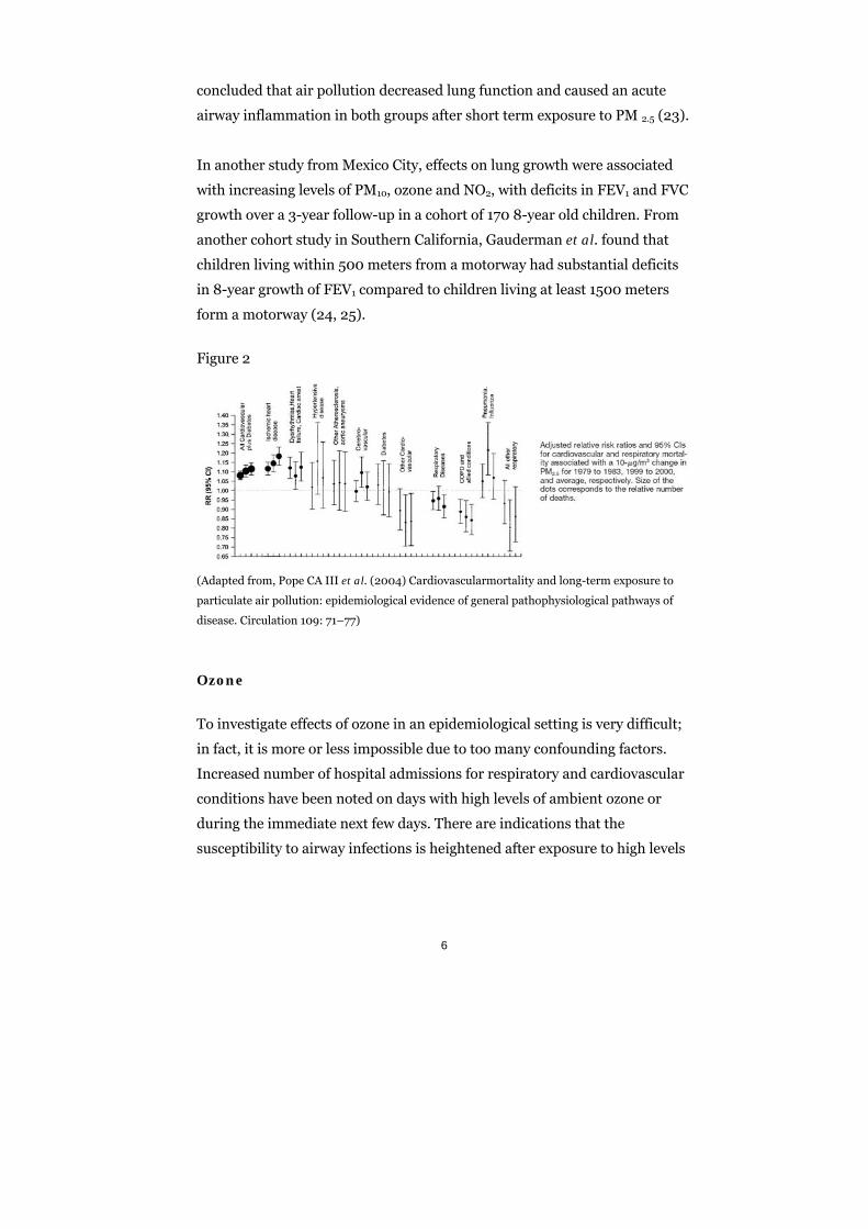
disease. Pope et al. have described an increase in both respiratory and
cardiovascular mortality for every increase in 10 µg/m3 PM10 concentration
(fig 2) (19). In a pivotal study by Peters et al., an increased risk of having a
myocardial infarction two hours after exposure to traffic was shown when
subjects were seeking medical help in emergency rooms after having spent
time in busy traffic (20). Acute exacerbations of COPD and chronic
bronchitis have been associated with short-term exposure to increased levels
of PM (21, 22). In a cohort study from Mexico City Barazza-Villarreal et al.
followed 158 asthmatic and 50 non-asthmatic children over 22 weeks and
6
concluded that air pollution decreased lung function and caused an acute
airway inflammation in both groups after short term exposure to PM 2.5 (23).
In another study from Mexico City, effects on lung growth were associated
with increasing levels of PM10, ozone and NO2, with deficits in FEV1 and FVC
growth over a 3-year follow-up in a cohort of 170 8-year old children. From
another cohort study in Southern California, Gauderman et al. found that
children living within 500 meters from a motorway had substantial deficits
in 8-year growth of FEV1 compared to children living at least 1500 meters
form a motorway (24, 25).
Figure 2
(Adapted from, Pope CA III et al. (2004) Cardiovascularmortality and long-term exposure to
particulate air pollution: epidemiological evidence of general pathophysiological pathways of
disease. Circulation 109: 71–77)
Ozone
To investigate effects of ozone in an epidemiological setting is very difficult;
in fact, it is more or less impossible due to too many confounding factors.
Increased number of hospital admissions for respiratory and cardiovascular
conditions have been noted on days with high levels of ambient ozone or
during the immediate next few days. There are indications that the
susceptibility to airway infections is heightened after exposure to high levels

7
of ozone or smog. Children with asthma are reporting worsening of
symptoms during the days following high levels of ozone.
In a recent study from Portugal, Almeida et al. reported a positive
association between short-term exposure to ozone and cardiovascular
mortality, showing an increase by 0.89 % in total mortality for every increase
in 8-hour average by 5 ppm (26).
Experimental Studies
PM
PM and studies in healthy subjects
Our research group has performed numerous exposure studies with DE and
ozone in healthy subjects. The PM mass concentration has varied from 100
µg/m3 to 350 µg/m3 in different studies. The same setup with an idling Volvo
diesel engine has been used. The studies have included investigations of the
respiratory tract with bronchial wash (BW), bronchoalveolar lavage (BAL)
and endobronchial mucosal biopsies but also of cardio-vascular function. In
the airways, analyses of mucosal biopsies have revealed an inflammatory
response with recruitment of the neutrophils and up-regulated expression of
various proinflammatory cytokines from 6 up to 24 hours after exposure
(27).
The work by Pourazar et al. has described the importance of the epidermal
growth factor receptor, (EGFR) and the signaling cascade of mitogen-
activated protein kinases (MAPKs) as probable trigger mechanism in
regulating the DE-induced airway inflammation (28, 29). Vascular
dysfunction with attenuation of vasodilatation after infusion of vasoactive
drugs and decreased tissue plasminogen activator (tPA) secretion has been
shown after DE exposure with 300 µg/m3 from an idling engine, indicating
that short term exposure to DE impairs two important aspects of vascular

8
function, vasomotor function and endogenous fibrinolysis, and provides a
possible link between air pollution and the pathogenesis of acute myocardial
infarction (MI) (30).
Short-term exposure to DE also enhanced thrombus formation within 2 h
after exposure, associated with increased platelet activation, which provides
an even stronger link between DE exposure and AMI (31).
In a recent study by Mills et al. heart rate variability (HRV) was assessed
after experimental exposure to DE in both healthy subjects and patients with
stable coronary heart disease. Neither patients nor healthy subjects showed
any serious arrhythmias or alterations in HRV (32).
PM and studies in subjects with respiratory disease
In controlled exposure studies in subjects with asthma, a different
inflammatory response has been shown compared to that seen in healthy
subject. An increase in the epithelial expression of the cytokine IL-10 was
found along with an increased bronchial responsiveness 24 hours after
exposure to DE, whereas the preexisting asthmatic inflammation was
unaffected (33). In subjects with COPD there is, to the best of our knowledge,
no published experimental exposure data as of yet. We have performed a
study in subjects with moderate COPD using induced sputum to investigate
airway inflammation and spirometry to assess lung function. Here we could
not find any signs of an aggravated airway inflammation induced by the DE
exposure or any deterioration of the lung function (data not published) (34).

9
PM and studies in subjects with cardiovascular disease
Exposure to DE generated by an idling engine impairs endogenous
fibrinolysis in individuals with a stable coronary heart disease measured as
decreased tPA secretion. When these patients exercised mildly on a bicycle
ergometer where the workload were standardized to maintain estimated
minute ventilation of 15 L/min/m2 body surface a ST-segment depression
was noted very rapidly after the onset of the exercise. All of the patients had
gone through previous PCI and were taking full relevant medication
according to recent guidelines but still a significant effect on the heart was
seen (35). This suggests that even mild exercise during exposure to curb side
levels of DE can cause “micro-ischemia” in the heart muscle. The same
population has been assessed regarding alterations in heart rhythm and
HRV and no such alterations were found (32).
Ozone
Ozone and studies in healthy subjects
There are many experimental exposure studies with healthy subjects exposed
to ozone. In bronchoscopy studies, BAL and BW have shown neutrophilia as
well as an increase of inflammatory mediators such as IL-6, IL-8 and PGE2.
Endobronchial mucosal biopsies have shown an up-regulation of adhesion
molecules such as P-selectin and ICAM-1 as evidence for an early
recruitment of neutrophils into the airways. Lung function studies have
shown reversible FEV1 and FVC decrements after exposure to ozone
concentrations both above and below air quality standards (120 ppb, 1 hour
average, http://www.epa.gov/glo/standards.html). The lung function has
been normalized within 24 hours (36-41).
10
Ozone and studies in subjects with respiratory disease
Studies in asthmatics have failed to show a more aggravated airway
inflammation of ozone compared to healthy subjects. No exaggerated
neutrophil recruitment or exacerbation of pre-existing allergic inflammation
was seen 6 hours after a 2-hour exposure to 200 ppb of ozone (42). This
study suggested that there might be a difference in time-kinetics between
healthy and asthmatic subjects in their response to ozone.
Ozone and studies in subjects with cardiovascular disease
There are to our knowledge no published experimental studies of exposure to
ozone in individuals with cardiovascular disease.
Studies of the combination of ozone and DE
In a study of healthy subjects in which airway responses were investigated by
induced sputum, a significant increase in neutrophilia was shown when
subjects were exposed to ozone preceded by an exposure to DE, but not when
the ozone exposure was preceded by an air exposure. This suggests that
ozone enhances the airway inflammatory response induced by DE (42).
In a vascular assessment of healthy subject using high resolution
ultrasonography of the brachial artery, alterations in the diameter of the
artery were measured after exposure to concentrated ambient fine particles
(CAP) at a concentration of 150 µg/m3 plus ozone 120 ppb and compared to
effects by exposure to filtered air. An acute arterial vasoconstriction by the
combined CAP + ozone exposure was registered (43). This indicates that an
exposure to curbside concentrations of the combination of these two
pollutants can cause alterations in vascular tone which in turn may be one of
the mechanisms explaining the observations from epidemiological studies
linking air pollution to acute cardiac events. So far no studies have addressed
effects on HRV in relation to ozone exposure.

11
Aims
Overall aim;
The aim of this thesis was to investigate the effects on respiratory and
cardiovascular functions in healthy subjects of two of the most powerful
oxidative air pollutants, DE and ozone.
Specific aims;
to investigate whether a real-life sequential exposure to DE and ozone
would enhance an already established airway inflammation caused by DE
to study whether DE generated by an diesel engine running at transient
load consistent with urban driving would induce similar cardiovascular
effects as DE generated at idling
to elucidate whether short-term exposure to ozone would cause
endothelial dysfunction in or affect heart rate variability in healthy
subjects
to evaluate whether FENO measurements at multiple flow rates could be
employed as a biomarker for air pollution-induced airway inflammation in
healthy subjects

12
Subjects and methods
Subjects
All subjects were healthy, nonsmoking individuals with a normal physical
examination, normal spirometry and negative skin prick tests against a
standard panel of ten common aeroallergens. In the first study, 5 women and
9 men were included; whereas study 2, 3 and 4 included men only. None
reported any symptoms from the respiratory tract at least six weeks prior to
the first visit.
All studies were approved by the local Ethics Review Board and all
volunteers gave their written informed consent and the studies were
performed in accordance to the Declaration of Helsinki. The ambition was to
include both men and women. However, this was unfortunately not to be
fully realized.
Study Design
All studies were carried out in a double-blind crossover fashion with two
visits at least two weeks apart, to reduce any possible carry over effects. Each
subject served as his or her own control. During all exposures, the subjects
alternated between rest and mild exercise on a bicycle ergometer in 15
minute intervals. The workload was individualized for each subject to give an
estimated minute ventilation of 20 L/min/m2 body surface.
Study 1
To this study fourteen healthy individuals were recruited. Five women and
nine men, with a mean age of 25 years, all without significant illness and free
from airway infections at least 6 weeks prior to the study were included. The

13
subjects were not allowed to use any non-steroid anti-inflammatory drugs or
vitamins within 2 weeks prior to the first visit and throughout the study.
Each subject was randomized to either DE or air exposure in the morning for
one hour, followed by a 2 hour ozone exposure 5 hours later.
Videobronchoshopy with BW and BAL were performed 24 hours after the
initial exposure. Two to three weeks later the second exposure visit with
subsequent bronchoshopy was performed.
Study 2 and 3
Study flow chart study 3
In total eighteen healthy, men were recruited, with a mean age of 27 years.
Women were not included due to the potential for cyclical hormones to affect
the vascular responses. All individuals were randomized to either air or DE
exposure for 1 hour. Vascular assessment using venous forearm
plethysmography was performed 6-hours after exposure according to a
protocol used in previous studies. A mean of 42 days, range 22-62 days, after
14
the first exposure, a second exposure to DE or air was performed in the same
manner.
All subjects were fitted with Holter electrocardiographic monitors (Reynolds
Medical Lifecard, Delmar Reynolds, United Kingdom) before exposures with
ECG monitoring continuing for 24 hours.
All individuals were asked to refrain from alcohol containing beverages for
24 hours and caffeine drinks for at least 4 hours prior to the exposure. This
was done to secure that uncontrolled effects of vasoconstriction or
vasodilatation caused by alcohol or caffeine was avoided.
Strenuous exercise during the day of exposure was not allowed and the
subjects were asked to stay indoors to avoid any uncontrolled DE exposure.
The vascular studies were carried out in a temperature-controlled room
maintaining a temperature of 22°C to 24°C to ensure as little effect as
possible on temperature-related blood flow alterations.
Study 4
In this study thirty-six men were exposed to ozone and ten to DE. The
unfortunate unequal distribution of subjects between the two exposures was
due to technical problems, beyond our control at the time. Standardized
duplicate Fraction of exhaled nitric oxide (FENO) was measured at the
expiratory flow rates of FENO10, FENO50, FENO100 and FENO270 mL/s before,
at 6 and 24 hours after the end of exposure by using a chemiluminescence
analyzer (NIOX-system; Aerocrine AB; Stockholm; Sweden). All exposures
took place in the morning and all individuals were asked to fast from
midnight. A standardized breakfast containing food without nitrates to
minimize the source for NO production was served after the exposure.
15
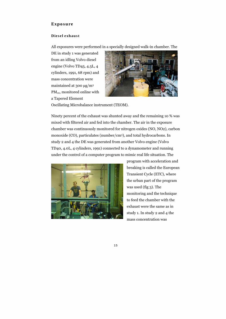
Exposure
Diesel exhaust
All exposures were performed in a specially designed walk-in chamber. The
DE in study 1 was generated
from an idling Volvo diesel
engine (Volvo TD45, 4.5L, 4
cylinders, 1991, 68 rpm) and
mass concentration were
maintained at 300 µg/m3
PM10, monitored online with
a Tapered Element
Oscillating Microbalance instrument (TEOM).
Ninety percent of the exhaust was shunted away and the remaining 10 % was
mixed with filtered air and fed into the chamber. The air in the exposure
chamber was continuously monitored for nitrogen oxides (NO, NO2), carbon
monoxide (CO), particulates (number/cm3), and total hydrocarbons. In
study 2 and 4 the DE was generated from another Volvo engine (Volvo
TD40, 4.0L, 4 cylinders, 1991) connected to a dynamometer and running
under the control of a computer program to mimic real life situation. The
program with acceleration and
breaking is called the European
Transient Cycle (ETC), where
the urban part of the program
was used (fig 3). The
monitoring and the technique
to feed the chamber with the
exhaust were the same as in
study 1. In study 2 and 4 the
mass concentration was

16
maintained at 250 µg/m3 PM10, the lower mass concentration is due to the
running cycle and the different combination of gases.
Figure 3
ETC (Directive 1999/96/EC of December 13, 1999)
Ozone
The ozone was generated by a Fischer’s ozone generator 500 MM (Fischer
Labor and Verfahrens-Technik, Bonn,
Germany) and the chamber
concentration was monitored and
maintained at 200 ppb. All ozone
exposure took place in a walk-in
chamber designed for this particular
purpose.

17
Bronchoscopy
A standard flexible video bronchoscope (Olympus BF 1T 160, Tokyo, Japan)
was introduced orally with the subjects in the supine position. All subjects
had been pre-medicated with 1 mg of atropine subcutaneously. For topical
anesthesia lidocain was used. The subjects received supplementary oxygen
(flow 2L/min) through a nasal cannula throughout the session.
BW was performed by an initial instillation of 2x20 mL sterile saline solution
followed by a BAL with 3x60 mL saline solution. The BAL was performed
either the in lingual or the right middle lobe, with sides randomly selected
and switched to the opposite side at the second visit. The recovered aspirate
from the first and second 20 mL instillations of BW and the collective BAL
fluid were accumulated and immediately placed on ice. The samples were
used for total and differential cell counts as well as examination of soluble
mediators. The BAL fluid was filtered to remove mucus (pore diameter 100
mm; Syntab Product Ab, Malmö, Sweden) and centrifuged at 400xg for 15
min in order to separate cellular components from the supernatant, which
was then divided into aliquots and stored at -80oC until analysis.
Forearm venous occlusion plethysmography
When assessing endothelial
dysfunction the coronary
arteries are of most interest.
However, coronary arteries are
difficult to investigate
invasively but there is a close
relationship between the
responses to the vasoactive
drugs in the peripheral vessels using forearm occlusion plethysmography.
This method can be used as a surrogate and has become the golden standard
18
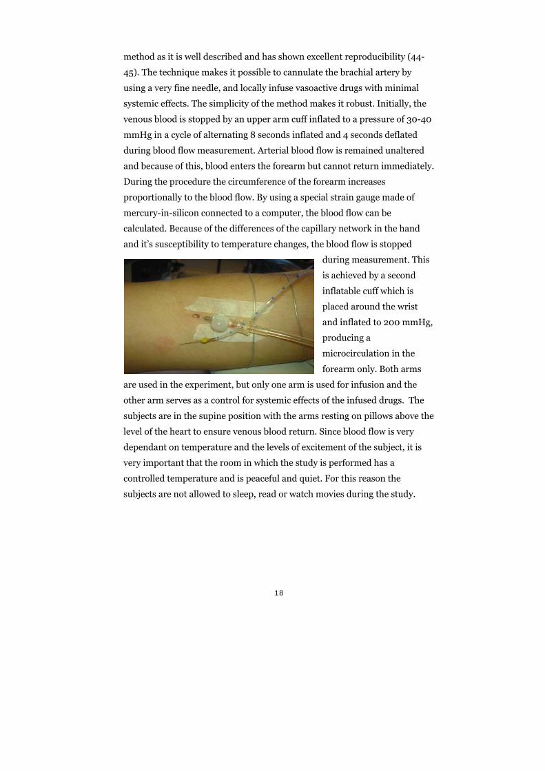
method as it is well described and has shown excellent reproducibility (44-
45). The technique makes it possible to cannulate the brachial artery by
using a very fine needle, and locally infuse vasoactive drugs with minimal
systemic effects. The simplicity of the method makes it robust. Initially, the
venous blood is stopped by an upper arm cuff inflated to a pressure of 30-40
mmHg in a cycle of alternating 8 seconds inflated and 4 seconds deflated
during blood flow measurement. Arterial blood flow is remained unaltered
and because of this, blood enters the forearm but cannot return immediately.
During the procedure the circumference of the forearm increases
proportionally to the blood flow. By using a special strain gauge made of
mercury-in-silicon connected to a computer, the blood flow can be
calculated. Because of the differences of the capillary network in the hand
and it’s susceptibility to temperature changes, the blood flow is stopped
during measurement. This
is achieved by a second
inflatable cuff which is
placed around the wrist
and inflated to 200 mmHg,
producing a
microcirculation in the
forearm only. Both arms
are used in the experiment, but only one arm is used for infusion and the
other arm serves as a control for systemic effects of the infused drugs. The
subjects are in the supine position with the arms resting on pillows above the
level of the heart to ensure venous blood return. Since blood flow is very
dependant on temperature and the levels of excitement of the subject, it is
very important that the room in which the study is performed has a
controlled temperature and is peaceful and quiet. For this reason the
subjects are not allowed to sleep, read or watch movies during the study.

19
Fraction of exhaled nitric oxide (FENO)
After an at least 4-hour fast,
all individuals were given a
standardized breakfast, free of
nitrates. FENO was then
measured before, at 6 hours
and 24 hours after exposure to
DE, ozone or air according to
the ATS/ERS guidelines (46).
The measurement was
performed by using a chemiluminescence analyzer (NIOX-system; Aerocrine
AB; Stockholm; Sweden) and exhaled nitric oxide was measured during a
slow single exhalation against an oral pressure of 5 cm of H2O. Four different
flow rates were used FENO10, FENO50, FENO100 and FENO270 mL/s (± 10%).
Each exhalation lasted for 10 seconds and FENO was measured during the
6th to the 10th second of the exhalation phase. Duplicate measurements were
secured, all within a 10% variation, and the mean concentration of nitric
oxide (NO) in parts per billion (ppb) was registered.
Heart rate variability (HRV)
All subjects were fitted with Holter electrocardiographic monitors (Reynolds
Medical Lifecard, Delmar Reynolds, United Kingdom) before exposures and
ECG monitoring continued for 24 hours.
To assess the acute effects of exposure on heart rate variability, the subjects
were asked to rest supine in a quiet, temperature controlled room
maintained at 22-24ºC for 20 min immediately prior to, and 2 and 6 hours
following the start of each exposure. All volunteers also underwent blood
pressure measurements and a vascular assessment.

20
Electrocardiographic recordings were analysed using the Reynolds Medical
Pathfinder Digital 700 Series Analysis System (Delmar Reynolds, United
Kingdom). An experienced single operator, blinded to both subject
characteristics and exposure, verified any abnormal rhythms and performed
manual editing of aberrant beats and electrical interference prior to
generating RR data tables. If more than 95% of the RR data was valid, the
recording was analysed. RRdata were analysed using the HRV Tools software
package (DelMar Reynolds, United Kingdom).
Standard time-domain measures were calculated including mean NN-
interval (time interval between consecutive sinus beats), standard deviation
of NN-interval values (SDNN; an index that expresses overall variability),
percent successive NN-interval differences >50 ms (PNN50), root-mean-
square of successive NN-interval differences (RMSSD) and the triangular
index (an estimate of overall heart rate variability). SDNN, PNN50 and
RMSSD are measures of high-frequency variation mediated primarily by the
vagus nerve. Frequency domain analysis determined the low frequency (LF;
0.1 Hz) and high frequency (HF; 0.25 Hz) components of the power
spectrum in absolute values of power (ms2). LF and HF were also expressed
in normalized units (LFn and HFn), to account for variation in the total
power and very low frequency components, as well as the HF/LF ratio.
21
Main results
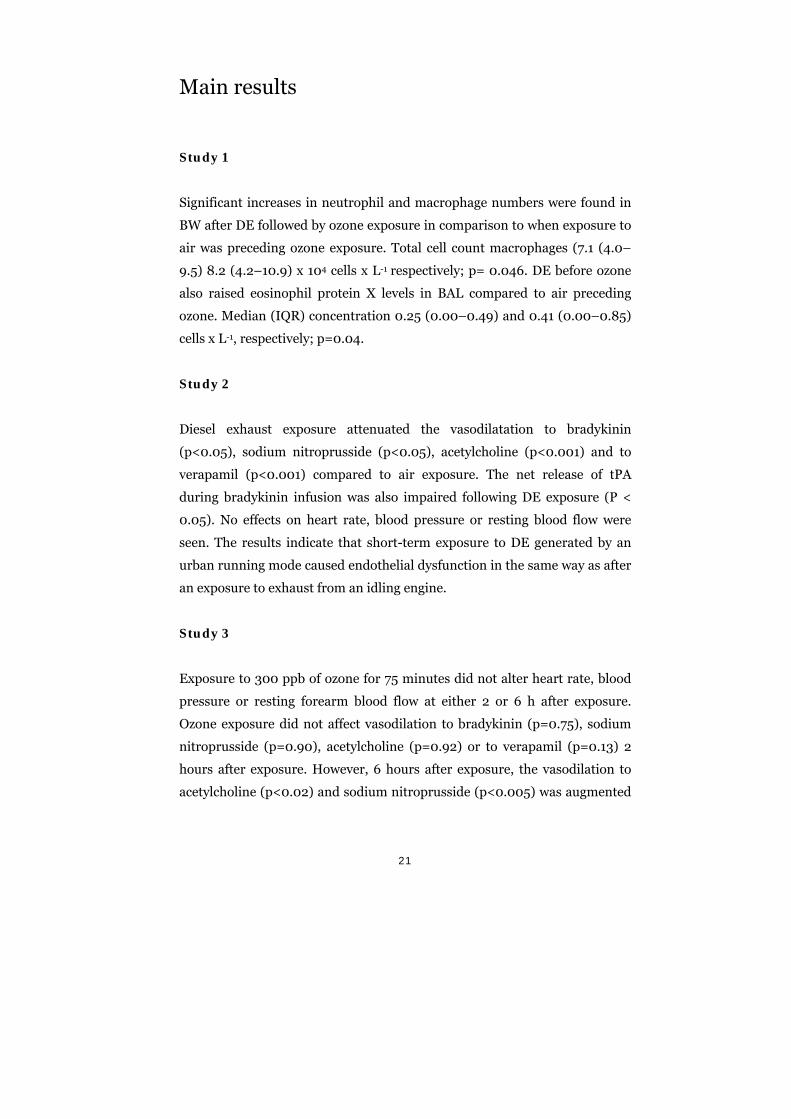
Study 1
Significant increases in neutrophil and macrophage numbers were found in
BW after DE followed by ozone exposure in comparison to when exposure to
air was preceding ozone exposure. Total cell count macrophages (7.1 (4.0–
9.5) 8.2 (4.2–10.9) x 104 cells x L-1 respectively; p= 0.046. DE before ozone
also raised eosinophil protein X levels in BAL compared to air preceding
ozone. Median (IQR) concentration 0.25 (0.00–0.49) and 0.41 (0.00–0.85)
cells x L-1, respectively; p=0.04.
Study 2
Diesel exhaust exposure attenuated the vasodilatation to bradykinin
(p<0.05), sodium nitroprusside (p<0.05), acetylcholine (p<0.001) and to
verapamil (p<0.001) compared to air exposure. The net release of tPA
during bradykinin infusion was also impaired following DE exposure (P <
0.05). No effects on heart rate, blood pressure or resting blood flow were
seen. The results indicate that short-term exposure to DE generated by an
urban running mode caused endothelial dysfunction in the same way as after
an exposure to exhaust from an idling engine.
Study 3
Exposure to 300 ppb of ozone for 75 minutes did not alter heart rate, blood
pressure or resting forearm blood flow at either 2 or 6 h after exposure.
Ozone exposure did not affect vasodilation to bradykinin (p=0.75), sodium
nitroprusside (p=0.90), acetylcholine (p=0.92) or to verapamil (p=0.13) 2
hours after exposure. However, 6 hours after exposure, the vasodilation to
acetylcholine (p<0.02) and sodium nitroprusside (p<0.005) was augmented

22
whilst the vascular response to bradykinin and verapamil were unaffected.
The subjects did not experience any symptoms or serious arrhythmias
during either exposure or during the 24 hour study period. Exposure to
ozone did not affect time or frequency domain measures of heart rate
variability over the 24-hour period. There were no differences in either time
or frequency domain measures of heart rate variability at 2 or 6 hours
between exposures.
Study 4
Exposure to diesel exhaust for one hour increased FENO at 6 hours
compared to filtered air at expiratory flow rates of 10 mL/s (mean±SEM
60.8 ± 6.0 ppb versus 50.2 ± 5.9 ppb; P=0.01) and at 50 mL/s (18.6 ± 1.6
ppb versus 15.9 ± 1.5 ppb; P=0.011), but concentrations had normalised at
24 hours. FENO concentrations were not affected by diesel exhaust exposure
or filtered air at FENO100 or FENO270, the two higher flow rates.
Exposure to ozone did not affect FENO at any flow rate or time point in
either cohort of healthy volunteers.

23
Statistics
Study 1
Cell count and soluble protein data of filtered air followed by ozone versus
DE followed by ozone were analyzed with Wilcoxon Signed-Rank Test, a
non-parametric test for paired observations. Correlations were assessed
using the Spearman correlation test. A p-value less than 0.05 were
considered statistical significant. Values are presented as medians with
interquartile ranges (IQRs).
Study 2 and 3
Plethysmographic data were analyzed as described previously. The net
release of tPA antigen was defined as the product of the infused forearm
plasma flow (based on the mean hematocrit and the infused FBF) and the
concentration difference between the infused and non-infused arms as
described previously (44). Data were analyzed by 2-way ANOVA with
repeated measures or 2-tailed Student’s t-testswhere appropriate, using
GraphPad Prism (GraphPad Software, Version 4 for Macintosh) and SPSS
(SPSS inc. Chicago, IL, USA, version 15).
Study 4
Data are presented as mean ± SEM. A repeated measures analysis of
variance (General Linear model) with two within-subject factors (time and
exposure) was used, with pre-exposure FENO data used as reference using
SPSS, version 16.0 (SPSS Inc., Chicago, IL, USA). In order to avoid type-I
errors due to two comparisons, the level of significance was adjusted by
dividing the set significance level by two (Bonferroni correction) and
therefore statistical significance was taken at P<0.025.
24
Discussion
Air pollution is of major global concern. In the years to come, it is evident
that air pollution will be an even larger problem when it comes to the “new”
economies and their potential to contribute to bad air quality. Efforts have
been made to find ways to reduce PM in ambient air. The introduction of
retrofit exhaust traps (47) and legislations against private driving in city
traffic are possibilities discussed. The London Summer Olympics in 2012 will
be a challenge for research on how to reduce PM in a large city, where smog
development is prone.
It is essential to elucidate the underlying mechanisms behind the reported
excess in mortality and morbidity associated with increasing air pollution. As
a scientific community, we need to provide the legislators with safe air
pollution threshold levels and evaluate risks of exposure to different air
pollutants and their possible interactions in ambient air. Epidemiology only
provides data on associations between air pollutants and health effects,
whereas experimental research is crucial to elucidate the underlying
biological mechanisms. The present thesis is based on research dealing with
associations provided by epidemiology.
The first study is based on the assumption that ozone and diesel exhaust
interact in causing airway inflammation. Since the two air pollutants usually
coexist and as ozone levels commonly peak in the afternoon, a previous study
protocol was designed to mimic ambient conditions. Subjects were exposed
to DE in morning (when they go to school or work) and ozone or air in the
afternoon (on returning home). In that study Bosson et al. showed that
ozone exposure magnified the DE-induced airway inflammation assessed by
induced sputum (42). In the present study, we exposed the subject to DE or
air in the morning followed by ozone exposure in the afternoon, in order to
elucidate whether pre-exposure to DE would enhance the ozone-induced
airway inflammatory responses. Bronchoscopy was performed the next

25
morning and showed an increase in BW-neutrophils when exposure to DE
was carried out pre-ozone exposure, indicating an additional effect. These
two studies cannot be fully compared, since the assessments were different
(induced sputum vs. bronchoscopy) but both studies show an increase in
inflammatory markers when subjects were exposed to an additional
pollutant. These results indicate the need for further studies on combined air
pollution exposures. The study presented in this thesis was designed as a
sequential exposure but it would have been interesting to perform an
experimental exposure study with both ozone and DE simultaneously, as in
real life. There are logistical and toxicological issues concerning a
simultaneous exposure study. The ozone would rapidly oxidize the surface
components in a chamber environment and probably change their oxidative
potential. Another problem would be to determine what exposure levels to
employ. Logistically, such a study design would be very complicated as it
would require four study arms in a double-blind and cross-over fashion; air,
air + DE, air + ozone, DE + ozone, which would be both expensive and time-
consuming.
The second study deals with the fact that, so far, all DE exposure studies
employing cardiovascular endpoints have been performed with the diesel
engine operating at idling conditions. In real-life however, exposure to DE
curbside mainly arises from moving trucks and cars and not primarily from
idling engines. The PM chemical and physiological properties differ between
the exhaust from these two running conditions. The PM from an idling
engine exhaust is richer in organic carbon, whereas the PM from transient
running engines mainly consists of elemental carbon, i.e. soot. These
different characteristics might have different effects on humans. Sehlstedt et
al. showed an increase in eosinophils in BAL after exposure to DE generated
during the ETC compared to air, something which has not been reported
after exposure to DE during idling (49). This study, although comparisons
are made to historical findings, suggests a different airway inflammatory
response between the two running conditions. We therefore designed a study
similar to the previous by Mills et al. but employed exposure to DE

26
generated during transient running, i.e. the urban part of the ETC (30) to
further investigate this issue.
The study showed that the vascular effects were similar between the two
engine running modes, however, not identical. In the previous studies,
vasodilatation during infusion of BK, ACH and SNP was attenuated after DE
exposure, whereas vasodilatation was not affected during VP-infusion. As
several studies employing DE exposure at idling engine conditions show no
attenuation of the vasodilatation during VP-infusion, this suggests, although
close to statistical significance in one study (p-value just above 0.05), that
this is a true lack of VP response to exposure to DE generated during idling
(32, 35, 47, 48, 50). The present study (study II) showed a similar vascular
response after DE exposure during transient running, but with attenuated
vasodilatation during VP-infusion as well. In a recent study by Lucking et al.
(47), the same protocol was employed and resulted in an identical
attenuation of vasodilatation during VP-infusion, indicating that this is a
true effect and a possible difference in response to DE generated by the two
different engine running modes.
Verapamil exerts its effects by blocking the calcium channels in smooth
muscles in the blood vessels (51). The VP effects seen after exposure to DE
generated during transient running could be associated with exhaust
properties and different chemical composition of the particles themselves,
for example, a higher content of soot and absorbed organic compounds.
Although the mass concentration between the two running modes was
similar, the particles were larger and fewer in number and had a lower
oxidative potential during the transient running mode. The PAH content was
also lower compared to the idling situation and the gaseous components
differed with higher NOx levels during the transient running mode. Whilst
there are some differences in the effects on the vasculature between idling
and transient running conditions, the main effects, impairment of vascular
function and endogenous fibrinolysis, were similar. This finding is clinically

27
relevant since it provides a possible link between increases in air pollution
and increased cardiovascular morbidity.
In a recent study by Mills et al., in which healthy subjects were exposed to
diesel exhaust, pure carbon nano-particulate, filtered DE and filtered air,
vascular effects were shown only after DE exposure, suggesting, that the
adverse vascular effects are mediated by combustion-derived DE particles
(50). In that study, the DE was generated by a generator engine powered by
diesel fuel and operated at idling. As in previous studies employing the idling
situation, there was no attenuation of vasodilatation during infusion of VP
(50).
It has been hypothesized that the adverse effects of DE exposure on the
vascular endothelium is caused by the oxidative stress exerted by the PM
itself or by its surface components (5). The oxidative stress produces
superoxide radicals which form peroxynitrite with endothelial NO. This
reaction leads to consumption of NO and thus, reduces NO-bioavailability
(30, 52).
Thus, as the effects on the vasculature are similar in five studies (30, 47, 48,
50) three studies employing an idling engine and two running at a transient
mode, it can be concluded that the adverse vascular effects are comparable
regardless of engine or running mode. This suggests that the particle fraction
of the exhaust is the culprit.
In the third study, it was found that a brief exposure to ozone did not impair
vascular function or affect heart rate variability. Rather, the vasomotor
response to two of the infused agonists was enhanced 6-8 hours after ozone
exposure. These findings are in contrast to previous DE studies and suggest
that the increase in cardiovascular events and mortality demonstrated after
exposure to ozone and motor engine exhaust in traffic is mediated through
different pathways and mechanisms.
28
Previously, experimental human studies have shown that exposure to ozone
increases the oxidative burden in the airways and trigger a pronounced
neutrophil-dominated inflammatory response (37, 53-55). Already at 1.5
hours after ozone exposure, the expression of P-selectin was up-regulated in
the bronchial endothelium. This was followed by the up-regulation of
endothelial ICAM-1 expression and inflammatory cell recruitment into the
bronchial mucosa and bronchoalveolar spaces at 6 hours, with the
inflammatory response peaking around 12-24 hours following exposure (37,
55). The ozone-induced airway responses coincide with the time course of
the development of airway inflammation following exposure to DE, and
include the up-regulation of vascular endothelial adhesion molecule
expression and a neutrophil-dominated inflammation, accompanied by
CD4+ and CD8+ cell influx (27).
The systemic inflammation seems to be more pronounced after ozone than
after diesel exhaust. In a study by Bosson et al. healthy subjects were
exposed to 200 ppb of ozone for 2 hours, and results showed a significant
increase in peripheral blood neutrophils 6 hours after exposure compared to
air (unpublished data) (56). This is in contrast to a study in which DE
exposure did not cause systemic neutrophilia but increased serum levels of
TNF-alfa and IL-6 24 hours after exposure (48).
We have previously reported that diesel engine exhaust generated during
idling or transient running conditions caused endothelial dysfunction,
increased arterial stiffness, impaired endogenous fibrinolysis, increased
platelet activation, and enhanced ex-vivo thrombus formation in human
subjects 6 hours after exposure, with some vascular effects maintained up to
24 hours (30, 48, 57). We have also demonstrated that patients with
coronary heart disease experienced ST-segment depression when exercising
during exposure to diesel exhaust as compared with air, despite stable
disease conditions and relevant preventive medication (35). These effects are
consistent with the reported immediate and delayed association between
traffic exposure and the onset of acute myocardial infarction (MI) (20).
29
The development of MI after ozone exposure has not been described in detail
as after traffic exposure. There are studies in which ozone has been linked
with MI and mortality during the day of exposure. Because of similarities in
the airway oxidative and inflammatory responses between ozone and diesel
exhaust, it has been proposed that vasomotor and thrombogenic functional
changes would potentially also occur in parallel (26, 58). Associations
between ozone and mortality are difficult to disentangle from the effects of
PM, and ozone and particles do indeed interact to enhance airway
inflammation as previously reported (42). Whilst effects on airway inflammation induced by ozone and diesel exhaust
exposure show several similarities, there are some important aspects to
address. The DE effect is thought to be mediated via epidermal growth
factors receptor (EGFR), tyrosine 1173 transphosphorylation, and redox-
sensitive elements causing NFκB activation and the subsequent up-
regulation of IL-8, GRO-alpha, ICAM-1 and NOS II in the bronchial mucosa
(29, 59). Ozone appears to react directly with components of the respiratory
tract lining fluid (RTLF) and leads to the production of secondary free-
radical-derived ozonation products that cause cellular damage (60). Exposure to ozone does not seem to involve NFκB-activation in the bronchial
epithelium in humans (61). In contrast to the study hypothesis, an ozone-induced increased vasomotor
response to acetylcholine and sodium nitroprusside was demonstrated. This
suggests a maintained or even improved NO bioavailability after ozone
exposure. In some studies, oxidative stress has been shown to increase NO
bioavailability through NOS III activation (62-63).
In the fourth study, FENO was, for the first time, introduced at multiple flow
rates in experimental exposure studies employing DE and ozone. The
rationale for this, is that the various flow rates are thought to reflect different
parts of the bronchial tree, with the lower flow rates (FENO10 and FENO50)

30
corresponding to the central or bronchial airways and the higher flow rate
(FENO270) to the distal airways (64).
Healthy subjects exposed to DE for one hour showed an increase in FENO at
6 hours after exposure. Only FENO concentrations obtained at lower flow
rates (FENO10 and FENO50) were affected, suggesting that the central
airways, but not the peripheral airways, are principally involved. This
observation is in accordance with previous studies, in which diesel exhaust
induced increases in inflammatory cells and cytokines in mucosal biopsies
and BW obtained from the central airways, but not in BAL (65-66).
In contrast, exposure to ozone did not affect FENO at any flow rate or any
time point. To increase the power of the study to detect a small ozone-
induced effect on FENO, the study was repeated and the number of subjects
was increased from 18 to 36, but still no consistent effect of ozone on FENO
in either cohort or in the combined data set could be detected. These findings
are consistent with two previous studies in healthy subjects, suggesting that
exposure to ozone does not affect FENO (67-68).
Associations reported between exposure to ozone and FENO in field studies
are in contrast with our findings (69). In real life, people can be exposed to
ozone repeatedly during many days but also to a mixture of many air
pollutants. As it is impossible to distinguish the effects of a single air
pollutant in field studies, it is not surprising that augmented effects on
airway inflammation are seen when exposures to different air pollutants are
combined (42). When it comes to FENO, a single ozone exposure does not
seem to cause a noticeable acute effect as seen after DE.
Exhaled NO production is thought to be under the regulation of three
endothelial NOS isoforms. NOS I and II are predominantly expressed in
healthy subjects, while NOS II, the corticoidsteroid-sensitive inducible NOS,
is up-regulated in patients with asthma. Recently, there is evidence that
exhaled NO is associated with a genetic variant of NOS III in patients with
31
asthma, suggesting that both NOS II and NOS III are important in
determining the exhaled NO in this patient group (70). Endothelial nitric
oxide synthetase (NOS III) is regulated under the influence of the oxidative
stress-sensitive transcription factor NFκB (71).
Both diesel exhaust and ozone are considered oxidant air pollutants and
exert their effects on the airways through oxidative stress (5, 72-73). The
ozone molecule is highly reactive and does not reach the airway epithelium
but reacts with components in the respiratory tract lining fluid and causes a
cascade of secondary free radical-derived ozonation products (60), whereas
the DEPs are deposited on the airway epithelium and induce a local
inflammatory response and may also translocate to affect the local vascular
endothelium (5).
NFκB activation and up-regulation of NOS III occur in endothelial cells
exposed to reactive oxygen species (74). NFκB, along with AP-1 and
MAPkinases, are activated by diesel exhaust exposure (10), and in turn this
might lead to up-regulation of NOS III. Exposure to ozone has not been
found to activate NFκB in human airway epithelium, suggesting a different
induction of airway inflammatory responses between ozone and DE (14).
These observations suggest that ozone exposure may not influence FENO, as
it does not by itself activate NFκB and up-regulate NOS III.
Mehta et al studied the levels of exhaled NO following infusion of the NO
precursor L-Arginine and found increased levels indicating that exhaled NO
may reflect endogenous production of NO (75). Interestingly, the baseline
concentrations and changes in exhaled NO were similar in that study to the
increase in FENO following exposure to diesel exhaust in the present study.
Previously, we have hypothesized that the DE-induced oxidative stress and
the subsequent adverse cardiovascular health effects are mediated through
reduced NO bioavailability (30). Oxidative stress caused by exposure to DE
and subsequent consumption of vascular NO may evoke homeostatic

32
mechanisms to normalize vascular function through the up-regulation of
NOS III, which in turn may increase FENO.
All the studies in this thesis contend with experimental exposure to oxidative
air pollutants. Sequential exposure of DE and ozone has been applied. To
mimic real-life urban driving a novel mode to generate DE for exposure has
been used. Both the cardiovascular system and the airways have been
investigated. In the airways, ozone-induced inflammation seems to be more
pronounced after sequential exposures, but ozone alone does not increase
the inflammatory marker FENO or cause endothelial dysfunction, as shown
after exposure to DE. This suggests different underlying mechanisms and
pathways and necessitates further research.
The time-points and exposure levels applied in this thesis have been chosen
based on previous investigations. All air pollutant exposures are equivalent
to high ambient levels, but realistic real-life levels, as they often can be
measured curbside or in tunnels etc. during rush hour traffic. All studies
were performed in healthy subjects. The first study included both men and
women, since it was a bronchoscopy and not a vascular assessment study. In
the second and third studies only men took part as it is well known that the
cyclic changes of hormones in women clearly affect vascular function,
especially fibrinolysis. Another important detail is the difficulty to cannulate
the brachial artery in women. As in other clinical research the fact that
women often are excluded limits the possibility to draw general conclusions.
An additional limiting factor is that only healthy subjects were included in
these studies and thus, it cannot be excluded that other effects might be
relevant in patients with asthma, COPD and cardiovascular diseases.

33
Final comments
The research group to which I belong has conducted human exposure studies
since the late 1980’ies. The studies have included exposure to sulphur
dioxide, nitrogen dioxide, ozone and diesel exhaust generated by a truck
engine, both during idling and transient running conditions. The exposures
have been performed in well validated walk-in exposure chambers and
because of the vast experience that our staff has acquired over the years, the
technical standard of the studies has been kept high.
In this thesis, exposures to the highly oxidative air pollutants ozone and
diesel exhaust have performed and both respiratory and cardiovascular
effects have been assessed. The respiratory effects were investigated by
means of bronchoscopy with bronchial wash and bronchoalveolar lavage,
methods which have been shown to be valid and highly reproducible.
The use of venous occlusion forearm plethysmography to assess vascular
function has been established through a very productive collaboration with
the Department of Cardiovascular Research in Edinburgh. This method is
thoroughly validated and robust as an invasive technique. The effects of
vasodilation and excretion of tPA in the forearm circulation, i.e. brachial
artery, is closely associated with the effects in the coronary circulation which
makes this method very appropriate to assess cardiovascular endpoints after
exposure to air pollution. A standard method for evaluating heart rate
variability has also been acquired.
The study results bring novel insights into this research field. Whilst the
airway inflammatory response after exposure to DE or ozone is similar, there
are significant differences in the response in the vasculature and in FENO
suggesting different mechanisms and pathways for inducing the airway
inflammation. It is important to reduce combustion-derived PM in ambient
air and regardless how the particulate is generated, it still has a big impact
on human health. An acute single exposure to ozone does not alter the heart

34
rate variability or rhythm, which is one of the hypotheses for the association
between mortality and increases in ambient ozone levels. This could be
crucial information for legislators when it comes to recommendations on air
quality.
For the first time, airway inflammation by means of fraction of exhaled nitric
oxide, FENO, measured at multiple flow-rates, was used in an experimental
ozone and DE exposure study. The significant results in FENO changes after
DE exposure in healthy humans are too small to have a significant clinical
impact. This raises the question of the mechanisms behind the association
between increased FENO and increased levels of air pollution. This study
emphasizes the importance of PM. Since there is a strong association
between PM and increasing levels of ozone and NO2, it cannot be excluded
that the measured air pollution-related effects on FENO are due to PM2.5-10 or
the combination of PM-ozone and not to ozone alone. For research purposes
and speculations on underlying mechanisms FENO still may play a role.
Studies on FENO are mainly performed in asthmatic subject showing an
augmentation of FENO in relation to increases in symptoms. Human
exposure studies employing different patient groups are needed to clarify the
mechanisms behind the reported adverse health effects. Vascular studies
following exposure to DE, ozone or the combination are needed, but
logistically problematic in patients. It is known that the ischemic burden
increases in patient with coronary heart disease when they are exercised
during exposure for DE. The cardiovascular impact of ozone in patients with
coronary disease is unknown, but given the lack of response in young healthy
subjects, perhaps such a study has a low priority at present.
Patients with COPD comprise a very heterogenetic group with different
clinical phenotypes. Therefore such studies would be difficult to interpret,
but it is a well known fact that COPD patients have an increased
cardiovascular morbidity and mortality compared to smokers without COPD
(76).
35
This thesis has determined that it is essential to reduce PM in ambient air.
Studies with intervention have already been conducted. Rudell et al. showed
an attenuation of symptoms using a cabin air-inlet filter in cars (77) and the
results have been confirmed in a very recent study (unpublished data).
Furthermore, Lucking et al. have shown that a retrofit particle trap reduces
the endothelial dysfunction and the increased prothrombotic effect seen
after exposure to DE (57).
This suggests that models for PM reduction are effective and that the
hypothesis that PM and its chemical and physiological properties are driving
the adverse health effects, is plausible and that ozone might act as a booster
of these effects.

36
Conclusions
It is hereby concluded that:
Exposure to ozone preceded by exposure to diesel exhaust at levels
that can be encountered curb-side, enhances the already established
DE-induced neutrophil-dominated airway inflammation in healthy
subjects
Exposure to diesel exhaust generated by an engine running at a
transient mode causes a similar vascular response as exposure to
diesel exhaust generated during idling
An acute single exposure to ozone does not cause endothelial
dysfunction or alterations in heart rate variability in healthy subjects
Fraction of exhaled nitric oxide, FENO, measured at multiple flow-
rates, may be used as a marker of acute diesel exhaust-induced, but
not ozone-induced, airway inflammation
37
Acknowledgements The work behind this thesis is of course not possible to achieve without the help of a great team. I hereby wish to thank everybody who has supported me throughout the years:
Ellinor Ädelroth, head of the Department of Public Health and Clinical Medicine, my main supervisor and colleague at the division for Respiratory Medicine and Allergy. Thank you for all support and help. Your knowledge in research is great and just to bring me a little of your insights has helped me a lot. Without your pads on the back I seriously don’t think this thesis would have been finished.
Anders Blomberg, my co-supervisor and colleague, your help has been enormous; I really appreciate that you always could find time for me.
David Newby, my co-supervisor, thank you for the inspiration and making this kind of research possible for us.
Thomas Sandström, head of our research group and colleague, thank you for inspiring me to do research and giving me the opportunity to meet all these wonderful people in the collaborating groups.
Nick Mills, thank you for all help in all practical things, the theoretical discussions as well as writing the papers.
Jeremy Langrish, for the friendship and help in both conducting studies and interpreting the results.
Andrew Lucking, thank you for all the help in learning new techniques and your generosity.
Ragnberth Helleday, head of the Department of Medicine and a colleague for making my research possible and for help with the layout.
All colleagues and staff at the Division for Respiratory Medicine and Allergy, without your support it wouldn’t have been possible to do research, my colleagues who do so much work and of course have to do a bit of mine as well during these years.

38
Anna-Carin Olin, thanks for all useful comments on exhaled nitric oxide, “en fena på FENO”
Jenny Bosson Damewood and Annelie Behndig, research colleagues, for support and fruitful discussions.
Jamshid Pourazar, Esther Roos-Engstrand, Maria Sehlstedt and Ann-Britt Lundström, for excellent laboratory work.
Maj-Cari Ledin and the staff at Svensk Maskinprovning for all technical support.
Annika Johansson, Frida Holmström, Margot Johansson and Veronika Sjögren, our research nurses, without you there wouldn’t be any research. Annika “the flame”, thank you for all the “frogs” that just jump out, especially on Thursdays.
Lena Åström, our excellent secretary who keeps track of just about everything.
Håkan Törnqvist, Magnus Lundbäck, Manuel Gonzales, Jon Unosson and Nirina Larsson, research colleagues over the years, thank you for all the fun we have had and mainly for all discussions.
Umeå Universitet
All friend and relatives: for just being there.
My daughter Alexandra and son Christian for being my joy in life
Frida and David for lighten up the day
Anna, my beautiful wife, thank you for putting up with me and all this work, your support through all these days when I’m not at home, I love you, with all my heart.
This work has been supported by grants from the Swedish Heart-Lung Foundation, the Swedish Research Council for Environment, Agricultural Sciences and Spatial Planning (FORMAS), the County Council of Västerbotten, the Swedish National Air Pollution Program and Umeå University.

39
References
1. Air pollution in the world's megacities: a report from the U.N. Environment Programme and WHO. Report No: 36.
2. Ministry of Health, Mortality and morbidity during the London fog of December 1952. London: HMSO, 1954. Reports on public health and medical subjects No 95.
3. Logan WP Mortality in the London fog incident, 1952. Lancet I 1953 336-338.
4. Brook RD Cardiovascular effects of Air pollution, Clinical Science (2008) 115 175-187.
5. Mills NL et al. Adverse cardiovascular effects of air pollution, Nature clinical practice cardiovascular medicine, January 2009 vol 6 no 1.
6. Dockery DW et al. An association between air pollution and mortality in six U.S. cities, N Engl J Med. 1993 Dec 9; 329 (24):1753-9.
7. Alfaro-Moreno E et al. Particulate matter in the environment: pulmonary and cardiovascular effects. Curr Opin Pulm Med. 2007 Mar; 13 (2):98-106.
8. Zelikoff JT et al. A role for associated transition metals in the immunotoxicity of inhaled ambient particulate matter, Environ Health Perspect. 2002 Oct; 110 Suppl 5:871-5.
9. Hesterberg TW et al. Non-cancer health effects of diesel exhaust: a critical assessment of recent human and animal toxicological literature. Crit Rev Toxicol. 2009; 39 (3):195-227.
10. Donaldson K et al. Combustion-derived nanoparticles: a review of their toxicology following inhalation exposure. Part Fibre Toxicol 2: 10 (2005).
11. Nel A et al. Toxic Potential of Materials at the Nanolevel, Science 311, 622 (2006).

40
12. Duffin R et al. Nanoparticles-a thoracic toxicology perspective, Yonsei Med J. 2007 Aug 31; 48(4):561-7.
13. Pope CA III and Dockery DW 1999. Epidemiology of particle effects. In: Air Pollution and Health (Holgate ST, Koren HS, Samet JM, Maynard RL, eds). London: Academic Press, 673–705.
14. U.S. EPA Air quality criteria for particulate matter (EPA/600/P-99/002aF), U.S Environmental Protection Agency, Washington DC (2004).
15. Pope CA III et al. Cardiovascular mortality and long-term exposure to particulate air pollution: epidemiological evidence of general pathophysiological pathways of disease. Circulation 109: 71–77 (2004).
16. Brook RD et al. Particulate Matter Air Pollution and Cardiovascular Disease: An Update to the Scientific Statement From the American Heart Association, Circulation 2010;121;2331-2378.
17. Schikowski T et al. Long-term air pollution exposure. Respir res. 2005.
18. Wallace J et al. Association between proximity to major roads and sputum cell counts. Can Respir J. 2011 Jan-Feb; 18 (1):13-8.
19. Pope, CA and Dockery DW Health effects of fine particulate air pollution: lines that connect. J. Air Waste Manage. Assoc. 56, 709–742 (2006).
20. Peters A et al. Exposure to traffic and the onset of myocaedial infarction N Engl J Med 2004; 351: 1721-30.
21. Ling SH and van Eeden SF, Particulate matter air pollution exposure: role in the development and exacerbation of chronic obstructive pulmonary disease. Int J Chron Obstruct Pulmon Dis. 2009; 4: 233-43. Epub 2009 Jun 11.
22. Sint T et al. Ambient air pollution particles and the acute exacerbation of chronic obstructive pulmonary disease. 2008 Inhal Toxicol. Jan; 20 (1):25-9.
41
23. Barraza-Villarreal A et al. Air Pollution, Airway Inflammation, and Lung Function in a Cohort Study of Mexico City Schoolchildren, Environ Health Perspect. 2008 Jun; 116 (6):832-8.
24. Rojas-Martinez R et al. Lung Function Growth in Children with Long-Term Exposure to Air Pollutants in Mexico City, Am J Respir Crit Care Med Vol 176. pp 377–384, 2007
25. Gauderman WJ et al. Effect of exposure to traffic on lung development from 10 to 18 years of age: a cohort study, Lancet. 2007 Feb 17;369(9561):571-7
26. Almeida SP et.al. Short-term association between exposure to ozone and mortality in Oporto, Portugal. Environ Res. 2011 Apr; 111(3): 406-10.
27. Salvi S et al. Acute inflammatory responses in the airways and peripheral blood after shortterm exposure to diesel exhaust in healthy human volunteers. Am J Respir Crit Care Med 1999 159: 702–709
28. Pourazar J et al. Diesel exhaust increases EGFR and phosphorylated C-terminal 1173 in the bronchial epithelium. Part Fibre Toxicol 2008; 5:8.
29. Pourazar J et al. Diesel Exhaust activates redox-sensitive transcription factors and kinases in human airways. Am J Physiol Lung Cell Mol Physiol 2005; 289: L724-30.
30. Mills NL et al. Diesel exhaust inhalation causes vascular dysfunction and impaired endogenous fibrinolysis. Circulation 2005 112: 3930–3936.
31. Lucking AJ et al. Diesel exhaust inhalation increases thrombus formation in man, Eur Heart J. 2008 Dec; 29 (24):3043-51.
32. Mills NL et al. Diesel exhaust inhalation does not affect heart rhythm or heart rate variability. Heart. 2011 Apr; 97 (7): 544-50.
33. Stenfors N et al. Different airway inflammatory responses in asthmatic and healthy humans exposed to diesel Eur Respir J. 2004 Jan; 23 (1):82-6.
42
34. Törnqvist H Respiratory and cardiovascular responses to diesel exhaust exposure. (2008) Medical Dissertation, Umeå University, 2008, paper I, Törnqvist H et al. Diesel exhaust exposure in subjects with chronic obstructive pulmonary disease.
35. Mills NL et al. Ischemic and thrombotic effects of dilute diesel-exhaust inhalation in men with coronary heart disease. N Engl J Med 2007 357: 1075–1082.
36. Balmes JR et al. Effects of ozone on normal and potentially sensitive human subjects. Part I: Airway inflammation and responsiveness to ozone in normal and asthmatic subjects, Res Rep Health Eff Inst. 1997 Jun;(78):1-37; discussion 81-99.
37. Krishna MT et al. Short-term ozone exposure upregulates P-selectin in normal human airways, Am J Respir Crit Care Med. 1997 May; 155 (5):1798-803.
38. Frampton MW et al. Effects of ozone on normal and potentially sensitive human subjects. Part II: Airway inflammation and responsiveness to ozone in nonsmokers and smokers, Res Rep Health Eff Inst. 1997 Jun; (78):39-72; discussion 81-99.
39. Koren HS et al. Ozone-induced inflammation in the lower airways of human subjects, Am Rev Respir Dis. 1989 Feb; 139 (2):407-15.
40. Shelegle ES et al. Time course of ozone-induced neutrophilia in normal humans, Am Rev Respir Dis. 1991 Jun; 143 (6):1353-8.
41. Stenfors N et al. Effect of ozone on bronchial mucosal inflammation in asthmatic and healthy subjects, Respir Med 2002: 96:352-8.
42. Bosson J et al. Ozone enhances the airway inflammation initiated by diesel exhaust, Respir Med. 2007 Jun; 101 (6):1140-6.
43. Brook RD et al. Inhalation of fine particulate air pollution and ozone causes acute arterial vasoconstriction in healthy adults. Circulation. 2002; 105: 1534-1536.
44. Anderson TJ et al. Close relation of endothelial function in the human coronary and peripheral circulations. J Am Coll Cardiol. 1995 Nov 1; 26 (5):1235-41.

43
45. Newby DE et al. Endothelial dysfunction, impaired endogenous fibrinolysis, and cigarette smoking: a mechanism for arterial thrombosis and myocardial infarction. Circulation. 1999 Mar 23;99(11):1411-5
46. ATS/ERS recommendations for standardized procedures for the on-line and off-line measurement of exhaled lower respiratory nitric oxide and nasal nitric oxide. Am J Respir Crit Care Med 2005; 171, 912-930.
47. Lucking AJ et al. Particle traps prevent adverse vascular and prothrombotic effects of diesel engine exhaust inhalation in men. Circulation. 2011 Apr 26; 123 (16):1721-8.
48. Törnqvist H et al. Persistent endothelial dysfunction in humans after diesel exhaust inhalation. Am J Respir Crit Care Med. 2007 Aug 15;176(4):395-400.
49. Sehlstedt M et al. Airway inflammatory response to diesel exhaust generated at urban cycle running conditions. Inhal Toxicol. 2010 Dec;22(14):1144-50
50. Mills NL et al. Combustion-derived nanoparticulate induces the adverse vascular effects of diesel exhaust inhalation. Eur Heart J. 2011 Jul 13.
51. Sperelakis N et al. Regulation of slow calcium channels of myocardial cells and vascular smooth muscle cells by cyclic nucleotides and phosphorylation Mol Cell Biochem. 1994 Nov 23; 140(2):103-17.
52. Miller MR et al. Direct impairment of vascular function by diesel exhaust particulate through reduced bioavailability of endothelium-derived nitric oxide induced by superoxide free radicalsEnviron Health Perspect. 2009 Apr; 117(4):611-6.
53. Blomberg A et al. Ozone-induced lung function decrements do not correlate with early airway inflammatory or antioxidant responses. Eur Respir J. 1999 (Jun; 13 (6):1418-28.
54. Stenfors N et al. Ozone exposure enhances mast-cell inflammation in asthmatic airways despite inhaled corticosteroid therapy. Inhal Toxicol. 2010 Feb; 22 (2):133-9.
44
55. Krishna MT et al. Effects of 0.2 ppm ozone on biomarkers of inflammation in bronchoalveolar lavage fluid and bronchial mucosa of healthy subjects. Eur Respir J. 1998 Jun; 11(6):1294-300.
56. Bosson J et al. Peripheral blood neutrophilia as a biomarker of ozone-induced pulmonary inflammation. Submitted manuscript.
57. Lucking AJ et al. Diesel exhaust inhalation increases thrombus formation in man. Eur Heart J. 2008 Dec; 29(24):3043-51.
58. Watkinson WP et al. Cardiovascular and systemic responses to inhaled pollutants in rodents: effects of ozone and particulate matter. Environ. Health Perspect. 2001 109, 539–546.
59. Salvi S et al. Acute exposure to diesel exhaust increases IL-8 and GRO-alpha production in healthy human airways. Am J Respir Crit Care Med. 2000 Feb; 161 (2 Pt 1):550-7.
60. Kelly FJ et al. The free radical basis of air pollution: focus on ozone. Respir Med. 1995 Nov; 89 (10):647-56.
61. Bosson J et al. Early suppression of NFkappaB and IL-8 in bronchial epithelium after ozone exposure in healthy human subjects. Inhal Toxicol. 2009 Sep; 21 (11):913-9.
62. Di Filippo C et al. Acute oxygen-ozone administration to rats protects the heart from ischemia reperfusion infarct. Inflamm Res. 2008 Oct;57(10):445-9.
63. Di Filippo C et al. Oxygen/ozone protects the heart from acute myocardial infarction through local increase of eNOS activity and endothelial progenitor cells recruitment. Naunyn Schmiedebergs Arch Pharmacol. 2010 Sep; 382(3):287-91.
64. Kerckx Y et al. Airway contribution to alveolar nitric oxide in healthy subjects and stable asthma patients. J Appl Physiol 2008;104:918-24
65. Behndig AF et al. Airway antioxidant and inflammatory responses to diesel exhaust exposure in healthy humans. Eur Respir J. 2006 Feb;27 (2):359-65.

45
66. Behndig AF et al. Pro-inflammatory doses of diesel exhaust in healthy subjects fail to elicit equivalent or augmented airway inflammation in subjects with asthma. Thorax. 2011 Jan; 66(1):12-9.
67. Nightingale J et al. Effect of inhaled ozone on exhaled nitric oxide, pulmonary function, and induced sputum in normal and asthmatic subjects. Thorax 1999; 54:1061–1069.
68. Olin AC et al. Nitric oxide (NO) in exhaled air after experimental ozone exposure in humans. Respiratory Medicine (2001) 95, 491–495.
69. Nickmilder M et al. Relationship between ambient ozone and exhaled nitric oxide in children. JAMA. 2003 Nov 19; 290(19):2546-7.
70. Ricciardolo F. et al. Nitric oxide in health and disease of the respiratory system. Physiol. Rew. 2004 84:731-765.
71. Kabe Y et al. Redox regulation of NF-kappaB activation: distinct redox regulation between the cytoplasm and the nucleus. Antioxid Redox Signal. 2005 Mar-Apr; 7(3-4):395-403.
72. Nemmar A et al. Evaluation of the direct systemic and cardiopulmonary effects of diesel particles in spontaneously hypertensive rats. Toxicology. 2009 Jul 28; 262(1):50-6.
73. Mudway IS and Kelly FJ Ozone and the lung: a sensitive issue. Mol Aspects Med. 2000 Feb-Apr; 21(1-2):1-48.
74. Zhen J et al. Upregulation of endothelial and inducible nitric oxide synthase expression by reactive oxygen species. Am J Hypertens 2008; 21:28-3.
75. Mehta S et al. The hypotensive effect of L-Arginine is associated with increased expired nitric oxide in humans. Chest 1996;109;1550-1555.
76. Ling SH et al. Particulate matter air pollution exposure: role in the development and exacerbation of chronic obstructive pulmonary disease Int J Chron Obstruct Pulmon Dis. 2009; 4:233-43.
77. Rudell B et al. Efficiency of automotive cabin air filters to reduce acute health effects of diesel exhaust in human subjects. Occup Environ Med. 1999 Apr; 56(4):222-31.

1