resistance reversing activity of plant-derived and …
TRANSCRIPT

RESISTANCE REVERSING ACTIVITY OF PLANT-DERIVED AND
NOVEL SYNTHETIC COMPOUNDS IN BACTERIAL MODELS
Ph.D. Thesis
Annamária Kincses
Supervisor: Gabriella Spengler, Ph.D.
Department of Medical Microbiology and Immunobiology
Faculty of Medicine
University of Szeged
Szeged
2019

2
CONTENTS
PUBLICATIONS .................................................................................................................... 4
ABBREVATIONS .................................................................................................................. 7
INTRODUCTION .................................................................................................................. 9
1. Antimicrobial resistance ........................................................................................................ 9
2. Horizontal gene transfer mechanisms ................................................................................ 10
3. Mechanisms of acquired antimicrobial resistance ............................................................ 12
4. Transporters in bacteria ...................................................................................................... 13
5. Biofilm formation and quorum sensing.............................................................................. 16
6. Background of resistance modulators applied in this study ............................................. 17
AIMS OF THE STUDY ....................................................................................................... 19
MATERIALS AND METHODS ......................................................................................... 21
1. Compounds studied .............................................................................................................. 21
2. Reagents and media ............................................................................................................. 22
3. Bacterial strains .................................................................................................................... 22
4. Determination of minimum inhibitory concentrations by microdilution method .......... 23
5. Real-time accumulation assay by Roche LightCycler real-time thermocycler ............... 23
6. Efflux assay using ethidium bromide ................................................................................. 24
7. Measuring biofilm formation using crystal violet ............................................................. 25
8. Interaction between antibiotics and resistance modifiers using checkerboard method 25
9. Interaction between antibiotics and resistance modifiers using minimum inhibitory
concentration reduction assay...................................................................................................... 26
10. Assay for quorum sensing inhibition .................................................................................. 26
11. Expression analyses of genes by RT-qPCR reaction ......................................................... 27
RESULTS .............................................................................................................................. 30
1. In vitro antibacterial activity of compounds ...................................................................... 30
1.1. Bioactive compounds from C. kirkii ........................................................................................... 30
1.2. Fluorinated β-diketo phosphorus ylides ...................................................................................... 31
1.3. Selenocompounds ....................................................................................................................... 31
2. Efflux pump inhibiting activity (accumulation assay) ...................................................... 31
2.1. Bioactive compounds from C. kirkii ........................................................................................... 31
2.2. Fluorinated β-diketo phosphorus ylides ...................................................................................... 33
2.3. Selenocompounds ....................................................................................................................... 34
3. Efflux pump inhibiting activity (efflux assay) ................................................................... 35
4. Anti-biofilm activity of selenocompounds .......................................................................... 36
5. Combined effects of chamanetin (CK2) and dichamanetin (CK4) with antibiotics ....... 37

3
6. Enhancement of the activity of antibiotics in the presence of selenocompounds ........... 38
7. Anti-quorum sensing activity .............................................................................................. 38
7.1. Bioactive compounds from C. kirkii ........................................................................................... 38
7.2. Fluorinated β-diketo phosphorus ylides ...................................................................................... 39
8. Relative expressions of genes related to antibiotic resistance, quorum sensing and efflux
pumps ............................................................................................................................................. 39
8.1. Bioactive compounds from C. kirkii ........................................................................................... 39
8.2. Fluorinated β-diketo phosphorus ylides ...................................................................................... 40
8.3. Selenocompounds ....................................................................................................................... 41
DISCUSSION ........................................................................................................................ 43
NEW FINDINGS .................................................................................................................. 47
SUMMARY ........................................................................................................................... 48
ÖSSZEFOGLALÁS ............................................................................................................. 49
REFERENCES ..................................................................................................................... 50
ACKNOWLEDGEMENTS ................................................................................................. 60
FINANCIAL SUPPORT ...................................................................................................... 61
APPENDIX ........................................................................................................................... 62
TÁRSSZERZŐI LEMONDÓ NYILATKOZAT .............................................................. 69
PUBLICATIONS .................................................................................................................. 69

4
PUBLICATIONS
1. Publications related to the thesis
I. Kincses A, Szabó ÁM, Saijo R, Watanabe G, Kawase M, Molnár J, Spengler G. Fluorinated β-diketo phosphorus ylides are novel efflux pump inhibitors in bacteria.
In Vivo. 30: 813-17, 2017.
IF: 0.953
II. Kincses A, Varga B, Csonka A, Sancha S, Mulhovo S, Madureira AM, Ferreira
MJU, Spengler G. Bioactive compounds from the African medicinal plant
Cleistochlamys kirkii as resistance modifiers in bacteria. Phytother Res. 32: 1039-46,
2018.
IF: 3.766
III. Mosolygó T, Kincses A, Csonka A, Tönki ÁS, Witek K, Sanmartín C, Marć MA, Handzlik J, Kieć-Kononowicz K, Domínguez-Álvarez E, Spengler G. Selenocompounds as novel antibacterial agents and bacterial efflux pump inhibitors.
Molecules. 24: E1487, 2019.
IF: 3.06
IV. Kincses A, Spengler G. Szelénvegyületek efflux pumpa és biofilm gátló hatásának vizsgálata Salmonella Typhimurium törzseken. Tudományos eredmények a nagyvilágból: Válogatás a Campus mundi ösztöndíjasok tanulmányaiból. ISBN 978-
615-5319-64-8, 2019.
IF: -
Total IF: 7.779
2. Publications not related to the thesis
I. Żesławska E, Kincses A, Spengler G, Nitek W, Wyrzuc K, Kieć-Kononowicz K,
Handzlik J. The 5-aromatic hydantoin-3-acetate derivatives as inhibitors of the
tumour multidrug resistance efflux pump P-glycoprotein (ABCB1): Synthesis,
crystallographic and biological studies. Bioorg Med Chem. 24: 2815-22, 2016.
IF: 2.93
II. Paterna A, Kincses A, Spengler G, Mulhovo S, Molnar J, Ferreira MJU. Dregamine
and tabernaemontanine derivatives as ABCBl modulators on resistant cancer cells.
Eur J Med Chem. 128: 247-57, 2017.
F: 4.816
III. Spengler G, Kincses A, Gajdács M, Amaral L. New roads leading to old destinations:
efflux pumps as targets to reverse multidrug resistance in bacteria. Molecules. 22:
E468, 2017.
IF: 3.098

5
IV. Magyari J, Barta Holló B, Vojinovic-Jesic L, Radanovic M, Armakovic S,
Armakovic S, Molnar J, Kincses A, Gajdács M, Spengler G, Mészáros Szécsényi K.
Interactions of Schiff base type compounds and their coordination complexes with
the anticancer drug cisplatin. New J Chem. 42: 5834-43, 2018.
IF: 3.069
V. Rédei D, Kúsz N, Sátori G, Kincses A, Spengler G, Burián K, Sarina Z, Hohmann J.
Bioactive segetane, ingenane, and jatrophane diterpenes from Euphorbia taurinensis.
Planta Med. 84:729-35, 2018.
IF: 2.746
VI. Żesławska E, Kincses A, Unger V, Tóth V, Spengler G, Nitek W, Tejchman W. Exocyclic sulfur and selenoorganic compounds towards their anticancer effects and
biological studies. Anticancer Res. 38: 4577-84, 2018.
IF: 1.935
VII. Nagyné-Kovács T, Studnicka L, Kincses A, Spengler G, Molnár M, Tolner M, Lukács IE, Szilágyi MI, Pokol Gy. Synthesis and characterization of Sr and Mg-
doped hydroxyapatite by a simple precipitation method. Ceramics Int. 44: 22976-82,
2018.
IF: 3.45
VIII. Ferreira RJ, Kincses A, Gajdács M, Spengler G, Dos Santos DJVA, Molnár J, Ferreira MJU. Terpenoids from Euphorbia pedroi as multidrug-resistance reversers.
J Nat Prod. 81: 2032-40, 2018.
IF: 4.257
IX. Spengler G, Kincses A, Rácz B, Varga B, Watanabe G, Saijo R, Sekiya H, Tamai E, Maki J, Molnár J, Kawase M. Benzoxazole-based Zn(II) and Cu(II) complexes
overcome multidrug-resistance in cancer. Anticancer Res. 38: 6181-7, 2018.
IF: 1.935
X. Żesławska E, Kincses A, Spengler G, Nitek W, Tejchman W, Handzlik J.
Pharmacophoric features for a very potent 5-spirofluorenehydantoin inhibitor of
cancer efflux pump ABCB1, based on X-ray analysis. Chem Biol Drug Des. 93: 844-
53, 2019.
IF: 2.256
XI. Nasim MJ, Witek K, Kincses A, Abdin AY, Żesławska E, Marć MA, Gajdács M, Spengler G et al. Pronounced activity of aromatic selenocyanates against multidrug
resistant ESKAPE bacteria. New J. Chem. 43: 6021-31, 2019.
IF: 3.069

6
XII. Mouwakeh A, Kincses A, Nové M, Mosolygó T, Mohácsi-Farkas C, Kiskó G, Spengler G. Nigella sativa essential oil and its bioactive compounds as resistance
modifiers against Staphylococcus aureus. Phytother Res. 33: 1010-18, 2019.
IF: 3.766
XIII. Csonka A, Kincses A, Nové M, Vadas Zs, Sanmartín C, Domínguez-Álvarez E, Spengler G. Selenoesters and selenoanhydrides as novel agents against resistant
breast cancer. Anticancer Res. 39: 3777-83, 2019.
IF: 1.935
XIV. Lentz F, Reiling N, Spengler G, Kincses A, Csonka A, Molnár J, Hilgeroth A. Dually acting nonclassical 1,4-dihydropyridines promote the anti-tuberculosis (Tb) activities
of clofazimine. Molecules. 24: E2873, 2019.
IF: 3.06
XV. Mosolygó T, Mouwakeh A, Hussein Ali M, Kincses A, Mohácsi-Farkas Cs, Kiskó G, Spengler G. Bioactive compounds of Nigella sativa essential oils as antibacterial
agents against Chlamydia trachomatis D. Microorganisms. 7: 370, 2019.
IF: 4.167
Total IF: 46.489
Cumulative IF: 54.268
7
ABBREVATIONS
ABC ATP-binding cassette family
ABCB1 ATP-binding cassette subfamily B member 1
AGE aminoglycoside-modifying enzyme
AHL N-acyl-homoserine lactone
AI autoinducer
AMR antimicrobial resistance
ATP adenosine triphosphate
CCCP carbonyl cyanide m-chlorophenyl hydrazone
CI combination index
CIP ciprofloxacin
CV crystal violet
DMSO dimethyl sulfoxide
DNA deoxyribonucleic acid
EB ethidium bromide
ED90 effective dose for 90% inhibition of bacterial growth
EP efflux pump
EPI efflux pump inhibitor
EPS extracellular polymeric substance
ESBL extended-spectrum-β-lactamase
HGT horizontal gene transfer
IM inner membrane
LB Luria-Bertani
MATE multidrug and toxic compound extrusion family
MDR multidrug resistant
MFS major facilitator superfamily
MH Mueller Hinton
MIC minimum inhibitory concentration
MRSA methicillin resistant Staphylococcus aureus
NBD nucleotide-binding domain
OD optical density
OM outer membrane

8
PACE proteobacterial antimicrobial compound efflux family
PBS phosphate buffered saline
PMF proton motive force
PMZ promethazine
PPB potassium phosphate buffer
QS quorum sensing
RF relative fluorescence
RFI relative fluorescence index
RNA ribonucleic acid
RND resistance-nodulation-division family
RT-qPCR reverse transcriptase quantitative polymerase chain reaction
SMR small multidrug resistance family
TET tetracycline
TMD transmembrane domain
TSB tryptic soy broth

9
INTRODUCTION
1. Antimicrobial resistance
In ancient cultures bacterial infections were treated using different materials such as
animal faeces, herbs and honey and one of the greatest successes was the use of moldy
bread. The beneficial effects of mold were also published in 1640 by John Parkinson.
Besides organic compounds, in the ancient Asian and Mediterranean civilizations the
antimicrobial properties of silver in wound care were known and applied1. In the 1600s
silver clips were used in surgery to prevent infection, then in the 1880s silver nitrate solution
was applied to the eyes of newborns to reduce the incidence of ophthalmia neonatorum2. A
major break-through in the treatment of bacterial infections was the discovery of antibiotics.
Penicillin was the first antibiotic discovered by Sir Alexander Fleming in 1928 and that
became widespread in 1944. With the introduction of penicillin began the golden age of
antibiotics. However, just a few new antibiotics had been discovered in recent decades (for
example linezolid in 2000, daptomycin in 2013 and ceftaroline in 2010)3. The evolution of
resistant pathogens and antimicrobial resistance (AMR) have begun to increase dramatically
after the introduction of antibiotics4–6. AMR is the ability of a microorganism to survive
treatment with an antibiotic or antimicrobial agent to which it was previously sensitive. The
rapid spread of AMR is a consequence of the inappropriate use of antibiotics, in particular in
medical practice, aquaculture, and agriculture7,8. Resistant bacteria are able to withstand the
attack of antimicrobials, consequently the standard therapy becomes ineffective leading to
increased treatment costs and may cause fatal outcomes9. The seriousness of AMR is
corroborated by several facts: according to the report of The European Centre for Disease
Prevention and Control (ECDC) Escherichia coli isolates resistant to aminopenicillins show
an alarming tendency in Latvia: ratio of resistant isolates was 48.4% in 2014, and three years
later was already 60.4%. In contrast the number of invasive isolates of methicillin resistant
Staphylococcus aureus (MRSA) experienced decreasing trend between 2014 (19.6%) and
2017 (16.9%)10. The O’Neill report estimates that by 2050 10 million lives and 100 trillion
USD of economic output are at risk from AMR11.
Multidrug resistant (MDR) bacteria show resistance against at least three different
classes of antibiotics that is also a serious problem for the treatment of bacterial infections3.
In Europe, 28.4% of the Acinetobacter isolates were resistant to aminoglycosides,
carbapenems and fluoroquinolones in 2017. Even challenging is the MDR Klebsiella

10
pneumoniae in Europe whereas 15% of the total number of tested isolates were resistant to
three antimicrobial groups10. One more fact from the United States of America: about 26,000
health-care acquired extended-spectrum-β-lactamase (ESBL)-producing Enterobacteriaceae
infections result in 1,700 deaths every year12.
Bacteria have evolved different mechanisms to prevent the harmful effect of
antibacterial drugs as a result of chromosomal mutations or horizontal gene transfer
(HGT)13. AMR may be acquired, adaptive or intrinsic14. Acquired resistance may develop
through the following mechanisms: inactivation, increased efflux, reduced uptake and target
alteration of antibiotics15,16. One of the most important resistance mechanisms is the
presence of MDR efflux pumps (EPs). EPs can transport antimicrobial agents out of the
bacterial cells17. Adaptive resistance means that bacteria are able to adapt to different
environmental conditions by differential gene expression pattern14. Examples of adaptive
resistance are the persister cells that can change their metabolism, stop their growth and thus
tolerate the antimicrobial treatments18. All Gram-negative bacteria have intrinsic resistance
due to the outer membrane (OM) that is a permeability barrier and prevents the entrance of
antibiotics19.
The spread of AMR is a global challenge and to overcome this problem the
combination therapy could be a solution (antibiotic plus adjuvants: for example EP inhibitor)
in order to improve the efficacy of antibacterial therapy, prevent the emergence of MDR
bacteria and decrease the costs of therapy20.
2. Horizontal gene transfer mechanisms
Emergence of AMR has become through the chromosomal mutations or the HGT
between bacterial species21. In the course of HGT bacteria acquire genetic information (for
example antibiotic resistance genes) from the environment by three canonical routes:
transformation, transduction and conjugation (Figure 1)9,21,22.

11
Figure 1 Mechanisms of horizontal gene transfer (HGT)21
Bacterial transformation
Transformation means that the bacterium is able to be competent namely it is able to
take up extracellular DNA from the environment and after that stably integrate it into its
genome using homologous recombination23 or autonomously replicating element22–24. For
example, Neisseria gonorrhoeae acquired penA (penicillin-binding protein 2) gene via
transformation which is implicated in ceftriaxone resistance25.
Bacterial transduction
Through transduction bacteriophages can transfer DNA from phage-infected donor
cell into a recipient bacterium during infection. After the infection, the transferable DNA can
be integrated into the genome of the new host. The DNA sequence encapsulated in the virus
particle may be chromosomal DNA molecules, plasmids, or large chromosomal regions such
as genomic islands9,22,26. Colomer-Llunch and his co-workers found that mecA gene is
present in bacteriophage from cattle farm slurry, furthermore pigs and poultry wastewater
abattoir. In addition mecA gene plays a role in the dissemination of MRSA27.
Bacterial conjugation
Conjugation is the progress when DNA is transferred on plasmids from a donor cell
to a recipient cell via a small tube namely pilus28. Conjugation is the most studied HGT
process29 and the most significant mechanism of AMR spreading30. Plasmids are responsible
for the dissemination of ESBL31 and New Delhi metallo-β-lactamase producing bacteria32.

12
3. Mechanisms of acquired antimicrobial resistance
Bacteria are able to become resistant to antibiotics by the following mechanisms: over-
expression of EPs, antibiotic inactivation, modification of target molecule, and reduced
influx of antibiotics (Figure 2)16,33,34.
Figure 2 Mechanisms of antimicrobial resistance (AMR) in bacteria35
Over-expression of efflux pumps
EPs are cytoplasmic membrane proteins which extrude various, structurally unrelated
agents from the bacterial cell to the environment. EPs are found in Gram-negative and
Gram-positive bacteria as well and these pumps contribute significantly to the development
of AMR because they can be involved in intrinsic and acquired resistance, furthermore they
can be responsible for transient, non-inheritable phenotypic resistance36.
Antibiotic inactivation
Bacteria can inactivate antibiotics by enzymes such as aminoglycoside-modifying
enzymes (AGE’s), β-lactamases and chloramphenicol acetyltransferases37. AGE’s have
already been described in Enterococcus faecalis, S. aureus and Streptococcus pneumoniae38,
furthermore they can provide resistance to fluoroquinolones and aminoglycosides39. The
members of β-lactamases (about 300) can develop AMR in Gram-negative bacteria by
hydrolyzing β-lactam antibiotics40. Clinically, the most important enzymes are the ESBLs
that can hydrolyze cephalosporins and penicillins in Enterobacteriaceae41. Furthermore,
resistance enzymes are the chloramphenicol acetyltransferases found in some Gram-negative
and Gram-positive strains. These enzymes acetylate the hydroxyl groups of chloramphenicol
and as a result chloramphenicol is not able to bind to the 50S subunit of ribosome42.
Modification of target molecule
The antibiotics have targets and bind to them with high affinity, however if a
spontaneous chromosomal mutation occurs in the target, the antibiotic is not able to bind37.

13
One example is the methicillin and oxacillin resistance in S. aureus. If the mecA resistance
gene is integrated into the chromosome of S. aureus, this gene codes a new penicillin-
binding protein contributing to antibiotic resistance in this strain43.
Reduced influx of antibiotics
Gram-negative bacteria have selective diffusion barriers that reduce the penetration
of the antibiotics into the cell, for example these barriers are the porins44–46 and the
composition of cell wall (can be responsible for the lower penetration rate of antibiotics)47.
The porins are located in the OM and through them the small hydrophilic drugs get into the
cells. The permeability of molecules decreases with the number of porin channels37.
4. Transporters in bacteria
Transporter proteins are common in bacterial species and their normal physiological
and protective function is to expel the noxious agents out of the cells48,49. Bacterial EPs are
located in the cytoplasmic membrane50. One of the most important mechanisms of multidrug
resistance is the over-expression of EPs which are able to export antimicrobials agents
outside the bacterium before they reach their targets within the bacterial cell50. In bacteria six
transporter families are distinguished: the ATP (adenosine triphosphate)-binding cassette
(ABC) family, the multidrug and toxic compound extrusion (MATE) family, the major
facilitator superfamily (MFS), the small multidrug resistance (SMR) family, the
proteobacterial antimicrobial compound efflux (PACE) family, and the resistance-
nodulation-division (RND) family51–53. Based on their energy source transporters can be
divided into two types: the primary EPs namely ABC transporters can bind and hydrolyze
ATP to remove small molecules while the secondary EPs use electrochemical gradients
namely proton motive force (PMF) or sodium ions to drive efflux (Figure 3)36,54. The EPs
are able to transport structurally unrelated molecules; they can drop one or more substrates to
the environment of bacteria55.
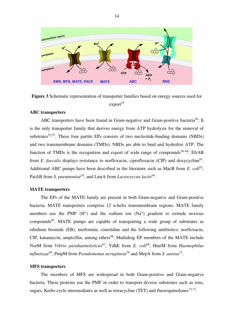
14
Figure 3 Schematic representation of transporter families based on energy sources used for
export55
ABC transporters
ABC transporters have been found in Gram-negative and Gram-positive bacteria56. It
is the only transporter family that derives energy from ATP hydrolysis for the removal of
substrates52,57. These four partite EPs consists of two nucleotide-binding domains (NBDs)
and two transmembrane domains (TMDs). NBDs are able to bind and hydrolize ATP. The
function of TMDs is the recognition and export of wide range of compounds58–60. EfrAB
from E. faecalis displays resistance to norfloxacin, ciprofloxacin (CIP) and doxycycline61.
Additional ABC pumps have been described in the literature such as MacB from E. coli62,
PatAB from S. pneumoniae63, and LmrA from Lactococcus lactis64.
MATE transporters
The EPs of the MATE family are present in both Gram-negative and Gram-positive
bacteria. MATE transporters comprise 12 α-helix transmembrane regions. MATE family
members use the PMF (H+) and the sodium ion (Na+) gradient to extrude noxious
compounds65. MATE pumps are capable of transporting a wide group of substrates as
ethidium bromide (EB), metformin, cimetidine and the following antibiotics: norfloxacin,
CIP, kanamycin, ampicillin, among others66. Multidrug EP members of the MATE include
NorM from Vibrio parahaemolyticus67, YdhE from E. coli68, HmrM from Haemophilus
influenzae69, PmpM from Pseudomonas aeruginosa70 and MepA from S. aureus71.
MFS transporters
The members of MFS are widespread in both Gram-positive and Gram-negative
bacteria. These proteins use the PMF in order to transport diverse substrates such as ions,
sugars, Krebs-cycle intermediates as well as tetracycline (TET) and fluoroquinolones72,73.

15
The MFS superfamily contains 12 or 14 transmembrane helices74. The EmrAB-TolC
tripartite EP75, EmrD76 and MdfA from E. coli are structurally characterized. MdfA
promotes the efflux of CIP, chloramphenicol and erythromycin77,78. Other members of this
family are NorA79, QacA, QacB80 and LmrS81 of S. aureus, LmrP82 of L. lactis, PmrA83 of S.
pneumoniae.
SMR transporters
SMR family is found in both Gram-positive and Gram-negative bacteria and these
proteins are the smallest known transporters. SMR transporters have a predicted function as
homodimers that contain 4 transmembrane helices. The efflux of noxious agents is mediated
by the PMF57,65. One of the best studied pump is EmrE84 from E. coli that is able to extrude
streptomycin, tobramycin85, erythromycin and TET86. Other representatives of SMR family:
EbrAB87 from Bacillus subtilis, QacC88 from S. aureus, YnfA89 from E. coli.
PACE transporters
PACE is the newest family of bacterial transporters, which has been described in
Gram-negative bacteria. It has 4 transmembrane helices based on its predicted topology. The
transporter AceI is the prototype protein from Acinetobacter baumannii and the homologue
of this transporter has been found in the genomes of Burkholderia, Enterobacter, Klebsiella,
Pseudomonas and Salmonella species. AceI and its homologues can mediate extrusion of
chlorhexidine, benzalkonium, acriflavine and proflavine53,90.
RND transporters
RND transporters are widely distributed and they form a tripartite efflux system from
the inner membrane (IM) to OM in Gram-negative bacteria91. The family uses PMF to
accelerate the extrusion of broad range of antibiotics and biocides92. In Gram-positive
bacteria (S. aureus, B. subtilis, Corynebacterium glutamicum and Clostridium difficile) only
a few RND pump monomers have been described54,93,94. The most well characterized RND
pump is the AcrAB-TolC multidrug transporter that consists of an IM transporter AcrB, a
periplasmic adaptor protein AcrA and an OM channel called TolC (Figure 4)95,96. The AcrB
EP consists of 12 transmembrane α-helices97.

16
AcrZ is 49 amino-acid length IM protein that associates
with the AcrAB-TolC system: it is directly bound to the AcrB
in order to regulate the substrate specificity of AcrB98. The
hexamer protein AcrA stabilizes the assembly of the pump and
the homotrimer TolC provides a channel to expel the
substrates from bacteria to the milieu99,100. The system can
transport EB, crystal violet (CV), erythromycin, fusidic acid,
novobiocin, fluoroquinolones, macrolides, chloramphenicol,
TET, rifampicin; detergents such as bile salts and
disinfectants101,102. Other RND drug transporters have been
discovered such as MexAB-OprM103, MexD, MexF and
MexY104 from P. aeruginosa, AcrD105 from E. coli, KexD106
from K. pneumoniae and MtrD107 from N. gonorrhoeae.
Figure 4 Structure of the AcrAB-TolC complex108
5. Biofilm formation and quorum sensing
Bacterial biofilm is a microbial community consisting of sessile bacterial cells attached
to each other and to a surface and is embedded in a self-produced extracellular matrix109.
Bacteria have the ability to form biofilms on both biotic (epithelial cells, tooth enamel etc.)
and abiotic (plastic, glass, medical devices such as pacemakers, implants, catheters etc.)
surfaces110,111. Biofilm-associated bacteria cause numerous infections, including
endocarditis, urinary tract infections, periodontitis and nosocomial infections112. Biofilm
formation has five stages: attachment, microcolonies and early biofilm formation, maturation
and dispersion (Figure 5)113. In the first stage the planktonic cells attach to the surface. At
the beginning, this attachment is initial and reversible and later becomes irreversible by
flagella, pili, curli, and fimbriae114. After adherence, the bacterial cells proliferate and form a
microcolony. The cells begin to grow and mature when these commence to synthesize
extracellular polymeric substance (EPS) matrix113, which includes exopolysaccharides,
proteins, nucleic acids115 and serves as a protective and diffusion barrier against
antimicrobial agents. When conditions become unfavorable (lack of nutrients, competition,
number of the bacterial population), a part of the biofilm will disperse and planktonic cells
will be released from the biofilm so they can colonize other surfaces116.

17
Figure 5 Schematic representation for the main stages of biofilm formation117
In the biofilm bacterial cells adapt to environmental conditions and bacteria show
increased (about 10-1000 times) resistance against antimicrobial agents compared to
planktonic bacteria118. Some factors play a role in the AMR of biofilm such as poor
penetration of the antibiotic due to the EPS119, slow growth and metabolism of biofilm
cells120, furthermore the transfer of AMR genes by conjugation is higher in biofilms121.
Another reason is the presence of EPs. In some bacterial strains, EP genes are up-regulated
showing that they contribute to biofilm formation35 and reduce susceptibility of the
biofilm122. EPs during biofilm formation extrude EPS and quorum sensing (QS) molecules.
Transporters influence genes important for biofilm formation indirectly, furthermore they
can pump out antibiotics and intermediates of metabolism35.
The expression of the genes required for biofilm formation have to be coordinated and
bacteria achieve this using the QS signal-response process. In bacteria the QS is a cell-to-cell
communication and regulatory mechanism that controls gene expression depending on
bacterial cell density123–125. For example, Gram-negative bacteria synthesize the extracellular
signaling molecules namely autoinducers (AIs) like N-acyl-homoserine lactone (AHL) by
Lux-I-type synthase enzymes. In the next step AHLs diffuse or are transported into the
environment to bind to their LuxR-type receptors (transcriptional regulator) in other nearby
cells. The AI and regulator complex binds to DNA to influence the expression of target
genes126.
In E. coli SdiA is a LuxR homolog that detects AHL signals from other bacteria and has a
great impact on the colonization of E. coli. SdiA represses the expression of virulence genes
by interacting with unknown stationary-phase signals in E. coli O157:H7127, and enhances
multidrug resistance by activating MDR EPs in E. coli109.
6. Background of resistance modulators applied in this study
Resistance to antibiotics has become a serious problem in the treatment of infectious
diseases because of the rapid spread of AMR. Consequently, there is a need to develop

18
alternative compounds that may be useful alone or in combination therapy129. The discovery
of plant-derived antimicrobials is advantageous because these plants are widely distributed
in nature130 and they could be used in order to overcome drug resistance in bacteria by
blocking QS, biofilm formation131 and multidrug EPs132. There are several studies in the
literature providing evidence of the EP inhibitory potential of plant-derived compounds. It
has been described that reserpine from the roots of Rauwolfia serpentina and R. vomitoria
inhibits TET efflux by Bmr transporter of B. subtilis133. Piperine isolated from black pepper
(Piper nigrum) has been identified as an inhibitor of NorA pump of MRSA134. Falcarindiol,
linoleic and oleic acids of Levisticum officinale reduce the efflux of AcrAB-TolC EP of
Salmonella enterica serovar Typhimurium135. Nigella sativa essential oil has shown activity
against the EP systems of methicillin sensitive S. aureus and MRSA strains136.
Cleistochlamys kirkii (Benth) Oliv. (Annonaceae) is an African medicinal plant traditionally
used in Mozambique for the treatment of wound infections, tuberculosis and rheumatism137.
Based on preliminary studies C. kirkii derivatives have a broad antimicrobial spectrum: they
had antifungal activity against Candida albicans and exhibited antibacterial effect against
methicillin sensitive S. aureus and MRSA138,139.
Organic compounds of phosphorus ylides (P-ylides) are a fascinating class of
compounds in organic chemistry140. The EP modulating activity of P-ylides has already been
described regarding the ATP-binding cassette subfamily B member (ABCB1) pump of MDR
mouse T-lymphoma cells141 and the further biological aspects of the phosphorus ylides in
bacteria have not been yet investigated in the literature.
Selenium is an important trace element, which plays a role in the prevention of
inflammatory diseases and cancer142. The antibacterial activity of selenium-containing
compounds has been found by many studies: research was mostly done using bacteria that
cause nosocomial infections. E. coli143 and S. aureus144 are often responsible for infections
during hospitalization145. Infections caused by these bacteria are difficult to treat due to their
biofilm forming ability. It has been demonstrated by a research group that a
perihydroselenoxantine compound showed antibacterial activity on S. aureus strain146.
Sodium selenite has been found to eradicate Helicobacter pylori and it showed antibacterial
effect against H. pylori at a low concentration in rats147. In another study, three polymers
(polyvinyl chloride, polyurethane and silicone) were coated with selenium nanoparticles.
The growth of S. aureus was prevented on selenium-coated polymers compared to the
uncoated materials148. The novel selenocompounds tested in this study have been previously
described as anticancer compounds against T-lymphoma and colon cancer cell lines149,150.

19
AIMS OF THE STUDY
Resistant bacteria are able to withstand the attack of antimicrobials, so that standard
treatments become ineffective and infections persist and may spread to others. Efflux
mechanisms of bacteria, namely, pumping of antimicrobial agents out of the cells play an
important role in antimicrobial resistance of pathogenic bacteria. The aim of our study was
to investigate the bioactive compounds of Cleistochlamys kirkii, synthesized fluorinated β-
diketo phosphorus ylides and selenocompounds on Gram-positive and Gram-negative model
bacterial strains in order to find resistance modifiers which could be applied later alone or in
combination with antibiotics.
The main goals of the study were the following:
1. Determination of the antibacterial activity of the compounds (five natural
compounds, ten fluorinated β-diketo phosphorus ylides and eleven
selenocompounds) on Gram-positive (Enterococcus faecalis ATCC 29212,
Staphylococcus aureus ATCC 25923, S. aureus 272123) and Gram-negative strains
(Escherichia coli AG100, AG100 A, Salmonella enterica serovar Typhimurium
SL1344 (wild-type), L644 (ΔacrB strain), 14028s (biofilm producing strain),
Chromobacterium violaceum 026 and Enterobacter cloacae 31298 by microdilution
method.
2. Investigation of the efflux pump (EP) inhibitory effect of the compounds (five
natural compounds, ten fluorinated β-diketo phosphorus ylides and eleven
selenocompounds) on Gram-positive (S. aureus ATCC 25923, S. aureus 272123) and
Gram-negative strains (E. coli AG100 expressing the AcrAB-TolC EP and its
AcrAB-TolC deleted mutant AG100 A strain) using real-time ethidium bromide (EB)
accumulation assay.
3. Evaluation of the EP inhibitory effect of eleven selenocompounds on Gram-
negative wild-type S. Typhimurium SL1344 strain expressing the AcrAB-TolC EP
and its AcrB deleted mutant L644 strain using EB efflux assay.
4. Determination of the anti-biofilm activity of eleven selenocompounds on Gram-
negative, biofilm producing S. Typhimurium 14028s strain using crystal violet (CV).

20
5. Characterization of the activity of five natural compounds of C. kirkii as
adjuvants in the presence of tetracycline (TET) and ciprofloxacin (CIP) on S. aureus
ATCC 25923 and S. aureus 27213 strains by checkerboard method.
6. Evaluation of the adjuvant role of eleven selenocompounds on E. coli AG100
strain by a two-fold broth microdilution method in the presence of TET and CIP.
7. Quorum sensing (QS) inhibition analysis of five natural compounds of C. kirkii
and ten fluorinated β-diketo phosphorus ylides using the sensor strain C.
violaceum 026 and the AHL producer strain E. cloacae 31298 by agar diffusion
method.
8. Monitoring the changes in relative gene expression of efflux (norA, mepA, acrA,
acrB), antibiotic resistance (marR) and QS (sdiA) genes in the presence of the most
effective EP inhibitors (natural compounds isolated from C. kirkii, fluorinated β-
diketo phosphorus ylides and selenocompounds) investigated by reverse transcriptase
quantitative polymerase chain reaction (RT-qPCR).

21
MATERIALS AND METHODS
1. Compounds studied
Five natural compounds (CK1-5) were isolated from the methanol extract of the root
barks of Cleistochlamys kirkii (Benth.) Oliv. (Annonaceae): triterpene polycarpol (CK1), C-
benzylated flavanones chamanetin (CK2), isochamanetin (CK3), dichamanetin (CK4) and
the heptane derivative acetylmelodorinol (CK5) were kindly provided by Prof. Dr. Maria-
José U. Ferreira (Universidade de Lisboa, Lisbon, Portugal; Figure 6). The stock solutions
(in 10 mM concentration) of compounds were prepared in dimethyl sulfoxide (DMSO).
Figure 6 Chemical structures of bioactive compounds (CK1–5)
Ten synthesized fluorinated β-diketo phosphorus ylides (P-ylides; PY1-10) were
kindly provided by Prof. Dr. Masami Kawase (Matsuyama University, Matsuyama, Japan)
(See Appendix 1 and 2). The stock solutions (in 10 mg/mL concentration) of compounds
were prepared in DMSO.
Eleven selenocompounds including a cyclic selenoanhydride (EDA1), heteroaryl
selenoesters (EDA2-3) and aryl selenoesters (EDA4-11) were kindly provided by Dr.
Enrique Domínguez-Álvarez (Consejo Superior de Investigaciones Científicas, Madrid,
Spain) and Prof. Dr. Carmen Sanmartín (University of Navarra, Pamplona, Spain)149,151

22
(Figure 7; See Appendix 3 and 4). The stock solutions (in 10 mM concentration) of
compounds were prepared in DMSO.
Figure 7 Chemical structures of tested selenoanhydride (EDA1) (A), heteroaryl selenoesters
(EDA2-3) (B) and aryl selenoesters (EDA4-11) (C)
2. Reagents and media
Promethazine (PMZ; EGIS), ethidium bromide (EB), carbonyl cyanide m-
chlorophenyl hydrazone (CCCP), verapamil, crystal violet (CV), tetracycline-hydrochloride
(TET), ciprofloxacin-hydrochloride (CIP), DMSO, Luria-Bertani (LB) broth, and LB agar
were purchased from Sigma-Aldrich Chemie GmbH (Steinheim, Germany). The modified
LB medium (LB*) was prepared from yeast extract 5 g/L, tryptone 10 g/L, NaCl 10 g/L,
K2HPO4 1 g/L, MgSO4 x 7H2O 0.3 g/L and FeNaEDTA 36 mg/L. In case of modified LB*
agar, the LB* medium was supplemented with agar 20 g/L (Difco, Detroit, USA). pH was
adjusted to 7.2. Tryptic soy broth (TSB), tryptic soy agar (TSA), and Mueller Hinton (MH)
broth were purchased from Scharlau Chemie S. A. (Barcelona, Spain).
3. Bacterial strains
Wild-type Escherichia coli K-12 AG100 strain [argE3 thi-1 rpsL xyl mtl Δ(gal-uvrB)
supE44], expressing the AcrAB-TolC efflux pump (EP) at its basal level and its
AcrAB-TolC deleted mutant E. coli AG100 A strain were used in the study. These strains
were kindly provided by Prof. Dr. Hiroshi Nikaido (University of California, Berkeley, CA,
USA).
Wild-type Salmonella enterica serovar Typhimurium SL1344 expressing the AcrAB-
TolC EP and its acrB gene inactivated mutant S. Typhimurium strain (L644)152, furthermore,
the biofilm producing S. Typhimurium 14028s strain were used in the study. These strains
were kindly provided by Dr. Jessica M. A. Blair (University of Birmingham, Birmingham,
United Kingdom).
A B C

23
Staphylococcus aureus ATCC (American Type Culture Collection) 25923, was used
as the methicillin susceptible reference strain, and the methicillin and ofloxacin resistant S.
aureus 272123 clinical isolate was kindly provided by Prof. Dr. Leonard Amaral (Institute of
Hygiene and Tropical Medicine, Lisbon, Portugal). In addition, Enterococcus faecalis ATCC
29212 strain was used in the assays.
For quorum sensing (QS) tests, the following strains were used: Chromobacterium
violaceum 026 (CV026) as sensor strain and Enterobacter cloacae 31298 as N-acyl-
homoserine lactone (AHL) producer strain (a clinical isolate from a wound)153.
4. Determination of minimum inhibitory concentrations by microdilution method
The minimum inhibitory concentrations (MICs) of all tested compounds were
determined according to the Clinical and Laboratory Standards Institute (CLSI) guidelines in
three independent assays154. The solvent DMSO had no antibacterial effect.
5. Real-time accumulation assay of ethidium bromide
The activity of compounds isolated from C. kirkii (CK1-5), P-ylides (PY1-10) and
selenocompounds (EDA1-11) on the real-time accumulation of EB was assessed by the
automated EB method155 using a LightCycler real-time thermocycler (LightCycler 1.5,
Roche, Indianapolis, USA). Briefly, an aliquot of an overnight culture of S. aureus strains
(ATCC 25923 and MRSA 272123) in TSB medium was transferred to fresh TSB medium,
and it was incubated until it reached an optical density (OD) of 0.6 at 600 nm. In case of E.
coli AG100 and AG100 A, the medium used in the assay was LB broth; the preparation of
the inoculum was similar to the one of S. aureus. The cells were washed with phosphate
buffered saline (PBS; pH 7.4) and centrifuged at 13,000 × g for 3 minutes, the pellets were
re-suspended in PBS (pH 7.4), and the OD was adjusted to 0.6 at 600 nm. The compounds
were added individually at different concentrations at ½ MIC, ⅓ MIC, ¼ MIC or ⅕ MIC (in
double concentrated form) to the EB solution in PBS. The final concentration of EB was
based on the MIC and the lowest fluorescent signal produced by this sub-MIC concentration
of EB. In case of S. aureus strains, the concentration of EB was 0.5 μg/mL, for E. coli
AG100 1 μg/mL, and in case of E. coli AG100 A it was 0.25 μg/mL. Then, 10 μL of the EB
solution containing the compound were transferred into standard glass capillary tubes of 20
µL maximum volume (Roche, Indianapolis, IN, USA), and 10 μL of bacterial suspension
(OD of 0.6 at 600 nm) were added to the capillaries. The capillaries containing the samples
were placed into the carousel (Roche), and the fluorescence was monitored at the FL-2
channel in every minute on a real-time basis.

24
From the real-time data, the activity of the compounds, namely the relative
fluorescence index (RFI) of the last time point (minute 30) of the EB accumulation assay,
was calculated according to the following formula:
Where RFtreated is the relative fluorescence (RF) at the last time point of EB retention curve
in the presence of an inhibitor, and RFuntreated is the RF at the last time point of the EB
retention curve of the untreated control having the solvent control (DMSO). Verapamil was
applied as a positive control on Gram-positive strains and PMZ was used on Gram-negative
strains.
6. Efflux assay using ethidium bromide
The activity of selenocompounds (EDA1-11) on the efflux of EB was determined on
wild-type (SL1344) and acrB gene inactivated (L644) S. Typhimurium strains. Briefly, an
aliquot of an overnight culture of S. Typhimurium strain in LB medium was transferred to
fresh LB medium, and it was incubated at 37°C with shaking at 150 rpm until it reached an
OD of 0.4 at 600 nm. The cultures were centrifuged at 3500 rpm for 10 min at 21°C. The
supernatant was removed, and the pellet was re-suspended in 20 mM potassium phosphate
buffer (PPB; pH 7.0) with 1 mM MgCl2 and the OD was adjusted to 0.2 at 600 nm. 100 μM
of CCCP was added to de-energize the cells and therefore EB was added at 50 μg/mL
concentration. The cultures were incubated at 23°C with shaking at 150 rpm for 1 h. After
the incubation period the cultures were centrifuged at 3500 rpm for 10 min at 21°C. The
supernatant was decanted, and the pellet was re-suspended in 20 mM PPB buffer (pH 7.0)
with 1 mM MgCl2 and 5% glucose to energize the cells. Following energization, 200 µL of
the bacterial culture were transferred into black 96-well microtiter plate (Corning,
Amsterdam) containing the compounds at 50 µM (½ MIC) and the fluorescence of EB was
measured over 2 hours at excitation and emission wavelengths of 530 and 600 nm,
respectively, using a FLUOstar Optima plate reader (BMG Labtech, United Kingdom)152.
During the evaluation the exact time was determined when the fluorescence dropped by 25%
and 50% of the starting value. CCCP was applied as a positive control and DMSO was used
as a negative control.
untreated
untreatedtreated
RF
RFRFRFI
−=
25
7. Measuring biofilm formation using crystal violet
The biofilm forming ability of S. Typhimurium 14028s strain was studied in 96-well
microtiter plates using LB broth without salt in the presence of selenocompounds (EDA1-
11). Initially, overnight cultures were diluted to an OD of 0.1 at 600 nm and then added to
each well with the exception of the medium control wells and compounds were added at 50
µM (½ MIC) concentration. The final volume was 200 μL in each well. Plates were
incubated at 30°C for 48 h with gentle agitation (100 rpm). After the incubation period the
medium was discarded, and the plate was washed with tap water to remove unattached cells.
200 μL CV (0.1% [v/v]) was added to the wells and incubated for 15 minutes at room
temperature. CV was removed from the wells and the plate was washed again with tap water.
200 μL of 70% ethanol was added to each well and the biofilm formation was determined by
measuring the OD at 600 nm using a FLUOstar Optima plate reader. The anti-biofilm effect
of selenocompounds was expressed in the percentage (%) of decrease in biofilm formation.
The results were analyzed using t-test and p-values of <0.05 were considered significant.
8. Interaction between antibiotics and resistance modifiers using checkerboard
method
The combined effect of chamanetin (CK2) and dichamanetin (CK4) and antibiotics
on the growth inhibition of S. aureus ATCC 25923 and methicillin resistant S. aureus
272123 strains was evaluated by checkerboard method. Two-fold serial dilutions of
antibiotics were prepared in MH broth on the horizontal rows of microtiter plate and then
cross-diluted vertically by two-fold serial dilutions of the compounds156. For this assay, only
the compounds with well-defined MIC values could be used. The dilutions of the antibiotics
(TET or CIP) were made in a horizontal direction in 100 µL, and the dilutions of compounds
were made vertically in the microtiter plate in 50 µL. After the dilution of an overnight
culture, bacterial cells were re-suspended in MH medium containing 1×10⁴ cells and
distributed into each well. The plates were incubated for 18 h at 37°C. The cell growth rate
was determined after MTT (3-(4,5-dimethylthiazol-2-yl)-2,5-diphenyltetrazolium bromide)
staining (See Appendix 5), as described elsewhere (Figure 8)156. The combination index (CI)
values at 90% growth inhibition (ED90) were determined by CompuSyn software to plot 4 or
5 data points for each ratio (www.combosyn.com, ComboSyn, Inc., Paramus, NJ. 07652
USA). CI values were calculated by means of the median-effect equation, where CI < 1, CI =
1 and CI > 1 represent synergism, an additive effect (or no interaction) and antagonism,
respectively157.

26
Figure 8 The layout of checkerboard plates
9. Interaction between antibiotics and resistance modifiers using minimum inhibitory
concentration reduction assay
The chemosensitizing effect of the selenocompounds (EDA1-11) was evaluated by
the determination of the MIC values of the antibiotics (TET and CIP), in the presence of sub-
inhibitory concentrations of the compounds (½ MIC) in E. coli AG100 strain by a two-fold
broth microdilution method in the 96-well plates, using serial dilutions of TET and CIP. The
first four rows contained two-fold dilutions of antibiotics, and the combinations of the
antibiotics and tested compounds were added into the last four rows. 10-4 dilution of an
overnight bacterial culture in 50 µL of MH was then added to each well, with the exception
of the medium control wells. The plates were then incubated at 37°C for 18 h. MIC values of
the antibiotics and their combination with the tested compounds were determined by naked
eyes.
10. Assay for quorum sensing inhibition
LB* was used for these experiments. The sensor strain C. violaceum 026 and the
AHL producer strains E. cloacae 31298 were inoculated as parallel lines and incubated at
room temperature (20C) for 24–48 h. QS inhibition was monitored by agar diffusion
method. Filter paper discs (7.0 mm in diameter) were impregnated with 10 μL of stock
solutions (10 mM or 10 mg/mL) of the CK and PY compounds in DMSO. The discs were

27
placed between the parallel lines of the sensor and the AHL producer strains on the surface
of the nutrient agar (Figure 9). The plates were incubated at room temperature for another
24–48 h, and the interactions between the strains and compounds were evaluated for the
reduction in the size of the zone of pigment production and the zone of growth inhibition of
the affected strains, in millimeters153. PMZ was applied as a positive control.
Figure 9 Positive (promethazine (PMZ; top)) and negative (DMSO; bottom) controls of
quorum sensing (QS) on C. violaceum 026 and E. cloacae 31298
11. Expression analyses of genes by RT-qPCR reaction
E. coli AG100 strain was cultured in LB broth, S. aureus ATCC 25923 and MRSA
272123 strains were cultured in TSB broth and were incubated overnight at 37°C with
shaking. On the day of RNA isolation, the bacterial suspensions (OD of 0.6 at 600 nm) were
transferred to 10 mL tubes in 3 mL aliquots, and the compounds (CK2, -4; PY2, -4, -5 and
EDA1, -4, -7) were added to the tubes at 50 µM (for EDA1, -4, -7), 50 µg/mL (for PY2, -4,
-5), 5 µM (for CK2) and 0.5 µM (for CK4) concentrations, which were incubated at 37°C.
After 4 hours (for CK2 and -4) or 4 and 18 hours (for PY2, -4, -5 and EDA1, -4, -7) of
culturing, the tubes were centrifuged at 12,000 × g for 2 min. Pellets were re-suspended in
100 µL Tris-EDTA buffer containing 1 mg/mL lysozyme by vigorous vortexing, and they
were incubated at 37°C for 10 min. The total RNA was isolated in an RNase-free
environment using NucleoSpin RNA kit (Macherey Nagel, Germany) according to the
manufacturer’s instructions. Purified RNA was stored in RNase-free water in nuclease-free
collection tubes and was maintained at −20°C until quantification was performed. The
concentration of the extracted RNA templates was assessed by SmartSpecTM Plus
Spectrophotometer at 260 nm (Bio-Rad, USA).
Expression of the EP genes norA and mepA in the presence of chamanetin (CK2) and
dichamanetin (CK4) was studied by reverse transcription of the total RNA of S. aureus

28
ATCC 25923 and MRSA 272123 strains. The data obtained for gene targets were
normalized against the S. aureus 16S ribosomal RNA gene measured in the same sample.
The effect of PY2, -4, -5 and EDA1, -4, -7 on the relative expression of the EP (acrA, acrB),
antibiotic resistance (marR) and QS (sdiA) genes were studied in E. coli AG100. The data
obtained for gene targets were normalized against the E. coli house-keeping gene
glyceraldehide-3-phosphate-dehydrogenase (gapdh) measured in the same sample. The full
names of the tested genes are presented in Table 1. The forward and reverse primers used in
this assay are shown in Appendix 6.
Gene Full name Reference
acrA Acridine resistance protein A
158 acrB Acridine resistance protein B
marR Multiple antibiotic resistance protein R
sdiA Quorum-sensing transcriptional activator This study
gapdh Glyceraldehyde-3-phosphate
dehydrogenase
158
norA Quinolone resistance protein NorA
159 mepA Multidrug export protein MepA
16S rRNA 16S Ribosomal RNA
Table 1 Full name of the tested genes investigated in the RT-qPCR assay
Real-time quantification of the RNA templates by one-step RT-qPCR was performed
in a CFX96 Touch real-time PCR detection system (Bio-Rad, USA) strictly adhered to the
manufacturer’s recommendations of the SensiFASTTM SYBR No-ROX One-Step Kit
(Bioline GmbH, Luckenwalde, Germany). Briefly, each well of the 96-well microtiter plates
in a final volume of 20 µL contained 10 µL of the 2x SensiFASTTM SYBR No-ROX One-
Step Mix, 0.2 µL reverse transcriptase, 0.4 µL ribosafe RNAase inhibitor, 5.4 µL
diethylpyrocarbonate-treated water, 500 nM of each primer and approximately 20 ng of the

29
total RNA in RNAase-free water. Thermal cycling was initiated with a denaturation step of 5
min at 95°C, followed by 40 cycles, each of 10 s at 95°C, 30 s at 57°C and 20 s at 72°C.
The relative quantities of the mRNA of each gene of interest were determined by the ΔΔCT
method160. Gene transcript levels were normalized against the previously mentioned
housekeeping genes. The formula 2-ΔΔCT allows the relative quantification of differences of
each gene’s expression level between two samples, the sample of interest and a calibrator or
reference sample.

30
RESULTS
1. In vitro antibacterial activity of compounds
1.1. Bioactive compounds from C. kirkii
Compounds (CK1–CK5) were assessed for their antibacterial activity against
methicillin susceptible Staphylococcus aureus ATCC 25923, and the methicillin and
ofloxacin resistant S. aureus 272123 clinical isolate. Wild-type Escherichia coli K-12
AG100 and E. coli AG100 A strains, over-expressing and lacking the AcrAB-TolC efflux
pump (EP) system, respectively, were used as Gram-negative models. In addition, the
antibacterial activity of the compounds was tested on quorum sensing (QS) strains
Chromobacterium violaceum 026 and Enterobacter cloacae 31298.
Concerning the antibacterial effect of the compounds, chamanetin (CK2) and dichamanetin
(CK4) had a potent antibacterial effect on both S. aureus strains. Minimum inhibitory
concentration (MIC) of compound CK2 was 12.5 µM on reference S. aureus strain;
however, the MIC in case of the methicillin and ofloxacin resistant strain was 25 µM.
Compound CK4 was the most effective flavanone because its MIC on S. aureus ATCC
25923 strain was 0.8 µM; furthermore, on the methicillin resistant strain it exhibited the MIC
of 1.56 µM. The compounds had no antibacterial effect on the Gram-negative E. coli
AG100, AG100 A, C. violaceum, and E. cloacae strains (MIC: >100 or 100 µM) (Table 2).
Compounds
MIC (µM)
S. aureus
ATCC
25923
S. aureus
272123
E. coli
AG100
E. coli
AG100 A
C.
violaceum
026
E.
cloacae
31298
CK1 >100 >100 >100 >100 >100 >100
CK2 12.5 25 >100 >100 >100 >100
CK3 100 >100 >100 >100 >100 >100
CK4 0.8 1.56 >100 >100 >100 >100
CK5 >100 >100 >100 >100 >100 >100
Table 2 Minimum inhibitory concentrations (MICs) of the CK compounds isolated from C.
kirkii on Gram-positive and Gram-negative bacteria

31
1.2. Fluorinated β-diketo phosphorus ylides
Compounds (PY1-10) were investigated for their antibacterial activity against wild-
type E. coli K-12 AG100 strain, and the AcrAB-TolC pump mutant E. coli AG100 A strain.
Furthermore, the antibacterial activity of the compounds was tested on QS strains C.
violaceum 026 and E. cloacae 31298.
Compounds PY1-10 did not have any antibacterial effect on the AcrAB-TolC expressing E.
coli AG100, C. violaceum and E. cloacae strain and the AcrAB-TolC deleted E. coli AG100
A strain (MIC: >100 g/mL), except for ethyl-4,4,4-trifluoro-3-oxo-2-(triphenyl
phosphoranylidene)butanoate (PY6), which had a mild effect on the EP deleted strain (MIC:
50 μg/mL) (See Appendix 7).
1.3. Selenocompounds
The antibacterial effect of selenocompounds (EDA1-11) were tested on Gram-
negative E. coli AG100, E. coli AG100 A, Salmonella enterica serovar Typhimurium
SL1344, acrB inactivated S. Typhimurium L644 and S. Typhimurium 14028s strains.
Furthermore, the following Gram-positive strains were used: S. aureus ATCC 25923 and
Enterococcus faecalis ATCC 29212.
The ketone-containing selenoesters (EDA9–11) showed a potent antibacterial activity
against the Gram-positive S. aureus ATCC 25923. The methylketone selenoester EDA9 was
the most active compound with noteworthy MIC value (3.12 µM). The chloro-substituted
tert-butylketone selenoester (EDA10) and dimethoxy-substituted tert-butylketone
selenoester (EDA11) showed lower antibacterial activity than EDA9 (25 and 50 µM,
respectively). The selenoanhydride EDA1 and the remaining selenoesters EDA2–8
evaluated were inactive as their MIC was equal or above 100 µM on S. aureus ATCC 25923.
EDA9 showed antibacterial activity towards E. faecalis (MIC: 12.5 µM). The compounds
had no antibacterial effect (MIC: 100 μM or >100 μM) on Gram-negative strains (See
Appendix 8).
2. Efflux pump inhibiting activity (accumulation assay)
2.1. Bioactive compounds from C. kirkii
The ethidium bromide (EB) accumulation assay provides information about the
intracellular accumulation of the general EP substrate EB. A potential efflux pump inhibitor
(EPI) increases the fluorescence level of EB because of its accumulation within the bacterial
cell. The EP inhibiting activity of the compounds was compared based on the relative
fluorescence index (RFI) of the real-time accumulation curves in E. coli AG100, E. coli

32
AG100 A, S. aureus ATCC 25923 and S. aureus 272123. In case of real-time EB
accumulation by the LightCycler thermocycler, the amount of EB accumulated by cells is
higher if the difference between RFtreated and RFuntreated is greater; therefore, the degree of
inhibition of the EP system by the compound becomes greater.
As shown in Figure 10, CK2 and CK4 had EP inhibiting activity compared to
verapamil (RFI: 0.13) on the S. aureus ATCC 25923 strain, and the most active compound
was CK2. However, compounds CK1–5 had no EP inhibitory activity on the methicillin
resistant S. aureus strain at the concentrations applied in the assay (See Appendix 9).
-0.3
-0.25
-0.2
-0.15
-0.1
-0.05
0
0.05
0.1
0.15
0.2
0.25
Rel
ati
ve
flu
ore
scen
ce i
nd
ex
CK compounds
Figure 10 Relative final fluorescence index (RFI) of compounds (CK1-5 isolated from C.
kirkii) on S. aureus ATCC 25923 at different concentrations
Concerning the inhibitory activity on Gram-negatives, triterpene polycarpol (CK1)
and CK5 could inhibit the AcrAB-TolC system of E. coli AG100 compared to promethazine
(PMZ; RFI: 0.15). CK1 proved to be the most effective EPI (Figure 11). Based on the real-
time accumulation data, CK1–5 had no effect on the E. coli AG100 A strain lacking the
AcrAB-TolC pump (See Appendix 10).

33
-0.8
-0.6
-0.4
-0.2
0
0.2
0.4
0.6
Rel
ati
ve
flu
ore
scen
ce i
nd
ex
CK compounds
Figure 11 Relative final fluorescence index (RFI) of compounds (CK1-5 isolated from C.
kirkii) on E. coli AG100 strain at 50 µM
2.2. Fluorinated β-diketo phosphorus ylides
The EP inhibitory effect was tested on the AcrAB-TolC expressing E. coli AG100
and the mutant AG100 A strains. The majority of the P-ylides were found to
inhibit the AcrAB-TolC system of E. coli except trifluoro-1-phenyl-2-
(triphenylphosphoranylidene)butane-1,3-dione (PY3), 4,4,5,5,5-pentafluoro-1-phenyl-2-
(triphenylphosphoranylidene)pentane-1,3-dione (PY7) and 4,4,5,5,6,6,6-heptafluoro-1-
phenyl-2-(triphenylphosphoranylidene)hexane-1,3-dione (PY8), which had little or no effect
on the intracellular EB accumulation in both strains. Among the P-ylide series, compounds
1,1,1-trifluoro-3-oxo-1-methoxy-3-(triphenylphosphoranylidene)propane-2-one (PY2),
4,4,4-trifluoro-3-oxo-2-(triphenylphosphoranylidene)butanal (PY4) and 1,1,1-trifluoro-3-
(triphenylphosphoranylidene)pentane-2,4-dione (PY5) exhibited strong AcrAB-TolC pump-
inhibiting properties compared to the AcrAB-TolC pump-deficient mutant strain. The most
potent derivative was PY4 and its effect was more pronounced on the multidrug resistant E.
coli strain compared to the pump deleted E. coli strain (Figure 12).

34
Figure 12 Relative final fluorescence index (RFI) of P-ylides (PY1-10) on the AcrAB-TolC
expressing E. coli AG100 and pump deleted E. coli AG100 A strains at 50 g/mL (except
PY6 (25 g/mL) on AG100 A strain)
2.3. Selenocompounds
The ability of the selenocompounds to inhibit the efflux of AcrAB-TolC transporter
was determined on E. coli AG100 and AG100 A strains. EDA1 and meta-substituted
benzene selenodiester (EDA4), strongly inhibited the efflux mediated by AcrAB-TolC in E.
coli AG100 compared to the positive control PMZ (RFI: 0.15). Methoxycarbonylmethyl
selenoester (EDA7) and EDA9–11 caused moderate inhibitory action, whereas thiophene
selenodiester (EDA2), pyridine selenodiester (EDA3), para-substituted benzene
selenodiester (EDA5), carbamoylmethyl selenoester (EDA6) and phenoxycarbonyl
selenoester (EDA8) showed weak or no activity on the intracellular EB accumulation in E.
coli AG100. Nevertheless, no EP inhibitory action was found in the E. coli AG100 A strain
in case of selenocompounds (EDA1–11) (Figure 13).

35
Figure 13 Relative final fluorescence index (RFI) of selenocompounds (EDA1-11) on the
AcrAB-TolC expressing E. coli AG100 and pump deleted E. coli AG100 A strains at 50 M
3. Efflux pump inhibiting activity (efflux assay)
In the EB efflux assay, after loading the wild-type S. Typhimurium SL1344 and acrB
mutant L644 strains with EB the active efflux of the dye was measured as the time taken for
fluorescence drops by 25% and 50% compared to the starting fluorescence level. Each
selenocompound showed a 25% and 50% decrease in fluorescence at an earlier time point
(25%: between 4.5th and 17.8th min; 50%: between 7.9th and 79.2nd min), compared with
positive control CCCP (25%: in 52.1st min; 50%: in 122.6th min) in S. Typhimurium SL1344
(Table 3). The EB efflux was most effectively inhibited in the presence of EDA9 as
fluorescence intensity of EB was reduced by half in the 79.2nd minutes. For the L644 strain
only EDA7 was able to prevent the efflux of EB more effectively than carbonyl cyanide m-
chlorophenyl hydrazone (CCCP) (Table 3).

36
Samples
Time taken (min) for percentage
S. Typhimurium
SL1344
S. Typhimurium
L644
-25% -50% -25% -50%
Untreated control =
Bacterial control 5.4 9.6 8 14.1
CCCP 52.1 122.6 42.1 117.6
EDA1 9.7 55.3 28 78.4
EDA2 17.8 56.8 31.5 88.6
EDA3 4.5 7.9 9.9 21
EDA4 17.2 56.1 21.6 83.2
EDA5 5.3 11.0 10.1 26.3
EDA6 5.4 11.0 11.8 30.5
EDA7 12.2 58.9 55.2 119.5
EDA8 5.5 9.5 9 16.8
EDA9 15.6 79.2 37.8 86.1
EDA10 17.1 65.6 18.8 80.8
EDA11 19.8 30.7 15 32
Table 3 Time taken for fluorescence to drop by 25% and 50% of starting value in wild-type
S. Typhimurium SL1344 and its acrB mutant L644 strains in the presence of
selenocompounds at 50 µM
4. Anti-biofilm activity of selenocompounds
The anti-biofilm effect of selenocompounds (EDA1-11) was evaluated by
microdilution method using crystal violet (CV) on S. Typhimurium 14028s strain. Except
compounds EDA6 and dimethoxy-substituted tert-butylketone selenoester (EDA11) all
derivatives showed significant (>45%; p<0.05) or higher biofilm inhibition at 50 μM on S.
Typhimurium. The most potent selenocompounds with anti-biofilm effect were EDA4 and -
5 at 50 μM showing 75% and 73% of inhibition, respectively (Figure 14).

37
Figure 14 Anti-biofilm effect of selenocompounds on S. Typhimurium 14028s at 50 μM
5. Combined effects of chamanetin (CK2) and dichamanetin (CK4) with antibiotics
The type of interaction between the antibacterial compounds CK2, CK4 and
tetracycline (TET) and the fluoroquinolone antibiotic ciprofloxacin (CIP) was evaluated on
reference (ATCC 25923) and methicillin resistant (MRSA 272123) S. aureus strains by
checkerboard assay. The results are presented in Tables 5 and 6 as combination index
values157. As it can be observed the combined effect of TET and compounds CK2 or CK4
on S. aureus ATCC 25923 resulted in synergism. The most effective ratio of antibiotic and
compound was 1:20 and 1:1, respectively. Similarly, CIP also acted synergistically with
compounds CK2 and CK4 and the most active ratio of antibiotic and compound was
1.3:12.5 and 1.3:1, respectively (Table 4).
Staphylococcus aureus ATCC 25923
Combination Best Ratio CI at ED90 SD (+/-) Interaction
TET + CK2 1:20 0.64 0.13 Synergism
TET + CK4 1:1 0.42 0.1 Synergism
CIP + CK2 1.3:12.5 0.82 0.24 Slight synergism
CIP + CK4 1.3:1 0.69 0.28 Synergism
Ratio: antibiotic and tested compound (µM). CI: combination index. CI < 1, CI = 1 and CI > 1 represent
synergism, additive effect (or no interaction), and antagonism, respectively.
Table 4 Combined effect of CK2 and CK4 with antibiotics on S. aureus ATCC 25923 strain
* p<0.05
** p<0.001
38
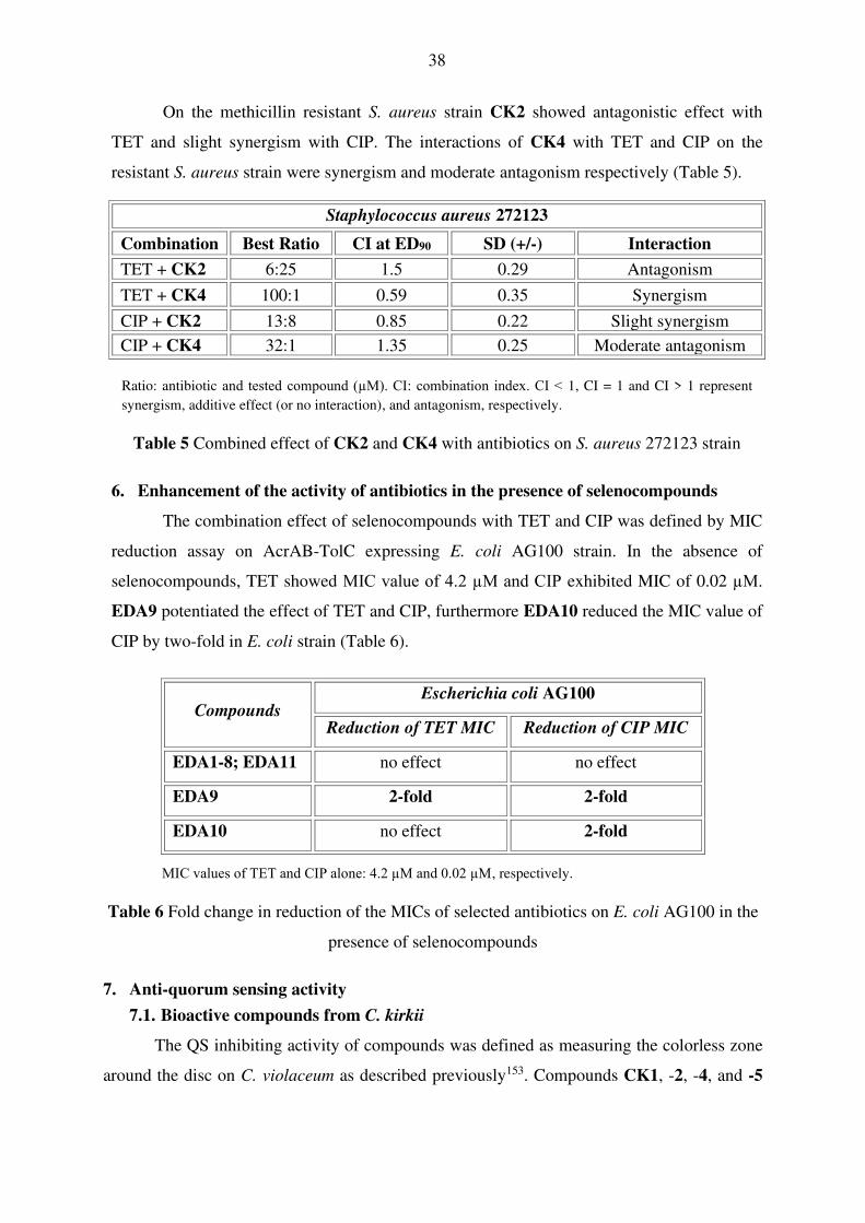
On the methicillin resistant S. aureus strain CK2 showed antagonistic effect with
TET and slight synergism with CIP. The interactions of CK4 with TET and CIP on the
resistant S. aureus strain were synergism and moderate antagonism respectively (Table 5).
Staphylococcus aureus 272123
Combination Best Ratio CI at ED90 SD (+/-) Interaction
TET + CK2 6:25 1.5 0.29 Antagonism
TET + CK4 100:1 0.59 0.35 Synergism
CIP + CK2 13:8 0.85 0.22 Slight synergism
CIP + CK4 32:1 1.35 0.25 Moderate antagonism
Ratio: antibiotic and tested compound (µM). CI: combination index. CI < 1, CI = 1 and CI > 1 represent
synergism, additive effect (or no interaction), and antagonism, respectively.
Table 5 Combined effect of CK2 and CK4 with antibiotics on S. aureus 272123 strain
6. Enhancement of the activity of antibiotics in the presence of selenocompounds
The combination effect of selenocompounds with TET and CIP was defined by MIC
reduction assay on AcrAB-TolC expressing E. coli AG100 strain. In the absence of
selenocompounds, TET showed MIC value of 4.2 µM and CIP exhibited MIC of 0.02 µM.
EDA9 potentiated the effect of TET and CIP, furthermore EDA10 reduced the MIC value of
CIP by two-fold in E. coli strain (Table 6).
MIC values of TET and CIP alone: 4.2 µM and 0.02 µM, respectively.
Table 6 Fold change in reduction of the MICs of selected antibiotics on E. coli AG100 in the
presence of selenocompounds
7. Anti-quorum sensing activity
7.1. Bioactive compounds from C. kirkii
The QS inhibiting activity of compounds was defined as measuring the colorless zone
around the disc on C. violaceum as described previously153. Compounds CK1, -2, -4, and -5
Compounds Escherichia coli AG100
Reduction of TET MIC Reduction of CIP MIC
EDA1-8; EDA11 no effect no effect
EDA9 2-fold 2-fold
EDA10 no effect 2-fold

39
were able to inhibit effectively the QS between C. violaceum and E. cloacae compared to the
positive control PMZ (Table 7).
Compounds QS inhibition zone in mm
CK1 51
CK2 50
CK3 -
CK4 53
CK5 52
Promethazine (PMZ) 46
10 μL of 10 mM stock solution was added onto the filter paper discs (10 μM/disc) and the colorless zone around
the disc was determined on C. violaceum. The inhibition was measured after incubation for 24–48 h at room
temperature
Table 7 Inhibitory effects of compounds CK1–5 on quorum sensing (QS) signal transmission.
7.2. Fluorinated β-diketo phosphorus ylides
P-ylides were not able to inhibit the QS (inhibition zone: 0 mm) in the applied systems
compared to the positive control PMZ.
8. Relative expressions of genes related to antibiotic resistance, quorum sensing and
efflux pumps
8.1. Bioactive compounds from C. kirkii
In order to evaluate the effect of compounds on the relative expression of EP genes in
both S. aureus strains, the most effective compounds CK2 and CK4 were selected for gene
expression studies. In the reverse transcriptase quantitative polymerase chain reaction (RT-
qPCR) assay the genes of NorA and MepA transporters were investigated. As shown in
Figure 15/A CK2 at 5 µM significantly up-regulated the expression of norA and mepA genes
after 4 h of exposure in methicillin resistant S. aureus strain. Compound CK4 at 0.5 µM also
significantly up-regulated both EP genes after 4 h of exposure in S. aureus 272123 as
presented in Figure 15/B. In S. aureus ATCC 25923 strain, the expression level of the mepA
gene was not influenced. Nevertheless, the norA gene was significantly up-regulated by
compounds CK2 (19.84-fold increase) at 5 µM and CK4 (2.39-fold increase) at 0.5 µM (data
not shown).

40
Figure 15 Relative gene expression levels of norA and mepA genes in the presence of CK2
(A) and CK4 (B) at the concentration of 5 µM (CK2) and 0.5 µM (CK4), respectively in S.
aureus 272123, after 4 h exposure. The line denotes the threshold value, which was set at a
two-fold increase in transcripts
8.2. Fluorinated β-diketo phosphorus ylides
Regarding the effect of P-ylides on the relative expression of EP and QS genes in E.
coli AG100 the most effective PY2, -4, and -5 compounds were selected for gene expression
studies. In the assay the gene of the multidrug EP subunit AcrB, the periplasmic AcrA
subunit, the component of the E. coli mar locus and the gene of the LuxR homologue SdiA
were investigated. As shown in Figure 16/A PY2 at 50 g/mL up-regulated all the genes
studied after 4 h of exposure, however, after 18 h the gene expression returned to basal levels.
PY4 also significantly up-regulated the secondary resistance-nodulation-division family
(RND) transporter gene acrB (approximately 2-fold increase) after 4 h and 18 h exposures as
well. Surprisingly, there was an up-regulation in the expression of sdiA after 18 h compared to
the expression level after 4 h implicating the ability of PY2 to influence sdiA, however, this
increase was not significant (Figure 16/B). PY5 up-regulated the expression levels of acrA
and acrB after 4 h, although after 18 h the up-regulation of these genes was not significant
(less than 2-fold increase) as presented by Figure 16/C.

41
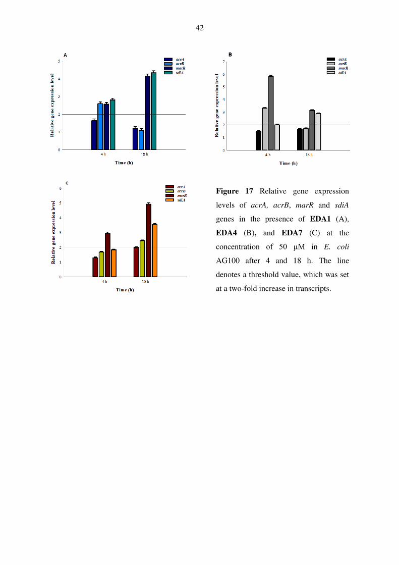
8.3. Selenocompounds
Regarding the effect of selenocompounds on the relative expression of EP, antibiotic
resistance and QS genes in E. coli AG100, the most effective EDA compounds -1, -4 and -7
were examined by gene expression analysis. In the assay, the gene of AcrB, AcrA, SdiA and
the component of the E. coli mar locus were investigated. As shown in Figure 17/A, EDA1 at
50 μM significantly up-regulated acrB, marR and sdiA genes studied after 4 h of exposure,
however, after 18 h, the expression of acrB gene returned to basal level and the marR and
sdiA genes increased significantly. EDA4 up-regulated the expression of acrB, marR and sdiA
after 4 h although after 18 h the expression levels of acrB and marR genes decreased. The QS
gene sdiA was significantly up-regulated after 18 h (Figure 17/B). EDA7 also significantly
up-regulated marR after 4 h and 18 h exposures. After 18 h the RND transporter subunit
genes (acrA, acrB) were significantly up-regulated in the presence of EDA7 (Figure 17/B).
Figure 16 Relative gene expression levels
of acrA, acrB, marR and sdiA in the
presence of PY2 (A), PY4 (B), and PY5 (C)
at the concentration of 50 µg/mL in E. coli
AG100 after 4 and 18 h. The line denotes a
threshold value, which was set at a two-fold
increase in transcripts
A B
C
42
Figure 17 Relative gene expression
levels of acrA, acrB, marR and sdiA
genes in the presence of EDA1 (A),
EDA4 (B), and EDA7 (C) at the
concentration of 50 µM in E. coli
AG100 after 4 and 18 h. The line
denotes a threshold value, which was set
at a two-fold increase in transcripts.

43
DISCUSSION
Multidrug resistance to antibiotics has become a serious problem in the treatment of
infectious diseases. One of the most important mechanisms causing multidrug resistance is
the over-expression of efflux pumps (EPs), whereby cells pump out toxic substances to the
exterior of the cells. Infections caused by multidrug resistant bacteria lead to increased
treatment costs and may result in fatal outcomes; consequently, it is a major challenge for
drug development in order to discover new efflux pump inhibitors (EPIs).
The natural, plant-derived or synthetic compounds may represent a valuable source of new
antibacterial agents because they can inhibit the growth of bacteria and the activity of
bacterial efflux systems which indirectly prevent the formation of biofilm and the bacterial
cell-to-cell communication system, furthermore, they can potentiate the efficacy of antibiotics
as well.
The main goal of our study was to evaluate the antibacterial and multidrug resistance
reversing effects of bioactive compounds from Chleistochlamys kirkii (CK1-5), fluorinated β-
diketo phosphorus ylides (PY1-10) and selenocompounds (EDA1-11) in different bacterial
models. The following methods were used in the studies: minimum inhibitory concentration
(MIC) determination, ethidium bromide (EB) accumulation and efflux assay, checkerboard
combination method, MIC reduction assay, biofilm, quorum sensing (QS) inhibitory test and
reverse transcriptase quantitative polymerase chain reaction (RT-qPCR).
1. Bioactive compounds from C. kirkii (CK1-5)
According to this study chamanetin (CK2) and dichamanetin (CK4) had the most
pronounced antibacterial activity in case of methicillin sensitive and resistant Staphylococcus
aureus strains. These results are supported by previous studies139. Dichamanetin showed
stronger effect than chamanetin, due to its higher lipophilic character resulting from the extra
benzyl group at C‐6. In contrast to chamanetin, isochamanetin (CK3), differing only in the
position of benzyl substituents, was inactive at the concentration tested. Thus, besides the
importance of lipophilicity, the presence of a benzyl moiety at C‐8 appears to play a decisive
role in the antibacterial activity of this type of compounds. In the combination assays,
chamanetin and dichamanetin showed synergism with tetracycline (TET) and ciprofloxacin
(CIP) on the S. aureus reference strain. Furthermore, on the methicillin resistant S. aureus
(MRSA) strain, chamanetin and dichamanetin, combined with TET and CIP, respectively,
also showed synergism, which indicates that these compounds could be potential adjuvants in

44
the therapy. Besides having an antibacterial effect, both chamanetin and dichamanetin could
inhibit the activity of EPs compared to the positive control. Furthermore, these compounds
inhibited the QS between Chromobacterium violaceum 026 and Enterobacter cloacae 31298,
and they caused over-expression of EP genes (norA and mepA) after 4 h of exposure in the
MRSA strain. The change in gene expression could be due to the stress response against
chamanetin and dichamanetin because these compounds as potential noxious agents for S.
aureus had to be extruded from the cytoplasm of the bacterium as soon as possible. This
stress response can be the explanation for the up‐regulation of the EP genes after 4 h.
Chamanetin and dichamanetin influenced the expression of mepA gene in the MRSA strain,
however, these compounds did not have any effect on the expression level of mepA in the
reference S. aureus strain. The difference between methicillin resistant S. aureus and
methicillin susceptible reference S. aureus strains is due to the over-expression of the mepA
gene in the resistant strain and it has low expression level in the reference strain. It has been
demonstrated by other studies that the over-expression of EPs confers a fitness cost for the
organism, for example, a resistant isolate over-expressing EPs shows reduced production of
virulence determinants. Usually the over-expression of pump genes is often related to global
effects on bacterial physiology influencing virulence161. In Gram‐negatives, compounds
CK1–5 did not show antibacterial activity. The highly lipophilic polycarpol (CK1) inhibited
the resistance-nodulation-division (RND) transporter AcrAB-TolC transport system in
Escherichia coli AG100 strain because it could increase the membrane permeability of
bacteria139. In contrast polycarpol had no EPI effect on the pump mutant E. coli AG100 A
strain confirming that it may have a direct EPI effect on the AcrAB-TolC EP. In addition,
apart from chamanetin and dichamanetin, polycarpol and acetylmelodorinol (CK5) were also
able to inhibit effectively the bacterial communication, suggesting that they could be applied
as anti‐QS agents.
2. Fluorinated β-diketo phosphorus ylides (PY1-10)
In our previous study trifluoro-1-phenyl-2-(triphenylphosphoranylidene)butane-1,3-
dione (PY3), 4,4,5,5,5-pentafluoro-1-phenyl-2-(triphenylphosphoranylidene)pentane-1,3-
dione (PY7) and 4,4,5,5,6,6,6-heptafluoro-1-phenyl-2-(triphenylphosphoranylidene)hexane-
1,3-dione (PY8) have been shown to have activity against the EPs of cancer cells141, but did
not show any activity against the EPs of E. coli strains. The aim of the previous work was to
examine the primary ATP-binding cassette subfamily B member (ABCB1) EP inhibitory
effect of PY compounds in cancer cells, in the present study the compounds have been tested

45
against the secondary AcrAB-TolC system in bacteria. It is important to note that ATP-
binding cassette (ABC) transporters derive their energy from the hydrolysis of ATP, however,
the AcrAB-TolC system is a proton motive force (PMF)-dependent multidrug efflux system.
The most effective compounds in E. coli AG100 were
1,1,1-trifluoro-3-oxo-1-methoxy-3-(triphenylphosphoranylidene)propane-2-one (PY2), 4,4,4-
trifluoro-3-oxo-2-(triphenylphosphoranylidene)butanal (PY4), and 1,1,1-trifluoro-3-
(triphenylphosphoranylidene)pentane-2,4-dione (PY5), which inhibited the AcrAB-TolC
system and influenced the expression of the transporter genes acrA and acrB. In addition,
although the compounds are not QS inhibitors, PY4 increased the expression of sdiA after 18
h exposure. The compounds in the R1 chain contain a trifluoromethyl ketone (COCF3) and the
R2 chain is a methoxy (OMe; PY2), formyl (CHO; PY4), and acetyl (COMe; PY5)
substituent. Thus, some structurally related fluorinated β-diketo phosphorus ylides differ in
their multidrug resistance reversal activities between cancer cells and bacterial strains,
indicating that the compounds act differently as inhibitors of ABCB1 and AcrB EPs because
these pumps differ in their structure and energy source driving the pump (ATP and PMF,
respectively)74.
3. Selenocompounds (EDA1-11)
These novel selenocompounds were studied previously for anticancer and ABCB1 EP
inhibitory activity in different cancer cells149–151,162. According to the results on bacteria it was
found that the ketone-containing selenoesters EDA9-11 showed antibacterial activity against
S. aureus reference strain, furthermore the methylketone selenoester (EDA9) was effective on
Enterococcus faecalis ATCC 29212. In the EB accumulation assay the cyclic selenoanhydride
(EDA1) and the meta-substituted benzene selenodiester (EDA4) could inhibit the activity of
the AcrAB-TolC EP system and they were more potent than the positive control promethazine
(PMZ) in E. coli AG100. Methylketone selenoester significantly inhibited the efflux
mechanism of wild-type Salmonella enterica serovar Typhimurium SL1344 strain. EP
inhibiting activity has been found for methoxycarbonylmethyl selenoester (EDA7) in the
acrB mutant S. Typhimurium strain. From the results obtained in the EB efflux assay, it can
be concluded that the same compounds had EP inhibitory activity in the wild-type and mutant
S. Typhimurium strains as well. The similarities observed in the efflux assay of compounds
mean that the derivatives do not directly inhibit the AcrAB-TolC system of S. Typhimurium.
Results showed that the meta-substituted benzene selenodiester (EDA4) and para-substituted
benzene selenodiester (EDA5), which contain a phenyl ring, were the strongest inhibitors of

46
biofilm formation in S. Typhimurium 14028s, indicating the promising potential of these two
difunctionalized derivatives against this Gram-negative strain assayed. EDA7-10 and the
thiophene selenodiester (EDA2) and pyridine selenodiester (EDA3) also exerted a biofilm
inhibiting activity higher than 50% and the inclusion of an amide group (carbamoylmethyl
selenoester; EDA6) or of a 3,5-dimethoxyphenyl moiety (dimethoxy-substituted tert-
butylketone selenoester; EDA11) significantly reduced the biofilm formation in S.
Typhimurium 14028s. Methylketone selenoester reduced the MIC value of TET by 2-fold,
furthermore this compound and chloro-substituted tert-butylketone selenoester potentiated the
activity of TET and CIP on E. coli AG100. In the gene expression analysis, we observed that
cyclic selenoanhydride, meta-substituted benzene selenodiester, and methoxycarbonylmethyl
selenoester influenced the gene expression of resistance (and transporter) genes because these
derivatives significantly up-regulated the marR gene after 4 and 18 h exposure, respectively.
Furthermore, the QS gene sdiA was significantly up-regulated in the presence of cyclic
selenoanhydride, after exposures of 4 h and 18 h.
According to the results these studies indicated that natural and synthetic compounds
could be used as potential antibacterial agents alone or combination with antibiotics for the
treatment of infectious diseases. In addition, our studies are suggesting the importance of the
substituent’s topology and moiety for the biological potency. In future studies, it will be
necessary to design and analyze systematically more compounds and understand the
structure-activity relationships in order to develop potent resistance modifiers.

47
NEW FINDINGS
1. Bioactive compounds from Cleistochlamys kirkii
• Chamanetin (CK2) and dichamanetin (CK4) with a benzyl moiety at C-8 showed
potent antibacterial effect against methicillin susceptible Staphylococcus aureus
ATCC 25923 and methicillin and ofloxacin resistant S. aureus 272123 strains.
Moreover, dichamanetin proved to be the most active compound isolated from C.
kirkii on these strains and has higher lipophilic character compared to chamanetin due
to its extra benzyl group at C‐6.
• Dichamanetin had synergistic activity with tetracycline and ciprofloxacin on S. aureus
reference strain. Furthermore, dichamanetin showed synergism in combination with
tetracycline on the methicillin and ofloxacin resistant S. aureus strain.
• Chamanetin and acetylmelodorinol (CK5) had potent efflux pump modulatory effects
in S. aureus reference strain. Polycarpol (CK1) was the most effective efflux pump
inhibitor on Escherichia coli AG100 inhibiting the AcrAB-TolC transporter system.
• All bioactive compounds of C. kirkii except isochamanetin (CK3) were able to inhibit
effectively the bacterial quorum sensing system.
2. Fluorinated β-diketo phosphorus ylides
• 1,1,1-trifluoro-3-oxo-1-methoxy-3-(triphenylphosphoranylidene)propane-2-one
(PY2), 4,4,4-trifluoro-3-oxo-2-(triphenylphosphoranylidene)butanal (PY4), and 1,1,1-
trifluoro-3-(triphenylphosphoranylidene)pentane-2,4-dione (PY5) inhibited directly
the AcrAB-TolC efflux transporter in E. coli AG100 strain compared to the AcrAB-
TolC pump mutant AG100 A strain.
3. Selenocompounds
• Methylketone selenoester (EDA9) had antibacterial activity against the reference S.
aureus ATCC 25923 and Enterococcus faecalis ATCC 29212 strains.
• Methylketone selenoester potentiated the effect of tetracycline and ciprofloxacin on
the AcrAB-TolC expressing E. coli AG100 strain.
• Almost all selenocompounds showed potent anti-biofilm activity against Salmonella
enterica serovar Typhimurium 14028s strain.
• Cyclic selenoanhydride (EDA1), meta-substituted benzene selenodiester (EDA4) and
methoxycarbonylmethyl selenoester (EDA7) were the most active inhibitors of the
AcrAB-TolC system in E. coli AG100 strain.

48
SUMMARY
Multidrug resistance is a major concern in the treatment of bacterial infections due to the
reduced or missing response of microorganisms to the applied antimicrobial agents. One of the
most important mechanisms of multidrug resistance is the increased expression of efflux pumps
(EPs) that can extrude distinct classes of antibiotics from bacteria to the environment. In order to
overcome this process, efflux pump inhibitors (EPIs) could be designed and applied alone or in
combination with antibiotics as adjuvants. For this reason, the objective of this study was to
investigate the antimicrobial and resistance modifying activity of natural compounds isolated
from Cleistochlamys kirkii, fluorinated β-diketo phosphorus ylides and selenocompounds on
different Gram-positive and Gram-negative strains. The effects of compounds on bacterial growth
were determined by microdilution method. The EP inhibiting activity of compounds on the
accumulation and/or efflux of the general EP substrate ethidium bromide was assessed by real-
time fluorimetry on Escherichia coli, Salmonella Typhimurium and Staphylococcus aureus
strains. The anti-biofilm effect of selenocompounds was investigated by crystal violet assay using
S. Typhimurium 14028s strain. The combined effects of antibiotics (tetracycline and
ciprofloxacin) and compounds were examined by minimum inhibitory concentration reduction
assay or checkerboard microdilution method. The quorum sensing inhibition of natural
compounds and phosphorus ylides was studied by agar diffusion method, furthermore the relative
gene expression level of EP and/or quorum sensing genes were determined by reverse
transcriptase quantitative polymerase chain reaction. Results showed that chamanetin (CK2) and
dichamanetin (CK4) from C. kirkii showed synergism with antibiotics on methicillin sensitive S.
aureus, all the natural compounds except isochamanetin (CK3) were able to inhibit the quorum
sensing. The most potent representative of phosphorus ylides was 4,4,4-trifluoro-3-oxo-2-
(triphenylphosphoranylidene)butanal (PY4) and its effect was more pronounced on the AcrAB-
TolC efflux system expressing E. coli strain. According to the results the methylketone
selenoester (EDA9) had remarkable antibacterial activity against Gram-positive strains. The most
selenocompounds were able to inhibit the biofilm formation of S. Typhimurium. The
selenoanhydride (EDA1) and the meta-substituted benzene selenodiester (EDA4) were active
inhibitors of the AcrAB-TolC system in E. coli AG100. These results suggested that natural
compounds could be effective adjuvants in the antibiotic treatment of infections. Phosphorus
ylides might be valuable EPI compounds to reverse efflux related multidrug resistance in bacteria
and selenocompounds can be effective antibacterials and EPIs.
49
ÖSSZEFOGLALÁS
A multidrog rezisztencia során az előzetesen alkalmazott antibakteriális szer hatástalanná
válik, mely súlyos problémát okoz a bakteriális fertőzések terápiájában. A multidrog rezisztencia
kialakulásának egyik legjelentősebb mechanizmusa az efflux pumpák (EPs) túltermelése, amelyek
a különböző hatásmechanizmusú antibiotikumokat képesek a baktériumsejtből a környezetbe
juttatni. Az efflux mechanizmusokhoz köthető rezisztencia leküzdéséhez fontos lenne olyan efflux
pumpa gátló (EPI) vegyületek tervezése, melyek önmagukban vagy antibiotikummal
kombinációban alkalmazhatóak a fertőzések kezelésében. A jelen disszertáció célja három
vegyületcsoport (Cleistochlamys kirkii-ből izolált természetes vegyületek, foszfor-ilidek, illetve
szelénvegyületek) antimikrobiális és rezisztencia visszafordító hatásának vizsgálata in vitro
Gram-pozitív, illetve Gram-negatív baktérium modellek felhasználásával. A vegyületek
antibakteriális hatását mikrodilúciós módszerrel határoztuk meg, az EP gátlást etídium-bromid
akkumulációs, illetve efflux vizsgálattal tanulmányoztuk valós idejű fluoreszcencia méréssel
Escherichia coli, Salmonella Typhimurium, illetve Staphylococcus aureus törzsek
felhasználásával. A szelénvegyületek biofilm gátló aktivitását kristályibolyás festéssel határoztuk
meg S. Typhimurium 14028s törzsön. A természetes vegyületeket és a szelénvegyületeket
kombinációban vizsgáltuk antibiotikumokkal (ciprofloxacinnal és tetraciklinnel), valamint a
foszfor-ilidek és a C. kirkii származékok quorum-sensing (QS) gátló hatását agardiffúziós
módszerrel határoztuk meg. Mind a három vegyületcsoportból a leghatásosabb EP gátló
aktivitással rendelkező vegyületek génexpresszióra gyakorolt hatását RT-qPCR reakcióval
teszteltük. Eredményeink alapján elmondható, hogy a C. kirkii-ből izolált kamanetin (CK2) és
dikamanetin (CK4) a kísérletben alkalmazott antibiotikumokkal szinergizmust mutatott a
methicillin érzékeny S. aureus törzsön, továbbá az izokamanetint (CK3) kivéve a vegyületek
hatékonyan gátolták a QS-et. A foszfor-ilidek közül a 4,4,4-trifluoro-3-oxo-2-
(trifenilfoszforanilidin)butanal (PY4) gátolta a leghatékonyabban az E. coli AcrAB-TolC efflux
rendszerét. A metilketon szelenoészter (EDA9) a vizsgált Gram-pozitív törzseken jelentős
antibakteriális hatással rendelkezett. A szelénvegyületek hatékonyan gátolták a S. Typhimurium
14028s biofilm képzését, illetve a ciklikus szelenoanhidrid (EDA1) és a meta-szubsztituált benzol
szelenodiészter (EDA4) erősen gátolta az E. coli AG100 AcrAB-TolC rendszerét. A mérési
eredményeink alapján megállapítható, hogy a C. kirkii-ből izolált vegyületek hatásosak lehetnek
adjuvánsként, a foszfor-ilidek EPI vegyületekként, a szelénszármazékok pedig antibakteriális,
illetve EPI vegyületként alkalmazva a bakteriális fertőzések kezelésében.

50
REFERENCES
1. Gould, K. Antibiotics: from prehistory to the present day. J. Antimicrob. Chemother. 71,
572–575 (2016).
2. Lansdown, A. B. Silver in Health Care: Antimicrobial effects and safety in use. Curr.
Probl. Dermatol. 33, 17–34 (2006).
3. Ventola, C. L. The antibiotic resistance crisis: part 1: causes and threats. P T. 40, 277–283 (2015).
4. Ferri, M., Ranucci, E., Romagnoli, P. & Giaccone, V. Antimicrobial resistance: a global
emerging threat to public health systems. Crit. Rev. Food Sci. Nutr. 57, 2857–2876
(2017).
5. Goossens, H., Ferech, M., Vander Stichele, R., Elseviers, M. & ESAC Project Group.
Outpatient antibiotic use in Europe and association with resistance: a cross-national
database study. Lancet Lond. Engl. 365, 579–587 (2005).
6. Sun, L., Klein, E. Y. & Laxminarayan, R. Seasonality and temporal correlation between
community antibiotic use and resistance in the United States. Clin. Infect. Dis. 55, 687–694 (2012).
7. Economou, V. & Gousia, P. Agriculture and food animals as a source of antimicrobial-
resistant bacteria. Infect. Drug Resist. 8, 49–61 (2015).
8. Cabello, F. C. Heavy use of prophylactic antibiotics in aquaculture: a growing problem
for human and animal health and for the environment. Environ. Microbiol. 8, 1137–1144 (2006).
9. von Wintersdorff, C. J. H. et al. Dissemination of antimicrobial resistance in microbial
ecosystems through horizontal gene transfer. Front. Microbiol. 7, 173 (2016).
10. The European Centre for Disease Prevention and Control (ECDC). Surveillance of
antimicrobial resistance in Europe. Annual report of the european antimicrobial
resistance surveillance Network (EARS-Net). (2017).
11. O’Neill, J. Antimicrobial resistance: tackling a crisis for the health and wealth of
nations. (2015). https://amr-review.org/
12. Centers for Disease Control and Prevention. Antibiotic resistance threats in the United
States, 2013. (2013). https://www.cdc.gov/drugresistance/pdf/ar-threats-2013-508.pdf
13. Livermore, D. M. Bacterial resistance: origins, epidemiology, and impact. Clin. Infect.
Dis. 36, S11-23 (2003).
14. Arzanlou, M., Chai, W. C. & Venter, H. Intrinsic, adaptive and acquired antimicrobial
resistance in Gram-negative bacteria. Essays Biochem. 61, 49–59 (2017).
15. Davies, J. & Davies, D. Origins and evolution of antibiotic resistance. Microbiol. Mol.
Biol. Rev. 74, 417–433 (2010).
16. Blair, J. M. A., Webber, M. A., Baylay, A. J., Ogbolu, D. O. & Piddock, L. J. V.
Molecular mechanisms of antibiotic resistance. Nat. Rev. Microbiol. 13, 42–51 (2015).
17. Webber, M. A. & Piddock, L. J. V. The importance of efflux pumps in bacterial
antibiotic resistance. J. Antimicrob. Chemother. 51, 9–11 (2003).
18. Lewis, K. Persister cells. Annu. Rev. Microbiol. 64, 357–372 (2010).

51
19. Zgurskaya, H. I., Löpez, C. A. & Gnanakaran, S. Permeability barrier of Gram-negative
cell envelopes and approaches to bypass it. ACS Infect. Dis. 1, 512–522 (2015).
20. González-Bello, C. Antibiotic adjuvants - A strategy to unlock bacterial resistance to
antibiotics. Bioorg. Med. Chem. Lett. 27, 4221–4228 (2017).
21. Furuya, E. Y. & Lowy, F. D. Antimicrobial-resistant bacteria in the community setting.
Nat. Rev. Microbiol. 4, 36–45 (2006).
22. Heuer, H. & Smalla, K. Horizontal gene transfer between bacteria. Environ. Biosafety
Res. 6, 3–13 (2007).
23. de Vries, J. & Wackernagel, W. Integration of foreign DNA during natural
transformation of Acinetobacter sp. by homology-facilitated illegitimate recombination.
Proc. Natl. Acad. Sci. U. S. A. 99, 2094–2099 (2002).
24. Thomas, C. M. & Nielsen, K. M. Mechanisms of, and barriers to, horizontal gene
transfer between bacteria. Nat. Rev. Microbiol. 3, 711–721 (2005).
25. Unemo, M. et al. High-level cefixime- and ceftriaxone-resistant Neisseria gonorrhoeae
in France: novel penA mosaic allele in a successful international clone causes treatment
failure. Antimicrob. Agents Chemother. 56, 1273–1280 (2012).
26. Brown-Jaque, M., Calero-Cáceres, W. & Muniesa, M. Transfer of antibiotic-resistance
genes via phage-related mobile elements. Plasmid 79, 1–7 (2015).
27. Colomer-Lluch, M., Imamovic, L., Jofre, J. & Muniesa, M. Bacteriophages carrying
antibiotic resistance genes in fecal waste from cattle, pigs, and poultry. Antimicrob.
Agents Chemother. 55, 4908–4911 (2011).
28. Hayes F. The horizontal gene pool: bacterial plasmids and gene spread. Editor: Thomas
CM. 86, 251-252 (Harwood Academic Publishers, 2000).
29. Guglielmini, J., de la Cruz, F. & Rocha, E. P. C. Evolution of conjugation and type IV
secretion systems. Mol. Biol. Evol. 30, 315–331 (2013).
30. Holmes, A. H. et al. Understanding the mechanisms and drivers of antimicrobial
resistance. Lancet Lond. Engl. 387, 176–187 (2016).
31. Dhanji, H. et al. Molecular epidemiology of fluoroquinolone-resistant ST131
Escherichia coli producing CTX-M extended-spectrum beta-lactamases in nursing
homes in Belfast, UK. J. Antimicrob. Chemother. 66, 297–303 (2011).
32. Walsh, T. R., Weeks, J., Livermore, D. M. & Toleman, M. A. Dissemination of NDM-1
positive bacteria in the New Delhi environment and its implications for human health:
an environmental point prevalence study. Lancet Infect. Dis. 11, 355–362 (2011).
33. Borges, A. et al. New perspectives on the use of phytochemicals as an emergent
strategy to control bacterial infections including biofilms. Molecules 21, pii: E877
(2016).
34. Wright, G. D. Molecular mechanisms of antibiotic resistance. Chem. Commun. 47, 4055
(2011).
35. Alav, I., Sutton, J. M. & Rahman, K. M. Role of bacterial efflux pumps in biofilm
formation. J. Antimicrob. Chemother. 73, 2003–2020 (2018).
36. Alcalde-Rico, M., Hernando-Amado, S., Blanco, P. & Martínez, J. L. Multidrug efflux
pumps at the crossroad between antibiotic resistance and bacterial virulence. Front.
Microbiol. 7, 1483 (2016).

52
37. Dockrell, H. M., Goering, R. V., Roitt, I., Wakelin, D., Zuckerman, M. Attacking the
enemy: antimicrobial agents and chemotherapy. In: Medical Microbiology.
Editors: Mims, C., Dockrell, H. M., Goering, R. V., Roitt, I., Wakelin, D., Zuckerman,
M. 473-507 (Elsevier Mosby) (2004).
38. Kapoor, G., Saigal, S. & Elongavan, A. Action and resistance mechanisms of
antibiotics: A guide for clinicians. J. Anaesthesiol. Clin. Pharmacol. 33, 300 (2017).
39. Maurice, F. et al. Enzyme structural plasticity and the emergence of broad-spectrum
antibiotic resistance. EMBO Rep. 9, 344–349 (2008).
40. Alekshun, M. N. & Levy, S. B. Molecular mechanisms of antibacterial multidrug
resistance. Cell 128, 1037–1050 (2007).
41. Doi, Y., Iovleva, A. & Bonomo, R. A. The ecology of extended-spectrum β-lactamases
(ESBLs) in the developed world. J. Travel Med. 24, S44–S51 (2017).
42. Tolmasky, M., E. Bacterial resistance to aminoglycosides and beta-lactams: the Tn1331
transposon paradigm. Front. Biosci. 5, D20-29 (1999).
43. Hiramatsu, K., Cui, L., Kuroda, M. & Ito, T. The emergence and evolution of
methicillin-resistant Staphylococcus aureus. Trends Microbiol. 9, 486–493 (2001).
44. Fernandez, L. & Hancock, R. E. W. Adaptive and mutational resistance: role of porins
and efflux pumps in drug resistance. Clin. Microbiol. Rev. 25, 661–681 (2012).
45. Masi, M., Winterhalter, M. & Pagès, J.-M. Outer membrane porins. In: Bacterial cell
walls and membranes. Editor: Kuhn, A. 92, 79–123 (Springer International Publishing),
(2019).
46. Pagès, J.M., James, C. E. & Winterhalter, M. The porin and the permeating antibiotic: a
selective diffusion barrier in Gram-negative bacteria. Nat. Rev. Microbiol. 6, 893–903
(2008).
47. Giedraitienė, A., Vitkauskienė, A., Naginienė, R. & Pavilonis, A. Antibiotic resistance
mechanisms of clinically important bacteria. Medicina 47, 137-146 (2011).
48. Nikaido, H. Multidrug resistance in bacteria. Annu. Rev. Biochem. 78, 119–146 (2009).
49. Li, X.-Z. & Nikaido, H. Efflux-mediated drug resistance in bacteria: an update. Drugs
69, 1555–1623 (2009).
50. Amaral, L., Martins, A., Spengler, G. & Molnar, J. Efflux pumps of Gram-negative
bacteria: what they do, how they do it, with what and how to deal with them. Front.
Pharmacol. 4, 168 (2014).
51. Poole, K. Efflux pumps as antimicrobial resistance mechanisms. Ann. Med. 39, 162–176
(2007).
52. Higgins, C. F. Multiple molecular mechanisms for multidrug resistance transporters.
Nature 446, 749–757 (2007).
53. Hassan, K. A., Liu, Q., Henderson, P. J. F. & Paulsen, I. T. Homologs of the
Acinetobacter baumannii AceI transporter represent a new family of bacterial multidrug
efflux systems. MBio. 6, (2015).
54. Schindler, B. D. & Kaatz, G. W. Multidrug efflux pumps of Gram-positive bacteria.
Drug Resist. Updat. 27, 1–13 (2016).
55. Mousa, J. J. & Bruner, S. D. Structural and mechanistic diversity of multidrug
transporters. Nat. Prod. Rep. 33, 1255–1267 (2016).

53
56. Lubelski, J., Konings, W. N. & Driessen, A. J. M. Distribution and physiology of ABC-
type transporters contributing to multidrug resistance in bacteria. Microbiol. Mol. Biol.
Rev. 71, 463–476 (2007).
57. Langton, K. P., Henderson, P. J. F. & Herbert, R. B. Antibiotic resistance: multidrug
efflux proteins, a common transport mechanism? Nat. Prod. Rep. 22, 439–451 (2005).
58. Kim, J. et al. Subnanometre-resolution electron cryomicroscopy structure of a
heterodimeric ABC exporter. Nature 517, 396–400 (2015).
59. Hollenstein, K., Frei, D. C. & Locher, K. P. Structure of an ABC transporter in complex
with its binding protein. Nature 446, 213–216 (2007).
60. Hohl, M. et al. Structural basis for allosteric cross-talk between the asymmetric
nucleotide binding sites of a heterodimeric ABC exporter. Proc. Natl. Acad. Sci. U. S.
A. 111, 11025–11030 (2014).
61. Lee, E.-W., Huda, M. N., Kuroda, T., Mizushima, T. & Tsuchiya, T. EfrAB, an ABC
multidrug efflux pump in Enterococcus faecalis. Antimicrob. Agents Chemother. 47,
3733–3738 (2003).
62. Kobayashi, N., Nishino, K. & Yamaguchi, A. Novel macrolide-specific ABC-type
efflux transporter in Escherichia coli. J. Bacteriol. 183, 5639–5644 (2001).
63. Boncoeur, E. et al. PatA and PatB form a functional heterodimeric ABC multidrug
efflux transporter responsible for the resistance of Streptococcus pneumoniae to
fluoroquinolones. Biochemistry 51, 7755–7765 (2012).
64. van Veen, H. W., Margolles, A., Müller, M., Higgins, C. F. & Konings, W. N. The homodimeric ATP-binding cassette transporter LmrA mediates multidrug transport by
an alternating two-site (two-cylinder engine) mechanism. EMBO J. 19, 2503–2514
(2000).
65. Du, D., van Veen, H. W., Murakami, S., Pos, K. M. & Luisi, B. F. Structure,
mechanism and cooperation of bacterial multidrug transporters. Curr. Opin. Struct. Biol.
33, 76–91 (2015).
66. Kuroda, T. & Tsuchiya, T. Multidrug efflux transporters in the MATE family. Biochim.
Biophys. Acta 1794, 763–768 (2009).
67. Jin, Y., Nair, A. & van Veen, H. W. Multidrug transport protein NorM from Vibrio
cholerae simultaneously couples to sodium- and proton-motive force. J. Biol. Chem.
289, 14624–14632 (2014).
68. Long, F., Rouquette-Loughlin, C., Shafer, W. M. & Yu, E. W. Functional cloning and
characterization of the multidrug efflux pumps NorM from Neisseria gonorrhoeae and
YdhE from Escherichia coli. Antimicrob. Agents Chemother. 52, 3052–3060 (2008).
69. Xu, X. J. et al. Molecular cloning and characterization of the HmrM multidrug efflux
pump from Haemophilus influenzae Rd. Microbiol. Immunol. 47, 937–943 (2003).
70. He, G. X. et al. An H+-coupled multidrug efflux pump, PmpM, a member of the MATE
family of transporters, from Pseudomonas aeruginosa. J. Bacteriol. 186, 262–265
(2004).
71. Banchs, C., Poulos, S., Nimjareansuk, W. S., Joo, Y. E. & Faham, S. Substrate binding
to the multidrug transporter MepA. Biochim. Biophys. Acta 1838, 2539–2546 (2014).
72. Putman, M., van Veen, H. W. & Konings, W. N. Molecular properties of bacterial
multidrug transporters. Microbiol. Mol. Biol. Rev. 64, 672–693 (2000).

54
73. Law, C. J., Maloney, P. C. & Wang, D. N. Ins and outs of major facilitator superfamily
antiporters. Annu. Rev. Microbiol. 62, 289–305 (2008).
74. Spengler, G., Kincses, A., Gajdács, M. & Amaral, L. New roads leading to old
destinations: efflux pumps as targets to reverse multidrug resistance in bacteria.
Molecules 22, E468 (2017).
75. Tanabe, M. et al. The multidrug resistance efflux complex, EmrAB from Escherichia
coli forms a dimer in vitro. Biochem. Biophys. Res. Commun. 380, 338–342 (2009).
76. Xianwei, T., Diannan, L. & Boxiong, W. Substrate transport pathway inside outward
open conformation of EmrD: a molecular dynamics simulation study. Mol. Biosyst. 12,
2634–2641 (2016).
77. Edgar, R. & Bibi, E. MdfA, an Escherichia coli multidrug resistance protein with an
extraordinarily broad spectrum of drug recognition. J. Bacteriol. 179, 2274–2280
(1997).
78. Adler, J. & Bibi, E. Determinants of substrate recognition by the Escherichia coli
multidrug transporter MdfA identified on both sides of the membrane. J. Biol. Chem.
279, 8957–8965 (2004).
79. Bhaskar, B. V., Babu, T. M. C., Reddy, N. V. & Rajendra, W. Homology modeling,
molecular dynamics, and virtual screening of NorA efflux pump inhibitors of
Staphylococcus aureus. Drug Des. Devel. Ther. 10, 3237–3252 (2016).
80. Paulsen, I. T., Brown, M. H., Littlejohn, T. G., Mitchell, B. A. & Skurray, R. A.
Multidrug resistance proteins QacA and QacB from Staphylococcus aureus: membrane
topology and identification of residues involved in substrate specificity. Proc. Natl.
Acad. Sci. U. S. A. 93, 3630–3635 (1996).
81. Floyd, J. L., Smith, K. P., Kumar, S. H., Floyd, J. T. & Varela, M. F. LmrS is a
multidrug efflux pump of the major facilitator superfamily from Staphylococcus aureus.
Antimicrob. Agents Chemother. 54, 5406–5412 (2010).
82. Roth, A. & Govaerts, C. LmrP from Lactoccoccus lactis: a tractable model to
understand secondary multidrug transport in MFS. Res. Microbiol. 169, 468–477
(2018).
83. Piddock, L. J., Johnson, M. M., Simjee, S. & Pumbwe, L. Expression of efflux pump
gene pmrA in fluoroquinolone-resistant and -susceptible clinical isolates of
Streptococcus pneumoniae. Antimicrob. Agents Chemother. 46, 808–812 (2002).
84. Kermani, A. A., Macdonald, C. B., Gundepudi, R. & Stockbridge, R. B. Guanidinium
export is the primal function of SMR family transporters. Proc. Natl. Acad. Sci. U. S. A.
115, 3060–3065 (2018).
85. Nasie, I., Steiner-Mordoch, S. & Schuldiner, S. New substrates on the block: clinically
relevant resistances for EmrE and homologues. J. Bacteriol. 194, 6766–6770 (2012).
86. Schuldiner, S., Lebendiker, M. & Yerushalmi, H. EmrE, the smallest ion-coupled
transporter, provides a unique paradigm for structure-function studies. J. Exp. Biol. 200,
335–341 (1997).
87. Kikukawa, T., Miyauchi, S., Araiso, T., Kamo, N. & Nara, T. Anti-parallel membrane
topology of two components of EbrAB, a multidrug transporter. Biochem. Biophys. Res.
Commun. 358, 1071–1075 (2007).

55
88. Seier-Petersen, M. A., Nielsen, L. N., Ingmer, H., Aarestrup, F. M. & Agersø, Y. Biocide susceptibility of Staphylococcus aureus CC398 and CC30 isolates from pigs
and identification of the biocide resistance genes, qacG and qacC. Microb. Drug Resist.
21, 527–536 (2015).
89. Sarkar, S. K., Bhattacharyya, A. & Mandal, S. M. YnfA, a SMR family efflux pump is
abundant in Escherichia coli isolates from urinary infection. Indian J. Med. Microbiol.
33, 139–142 (2015).
90. Hassan, K. A. et al. Pacing across the membrane: the novel PACE family of efflux
pumps is widespread in Gram-negative pathogens. Res. Microbiol. 169, 450–454
(2018).
91. Daury, L. et al. Tripartite assembly of RND multidrug efflux pumps. Nat. Commun. 7,
10731 (2016).
92. Nikaido, H. & Takatsuka, Y. Mechanisms of RND multidrug efflux pumps. Biochim.
Biophys. Acta 1794, 769–781 (2009).
93. Yang, L. et al. RND transporters protect Corynebacterium glutamicum from antibiotics
by assembling the outer membrane. Microbiologyopen 3, 484–496 (2014).
94. Alnaseri, H. et al. Inducible expression of a resistance-nodulation-division-type efflux
pump in Staphylococcus aureus provides resistance to linoleic and arachidonic acids. J.
Bacteriol. 197, 1893–1905 (2015).
95. Kim, J. S. et al. Structure of the tripartite multidrug efflux pump AcrAB-TolC suggests
an alternative assembly mode. Mol. Cells 38, 180–186 (2015).
96. Li, X.-Z., Plésiat, P. & Nikaido, H. The challenge of efflux-mediated antibiotic
resistance in Gram-negative bacteria. Clin. Microbiol. Rev. 28, 337–418 (2015).
97. Venter, H., Mowla, R., Ohene-Agyei, T. & Ma, S. RND-type drug efflux pumps from Gram-negative bacteria: molecular mechanism and inhibition. Front. Microbiol. 6, 377
(2015).
98. Hobbs, E. C., Yin, X., Paul, B. J., Astarita, J. L. & Storz, G. Conserved small protein
associates with the multidrug efflux pump AcrB and differentially affects antibiotic
resistance. Proc. Natl. Acad. Sci. U. S. A. 109, 16696–16701 (2012).
99. Du, D. et al. Structure of the AcrAB-TolC multidrug efflux pump. Nature 509, 512–515
(2014).
100. Piddock, L. J. Multidrug-resistance efflux pumps - not just for resistance. Nat. Rev.
Microbiol. 4, 629–636 (2006).
101. Ruggerone, P., Murakami, S., Pos, K. M. & Vargiu, A. V. RND efflux pumps:
structural information translated into function and inhibition mechanisms. Curr. Top.
Med. Chem. 13, 3079–3100 (2013).
102. Nikaido, H. & Zgurskaya, H. I. AcrAB and related multidrug efflux pumps of
Escherichia coli. J. Mol. Microbiol. Biotechnol. 3, 215–218 (2001).
103. Pan, Y.-P., Xu, Y.-H., Wang, Z.-X., Fang, Y.-P. & Shen, J.-L. Overexpression of
MexAB-OprM efflux pump in carbapenem-resistant Pseudomonas aeruginosa. Arch.
Microbiol. 198, 565–571 (2016).
104. Terzi, H. A., Kulah, C. & Ciftci, I. H. The effects of active efflux pumps on antibiotic
resistance in Pseudomonas aeruginosa. World J. Microbiol. Biotechnol. 30, 2681–2687
(2014).

56
105. Rosenberg, E. Y., Ma, D. & Nikaido, H. AcrD of Escherichia coli is an aminoglycoside
efflux pump. J. Bacteriol. 182, 1754–1756 (2000).
106. Srinivasan, V. B., Singh, B. B., Priyadarshi, N., Chauhan, N. K. & Rajamohan, G. Role
of novel multidrug efflux pump involved in drug resistance in Klebsiella pneumoniae.
PLoS One 9, e96288 (2014).
107. Hagman, K. E. et al. The MtrD protein of Neisseria gonorrhoeae is a member of the
resistance-nodulation-division protein family constituting part of an efflux system.
Microbiol. Read. Engl. 143, 2117–2125 (1997).
108. Wang, Z. et al. An allosteric transport mechanism for the AcrAB-TolC multidrug efflux
pump. Elife 6, e24905 (2017).
109. Suresh, M. K., Biswas, R. & Biswas, L. An update on recent developments in the
prevention and treatment of Staphylococcus aureus biofilms. Int. J. Med. Microbiol.
309, 1–12 (2019).
110. Jamal, M. et al. Bacterial biofilm and associated infections. J. Chin. Med. Assoc. 81, 7–11 (2018).
111. Steenackers, H., Hermans, K., Vanderleyden, J. & De Keersmaecker, S. C. J.
Salmonella biofilms: An overview on occurrence, structure, regulation and eradication.
Food Res. Int. 45, 502–531 (2012).
112. del Pozo, J. L. & Patel, R. The challenge of treating biofilm-associated bacterial
infections. Clin. Pharmacol. Ther. 82, 204–209 (2007).
113. Soto, S. M. Role of efflux pumps in the antibiotic resistance of bacteria embedded in a
biofilm. Virulence 4, 223–229 (2013).
114. Renner, L. D. & Weibel, D. B. Physicochemical regulation of biofilm formation. MRS
Bull. 36, 347–355 (2011).
115. Flemming, H. C., Neu, T. R. & Wozniak, D. J. The EPS matrix: the ‘house of biofilm cells’. J. Bacteriol. 189, 7945–7947 (2007).
116. Rabin, N. et al. Biofilm formation mechanisms and targets for developing antibiofilm
agents. Future Med. Chem. 7, 493–512 (2015).
117. Sauer, K. The genomics and proteomics of biofilm formation. Genome Biol. 4, 219
(2003).
118. Wu, H., Moser, C., Wang, H.-Z., Høiby, N. & Song, Z. J. Strategies for combating
bacterial biofilm infections. Int. J. Oral Sci. 7, 1–7 (2015).
119. Suci, P. A., Mittelman, M. W., Yu, F. P. & Geesey, G. G. Investigation of ciprofloxacin
penetration into Pseudomonas aeruginosa biofilms. Antimicrob. Agents Chemother. 38,
2125–2133 (1994).
120. Del Pozo, J. L. Biofilm-related disease. Expert Rev. Anti Infect. Ther. 16, 51–65 (2018).
121. Molin, S. & Tolker-Nielsen, T. Gene transfer occurs with enhanced efficiency in
biofilms and induces enhanced stabilisation of the biofilm structure. Curr. Opin.
Biotechnol. 14, 255–261 (2003).
122. Zhang, L. & Mah, T. F. Involvement of a novel efflux system in biofilm-specific
resistance to antibiotics. J. Bacteriol. 190, 4447–4452 (2008).
123. Lyon, G. J. & Muir, T. W. Chemical signaling among bacteria and its inhibition. Chem.
Biol. 10, 1007–1021 (2003).

57
124. Lipa, P., Kozieł, M. & Janczarek, M. Quorum sensing in Gram-negative bacteria: signal
molecules, inhibitors and their potential therapeutic application. Postepy Biochem. 63,
242–260 (2017).
125. Brunel, A.-S. & Guery, B. Multidrug resistant (or antimicrobial-resistant) pathogens -
alternatives to new antibiotics? Swiss Med. Wkly. 147, w14553 (2017).
126. Papenfort, K. & Bassler, B. L. Quorum sensing signal-response systems in Gram-
negative bacteria. Nat. Rev. Microbiol. 14, 576–588 (2016).
127. Kanamaru, K., Kanamaru, K., Tatsuno, I., Tobe, T. & Sasakawa, C. SdiA, an
Escherichia coli homologue of quorum-sensing regulators, controls the expression of
virulence factors in enterohaemorrhagic Escherichia coli O157:H7. Mol. Microbiol. 38,
805–816 (2000).
128. Tavío, M. M. et al. Quorum-sensing regulator sdiA and marA overexpression is
involved in in vitro-selected multidrug resistance of Escherichia coli. J. Antimicrob.
Chemother. 65, 1178–1186 (2010).
129. Sweeney, L. C. Antibiotic resistance in general dental practice – a cause for concern? J.
Antimicrob. Chemother. 53, 567–576 (2004).
130. Upadhyay, A. et al. Inhibiting microbial toxins using plant-derived compounds and
plant extracts. Medicines 2, 186–211 (2015).
131. Savoia, D. Plant-derived antimicrobial compounds: alternatives to antibiotics. Future
Microbiol. 7, 979–990 (2012).
132. Stavri, M., Piddock, L. J. & Gibbons, S. Bacterial efflux pump inhibitors from natural
sources. J. Antimicrob. Chemother. 59, 1247–1260 (2007).
133. Neyfakh, A. A., Bidnenko, V. E. & Chen, L. B. Efflux-mediated multidrug resistance in
Bacillus subtilis: similarities and dissimilarities with the mammalian system. Proc. Natl.
Acad. Sci. 88, 4781–4785 (1991).
134. Khan, I. A., Mirza, Z. M., Kumar, A., Verma, V. & Qazi, G. N. Piperine, a
phytochemical potentiator of ciprofloxacin against Staphylococcus aureus. Antimicrob.
Agents Chemother. 50, 810–812 (2006).
135. Garvey, M. I., Rahman, M. M., Gibbons, S. & Piddock, L. J. V. Medicinal plant extracts
with efflux inhibitory activity against Gram-negative bacteria. Int. J. Antimicrob. Agents
37, 145–151 (2011).
136. Mouwakeh, A. et al. Nigella sativa essential oil and its bioactive compounds as
resistance modifiers against Staphylococcus aureus. Phytother. Res. 33, 1010–1018
(2019).
137. Verzár, R. & Petri, G. Medicinal plants in Mozambique and their popular use. J.
Ethnopharmacol. 19, 67–80 (1987).
138. Samwel, S. et al. Cleistenolide and cleistodienol: novel bioactive constituents of
Cleistochlamys kirkii. Nat. Prod. Commun. 2, 737-741 (2007).
139. Pereira, F. et al. Cleistenolide and cleistodienol: novel bioactive constituents of
Cleistochlamys kirkii. chemical constituents: antibacterial activity and synergistic
effects against resistant Staphylococcus aureus strains. J. Ethnopharmacol. 178, 180–187 (2016).

58
140. El-Batta, A. et al. Wittig reactions in water media employing stabilized ylides with
aldehydes. Synthesis of α, β -unsaturated esters from mixing aldehydes, alpha-
bromoesters, and Ph3P in aqueous NaHCO3. J. Org. Chem. 72, 5244–5259 (2007).
141. Spengler, G. et al. Fluorinated β-diketo phosphorus ylides are novel inhibitors of the
ABCB1 efflux pump of cancer cells. Anticancer Res. 35, 5915–5919 (2015).
142. Lobanov, A. V., Hatfield, D. L. & Gladyshev, V. N. Eukaryotic selenoproteins and
selenoproteomes. Biochim. Biophys. Acta 1790, 1424–1428 (2009).
143. Vila, J. et al. Escherichia coli: an old friend with new tidings. FEMS Microbiol. Rev.
40, 437–463 (2016).
144. Tong, S. Y. C., Davis, J. S., Eichenberger, E., Holland, T. L. & Fowler, V. G.
Staphylococcus aureus infections: epidemiology, pathophysiology, clinical
manifestations, and management. Clin. Microbiol. Rev. 28, 603–661 (2015).
145. Mancini, A. et al. Retrospective analysis of nosocomial infections in an Italian tertiary
care hospital. New Microbiol. 39, 197–205 (2016).
146. Sonkusre, P. & Singh Cameotra, S. Biogenic selenium nanoparticles inhibit
Staphylococcus aureus adherence on different surfaces. Colloids Surf. B Biointerfaces
136, 1051–1057 (2015).
147. Kumar, B. S. et al. Anti-ulcer and antimicrobial activities of sodium selenite against
Helicobacter pylori: in vitro and in vivo evaluation. Scand. J. Infect. Dis. 42, 266–274
(2010).
148. Tran, P. A. & Webster, T. J. Antimicrobial selenium nanoparticle coatings on polymeric
medical devices. Nanotechnology 24, 155101 (2013).
149. Domínguez-Álvarez, E. et al. Identification of selenocompounds with promising
properties to reverse cancer multidrug resistance. Bioorg. Med. Chem. Lett. 26, 2821–2824 (2016).
150. Gajdács, M. et al. Selenoesters and selenoanhydrides as novel multidrug resistance
reversing agents: a confirmation study in a colon cancer MDR cell line. Bioorg. Med.
Chem. Lett. 27, 797–802 (2017).
151. Domínguez-Álvarez, E. et al. Synthesis and antiproliferative activity of novel
selenoester derivatives. Eur. J. Med. Chem. 73, 153–166 (2014).
152. Smith, H. E. & Blair, J. M. A. Redundancy in the periplasmic adaptor proteins AcrA
and AcrE provides resilience and an ability to export substrates of multidrug efflux. J.
Antimicrob. Chemother. 69, 982–987 (2014).
153. Varga, Z. G. et al. Inhibition of quorum sensing and efflux pump system by
trifluoromethyl ketone proton pump inhibitors. In Vivo 26, 277–285 (2012).
154. Clinical and Laboratory Standards Institute (CLSI). Performance standards for
antimicrobial susceptibility testing. 28th ed. CLSI supplement M100. (2018).
155. Viveiros, M. et al. Demonstration of intrinsic efflux activity of Escherichia coli K-12
AG100 by an automated ethidium bromide method. Int. J. Antimicrob. Agents 31, 458-
462 (2008).
156. Wolfart, K. et al. Synergistic interaction between proton pump inhibitors and resistance
modifiers: promoting effects of antibiotics and plasmid curing. In Vivo 20, 367–372
(2006).

59
157. Chou, T. C. Theoretical basis, experimental design, and computerized simulation of
synergism and antagonism in drug combination studies. Pharmacol. Rev. 58, 621–681
(2006).
158. Couto, I., Costa, S. S., Viveiros, M., Martins, M. & Amaral, L. Efflux-mediated
response of Staphylococcus aureus exposed to ethidium bromide. J. Antimicrob.
Chemother. 62, 504–513 (2008).
159. Viveiros, M. et al. Antibiotic stress, genetic response and altered permeability of E. coli.
PloS One 2, e365 (2007).
160. Livak, K. J. & Schmittgen, T. D. Analysis of relative gene expression data using real-
time quantitative PCR and the 2-ΔΔCT method. Methods 25, 402–408 (2001).
161. Ledda, A. et al. Re-emergence of methicillin susceptibility in a resistant lineage of
Staphylococcus aureus. J. Antimicrob. Chemother. 72, 1285–1288 (2017).
162. Spengler, G., Gajdács, M., Marć, M. A., Domínguez-Álvarez, E. & Sanmartín, C. Organoselenium compounds as novel adjuvants of chemotherapy drugs-a promising
approach to fight cancer drug resistance. Molecules 24, e336 (2019).
163. Grela, E., Kozłowska, J. & Grabowiecka, A. Current methodology of MTT assay in
bacteria – A review. Acta Histochem. 120, 303-311 (2018).

60
ACKNOWLEDGEMENTS
I would like to express my deep and sincere gratitude to my supervisor, Dr. Gabriella
Spengler for the opportunity to work in her research group and for opening the world to me. I
am grateful to her for introducing me to the scientific research, for her excellent support and
encouragement.
I would like to thank Dr. Katalin Burián, chair of the Department of Medical Microbiology
and Immunobiology, for the possibility to work at the department.
I am extremly grateful to our collaborators Dr. Enrique Domínguez-Álvarez, Prof. Dr.
Carmen Sanmartín, Prof. Dr. Maria-José U. Ferreira and Prof. Dr. Masami Kawase for
providing us compounds for biological evaluation, furthermore their help and support resulted
in fruitful scientific collaborations, projects and research grants.
I would like to give my special thanks to Mrs Anikó Vigyikánné Váradi for her technical
assistance. I am grateful to Mrs Györgyi Müllerné Deák for the help with measuring the
concentration of RNA.
I would like to thank all undergraduate students working in our research group who helped
me during my studies.
I am grateful to all the members of the department for their support and for creating a pleasant
working environment during my Ph.D studies.
I am likewise grateful to Dr. Jessica M. A. Blair and Dr. Helen McNeil for the help with the
biofilm and efflux assays at the University of Birmingham.
I would like to thank to Dr. Márió Gajdács for the help in naming compounds.
I am deeply grateful to my best friend Dr. Tímea Mosolygó and my friends Márta Bozóki-Nové, Dóra Kovács, Csilla Abonyi and Dr. Anita Varga-Bogdanov for their selfless help,
encouragment and friendship.
Lastly, I feel a deep sense of gratitude to my parents, my sisters and my boyfriend for their
love, patience and encouragement.

61
FINANCIAL SUPPORT
The work on which this thesis was based on was supported by the following organizations
and grants:
• New National Excellence Program of the Ministry of Human Capacities (ÚNKP-17-3;
ÚNKP-18-3)
• Tempus Public Foundation: Campus mundi student mobility, short study grant
• Talent Scholarship and Excellence List of the University of Szeged (2016/2017:
Bronze level; 2017/2018: Gold level)
• Travel Award of the European Society of Clinical Microbiology and Infectious
Diseases
• SZTE ÁOK-KKA 2018/270-62-2 of the University of Szeged, Faculty of Medicine,
Hungary: Selenium derivatives as novel promising antimicrobial agents
• GINOP-2.3.2-15-2016-00012: New ways in natural medicine-based drug research:
System-metabolomic approaches to screen for bioactive terpenoids of plant and
microbial origin
• 2019-2021: Hungarian-Portuguese Intergovernmental S&T Cooperation Programme.
Title: Plant-derived compounds as anticancer agents: a promising approach for
overcoming multidrug resistance

62
APPENDIX
Appendix 1 IUPAC names of the P-ylides compounds
PY1: 4,4,4-trifluoro-3-oxo-2-(triphenylphosphoranylidene)butanenitrile
PY2: 1,1,1-trifluoro-3-oxo-1-methoxy-3-(triphenylphosphoranylidene)propane-2-one
PY3: trifluoro-1-phenyl-2-(triphenylphosphoranylidene)butane-1,3-dione
PY4: 4,4,4-trifluoro-3-oxo-2-(triphenylphosphoranylidene)butanal
PY5: 1,1,1-trifluoro-3-(triphenylphosphoranylidene)pentane-2,4-dione
PY6: ethyl-4,4,4-trifluoro-3-oxo-2-(triphenylphosphoranylidene)butanoate
PY7: 4,4,5,5,5-pentafluoro-1-phenyl-2-(triphenylphosphoranylidene)pentane-1,3-dione
PY8: 4,4,5,5,6,6,6-heptafluoro-1-phenyl-2-(triphenylphosphoranylidene)hexane-1,3-dione
PY9: 3-oxo-2-(triphenylphosphoranylidene)butanenitrile
P10: 2-(triphenylphosphoranylidene)butanenitrile
Ph3P
R1
R2
Ph3P
H
R2
R12O
1-9
Compound R1 R2
PY1 COCF3 CN
PY2 COCF3 OMe
PY3 COCF3 COPh
PY4 COCF3 CHO
PY5 COCF3 COMe
PY6 COCF3 CO2Et
PY7 COC2F5 COPh
PY8 COC3F7 COPh
PY9 COCH3 CN
PY10 H CN
Appendix 2 Structures of P-ylides (PY1-10)

63
Appendix 3 IUPAC names of the selenocompounds
EDA1: Benzo[c]selenophen-1,3-dione (cyclic selenoanhidride)
EDA2: Dimethyl thiophene-2,5-dicarboselenoate (thiophene selenodiester)
EDA3: Dimethyl pyridine-2,6-dicarboselenoate (pyridine selenodiester)
EDA4: Dimethyl benzene-1,3-dicarboselenoate (meta-substituted benzene selenodiester)
EDA5: Dimethyl benzene-1,4-dicarboselenoate (para-substituted benzene selenodiester)
EDA6: Carbamoylmethyl benzoselenoate (carbamoylmethyl selenoester)
EDA7: Methoxycarbonylmethyl p-chlorobenzoselenoate (methoxycarbonylmethyl
selenoester)
EDA8: Phenoxycarbonylmethyl benzoselenoate (phenoxycarbonyl selenoester)
EDA9: 2-Oxopropyl 4-chlorobenzoselenoate (methylketone selenoester)
EDA10: 3,3-Dimethyl-2-oxobutyl 4-chlorobenzoselenoate (chloro-substituted tert-
butylketone selenoester)
EDA11: 3,3-Dimethyl-2-oxobutyl 3,5-dimethoxybenzoselenoate (dimethoxy-substituted tert-
butylketone selenoester)
I. Cyclic selenoanhydride (EDA1) II. Selenoesters (EDA2-11)
Compound R1 X n R2
EDA1 - - - -
EDA2 5-COSeCH3 S 0 -CH3
EDA3 6-COSeCH3 N 1 -CH3
EDA4 3-COSeCH3 C 1 -CH3
EDA5 4-COSeCH3 C 1 -CH3
EDA6 -H C 1 -CH2CONH2
EDA7 4−Cl C 1 -CH2COOCH3
EDA8 -H C 1 -CH2COOPh
EDA9 4−Cl C 1 -CH2COCH3
EDA10 4−Cl C 1 -CH2COC(CH3)3
EDA11 3,5-diOCH3 C 1 -CH2COC(CH3)3
Appendix 4 Structures of the selenoanhydride (EDA1) and selenoesters (EDA2-
11)

64
Appendix 5 MTT assay: reduction of MTT bromide to MTT formazan163
Gene Primer sequence (5’-3’) Amplicon size (bp)
acrA CTTAGCCCTAACAGGATGTG
TTGAAATTACGCTTCAGGAT
189
acrB CGTACACAGAAAGTGCTCAA
CGCTTCAACTTTGTTTTCTT
183
marR AGCGATCTGTTCAATGAAAT
TTCAGTTCAACCGGAGTAAT
170
sdiA CTGATGGCTCTGATGCGTTTA
TCTGGTGGAAATTGACCGTATT
163
gapdh ACTTACGAGCAGATCAAAGC
AGTTTCACGAAGTTGTCGTT
170
norA TCGTCTTAGCGTTCGGTTTA
TCCAGTAACCATCGGCAATA
246
mepA TGCTGCTGCTCTGTTCTTTA
GCGAAGTTTCCATAATGTGC
198
16S rRNA AGAGTTTGATCMTGGCTCAG
GWATTACCGCGGCKGCTG
492
Appendix 6 Forward and reverse primers used in RT-qPCR reaction

65
Compounds
MIC (µg/mL)
E. coli
AG100
E. coli
AG100 A
C. violaceum
026
E. cloacae
31298
PY1 >100 >100 >100 >100
PY2 >100 >100 >100 >100
PY3 >100 >100 >100 >100
PY4 >100 >100 >100 >100
PY5 >100 >100 >100 >100
PY6 >100 50 >100 >100
PY7 >100 >100 >100 >100
PY8 >100 >100 >100 >100
PY9 >100 >100 >100 >100
PY10 >100 >100 >100 >100
Appendix 7 Minimum inhibitory concentrations (MICs) of fluorinated P-ylides (PY1-10) on
the Gram-negative strains

66
Compounds
MIC (µM)
25923 29212 AG100 AG100 A 14028s SL1344 L644
EDA1 >100 >100 >100 >100 >100 >100 >100
EDA2 100 >100 >100 >100 >100 >100 >100
EDA3 100 >100 >100 >100 >100 >100 >100
EDA4 100 >100 >100 >100 >100 >100 >100
EDA5 >100 >100 >100 >100 >100 >100 >100
EDA6 >100 >100 >100 >100 >100 >100 >100
EDA7 100 >100 >100 >100 >100 >100 >100
EDA8 100 >100 >100 >100 >100 >100 >100
EDA9 3.12 12.5 >100 >100 >100 >100 >100
EDA10 25 >100 >100 >100 >100 >100 >100
EDA11 50 >100 >100 >100 >100 >100 >100
25923: S. aureus ATCC 25923; 29212: E. faecalis ATCC 29212; AG100: E. coli AG100; AG100 A: E. coli
AG100 A; 14028s: S. Typhimurium 14028s; SL1344: S. Typhimurium SL1344; L644: S. Typhimurium L644
(ΔacrB)
Appendix 8 Minimum inhibitory concentrations (MICs) of selenocompounds (EDA1-11) on
Gram-positive and Gram-negative strains

67
Appendix 9 Relative final fluorescence index (RFI) of CK compounds (CK1-5) on
methicillin and ofloxacin resistant S. aureus 272123 at different concentrations
Appendix 10 Relative final fluorescence index (RFI) CK compounds (CK1-5) on E. coli
AG100 A at 50 µM

68
TÁRSSZERZŐI LEMONDÓ NYILATKOZAT
Alulírott Dr. Mosolygó Tímea kijelentem, hogy Kincses Annamária PhD értekezésének
tézispontjaiban bemutatott - közösen publikált - tudományos eredmények elérésében a
pályázónak meghatározó szerepe volt, ezért ezeket a téziseket más a PhD fokozat
megszerzését célzó minősítési eljárásban nem használta fel, illetve nem kívánja felhasználni.
....................................... ..............................
dátum Dr. Mosolygó Tímea
szerző
..............................
Dr. Spengler Gabriella
témavezető
A pályázó tézispontjaiban érintett, közösen publikált közlemény:
Mosolygó T, Kincses A, Csonka A, Tönki ÁS, Witek K, Sanmartín C, Marć MA, Handzlik J,
Kieć-Kononowicz K, Domínguez-Álvarez E, Spengler G. Selenocompounds as novel
antibacterial agents and bacterial efflux pump inhibitors. Molecules. 24: E1487, 2019.

69
PUBLICATIONS
I.
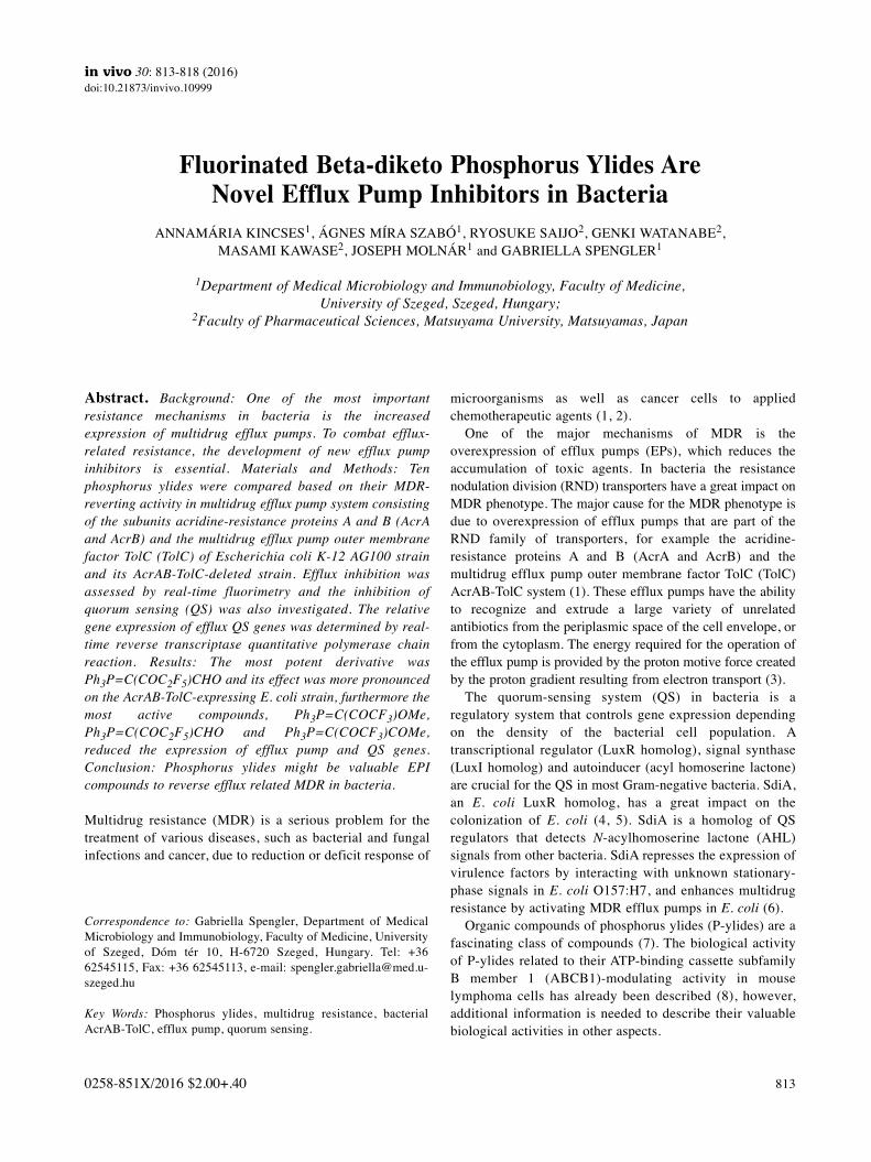
Abstract. Background: One of the most important
resistance mechanisms in bacteria is the increased
expression of multidrug efflux pumps. To combat efflux-
related resistance, the development of new efflux pump
inhibitors is essential. Materials and Methods: Ten
phosphorus ylides were compared based on their MDR-
reverting activity in multidrug efflux pump system consisting
of the subunits acridine-resistance proteins A and B (AcrA
and AcrB) and the multidrug efflux pump outer membrane
factor TolC (TolC) of Escherichia coli K-12 AG100 strain
and its AcrAB-TolC-deleted strain. Efflux inhibition was
assessed by real-time fluorimetry and the inhibition of
quorum sensing (QS) was also investigated. The relative
gene expression of efflux QS genes was determined by real-
time reverse transcriptase quantitative polymerase chain
reaction. Results: The most potent derivative was
Ph3P=C(COC2F5)CHO and its effect was more pronounced
on the AcrAB-TolC-expressing E. coli strain, furthermore the
most active compounds, Ph3P=C(COCF3)OMe,
Ph3P=C(COC2F5)CHO and Ph3P=C(COCF3)COMe,
reduced the expression of efflux pump and QS genes.
Conclusion: Phosphorus ylides might be valuable EPI
compounds to reverse efflux related MDR in bacteria.
Multidrug resistance (MDR) is a serious problem for thetreatment of various diseases, such as bacterial and fungalinfections and cancer, due to reduction or deficit response of
microorganisms as well as cancer cells to appliedchemotherapeutic agents (1, 2).
One of the major mechanisms of MDR is theoverexpression of efflux pumps (EPs), which reduces theaccumulation of toxic agents. In bacteria the resistancenodulation division (RND) transporters have a great impact onMDR phenotype. The major cause for the MDR phenotype isdue to overexpression of efflux pumps that are part of theRND family of transporters, for example the acridine-resistance proteins A and B (AcrA and AcrB) and themultidrug efflux pump outer membrane factor TolC (TolC)AcrAB-TolC system (1). These efflux pumps have the abilityto recognize and extrude a large variety of unrelatedantibiotics from the periplasmic space of the cell envelope, orfrom the cytoplasm. The energy required for the operation ofthe efflux pump is provided by the proton motive force createdby the proton gradient resulting from electron transport (3).
The quorum-sensing system (QS) in bacteria is aregulatory system that controls gene expression dependingon the density of the bacterial cell population. Atranscriptional regulator (LuxR homolog), signal synthase(LuxI homolog) and autoinducer (acyl homoserine lactone)are crucial for the QS in most Gram-negative bacteria. SdiA,an E. coli LuxR homolog, has a great impact on thecolonization of E. coli (4, 5). SdiA is a homolog of QSregulators that detects N-acylhomoserine lactone (AHL)signals from other bacteria. SdiA represses the expression ofvirulence factors by interacting with unknown stationary-phase signals in E. coli O157:H7, and enhances multidrugresistance by activating MDR efflux pumps in E. coli (6).
Organic compounds of phosphorus ylides (P-ylides) are afascinating class of compounds (7). The biological activityof P-ylides related to their ATP-binding cassette subfamilyB member 1 (ABCB1)-modulating activity in mouselymphoma cells has already been described (8), however,additional information is needed to describe their valuablebiological activities in other aspects.
813
Correspondence to: Gabriella Spengler, Department of MedicalMicrobiology and Immunobiology, Faculty of Medicine, Universityof Szeged, Dóm tér 10, H-6720 Szeged, Hungary. Tel: +3662545115, Fax: +36 62545113, e-mail: [email protected]
Key Words: Phosphorus ylides, multidrug resistance, bacterialAcrAB-TolC, efflux pump, quorum sensing.
in vivo 30: 813-818 (2016)doi:10.21873/invivo.10999
Fluorinated Beta-diketo Phosphorus Ylides Are Novel Efflux Pump Inhibitors in Bacteria
ANNAMÁRIA KINCSES1, ÁGNES MÍRA SZABÓ1, RYOSUKE SAIJO2, GENKI WATANABE2, MASAMI KAWASE2, JOSEPH MOLNÁR1 and GABRIELLA SPENGLER1
1Department of Medical Microbiology and Immunobiology, Faculty of Medicine,
University of Szeged, Szeged, Hungary;2Faculty of Pharmaceutical Sciences, Matsuyama University, Matsuyamas, Japan
0258-851X/2016 $2.00+.40

In this article, we report the MDR-modulating activitiesof P-ylides in bacteria related to the inhibition of effluxactivity and QS.
Materials and Methods
Compounds. The synthesis of P-ylides was described previously (8)and the structures of the P-ylides (compounds 1-10) screened fortheir MDR-modulating activities are shown in Table I. Thecompounds were dissolved in dimethyl sulfoxide (DMSO) for theexperiments.
Acridine orange (AO), ethidium bromide (EB), and LB brothwere purchased from Sigma-Aldrich Chemie GmbH (Steinheim,Germany). Modified LB agar (LB*) contained 5 g yeast extract, 10 g trypton, 10 g NaCl, 1 g K2HPO4, 0.3 g MgSO4.7H2O and 36mg FeNaEDTA in 1.0 l of distilled water. Mueller Hinton broth waspurchased from Scharlau Chemie S.A. (Barcelona, Spain).
Bacterial strains. Wild-type E. coli K-12 AG100 strain [argE3thi-1 rpsL xyl mtl Δ(gal-uvrB) supE44] expressing the AcrAB-TolC EP at its basal level and its AcrAB-TolC-deleted mutantstrain (E. coli K-12 AG100A). The strains were kindly providedby Professor Dr. Hiroshi Nikaido (Department of Molecular andCell Biology and Chemistry, University of California, Berkeley,CA, USA).
Strains used for QS tests: Chromobacterium violaceum 026(CV026) as sensor strain; Sphingomonas spp. EZF 10-17(Sphingomonadaceae) isolated from a grapevine crown gall tumor(used as AHL producer), this strain induced pigment production byCV026 and proved to be efficient for the study of QS interactions;Enterobacter cloaceae 31298 (clinical wound isolate, used as AHLproducer).
Determination of minimum inhibitory concentrations. The minimuminhibitory concentrations (MICs) of P-ylides were tested accordingto Clinical and Laboratory Standard Institute guidelines (9).
Real-time accumulation assay by Roche LightCycler real-time
thermocycler. The effect of the studied compounds on the real-time accumulation of EB was assessed by an automated EBmethod (10), using a LightCycler real-time thermocycler(LightCycler 1.5; Roche, Indianapolis, IN, USA). Briefly, analiquot of an overnight culture of the strain in LB medium wastransferred to fresh LB medium and incubated until it reached anoptical density (OD) of 0.6 at 600 nm. It was then washed withphosphate-buffered saline (PBS; pH 7.4) and centrifuged at13,000 × g for 3 min, the pellets re-suspended in PBS (pH 7.4)and the OD adjusted to 0.6 at 600 nm. The compounds wereadded individually at different concentrations (in doubleconcentrated form) in 5 μl volumes of their stock solutions to 45μl of EB solution of 2 mg/l in PBS. Then, 10 μl of the EBsolution containing the compound were transferred into standardglass capillary tubes of 20 μl maximum volume (Roche) and 10μl of bacterial suspension (OD of 0.6 at 600 nm) was added tothe capillaries. The capillaries containing the samples were placedinto a carousel (Roche) and the fluorescence was monitored at theFL-2 channel every minute on a real-time basis.
From the real-time data, the activity of the compound, namelythe relative fluorescence index (RFI) of the last time point (minute
30) of the EB accumulation assay was calculated according to theformula:
where RFtreated is the relative fluorescence at the last time point ofthe EB retention curve in the presence of an inhibitor, andRFuntreated is the relative fluorescence at the last time point of theEB retention curve of the untreated solvent control (DMSO).
Assay for QS inhibition. LB* was used for these experiments. Thesensor strain C. violaceum 026 (CV026) and the AHL producerstrains EZF 10-17 Sphingomonas spp. (Sphingomonadaceae) or E.
cloaceae 31298 were inoculated as parallel lines and incubated atroom temperature (20˚C) for 24-48 h. QS inhibition was monitoredby agar diffusion method. Filter paper discs (7.0 mm in diameter)were impregnated with 10 μl of stock solutions of the compoundsin DMSO (10 mg/ml in DMSO). The discs were placed between theparallel lines of sensor and AHL producer strains on the surface ofthe nutrient agar. The plates were incubated at room temperature fora further 24-48 h and the interactions between the strains andcompounds were evaluated for the reduction in size of the zone ofpigment production and the zone of growth inhibition of the affectedstrains, in millimeters. AO was applied as positive control (11).
Expression analyses of genes by real-time reverse transcriptase
quantitative polymerase chain (RT-qPCR) reaction. Bacteria werecultured in LB broth and were incubated overnight at 37˚C withshaking. On the day of RNA isolation, the bacterial suspensions(OD of 0.6 at 600 nm) were transferred to 10 ml tubes in 3 mlaliquots and 50 μg/ml of compounds were added to the tubeswhich were the incubated at 37˚C. At intervals of 4 and 18 h ofculture, the tubes were centrifuged at 12,000 × g for 2 min. Pelletswere suspended in 100 μl TE buffer containing 1 mg/ml lysozymeby vigorous vortexing and were incubated at 37˚C for 10 min. Thetotal RNA was isolated in an RNase-free environment usingNucleoSpin RNA kit (Macherey Nagel, Germany) according to themanufacturer’s instructions. Purified RNA was stored in RNase-free water in nuclease-free collection tubes and was maintained at−20˚C until quantification was performed. The concentration ofthe extracted RNA templates was assessed by spectrophotometryat 260 nm.
Expression of the acrA, acrB, multiple antibiotic resistance proteinR (marR) and quorum-sensing transcriptional activator (sdiA) geneswas studied by reverse transcription of total RNA. The data obtainedfor gene targets were normalized against the E. coli house-keepinggene glyceraldehyde-3-phosphate-dehydrogenase (gapdh) measured inthe same sample. Real-time quantification of the RNA templates byreal-time one-step RT-qPCR was performed in a CFX96 Touch real-time PCR detection system (Bio-Rad, Hercules, CA, USA) strictlyadhering to the manufacturer recommendations of the SensiFAST™SYBR No-ROX One-Step Kit (Bioline GmbH, Luckenwalde,Germany). Briefly, each well of 96-well microtiter plates contained,in a final volume of 20 μl, 10 μl of the 2x SensiFAST™ SYBR No-ROX One-Step Mix, 0.2 μl Reverse Transcriptase, 0.4 μl RiboSafeRNase Inhibitor, 5.4 μl diethylpyrocarbonate-treated water, 500 nM ofeach primer and approximately 20 ng of total RNA in RNAase-freewater. Thermal cycling was initiated with a denaturation step of 5 min
in vivo 30: 813-818 (2016)
814
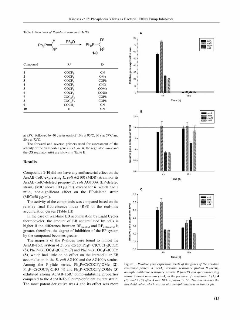
at 95˚C, followed by 40 cycles each of 10 s at 95˚C, 30 s at 57˚C and20 s at 72˚C.
The forward and reverse primers used for assessment of theactivity of the transporter genes acrA, acrB, the regulator marR andthe QS regulator sdiA are shown in Table II.
Results
Compounds 1-10 did not have any antibacterial effect on theAcrAB-TolC-expressing E. coli AG100 (MDR) strain nor itsAcrAB-TolC-deleted progeny E. coli AG100A (EP-deletedstrain) (MIC above 100 μg/ml), except for 6, which had amild, non-significant effect on the EP-deleted strain(MIC=50 μg/ml).
The activity of the compounds was compared based on therelative final fluorescence index (RFI) of the real-timeaccumulation curves (Table III).
In the case of real-time EB accumulation by Light Cyclerthermocycler, the amount of EB accumulated by cells ishigher if the difference between RFtreated and RFuntreated isgreater, therefore, the degree of inhibition of the EP systemby the compound becomes greater.
The majority of the P-ylides were found to inhibit theAcrAB-TolC system of E. coli except Ph3P=C(COCF3)COPh(3), Ph3P=C(COC2F5)COPh (7) and Ph3P=C(COC3F7)COPh(8), which had little or no effect on the intracellular EBaccumulation in the E. coli AG100 and the AG100A strains.Among the P-ylide series, Ph3P=C(COCF3)OMe (2),Ph3P=C(COCF3)CHO (4) and Ph3P=C(COCF3)COMe (5)exhibited strong AcrAB-TolC pump-inhibiting propertiescompared to the AcrAB-TolC pump-deficient mutant strain.The most potent derivative was 4 and its effect was more
Kincses et al: Phosphorus Ylides as Bacterial Efflux Pump Inhibitors
815
Table I. Structures of P-ylides (compounds 1-10).
Compound R1 R2
1 COCF3 CN2 COCF3 OMe3 COCF3 COPh4 COCF3 CHO5 COCF3 COMe6 COCF3 CO2Et7 COC2F5 COPh8 COC3F7 COPh9 COCH3 CN10 H CN
Figure 1. Relative gene expression levels of the genes of the acridine
resistance protein A (acrA), acridine resistance protein B (acrB),
multiple antibiotic resistance protein R (marR) and quorum-sensing
transcriptional activator (sdiA) in the presence of compounds 2 (A), 4
(B), and 5 (C) after 4 and 18 h exposure in LB. The line denotes the
threshold value, which was set at a two-fold increase in transcripts.

pronounced on the MDR E. coli strain compared to thepump-deleted E. coli strain. This activity suggests that thecompound may interfere with the proton motive forcebecause AcrB utilizes proton motive force as energy for itstransport function.
The P-ylides were not able to inhibit the QS in the appliedsystems compared to the positive control AO (data not shown).
Regarding the effect of P-ylides on the relative expressionof efflux pump and QS genes in E. coli AG100, the mosteffective compounds 2, 4, and 5 were selected for gene-expression studies. In the assay, the gene of the multidrugefflux pump subunit AcrB, the gene of the periplasmic AcrAsubunit, the component of the E. coli mar locus (multipleantibiotic resistance), and the gene of the LuxR homologSdiA were investigated. As shown in Figure 1A, compound2 at 50 μg/ml up-regulated all the genes studied after 4 h ofexposure, however, after 18 h, the gene expression returnedto basal levels. Compound 4 also significantly up-regulatedthe secondary RND transporter gene acrB (approximately 2-fold increase) after 4 h and 18 h exposures as well.Surprisingly, there was up-regulation of sdiA expression after18 h compared to the expression level after 4 h implicatingthe ability of compound 2 to influence the QS gene sdiA,however, this increase was not significant (Figure 1B).Compound 5 up-regulated the expression levels of acrA andacrB after 4 h, although after 18 h, the up-regulation of thesegenes was not significant (less than 2-fold increase) aspresented by Figure 1C.
Discussion
Some phenothiazines and hydantoins are known to be EPIsagainst both bacteria and cancer cells (13). However, P-ylides 3, 7 and 8 have been shown to have activity againstthe EPs of cancer cells (8), but not to have activity againstthe EP of bacteria. It is important to note that ABCtransporters are primary efflux pumps deriving their energyfrom the hydrolysis of ATP, however, the AcrAB-TolCsystem is a three-component proton motive force-dependentmultidrug efflux system. The most effective compounds in
the present bacterial system were compounds 2, 4, and 5,which inhibited the AcrAB-TolC system and influenced theexpression of the genes acrA and acrB, which areconstituents of the AcrAB-TolC system. In addition, althoughthe compounds are not QS inhibitors, compound 4 didincrease the expression of sdiA after 18 h exposure.
It can be concluded that in the COCF3 series (compounds1-6), the MDR-reverting activity in the MDR E. coli strainwas intensified in the following order: CHO (4) > OMe (2),COMe (5) >> CO2Et (6), COPh (3), CN (1).
in vivo 30: 813-818 (2016)
816
Table II. Forward and reverse primers used for assessment of the activity of the transporter genes acrA and acrB, the regulator marR and the
quorum-sensing regulator sdiA of Escherichia coli AG100.
Gene Full name Primer sequence (5’-3’) Amplicon Reference size (bp)
acrA Acridine-resistance protein A CTTAGCCCTAACAGGATGTGTTGAAATTACGCTTCAGGAT 189 (12)acrB Acridine-resistance protein B CGTACACAGAAAGTGCTCAACGCTTCAACTTTGTTTTCTT 183 (12)marR Multiple antibiotic-resistance protein R AGCGATCTGTTCAATGAAATTTCAGTTCAACCGGAGTAAT 170 (12)sdiA Quorum-sensing transcriptional activator CTGATGGCTCTGATGCGTTTATCTGGTGGAAATTGACCGTATT 163 This studygapdh Glyceraldehyde-3-phospate dehydrogenase ACTTACGAGCAGATCAAAGCAGTTTCACGAAGTTGTCGTT 170 (12)
Table III. Relative final fluorescence index (RFI) for the effect of P-ylides
(compounds 1-10) on Escherichia coli AG100 expresing acridine-resistance
proteins A and B and the multidrug efflux pump outer membrane factor
TolC, and the pump-deleted E. coli AG100A strain at 50 μg/ml.
Compound RFIa
AG100 AG100A
1 0.36 0.462 0.64 0.493 0.04 0.124 0.73 0.465 0.42 0.276 0.25 0.497 0.02 08 0 0.199 0.29 0.3210 0.42 0.44
aRFI was calculated according to the formula:
where RFtreated is the relative fluorescence at the last point (30 min) ofthe ethidium bromide (EB) retention curve in the presence an P-ylideand RFuntreated is the relative fluorescence at the last point (30 min) ofthe EB retention curve of the untreated solvent control dimethylsulfoxide.

Thus, some structurally related trifluoroacetylated P-ylidesdiffer in their MDR-reversal activities between cancer cellsand bacterial strains, indicating that the compoundsdifferently act as inhibitors of primary (ABCB1) andsecondary (AcrB) efflux pumps because these pumps differin their energy source for driving the pump (ATP and PMF,respectively).
The present study demonstrated that trifluoroacetylated P-ylides may be attractive lead EPIs for further developmentas a MDR-reversing agents, however, their mode of actionshould be elucidated by structure–activity relationshipstudies.
Acknowledgements
This research was supported by the Szeged Foundation for CancerResearch, the European Union and the State of Hungary, co-financed by the European Social Fund in the framework of TÁMOP4.2.4. A/2-11-1-2012-0001 ‘National Excellence Program’. Thisarticle was supported by the János Bolyai Research Scholarship ofthe Hungarian Academy of Sciences.
References
1 Tanwar J, Das S, Fatima Z and Hameed S: Multidrug resistance:an emerging crisis. Interdiscip Perspect Infect Dis 541340. doi:10.1155/2014/541340, 2014.
2 Nikaido H: Multidrug resistance in bacteria. Annu Rev Biochem78: 119-146, 2009.
3 Nikaido H and Pagès JM: Broad-specificity efflux pumps andtheir role in multidrug resistance of Gram-negative bacteria.FEMS Microbiol Rev 36: 340-363, 2012.
4 Kanamaru K, Kanamaru K, Tatsuno I, Tobe T and Sasakawa C:SdiA, an Escherichia coli homologue of quorum-sensingregulators, controls the expression of virulence factors inenterohaemorrhagic Escherichia coli O157:H7. Mol Microbiol38: 805-816, 2000.
5 Tavío MM, Aquili VD, Poveda JB, Antunes NT, Sánchez-Céspedes J and Vila J: Quorum-sensing regulator sdiA and marAoverexpression is involved in in vitro-selected multidrugresistance of Escherichia coli. J Antimicrob Chemother 65:1178-1186, 2010.
6 Lee J, Maeda T, Hong SH and Wood TK: Reconfiguring thequorum-sensing regulator SdiA of Escherichia coli to controlbiofilm formation via indole and N-acylhomoserine lactones.Appl Environ Microbiol 75: 1703-1716, 2009.
7 El-Batta A, Jiang C, Zhao W, Anness R, Cooksy AL andBergdahl M: Wittig reactions in water media employingstabilized ylides with aldehydes. Synthesis of alpha,beta-unsaturated esters from mixing aldehydes, alpha-bromoesters,and Ph3P in aqueous NaHCO3. J Org Chem 72: 5244-5259,2007.
8 Spengler G, Ocsovszki I, Tönki ÁS, Saijo R, Watanabe G,Kawase M and Molnár J: Fluorinated β-diketo phosphorus ylidesare novel inhibitors of the ABCB1 efflux pump of cancer cells.Anticancer Res 35: 5915-5919, 2015.
9 Clinical and Laboratory Standard Institute guidelines.http://clsi.org/
10 Viveiros M, Martins A, Paixão L, Rodrigues L, Martins M,Couto I, Fähnrich E, Kern WV and Amaral L: Demonstration ofintrinsic efflux activity of Escherichia coli K-12 AG100 by anautomated ethidium bromide method. Int J Antimicrob Agents31: 458-462, 2008.
11 Varga GZ, Szabo MA, Schelz Zs, Szegedi E, Amaral L andMolnár J: Quorum sensing inhibition by phenothiazines andrelated compounds. Lett Drug Design Discovery 8: 133-137,2011.
12 Viveiros M, Dupont M, Rodrigues L, Couto I, Davin-Regli A,Martins M, Pagès JM and Amaral L: Antibiotic stress, geneticresponse and altered permeability of E. coli. PLoS One 2: e365,2007.
13 Amaral L, Spengler G, Martins A, Armada A, Handzlik J, Kiec-Kononowicz K and Molnar J: Inhibitors of bacterial effluxpumps that also inhibit efflux pumps of cancer cells. AnticancerRes 32: 2947-2957, 2012.
Received July 22, 2016
Revised August 29, 2016
Accepted September 5, 2016
Kincses et al: Phosphorus Ylides as Bacterial Efflux Pump Inhibitors
817

70
II.

R E S E A R CH AR T I C L E
Bioactive compounds from the African medicinal plantCleistochlamys kirkii as resistance modifiers in bacteria
Annamária Kincses1 | Borisz Varga1 | Ákos Csonka1,2 | Shirley Sancha3 | Silva Mulhovo4 |
Ana Margarida Madureira3 | Maria‐José U. Ferreira3 | Gabriella Spengler1
1Department of Medical Microbiology and
Immunobiology, Faculty of Medicine,
University of Szeged, Dóm tér 10, Szeged
6720, Hungary
2Department of Traumatology, Faculty of
Medicine, University of Szeged, Semmelweis
utca 6, Szeged 6725, Hungary
3Research Institute for Medicines (iMed.
Ulisboa), Faculty of Pharmacy, University of
Lisbon, Avenue Professor Gama Pinto, Lisbon
1649‐003, Portugal
4Mozambican and Ethnoscience Study Center
(CEMEC), Faculty of Mathematics and Natural
Sciences, Pedagogic University, Lhanguene
Campus, Av. de Moçambique, Maputo
21402161, Mozambique
Correspondence
Gabriella Spengler, Department of Medical
Microbiology and Immunobiology, Faculty of
Medicine, University of Szeged, Dóm tér 10,
Szeged 6720, Hungary.
Email: [email protected]‐szeged.hu
Funding information
Portuguese Foundation for Science and Tech-
nology (FCT), Grant/Award Numbers: UID/
DTP/04138/2013 and PTDC/QEQ‐MED/
0905/2012; European Social Fund, Grant/
Award Number: TÁMOP 4.2.4. A/2‐11‐1‐
2012‐0001; ÚNKP‐17‐3 New National Excel-
lence Program of the Ministry of Human
Capacities of Hungary; János Bolyai Research
Scholarship of the Hungarian Academy of Sci-
ences; University of Szeged, Hungary, Grant/
Award Number: GINOP‐2.3.2‐15‐2016‐
00012; European Union and the State of
Hungary
Cleistochlamys kirkii (Benth) Oliv. (Annonaceae) is a medicinal plant traditionally used in
Mozambique to treat infectious diseases. The aim of this study was to find resistance modifiers
in C. kirkii for Gram‐positive and Gram‐negative model bacterial strains. One of the most impor-
tant resistance mechanisms in bacteria is the efflux pump‐related multidrug resistance. Therefore,
polycarpol (1), three C‐benzylated flavanones (2–4), and acetylmelodorinol (5) were evaluated for
their multidrug resistance‐reverting activity on methicillin‐susceptible and methicillin‐resistant
Staphylococcus aureus and Escherichia coli AG100 and AG100 A strains overexpressing and lacking
the AcrAB–TolC efflux pump system. The combined effects of antibiotics and compounds (2 and
4) were also assessed by using the checkerboard microdilution method in both S. aureus strains.
The relative gene expression of the efflux pump genes was determined by real‐time reverse
transcriptase quantitative polymerase chain reaction. The inhibition of quorum sensing was also
investigated. The combined effect of the antibiotics and compound 2 or 4 on the methicillin‐
sensitive S. aureus resulted in synergism. The most active compounds 2 and 4 increased the
expression of the efflux pump genes. These results suggested that C. kirkii constituents could
be effective adjuvants in the antibiotic treatment of infections.
KEYWORDS
C‐benzylated flavanones, Cleistochlamys kirkii, efflux pump, Escherichia coli AG100, methicillin‐
resistant Staphylococcus aureus, quorum sensing
1 | INTRODUCTION
The increasing resistance of bacterial isolates is a high concern in the
therapy of infectious diseases. The widespread and inappropriate use
of antibiotics has contributed to the selection of resistant bacteria. Mul-
tidrug‐resistant (MDR) bacteria show resistance against a broad range of
antimicrobials, and one of the most important resistance mechanisms is
thepresenceofmultidrug‐resistant efflux pumps (EPs). Thesemembrane
proteins have physiological functions, and they are involved in the
extrusion of toxic substances into the environment (Aparna,
Dineshkumar, Mohanalakshmi, Velmurugan, & Hopper, 2014; Webber
& Piddock, 2003).
In Staphylococcus aureus, the most studied MDR pump is the chro-
mosomally encoded NorA, which is a member of the major facilitator
superfamily. NorA can transport hydrophilic compounds, quaternary
ammonium compounds, and dyes. The MepA chromosomally encoded
efflux transporter described in S. aureus belongs to the multidrug and
toxic compound extrusion family.MepA can recognize fluoroquinolones,
Received: 31 August 2017 Revised: 19 December 2017 Accepted: 10 January 2018
DOI: 10.1002/ptr.6042
Phytotherapy Research. 2018;1–8. Copyright © 2018 John Wiley & Sons, Ltd.wileyonlinelibrary.com/journal/ptr 1

glycylcyclines, dyes, and quaternary ammonium compounds (Costa,
Viveiros, Amaral, & Couto, 2013).
The discovery of plant‐derived antimicrobials has drawn particular
attention (Balogh et al., 2014; Hintz, Matthews, & Di, 2015). Numerous
phytochemicals have minimal toxic activity, and they could be used in
order to overcome drug resistance in bacteria by blocking multidrug
EPs (Stavri, Piddock, & Gibbons, 2007). It is important to note that EP
inhibitors (EPIs) from plant sources can inhibit the activity of bacterial
efflux systems; furthermore, they can potentiate the efficacy of antibi-
otics aswell. It has been described that reserpine, isolated from the roots
of Rauwolfia vomitoria, inhibits the Bmr EP of Bacillus subtilis (Klyachko,
Schuldiner, &Neyfakh, 1997). SeveralBerberis species producing berber-
ine were also found to synthesize 5′‐methoxyhydnocarpin, an inhibitor
of theNorAMDRpumpof S. aureus (Stermitz, Lorenz, Tawara, Zenewicz,
& Lewis, 2000). Cucurbitane‐type triterpenoids, isolated from the aerial
parts of the African medicinal plant Momordica balsamina, have shown
activity against the EP systems of Gram‐positive bacteria (Ramalhete,
Da Cruz, et al., 2011a; Ramalhete, Lopes, et al., 2011b; Ramalhete,
Spengler, et al., 2011c).
The bacterial quorum sensing (QS) is a cell‐to‐cell communication
system that is based on chemical signals, namely, autoinducers (AIs). In
Gram‐negative bacteria, the most common AI is the N‐acyl homoserine
lactone (AHL; Varga et al., 2011). EPs and QS signals play an important
role in the development of bacterial virulence. The QS system and the
AIs are able to influence the expression of transporter genes. These
EPs have the ability to transport AI molecules to the external environ-
ment of bacteria, thus facilitating cell‐to‐cell communication (Spengler,
Kincses, Gajdács, & Amaral, 2017).
Cleistochlamys kirkii (Benth) Oliv. (Annonaceae) is an African medic-
inal plant traditionally used in Mozambique for the treatment of wound
infections, tuberculosis, and rheumatism (Verzár & Petri, 1987).
Previously, in a research for antimicrobial compounds from African
medicinal plants (Cabral et al., 2015; Kolaczkowski et al., 2010;
Madureira, Ramalhete, Mulhovo, Duarte, & Ferreira, 2012; Pereira
et al., 2016; Ramalhete et al., 2010), bioassay‐guided fractionation of
the methanol extract of the root barks of C. kirkii led to the isolation
of several compounds with different scaffolds. Among them,
C‐benzylated flavanones, namely, dichamanetin and the α,β‐unsatu-
rated lactone (−)‐cleistenolide, have shown relevant antibacterial activ-
ity against Gram‐positive bacteria, including drug‐resistant strains
(Pereira et al., 2016). Moreover, in the combination with β‐lactam anti-
biotics and vancomycin, polycarpol bearing a triterpenic scaffold exhib-
ited a strong synergistic effect against Gram‐positive resistant strains.
In this study, polycarpol (1) and C‐benzylated flavanones 2–4 and a
heptane derivative (5) were further investigated for their ability as both
EP and QS inhibitors of the efflux systems of Gram‐negative and
Gram‐positive bacteria. Furthermore, the ability of compounds 2 and
4 to potentiate the effect of tetracycline and the fluoroquinolone anti-
biotic ciprofloxacin on methicillin‐susceptible and methicillin‐resistant
S. aureus (MRSA) strains was also described.
2 | MATERIALS AND METHODS
2.1 | Compounds tested
Triterpene polycarpol (1), C‐benzylated flavanones chamanetin (2),
isochamanetin (3), dichamanetin (4), and the heptane derivative
acetylmelodorinol (5; Figure 1) were isolated from the methanol
extract of the root barks of C. kirkii, as it has previously been described
(Pereira et al., 2016). The purity of all the compounds was higher than
95% based on high‐performance liquid chromatography analysis and
nuclear magnetic resonance spectroscopy. The compounds were dis-
solved in dimethyl sulfoxide (DMSO).
2.2 | Reagents and media
Promethazine (PMZ; EGIS), ethidium bromide (EB), verapamil, tetracy-
cline‐hydrochloride (TET), ciprofloxacin‐hydrochloride (CIP), and
Luria–Bertani (LB) broth, and LB agar were purchased from Sigma‐
Aldrich Chemie GmbH (Steinheim, Germany). The modified LB medium
(LB*) was prepared from yeast extract 5 g/L, tryptone 10 g/L, NaCl
10 g/L, K2HPO4 1 g/L, MgSO4 × 7H2O 0.3 g/L, and FeNaEDTA
FIGURE 1 Chemical structures of compounds1–5
2 KINCSES ET AL.

36 mg/L. In case of modified LB* agar, the LB* medium was supple-
mented with agar 20 g/L (Difco). pH was adjusted to 7.2. Tryptic soy
broth (TSB), tryptic soy agar, and Mueller–Hinton (MH) broth were
purchased from Scharlau Chemie S. A. (Barcelona, Spain).
2.3 | Bacterial strains
Compounds were evaluated against the Gram‐negative wild‐type
Escherichia coli K‐12 AG100 strain [argE3 thi‐1 rpsL xyl mtl
Δ(gal‐uvrB) supE44], expressing the AcrAB–TolC EP at its basal level
and its AcrAB–TolC‐deleted mutant E. coli AG100 A strain. These
strains were kindly provided by Prof. Dr. Hiroshi Nikaido (Department
of Molecular and Cell Biology and Chemistry, University of California,
Berkeley, CA, USA).
The compounds were further evaluated against two Gram‐positive
strains, S. aureus American Type Culture Collection (ATCC) 25923,
used as the methicillin‐susceptible reference strain, and the methicillin
and ofloxacin‐resistant S. aureus 272123 clinical isolate, which was
kindly provided by Prof. Dr. Leonard Amaral (Institute of Hygiene
and Tropical Medicine, Lisbon, Portugal).
For QS tests, the following strains were used: Chromobacterium
violaceum 026 (CV026) as sensor strain and Enterobacter cloaceae
31298 as AHL producer strain (a clinical isolate from a wound). When
C. violaceum reaches a high cell density, it produces a purple pigment,
namely, violacein (Ballantine, Beer, Crutchley, Dodd, & Palmer, 1958).
2.4 | Determination of minimum inhibitory
concentrations by microdilution method
The minimum inhibitory concentrations (MICs) of compounds were
determined according to the Clinical and Laboratory Standard Institute
guidelines (2017) in three independent assays. The solvent DMSO had
no antibacterial effect.
2.5 | Interaction between antibiotics and compounds
The combined effect of compounds and antibiotics on the growth inhi-
bition of S. aureus was evaluated by the checkerboard method. Two-
fold serial dilutions of antibiotics were prepared in MH broth on the
horizontal rows of microtiter plate and then cross‐diluted vertically
by twofold serial dilutions of the compounds (Wolfart et al., 2006).
For this assay, only the compounds with well‐definedMIC values could
be used. Consequently, the combination assays were carried out on
methicillin‐susceptible and MRSA strains. The dilutions of the antibi-
otics (TET or CIP) were made in a horizontal direction in 100 μl, and
the dilutions of compounds were made vertically in the microtiter plate
in 50 μl. After the dilution of an overnight culture, bacterial cells were
resuspended in MH medium containing 1 × 10⁴ cells and distributed
into each well. The plates were incubated for 18 hr at 37 °C. The cell
growth rate was determined after MTT (3‐(4,5‐dimethylthiazol‐2‐yl)‐
2,5‐diphenyltetrazolium bromide) staining, as described elsewhere
(Wolfart et al., 2006). The combination index (CI) values at 90% growth
inhibition (ED90) were determined by using CompuSyn software to
plot 4 or 5 data points for each ratio (www.combosyn.com, ComboSyn,
Inc., Paramus, NJ, USA). CI values were calculated by means of the
median‐effect equation, where CI < 1, CI = 1, and CI > 1 represent
synergism, an additive effect (or no interaction), and antagonism,
respectively (Chou & Martin, 2005).
2.6 | Real‐time accumulation assay by Roche
LightCycler real‐time thermocycler
The activity of compounds on the real‐time accumulation of EB was
assessed by the automated EB method (Viveiros et al., 2008) using a
LightCycler real‐time thermocycler (LightCycler 1.5, Roche, Indianapo-
lis, IN, USA). Briefly, an aliquot of an overnight culture of the S. aureus
strain inTSB medium was transferred to freshTSB medium, and it was
incubated until it reached an optical density (OD) of 0.6 at 600 nm. In
case of E. coli, the medium used in the assay was LB broth; the prepa-
ration of the inoculum was similar to the one of S. aureus. The cells
were washed with phosphate‐buffered saline (PBS; pH 7.4) and centri-
fuged at 13,000 × g for 3 min, the pellets were resuspended in PBS
(pH 7.4), and the OD was adjusted to 0.6 at 600 nm. The compounds
were added individually at different concentrations at MIC/2, MIC/3,
MIC/4, or MIC/5 (in double concentrated form) to the EB solution in
PBS. The final concentration of EB was based on the MIC and the fluo-
rescent signal produced by this amount of EB. In case of S. aureus
strains, the concentration of EB was 0.5 μg/ml, for E. coli AG100
1 μg/ml, and in case of E. coli AG100 A, it was 0.25 μg/ml. Then,
10 μl of the EB solution containing the compound was transferred into
standard glass capillary tubes of 20 μl maximum volume (Roche), and
10 μl of bacterial suspension (OD of 0.6 at 600 nm) was added to
the capillaries. The capillaries containing the samples were placed into
the carousel (Roche), and the fluorescence was monitored at the FL‐2
channel in every minute on a real‐time basis.
From the real‐time data, the activity of the compound, namely, the
relative final fluorescence index (RFI) of the last time point (minute 30)
of the EB accumulation assay, was calculated according to the follow-
ing formula:
RFI ¼RFtreated−RFuntreated
RFuntreated
where RFtreated is the relative fluorescence at the last time point of
EB retention curve in the presence of an inhibitor and RFuntreated is the
relative fluorescence at the last time point of the EB retention curve of
the untreated control having the solvent control (DMSO). Verapamil
was applied as positive control on Gram‐positive strains, and PMZ
was used on Gram‐negative strains.
2.7 | Assay for quorum sensing inhibition
LB* was used for these experiments. The sensor strain C. violaceum
026 and the AHL producer strains E. cloaceae 31298 were inoculated
as parallel lines and incubated at room temperature (20 °C) for
24–48 hr. QS inhibition was monitored by the agar diffusion method.
Filter paper discs (7.0 mm in diameter) were impregnated with 10 μl
of stock solutions (10 mM) of the compounds in DMSO. The discs
were placed between the parallel lines of the sensor and the AHL
producer strains on the surface of the nutrient agar. The plates were
incubated at room temperature for another 24–48 hr, and the interac-
tions between the strains and compounds were evaluated for the
KINCSES ET AL. 3
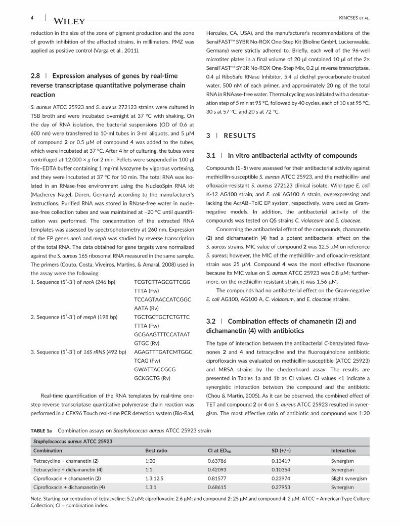
reduction in the size of the zone of pigment production and the zone
of growth inhibition of the affected strains, in millimeters. PMZ was
applied as positive control (Varga et al., 2011).
2.8 | Expression analyses of genes by real‐time
reverse transcriptase quantitative polymerase chain
reaction
S. aureus ATCC 25923 and S. aureus 272123 strains were cultured in
TSB broth and were incubated overnight at 37 °C with shaking. On
the day of RNA isolation, the bacterial suspensions (OD of 0.6 at
600 nm) were transferred to 10‐ml tubes in 3‐ml aliquots, and 5 μM
of compound 2 or 0.5 μM of compound 4 was added to the tubes,
which were incubated at 37 °C. After 4 hr of culturing, the tubes were
centrifuged at 12,000 × g for 2 min. Pellets were suspended in 100 μl
Tris–EDTA buffer containing 1 mg/ml lysozyme by vigorous vortexing,
and they were incubated at 37 °C for 10 min. The total RNA was iso-
lated in an RNase‐free environment using the NucleoSpin RNA kit
(Macherey Nagel, Düren, Germany) according to the manufacturer's
instructions. Purified RNA was stored in RNase‐free water in nucle-
ase‐free collection tubes and was maintained at −20 °C until quantifi-
cation was performed. The concentration of the extracted RNA
templates was assessed by spectrophotometry at 260 nm. Expression
of the EP genes norA and mepA was studied by reverse transcription
of the total RNA. The data obtained for gene targets were normalized
against the S. aureus 16S ribosomal RNA measured in the same sample.
The primers (Couto, Costa, Viveiros, Martins, & Amaral, 2008) used in
the assay were the following:
1. Sequence (5′‐3′) of norA (246 bp) TCGTCTTAGCGTTCGG
TTTA (Fw)
TCCAGTAACCATCGGC
AATA (Rv)
2. Sequence (5′‐3′) of mepA (198 bp) TGCTGCTGCTCTGTTC
TTTA (Fw)
GCGAAGTTTCCATAAT
GTGC (Rv)
3. Sequence (5′‐3′) of 16S rRNS (492 bp) AGAGTTTGATCMTGGC
TCAG (Fw)
GWATTACCGCG
GCKGCTG (Rv)
Real‐time quantification of the RNA templates by real‐time one‐
step reverse transcriptase quantitative polymerase chain reaction was
performed in a CFX96 Touch real‐time PCR detection system (Bio‐Rad,
Hercules, CA, USA), and the manufacturer's recommendations of the
SensiFAST™ SYBR No‐ROXOne‐Step Kit (Bioline GmbH, Luckenwalde,
Germany) were strictly adhered to. Briefly, each well of the 96‐well
microtiter plates in a final volume of 20 μl contained 10 μl of the 2×
SensiFAST™ SYBR No‐ROX One‐Step Mix, 0.2 μl reverse transcriptase,
0.4 μl RiboSafe RNase Inhibitor, 5.4 μl diethyl pyrocarbonate‐treated
water, 500 nM of each primer, and approximately 20 ng of the total
RNA inRNAase‐freewater. Thermal cyclingwas initiatedwith a denatur-
ation step of 5 min at 95 °C, followed by 40 cycles, each of 10 s at 95 °C,
30 s at 57 °C, and 20 s at 72 °C.
3 | RESULTS
3.1 | In vitro antibacterial activity of compounds
Compounds (1–5) were assessed for their antibacterial activity against
methicillin‐susceptible S. aureus ATCC 25923, and the methicillin‐ and
ofloxacin‐resistant S. aureus 272123 clinical isolate. Wild‐type E. coli
K‐12 AG100 strain, and E. coli AG100 A strain, overexpressing and
lacking the AcrAB–TolC EP system, respectively, were used as Gram‐
negative models. In addition, the antibacterial activity of the
compounds was tested on QS strains C. violaceum and E. cloaceae.
Concerning the antibacterial effect of the compounds, chamanetin
(2) and dichamanetin (4) had a potent antibacterial effect on the
S. aureus strains. MIC value of compound 2 was 12.5 μM on reference
S. aureus; however, the MIC of the methicillin‐ and ofloxacin‐resistant
strain was 25 μM. Compound 4 was the most effective flavanone
because its MIC value on S. aureus ATCC 25923 was 0.8 μM; further-
more, on the methicillin‐resistant strain, it was 1.56 μM.
The compounds had no antibacterial effect on the Gram‐negative
E. coli AG100, AG100 A, C. violaceum, and E. cloaceae strains.
3.2 | Combination effects of chamanetin (2) and
dichamanetin (4) with antibiotics
The type of interaction between the antibacterial C‐benzylated flava-
nones 2 and 4 and tetracycline and the fluoroquinolone antibiotic
ciprofloxacin was evaluated on methicillin‐susceptible (ATCC 25923)
and MRSA strains by the checkerboard assay. The results are
presented in Tables 1a and 1b as CI values. CI values <1 indicate a
synergistic interaction between the compound and the antibiotic
(Chou & Martin, 2005). As it can be observed, the combined effect of
TET and compound 2 or 4 on S. aureus ATCC 25923 resulted in syner-
gism. The most effective ratio of antibiotic and compound was 1:20
TABLE 1a Combination assays on Staphylococcus aureus ATCC 25923 strain
Staphylococcus aureus ATCC 25923
Combination Best ratio CI at ED90 SD (+/−) Interaction
Tetracycline + chamanetin (2) 1:20 0.63786 0.13419 Synergism
Tetracycline + dichamanetin (4) 1:1 0.42093 0.10354 Synergism
Ciprofloxacin + chamanetin (2) 1.3:12.5 0.81577 0.23974 Slight synergism
Ciprofloxacin + dichamanetin (4) 1.3:1 0.68615 0.27953 Synergism
Note. Starting concentration of tetracycline: 5.2 μM; ciprofloxacin: 2.6 μM; and compound 2: 25 μM and compound 4: 2 μM. ATCC = AmericanType CultureCollection; CI = combination index.
4 KINCSES ET AL.

and 1:1, respectively. Similarly, CIP also acted synergistically with
compounds 2 and 4, being the most active ratio of antibiotic and com-
pound 1.3:12.5 and 1.3:1, respectively (Table 1a).
Against the MRSA strain, compound 2 showed antagonistic effect
withTET and slight synergism with CIP. The interactions of compound
4 withTET and CIP on the MRSA strain were synergism and moderate
antagonism, respectively (Table 1b).
3.3 | Efflux pump‐inhibiting activity
The EB accumulation assay provides information about the intracellu-
lar accumulation of the general EP substrate EB. A potential EPI
increases the fluorescence level of EB because of its accumulation
within the bacterial cell. The EP‐inhibiting activity of the compounds
was compared on the basis of the RFI of the real‐time accumulation
curves in Gram‐positive and Gram‐negative strains (Figure 2a). In case
of real‐time EB accumulation by the LightCycler thermocycler, the
amount of EB accumulated by cells is higher if the difference between
RFtreated and RFuntreated is greater; therefore, the degree of inhibition of
the EP system by the compound becomes greater.
As shown in Figure 2a, compounds 2, 3, and 5 had EP‐inhibiting
activity compared with verapamil (RFI: 0.29) on the S. aureus ATCC
25923 strain, and the most active compound was compound 2. How-
ever, compounds 1–5 had no EPI activity on the MRSA strain at the
concentrations applied in the assay.
Concerning the inhibitory activity on Gram‐negatives, triterpene
polycarpol (1) and acetylmelodorinol (5) compared with the PMZ
(RFI: 0.15) could inhibit the AcrAB–TolC system of E. coli AG100. Com-
pound 1 proved to be the most effective EPI (Figure 2a). On the basis
of the real‐time accumulation data, compounds 1–5 had no effect on
the E. coli AG100 A strain lacking the AcrAB–TolC pump.
3.4 | Anti‐quorum sensing activity
The QS inhibition activity of compounds was defined measuring the
colorless zone around the disc on C. violaceum as described previously
(Varga et al., 2011). Compounds 1, 2, 4, and 5 were able to inhibit
effectively the QS between CV026 and E. cloaceae compared with
the positive control PMZ (Table 2).
3.5 | Relative expression of efflux pump genes
In order to evaluate the effect of compounds on the relative expres-
sion of EP genes in both S. aureus strains, the most effective
compounds 2 and 4 were selected for gene expression studies. In the
real‐time quantitative RT‐PCR assay, the genes of NorA and MepA
transporters were investigated. As shown in Figure 2bA, compound 2
at 5 μM significantly up‐regulated the expression of norA and mepA
genes after 4 hr of exposure in the MRSA strain. Compound 4 at
0.5 μM also significantly up‐regulated both EP genes after 4 hr of
exposure in the MRSA as presented in Figure 2bB. In the S. aureus
ATCC strain, the expression level of the mepA gene was not influ-
enced; nevertheless, the norA gene was significantly up‐regulated by
compounds 2 (19.84‐fold increase) at 5 μM and 4 (2.39‐fold increase)
at 0.5 μM (data not shown).
4 | DISCUSSION
Natural compounds isolated from plants represent a valuable source of
new antibacterial agents. When analyzing the results, the potent anti-
bacterial activity found for dichamanetin (4) and also the significant
activity of chamanetin (2) corroborated our previous studies on this
set of rare C‐benzylated flavanones (Pereira et al., 2016). The strongest
activity of dichamanetin (4), when compared with chamanetin (2),
seems to be due to its higher lipophilic character resulting from the
extra benzyl group at C‐6. In contrast to chamanetin (2), isochamanetin
(3), differing only in the position of the benzyl group, was inactive at
the concentration tested. Thus, besides the importance of lipophilicity,
the presence of a benzyl moiety at C‐8 appears to play a decisive role
in the antibacterial activity of this type of compounds.
In the combination assays, chamanetin (2) and dichamanetin (4)
had synergistic activity with tetracycline and ciprofloxacin on the
S. aureus ATCC 25923 strain. Furthermore, on the MRSA strain,
dichamanetin (4) and chamanetin (2), combined with tetracycline and
ciprofloxacin, respectively, also showed synergism, which indicates
that these compounds could be potential adjuvants in the therapy.
Besides having an antibacterial effect, both compounds (2 and 4)
could inhibit the activity of EPs, they were QS inhibitors, and they sig-
nificantly increased the expression of EP genes norA and mepA after
4 hr of exposure in the MRSA strain. Furthermore, the NorA and MepA
pumps of the MRSA strain could not be inhibited because of the over-
expression of these pumps. The change in gene expression could be
due to the stress response against compounds 2 and 4 because these
compounds as potential noxious agents for S. aureus had to be
extruded from the cytoplasm as soon as possible. This stress response
can be the explanation for the up‐regulation of the EP genes after 4 hr.
The difference between MRSA and ATCC is due to the overexpression
TABLE 1b Combination assays on MRSA 272123 strain
Staphylococcus aureus 272123
Combination Best ratio CI at ED90 SD (+/−) Interaction
Tetracycline + chamanetin (2) 6:25 1.4595 0.29390 Antagonism
Tetracycline + dichamanetin (4) 100:1 0.59402 0.34521 Synergism
Ciprofloxacin + chamanetin (2) 13:8 0.85030 0.21648 Slight synergism
Ciprofloxacin + dichamanetin (4) 32:1 1.35064 0.24946 Moderate antagonism
Note. Starting concentration of tetracycline: 100 μM; ciprofloxacin: 64 μM; and compound 2: 50 μM and compound 4: 3 μM. Ratio: antibiotic and testedcompound (μM). CI < 1, CI = 1, and CI > 1 represent synergism, an additive effect (or no interaction), and antagonism, respectively. CI = combination index;MRSA = methicillin‐resistant Staphylococcus aureus.
KINCSES ET AL. 5

of themepA gene in the resistant strain and it has low expression level in
the ATCC strain. Compounds 2 and 4 increased the expression of mepA
gene in the MRSA, but in the ATCC strain, these compounds did not
influence the expression level of mepA. It has been demonstrated by
other studies that the overexpression of EPs confers a fitness cost
for the organism, for example, a resistant isolate overexpressing
EPs shows reduced production of virulence determinants. Usually the
overexpression of pump genes is often related to global effects on
bacterial physiology influencing virulence (Ledda et al., 2017).
In Gram‐negatives, compounds 1–5 had no antibacterial activity.
Polycarpol (1) was the most effective EPI, inhibiting the AcrAB–TolC
transport system in the E. coli AG100 strain. The AcrAB–TolC system
belongs to the resistance nodulation division transporters, utilizing
the proton motive force to extrude toxic agents out of the bacterial
TABLE 2 Inhibitory effects of compounds 1–5 on QS signaltransmission
Compound QS inhibition zone in mm
1 51
2 50
3 —
4 53
5 52
Promethazine 46
Note. Ten microliters of 10 mM stock solution was added into the filterpaper discs (10 μM per disc), and the colorless zone around the disc mea-sured on Chromobacterium violaceum was measured after incubation for24–48 hr at room temperature. QS = quorum sensing.
FIGURE 2 Theminimum inhibitory concentration(MIC) and efflux pump modulating effectof compounds 1–5. (a) Relative finalfluorescence index (RFI) for the effect ofcompounds 1–5 on the AcrAB–TolC‐expressing Escherichia coli AG100 andStaphylococcus aureus American Type CultureCollection (ATCC) 25923 strains. (b) Relativegene expression levels of norA and mepA
genes in the presence of (A) chamanetin(2) and (B) dichamanetin (4) in methicillinresistant Staphylococcus aureus (MRSA)272123, after 4‐hr exposure. The linedenotes the threshold value, which was set ata twofold increase in transcripts
6 KINCSES ET AL.

cell. Since compound 1 had no EPI effect on the AcrAB–TolC pump‐
deleted E. coli strain, it may have a direct EPI effect on the AcrAB–TolC
transporter system. The effect of polycarpol could be due to its high
lipophilicity (Pereira et al., 2016), which can increase the membrane
permeability of bacteria.
In addition to flavanones 2 and 4, polycarpol (1) and
acetylmelodorinol (5) were also able to inhibit effectively the
bacterial communication, suggesting that they could be applied as
anti‐QS agents.
5 | CONCLUSIONS
To summarize our findings, C. kirkii constituents, chamanetin (2) and
dichamanetin (4), inhibited the growth of both S. aureus strains. The
three flavanones tested (2–4) increased the accumulation of the EP
substrate EB on sensitive S. aureus strain, and compounds 1, 2, 4,
and 5 prevented the cell‐to‐cell communication. Polycarpol (1) was
an effective inhibitor of the AcrAB–TolC system of E. coli, which might
be explained by its interference with membrane permeability and mod-
ulation of the function of the pump. Dichamanetin (4) and chamanetin
(2) showed synergistic effect on MRSA and sensitive S. aureus strains
implying that they could restore and increase the efficacy of antibi-
otics, especially in MDR infections. It can be assumed that the advan-
tageous properties of C. kirkii constituents can influence the QS system
and operation of EPs decreasing virulence and antibiotic resistance
(Varga et al., 2011).
According to the results, the most active compounds,
chamanetin (2) and dichamanetin (4), could be possible resistance
modifiers because they possess antibacterial, EPI, and anti‐QS
properties; moreover, these compounds might be used as adjuvants
in the antibiotic therapy because they may be able to reduce the
bacterial virulence. Furthermore, they can potentiate the activity of
antibiotics.
ACKNOWLEDGEMENTS
This research was supported by the European Union and the State of
Hungary and by the Portuguese Foundation for Science and Technol-
ogy (FCT), Projects PTDC/QEQ‐MED/0905/2012 and UID/DTP/
04138/2013, and co‐financed by the European Social Fund in the
framework of TÁMOP 4.2.4. A/2‐11‐1‐2012‐0001 “National Excel-
lence Program.” This study was supported by the GINOP‐2.3.2‐15‐
2016‐00012 project (University of Szeged, Hungary), and Gabriella
Spengler was supported by the János Bolyai Research Scholarship of
the Hungarian Academy of Sciences. Annamária Kincses was
supported by the ÚNKP‐17‐3 New National Excellence Program of
the Ministry of Human Capacities of Hungary. The authors thank the
Portuguese Embassy in Mozambique, as well as the Portuguese Office
of International Affairs, for plant transport.
CONFLICT OF INTEREST
The authors declare no conflict of interest.
ORCID
Maria‐José U. Ferreira http://orcid.org/0000-0002-8742-1486
Gabriella Spengler http://orcid.org/0000-0001-8085-0950
REFERENCES
Aparna, V., Dineshkumar, K., Mohanalakshmi, N., Velmurugan, D., &Hopper, W. (2014). Identification of natural compound inhibitors formultidrug efflux pumps of Escherichia coli and Pseudomonas aeruginosa
using in silico high‐throughput virtual screening and in vitro validation.PLoS One, 9, e101840.
Ballantine JA, Beer RJS, Crutchley DJ, Dodd GM, Palmer DR. 1958. Thesynthesis of violacein and related compounds. Proc Chem Soc 232–33.
Balogh, E. P., Mosolygó, T., Tiricz, H., Szabó, A. M., Karai, A., Kerekes, F., …Burián, K. (2014). Anti‐chlamydial effect of plant peptides. Acta
Microbiol Immunol, 61, 229–239.
Cabral, V., Luo, X., Junqueira, E., Costa, S. S., Mulhovo, S., DuarteA, C. I., …Ferreira, M. J. U. (2015). Enhancing activity of antibiotics against Staph-ylococcus aureus: Zanthoxylum capense constituents and derivatives.Phytomedicine, 22, 469–476.
Chou, T. C., & Martin, N. (2005). Compusyn for drug combinations: PC soft-
ware and user's guide: A Computer program for quantitation of synergism
and antagonism in drug combinations, and the determination of IC50 and
ED50 and LD50 values. Paramus, NJ, USA: ComboSyn Inc.
Clinical and Laboratory Standard Institute guidelines (2017). Accessed on24 July 2017. Retrieved from http://clsi.org/
Costa, S. S., Viveiros, M., Amaral, L., & Couto, I. (2013). Multidrug effluxpumps in Staphylococcus aureus: An update. Open Microbiol J, 7, 59–71.
Couto, I., Costa, S. S., Viveiros, M., Martins, M., & Amaral, L. (2008). Efflux‐mediated response of Staphylococcus aureus exposed to ethidium bro-mide. The Journal of Antimicrobial Chemotherapy, 62, 504–513.
Hintz, T., Matthews, K. K., & Di, R. (2015). The use of plant antimicrobialcompounds for food preservation. BioMed Research International,2015, 246264.
Klyachko, K. A., Schuldiner, S., & Neyfakh, A. A. (1997). Mutations affectingsubstrate specificity of the Bacillus subtilis multidrug transporter Bmr.Journal of Bacteriology, 179, 2189–2193.
Kolaczkowski, M., Kolaczkowska, A., Środa, K., Ramalhete, C., Michalak, K.,Mulhovo, S., & Ferreira, M. J. U. (2010). Substrates and modulators ofthe multidrug transporter Cdr1p of Candida albicans in antifungalextracts of medicinal plants. Mycoses, 53, 305–310.
Ledda, A., Price, J. R., Cole, K., Llewelyn, M. J., Kearns, A. M., Crook, D. W.,… Didelot, X. (2017). Re‐emergence of methicillin susceptibility in aresistant lineage of Staphylococcus aureus. The Journal of Antimicrobial
Chemotherapy, 72, 1285–1288.
Madureira, A. M., Ramalhete, C., Mulhovo, S., Duarte, A., & Ferreira, M. J. U.(2012). Antibacterial activity of some African medicinal plants usedtraditionally against infectious diseases. Pharmaceutical Biology, 50,481–489.
Pereira, F., Madureira, A. M., Sancha, S., Mulhovo, S., Luo, X., Duarte, A., &Ferreira, M. J. U. (2016). Cleistochlamys kirkii chemical constituents:Antibacterial activity and synergistic effects against resistant Staphylo-coccus aureus strains. Journal of Ethnopharmacology, 178, 180–187.
Ramalhete, C., Da Cruz, F. P., Lopes, D., Mulhovo, S., Rosário, V. E.,Prudêncio, M., & Ferreira, M. J. U. (2011a). Triterpenoids as inhibitorsof erythrocytic and liver stages of Plasmodium infections. BioorganicMed Chem, 19, 7474–7481.
Ramalhete, C., Lopes, D., Molnár, J., Mulhovo, S., Rosário, V. E., & Ferreira,M. J. U. (2011b). Karavilagenin C derivatives as antimalarials. BioorganicMed Chem, 19, 330–338.
Ramalhete, C., Lopes, D., Mulhovo, S., Molnár, J., Rosário, V. E., & Ferreira,M. J. U. (2010). New antimalarials with a triterpenic scaffold fromMomordica balsamina. Bioorganic Med Chem, 18, 5254–5260.
Ramalhete, C., Spengler, G., Martins, A., Martins, M., Viveiros, M., Mulhovo,S., … Amaral, L. (2011c). Inhibition of efflux pumps in methicillin‐resis-tant Staphylococcus aureus and Enterococcus faecalis resistant strainsby triterpenoids from Momordica balsamina. International Journal of
Antimicrobial Agents, 37, 70–74.
KINCSES ET AL. 7

Spengler, G., Kincses, A., Gajdács, M., & Amaral, L. (2017). New roads lead-ing to old destinations: Efflux pumps as targets to reverse multidrugresistance in bacteria. Molecules, 22, E468.
Stavri, M., Piddock, L. J., & Gibbons, S. (2007). Bacterial efflux pump inhib-itors from natural sources. The Journal of Antimicrobial Chemotherapy,59, 1247–1260.
Stermitz, F. R., Lorenz, P., Tawara, J. N., Zenewicz, L. A., & Lewis, K. (2000).Synergy in a medicinal plant: Antimicrobial action of berberinepotentiated by 5′‐methoxyhydnocarpin, a multidrug pump inhibitor.Proceedings of the National Academy of Sciences of the United States of
America, 97, 1433–1437.
Varga, G. Z., Szabó, M. A., Schelz, Zs., Szegedi, E., Amaral, L., & Molnár, J.(2011). Quorum sensing inhibition by phenothiazines and relatedcompounds. Lett Drug Design Discovery, 8, 133–137.
Verzár, R., & Petri, G. (1987). Medicinal plants in Mozambique and theirpopular use. Journal of Ethnopharmacology, 19, 67–80.
Viveiros, M., Martins, A., Paixão, L., Rodrigues, L., Martins, M., Couto, I., …Amaral, L. (2008). Demonstration of intrinsic efflux activity of
Escherichia coli K‐12 AG100 by an automated ethidium bromidemethod. International Journal of Antimicrobial Agents, 31, 458–462.
Webber, M. A., & Piddock, L. J. (2003). The importance of efflux pumps inbacterial antibiotic resistance. The Journal of Antimicrobial Chemother-
apy, 51, 9–11.
Wolfart, K., Spengler, G., Kawase, M., Motohashi, N., Molnár, J., Viveiros,M., & Amaral, L. (2006). Synergistic interaction between proton pumpinhibitors and resistance modifiers: Promoting effects of antibioticsand plasmid curing. In Vivo, 20, 367–372.
How to cite this article: Kincses A, Varga B, Csonka Á, et al.
Bioactive compounds from the African medicinal plant
Cleistochlamys kirkii as resistance modifiers in bacteria.
Phytotherapy Research. 2018;1–8. https://doi.org/10.1002/
ptr.6042
8 KINCSES ET AL.

71
III.

molecules
Article
Selenocompounds as Novel Antibacterial Agents andBacterial Efflux Pump Inhibitors
Tímea Mosolygó 1 , Annamária Kincses 1, Andrea Csonka 1,2, Ádám Szabó Tönki 1,
Karolina Witek 3, Carmen Sanmartín 4,5, Małgorzata Anna Marc 3,6, Jadwiga Handzlik 3,
Katarzyna Kiec-Kononowicz 3, Enrique Domínguez-Álvarez 7,*,† and Gabriella Spengler 1,*,†
1 Department of Medical Microbiology and Immunobiology, Faculty of Medicine, University of Szeged,Dóm tér 10, 6720 Szeged, Hungary; [email protected] (T.M.);[email protected] (A.K.); [email protected] (A.C.);[email protected] (Á.S.T.)
2 Department of Obstetrics and Gynecology, Faculty of Medicine, University of Szeged, Semmelweis utca 1,6725 Szeged, Hungary
3 Department of Technology and Biotechnology of Drugs, Jagiellonian University Medical College,Medyczna 9, 30-688 Kraków, Poland; [email protected] (K.W.);[email protected] (M.A.M.); [email protected] (J.H.); [email protected] (K.K.-K.)
4 Department of Pharmaceutical Technology and Chemistry, School of Pharmacy and Nutrition,University of Navarra, Irunlarrea 1, 31008 Pamplona, Spain; [email protected]
5 Instituto de Investigación Sanitaria de Navarra (IdiSNA), Irunlarrea 3, 31008 Pamplona, Spain6 Interdisciplinary Excellence Centre, Department of Inorganic and Analytical Chemistry,
University of Szeged, Dóm tér 7, 6720 Szeged, Hungary7 Instituto de Química Orgánica General (IQOG-CSIC), Consejo Superior de Investigaciones Científicas,
Juan de la Cierva 3, 28006 Madrid, Spain* Correspondence: [email protected] (E.D-Á.); [email protected] (G.S.);
Tel.: +34-91-258-7661 (E.D.-Á.); +36-62-445-115 (G.S.)† These authors contributed equally to this work.
Academic Editor: Peter J. RutledgeReceived: 12 March 2019; Accepted: 13 April 2019; Published: 16 April 2019
�����������������
Abstract: Bacterial multidrug resistance is becoming a growing problem for public health, due tothe development and spreading of bacterial strains resistant to antimicrobials. In this study, theantibacterial and multidrug resistance reversing activity of a series of seleno-carbonyl compoundshas been evaluated. The effects of eleven selenocompounds on bacterial growth were evaluated inStaphylococcus aureus, methicillin resistant S. aureus (MRSA), Enterococcus faecalis, Escherichia coli, andChlamydia trachomatis D. The combination effect of compounds with antibiotics was examined by theminimum inhibitory concentration reduction assay. Their efflux pump (EP) inhibitory propertieswere assessed using real-time fluorimetry. Relative expressions of EP and quorum-sensing geneswere studied by quantitative PCR. Results showed that a methylketone selenoester had remarkableantibacterial activity against Gram-positive bacteria and potentiated the activity of oxacillin in MRSA.Most of the selenocompounds showed significant anti-chlamydial effects. The selenoanhydrideand the diselenodiester were active inhibitors of the AcrAB-TolC system. Based on these resultsit can be concluded that this group of selenocompounds can be attractive potential antibacterialsand EP inhibitors. The discovery of new derivatives with a significant antibacterial activity as novelselenocompounds, is of high impact in the fight against resistant pathogens.
Keywords: selenocompounds; selenoesters; AcrAB-TolC efflux pump; Chlamydia trachomatis D;Escherichia coli K-12 AG100; Staphylococcus aureus
Molecules 2019, 24, 1487; doi:10.3390/molecules24081487 www.mdpi.com/journal/molecules

Molecules 2019, 24, 1487 2 of 13
1. Introduction
Multidrug resistance is becoming a serious problem in the treatment of resistant bacterial infections.The discovery of novel antibacterial or multidrug resistance reversing agents is extremely urgent assoon we may lack effective drugs to treat bacterial infections caused by the arising superbugs resistantto the majority of the clinically available antibiotics [1]. Selenium (Se)-containing molecules could bepossible alternatives in the development of a new approach to combat infections caused by multidrugresistant (MDR) pathogens. Se is an important element in biological molecules in archea, bacteria, andeukaryotes [2]. In humans, Se is an essential trace element and also has chemopreventive effects [3].
In this context, a few studies have reported that certain selenocompounds have shown aninteresting antibacterial activity. First, a series of selenides-bearing benzenesulfonamide moietieshas been found to strongly inhibit the carbonic anhydrases VchCAα and VchCAβ of Vibrio cholerae,thus, exerting an inhibition on the growth and pathogenicity of this bacterium [4]. In addition, adegraded selenide polysaccharide, extracted from Enteromorpha prolifera, has been found to showantibacterial activity against Escherichia coli [5]. Additionally, a series of fused selenazolinium salts havebeen shown to have a potent activity against ESKAPE pathogens, which are: vancomycin-resistantEnterococci, methicillin-resistant Staphylococcus aureus (MRSA), Klebsiella pneumoniae, Acinetobacter
baumannii, Pseudomonas aeruginosa, and carbapenem-resistant Enterobacteriaceae. The majority of thesecompounds have minimum inhibitory concentration (MIC) values below 1 µg/mL, in resistant bacterialstrains of MRSA, K. pneumoniae, A. baumanii, and P. aeruginosa [6]. This last work highlights the potentialapplications of selenocompounds, in the treatment of infections caused by the MDR bacterial strains.Our previous studies have demonstrated that the selenoanhydride 1 and selected selenoesters 2–11
(Table 1) have shown potent anticancer activity against ATP-Binding cassette sub-family B member 1(ABCB1)-overexpressing MDR mouse T-lymphoma cells and MDR colon adenocarcinoma cells [7,8].The ABC family of protein transporters also plays an important role in bacterial multidrug resistance [9].Several members of the ABC family, e.g., MsrA in staphylococci [10,11] or Msr(D) in Streptococcus
pneumoniae [12], significantly contribute to the efflux of antibiotics, and are considered as attractiveprotein targets in experimental chemotherapy. A major factor of bacterial and cancer drug resistanceis assigned to the MDR efflux transporter proteins, expelling toxic compounds and drugs out of thecells. Based on the energy source of these pumps, the primary transporter derives their energy fromthe hydrolysis of ATP (ABC-transporters) and secondary transporters use proton or ion gradients todrive the extrusion of toxic compounds. Using selenocompounds, such as chemosensitizers, thesecompounds have been shown to inhibit the ABCB1 in cancer cells [7,8], and based on these results,our aim was to investigate the efflux pump inhibitory properties of these selenocompounds on therepresentative bacterial efflux system AcrAB-TolC. RND (Resistance–Nodulation–Division) familytransporters are widespread, especially among Gram-negative bacteria, and catalyse the efflux ofantibiotics and biocides. This tripartite efflux system consists of an outer membrane channel andperiplasmic adaptor proteins, and the inner membrane transporter AcrB [13]. The MarR transcriptionfactor regulates resistance to diverse antibiotics, organic solvents and oxidative stress agents bycontrolling the expression of efflux pumps (including AcrAB-TolC) through the repression of theoperon that encodes the transcriptional activator MarA. The antibiotic resistance arises when the MarRprotein is inactivated or the expression of marR genes is inhibited [14]. Although the expression ofAcrAB-TolC efflux pump is regulated at several levels, the MarR the AcrR also regulates it negatively,meanwhile, the MarA, SoxS, and Rob are activators of this efflux pump [15]. In addition, the quorumsensing (QS) regulators, such as SdiA could also affect the expression of AcrAB-TolC efflux pump inE. coli, since AcrAB-TolC has been proposed to pump out QS signals [16].

Molecules 2019, 24, 1487 3 of 13
Table 1. Selenocompounds evaluated as antibacterial and as multidrug resistance reversingagents—selenoanhydride (1) and selenoesters (2–11).
—–
–
4−Cl
4−Cl4−Cl
–
–
–
–
—–
–
4−Cl
4−Cl4−Cl
–
–
–
–
I. Cyclic selenoanhydride (1) II. Selenoesters (2–11)
Compound Group R1 X n R2
1 I - - - -2 II 5-COSeCH3 S 0 -CH33 II 6-COSeCH3 N 1 -CH34 II 3-COSeCH3 C 1 -CH35 II 4-COSeCH3 C 1 -CH36 II -H C 1 -CH2CONH27 II 4−Cl C 1 -CH2COOCH38 II -H C 1 -CH2COOPh9 II 4−Cl C 1 -CH2COCH3
10 II 4−Cl C 1 -CH2COC(CH3)311 II 3,5-diOCH3 C 1 -CH2COC(CH3)3
Furthermore, coating surfaces with Se could reduce the bacterial attachment to prostheticdevices [17], whereas sodium selenite exhibited ulcer healing and antibacterial activity againstHelicobacter pylori [18]. Various studies have highlighted the antimicrobial properties of elemental Se,in the form of nanoparticles (SeNP) against S. aureus [19–22], Staphylococcus epidermidis, K. pneumoniae,Bacillus subtilis [23], P. aeruginosa, E. coli, and A. baumannii [24]. Additionally, biogenic SeNPs,synthesized by different non-pathogenic bacterial strains and stabilized with bacterial proteins, haveshown activity against pathogenic bacteria [25,26].
In addition, there is an emerging evidence that Chlamydia trachomatis is developing resistance toantibiotics, as certain clinical isolates have shown single- or multidrug resistance [27,28]. Consequently,the development of new antibacterials and multidrug resistance reversing compounds is required toovercome this emerging problem. Although there are numerous studies that have investigated theantibacterial activity of Se-containing (in)organic compounds and SeNPs, according to our knowledge,no report has been described regarding anti-chlamydial activity of selenocompounds. Furthermore,the selenocompounds found as anticancer agents and cancer efflux pump inhibitors have not yet beentested on any bacterial strains.
Herein, we report the antibacterial effects of selenocompounds 1–11 on Gram-negative andGram-positive bacteria, such as E. coli, C. trachomatis D, Enterococcus faecalis, and S. aureus (includingmethicillin resistant strain, MRSA).
2. Results
2.1. Antibacterial Activity: Determination of the MIC
The ketone-containing selenoesters 9–11 showed a potent antibacterial activity against theGram-positive S. aureus ATCC 25923 and MRSA HEMSA 5. The methylketone selenoester 9 wasthe most active agent with noteworthy MIC values in the low micromolar range (3.12 and 3.91 µM).The tert-butylketone selenoesters 10 and 11 showed lower antibacterial activity than methylketoneselenoester 9, but was still significant (25 and 50 µM). The selenoanhydride 1 and the remainingselenoesters 2–8 evaluated were inactive as their MIC were equal or above 100 µM. The selenoester 9
showed also significant antibacterial activity towards E. faecalis, but this Gram-positive strain was less
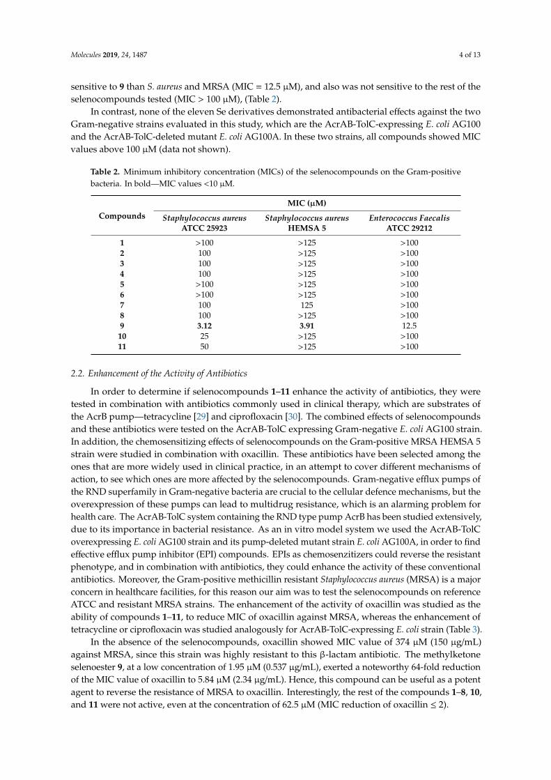
Molecules 2019, 24, 1487 4 of 13
sensitive to 9 than S. aureus and MRSA (MIC = 12.5 µM), and also was not sensitive to the rest of theselenocompounds tested (MIC > 100 µM), (Table 2).
In contrast, none of the eleven Se derivatives demonstrated antibacterial effects against the twoGram-negative strains evaluated in this study, which are the AcrAB-TolC-expressing E. coli AG100and the AcrAB-TolC-deleted mutant E. coli AG100A. In these two strains, all compounds showed MICvalues above 100 µM (data not shown).
Table 2. Minimum inhibitory concentration (MICs) of the selenocompounds on the Gram-positivebacteria. In bold—MIC values <10 µM.
Compounds
MIC (µM)
Staphylococcus aureusATCC 25923
Staphylococcus aureusHEMSA 5
Enterococcus FaecalisATCC 29212
1 >100 >125 >1002 100 >125 >1003 100 >125 >1004 100 >125 >1005 >100 >125 >1006 >100 >125 >1007 100 125 >1008 100 >125 >1009 3.12 3.91 12.510 25 >125 >10011 50 >125 >100
2.2. Enhancement of the Activity of Antibiotics
In order to determine if selenocompounds 1–11 enhance the activity of antibiotics, they weretested in combination with antibiotics commonly used in clinical therapy, which are substrates ofthe AcrB pump—tetracycline [29] and ciprofloxacin [30]. The combined effects of selenocompoundsand these antibiotics were tested on the AcrAB-TolC expressing Gram-negative E. coli AG100 strain.In addition, the chemosensitizing effects of selenocompounds on the Gram-positive MRSA HEMSA 5strain were studied in combination with oxacillin. These antibiotics have been selected among theones that are more widely used in clinical practice, in an attempt to cover different mechanisms ofaction, to see which ones are more affected by the selenocompounds. Gram-negative efflux pumps ofthe RND superfamily in Gram-negative bacteria are crucial to the cellular defence mechanisms, but theoverexpression of these pumps can lead to multidrug resistance, which is an alarming problem forhealth care. The AcrAB-TolC system containing the RND type pump AcrB has been studied extensively,due to its importance in bacterial resistance. As an in vitro model system we used the AcrAB-TolCoverexpressing E. coli AG100 strain and its pump-deleted mutant strain E. coli AG100A, in order to findeffective efflux pump inhibitor (EPI) compounds. EPIs as chemosenzitizers could reverse the resistantphenotype, and in combination with antibiotics, they could enhance the activity of these conventionalantibiotics. Moreover, the Gram-positive methicillin resistant Staphylococcus aureus (MRSA) is a majorconcern in healthcare facilities, for this reason our aim was to test the selenocompounds on referenceATCC and resistant MRSA strains. The enhancement of the activity of oxacillin was studied as theability of compounds 1–11, to reduce MIC of oxacillin against MRSA, whereas the enhancement oftetracycline or ciprofloxacin was studied analogously for AcrAB-TolC-expressing E. coli strain (Table 3).
In the absence of the selenocompounds, oxacillin showed MIC value of 374 µM (150 µg/mL)against MRSA, since this strain was highly resistant to this β-lactam antibiotic. The methylketoneselenoester 9, at a low concentration of 1.95 µM (0.537 µg/mL), exerted a noteworthy 64-fold reductionof the MIC value of oxacillin to 5.84 µM (2.34 µg/mL). Hence, this compound can be useful as a potentagent to reverse the resistance of MRSA to oxacillin. Interestingly, the rest of the compounds 1–8, 10,and 11 were not active, even at the concentration of 62.5 µM (MIC reduction of oxacillin ≤ 2).

Molecules 2019, 24, 1487 5 of 13
However, none of the tested selenocompounds were able to improve the efficacy of antibioticsagainst the AcrAB-TolC-overexpressing strain of E. coli with the above-mentioned excellent 64-foldfactor. The highest reductions observed in E. coli were a 2-fold reduction of the MIC values oftetracycline (compound 9) and of ciprofloxacin (compounds 9, 10).
Table 3. Numerical value of the reduction of the MICs of selected antibiotics in methicillin resistantS. aureus (MRSA) or in E. coli AG100 exerted by selenocompounds when administered in combinationwith antibiotics.
Cpd1
MRSA HEMSA 5 Escherichia coli AG100
Concentration ofCompound [µM]2
Reduction ofOxacillin MIC
Concentration ofCompound [µM]
Reduction ofTetracycline mic
Reduction ofCiprofloxacin mic
1 62.5 no effect 50 no effect no effect2 ND ND 3 50 no effect no effect3 62.5 2-fold 50 no effect no effect4 62.5 no effect 50 no effect no effect5 62.5 no effect 50 no effect no effect6 62.5 no effect 50 no effect no effect7 62.5 ≥ 2-fold 50 no effect no effect8 62.5 2-fold 50 no effect no effect9 1.95 64-fold 25 2-fold 2-fold
10 62.5 no effect 50 no effect 2-fold11 62.5 no effect 50 no effect no effect1 Cpd: Compound. 2 Starting concentration of tetracycline: 8.4 µM; ciprofloxacin: 1.4 µM; and oxacillin: 747 µM.3 ND: Not determined.
2.3. Anti-Chlamydial Activity
Before the assessment of the anti-chlamydial activity of the selenocompounds, a cytotoxicity assaywas performed on HeLa cells to determine the ranges of concentrations at which the selenocompoundscan be evaluated without showing direct toxic effects to HeLa cells. Selenocompounds 2, 3, 5, 7,and 9–11, significantly inhibited the formation of chlamydial inclusions at selected concentrations(Figure 1).
effect of selenocompounds at 1.25 and 2.5 μM ( ), and at 0.25 and 0.5 μM (
at 2.5 μMat 1.25 μM, whereas
of inclusions at low submicromolar concentrations of 0.5 μM. The most potent anti
concentration of 0.25 μM (0.0689 and 0.0858 μg/
–
–
–
Figure 1. Anti-chlamydial effect of selenocompounds at 1.25 and 2.5 µM (A), and at 0.25 and 0.5 µM (B).
Compounds 2 and 7 at 2.5 µM, showed 82% and 71% inhibition, compared to the control,respectively. In addition, 2 and 7 were effective at 1.25 µM, whereas 9 and 10 inhibited the formationof inclusions at low submicromolar concentrations of 0.5 µM. The most potent anti-chlamydialselenocompounds were 9 and 11, as they inhibited more than 50% of the growth of C. trachomatis D, ata concentration of 0.25 µM (0.0689 and 0.0858 µg/mL, respectively).

Molecules 2019, 24, 1487 6 of 13
2.4. Real-Time Accumulation Assay
Since ethidium bromide (EB) is a substrate of the AcrB efflux pump, the intracellular accumulationof EB provides information about the inhibition of the AcrAB-TolC system, in the presence ofselenocompounds, in a time-dependent manner. The assay records the real-time accumulation of EB,using a real-time thermocycler, by monitoring the fluorescence of EB inside the cells [31]. The activitiesof compounds 1–11 in the real-time EB accumulation assay, were given in terms of the relativefluorescence index (RFI) of the real-time accumulation curves (Table 4). In case of the real-time EBaccumulation, the amount of EB accumulated by cells was higher if the difference between RFtreated andRFuntreated was greater, therefore, the degree of inhibition of the efflux pump system by the compoundbecame greater. Compounds 9 and 10 possessed EPI activity and decreased the MIC of ciprofloxacinon E. coli AG100. However, the selenoanhydride 1 and the selenoester 4, compared with the positivecontrol promethazine (PMZ, RFI: 0.15), strongly inhibited the efflux of AcrAB-TolC in E. coli AG100;they had no effect in combination with the antibiotics, suggesting that other cellular mechanisms mightalso be involved in the mode of action, such as interaction with cell wall components, formation ofreactive oxygen species (ROS), or membrane destabilizing effects. Without investigating the possiblemetabolites of the selenocompounds, no further conclusions can be drawn, for this reason we areplanning to study the metabolites of these compounds in future works. Derivatives 7 and 9–11 causedmoderate inhibitory action, whereas 2, 3, 5, 6 and 8 showed weak or no activity on the intracellular EBaccumulation in E. coli AG100. Among derivatives 7 and 9–11, compound 7, which contained a methyloxoester in the alkyl moiety bound to Se, was the most active agent (RFI = 0.13).
Table 4. Relative fluorescence index (RFI) for the effect of selenocompounds and positive controlpromethazine (PMZ) on the AcrAB-TolC-expressing Escherichia coli AG100 strain.
CompoundRFIa
CompoundRFIa
CompoundRFIa
Escherichia coli AG100 Escherichia coli AG100 Escherichia coli AG100
1 0.28 5 0.04 9 0.112 0.03 6 0.06 10 0.123 0.04 7 0.13 11 0.114 0.18 8 0.08 PMZ 0.15
Nevertheless, no efflux pump inhibitory action of selenocompounds (1–11) was found in theE. coli AG100A strain.
2.5. Gene Expression Analysis by Quantitative PCR
For the effect of the selenocompounds on the relative expression of the efflux pump, antibioticresistance and QS genes in E. coli AG100—the most effective compounds in the EB real-time accumulationassay—were examined (compounds 1, 4, 7; Figure 2). In this assay, the genes of the multidrug effluxpump (AcrAB), the component of the E. coli mar locus (multiple antibiotic resistance), and the gene ofSdiA were investigated. The changes in gene expression from reverse transcription quantitative PCRexperiments were normalized to the expression of gapdh (internal control), in the same sample, andcompared to the expression of the examined genes obtained from the untreated, control samples.
As shown in Figure 2A, compound 1 at 50 µM concentration significantly up-regulated the acrB,marR, and sdiA genes, after 4 h of exposure. However, after 18 h, the expression of the acrB genereturned to the basal levels and the expression of the marR and sdiA genes, significantly increased.Compound 4 up-regulated the expression levels of acrB, marR, and sdiA, after 4 h, although after18 h, the expression levels of the acrB and marR genes decreased. The QS gene sdiA was significantlyup-regulated after 18 h (Figure 2B).
Compound 7 also significantly up-regulated marR, after exposures of 4 h and of 18 h. After 18 h,the expression level of the RND transporters subunit genes (acrA, acrB) was significantly increased(Figure 2C).

Molecules 2019, 24, 1487 7 of 13
–
——
at 50 μM co
–
–
– – – –
Figure 2. Relative gene expression levels of the genes of the acrA, acrB, marR, and sdiA in the presenceof compounds 1 (A), 4 (B), and 7 (C), after 4 and 18 h exposure. The line denotes a threshold value,which was set at a two-fold increase in transcripts.
3. Discussion
Results of these studies indicate that selenoesters and selenoanhydrides, previously found asactive anticancer or ABCB1 efflux pump inhibitors in cancer cells [7,8,32–35], also displayed a promisingantimicrobial potential against the MDR bacterial strains.
3.1. Antibacterial Activity
The evaluation of the compounds proved that the ketone-containing selenoesters 9–11 showed anantibacterial activity against the Gram-positive reference S. aureus strain, whereas, the methylketoneselenoester 9 was also active against the MRSA HEMSA 5 and E. faecalis. However, none of thecompounds were active against the Gram-negative E. coli ag100. The background of the differentantibacterial activity of 9 against the tested Gram-positives and Gram-negatives was unknown;further experiments are required to clarify whether the mechanism of action of the methylketoneselenoester could be related with any kind of interaction between this compound and the bacterialcell wall that is typical for gram-positive bacteria. In contrast, the remaining alkyl groups (–CH3) oralkyl-functionalized moieties (–CH2CONH2, –CH2COOCH3 and –CH2COOPh, Table 1) bound tothe Se atom rendered selenoesters that were ineffective against the tested strains. Interestingly, thecompounds 9–11 were also the most potent anticancer agents in previous works [7,8,33], and they alsoshowed a good selectivity towards cancer cells, with respect to non-tumour cell lines, as they showedselectivity indexes ranging from 8.4 to 14.4 [8].
In previous works, it was hypothesized that the possible mechanism of action of these compoundscould be the hydrolysis of the compound and the subsequent liberation of the ionic species of Se, whichcould be responsible for the activity of the compounds [33]. In this case, this phenomenon enables usto hypothesize that the CH3COCH2SeH, or its anionic form, are the chemical forms of Se that could bebehind the observed activities. The lack of activity of the non-ketone selenoesters, directs a specialattention to this –SeCH2COCH3 ketone-containing moiety.
3.2. Enhancement of the Activity of Antibiotics
The activity of 9 on the MDR clinical isolate (MRSA) was very promising, because compound 9
reduced the MIC of oxacillin in 64-fold (from 374 µM to 5.84 µM). These results supported the potentialapplications of the methylketone selenoesters, such as antimicrobials, and the multidrug resistancereversing agents. These results were in accordance with the activity shown by these compoundsas enhancers of the anticancer activity of chemotherapy drugs [35], suggesting that these seleniumderivatives have the ability to effectively interact with the resistance mechanisms developed by theresistant bacterial strains and by the resistant cancer cells. This work intends to carry out a screeningof the potential applications of the selenocompounds, and in future works we will attempt to ascertainthe possible mechanisms of actions of the activities described herein, as this observed potentialenhancement of the activity of oxacillin exerted by compound 9 in an MRSA clinical isolate.

Molecules 2019, 24, 1487 8 of 13
3.3. Anti-Chlamydial Activity
Previous studies have reported that selected selenocompounds, such as certain selenocyanates,selenoureas, and diselenides, showed antiproliferative activities against the intracellular forms ofLeishmania spp. [36,37]. Taking those results into account, this study provided a new line of evidence forthe action of selenoanhydride/selenoesters on an obligate intracellular chlamydial strain. In particular,different selenoesters, such as 2, 3, 5, 7, and 9–11, have exerted a noteworthy activity againstC. trachomatis D. Furthermore, the activities of the methyl (9) and the tert-butyl (11) derivatives werevery promising, as they inhibited the formation of more than 50% of the chlamydial inclusions, at avery low concentration (0.25 µM). However, their mode of action has not been ascertained in this study.
Regarding the observed structure activity relationships of the anti-chlamydial assays, the ketoneselenoesters 9–11 showed noteworthy activity at lower concentrations (0.25 µM, 0.5 µM), compared tothe rest of the series (1.25 µM, 2.5 µM). Among the remaining selenoesters, the symmetric dimethylselenodiester, which contains a thiophene ring 2, and the methyl oxoester derivative 7 showed a betteractivity, and the activities of the symmetric dimethyl selenodiesters 3 and 5 were also remarkable.These fact highlights the importance of the symmetry for the activity against intracellular pathogens [36].
3.4. Interaction of the Compounds with Bacterial Efflux Pumps
The resistance to the current antibacterial drugs is one of the major therapeutic challenges in thetreatment of bacterial infections, and knowing the potential of these derivatives as multidrug resistancereversing agents (proved both by the capacity to enhance the activity of antibiotics described aboveand by the enhancement of anticancer drugs reported in previous works), we have studied here theprocedure through which selenocompounds interact with the bacterial AcrAB-TolC system in the E. coli
AG100 strain.The results obtained revealed that the cyclic selenoanhydride 1 significantly inhibited this bacterial
AcrAB-TolC efflux pump in the E. coli AG100 strain. Similarly, EP inhibiting activity has been foundfor compounds 4 and 7. The second most potent inhibitor was the symmetrical benzene derivative1,3-disubstituted with methylselenoester moieties (4). Interestingly, its 1,4-disubstituted analogue(5) showed an EP-inhibitory activity, 4.5-fold lower, suggesting the importance of the substituents’topology for the expected biological effect. Taking into account the distinct difference in electrondensity properties between m- and p-substituted phenyl rings, this factor seems to have affected themechanisms of EP inhibition.
The well-characterized RND-type transporter, AcrB is associated with TolC and AcrA and isthe major efflux pump of E. coli [38]. These efflux pumps recognize and extrude a large variety ofantibiotics from the cytoplasm. The energy required for the operation of the efflux pump is providedby the proton motive force, created by the proton gradient resulting from electron transport [39].This fact suggests that those selenocompounds, which possessed EP inhibitor activity, might interferewith the proton motive force. Surprisingly, compounds 1 and 4, which inhibited the AcrAB-TolCsystem, influenced the expression of the gene acrB, which is a constituent of the AcrAB-TolC system.In addition, the compounds increased the expression of the QS gene sdiA, after 18 h of exposure, whichsuggests their roles in QS, although their QS inhibitory activities were not investigated in this study.
4. Materials and Methods
4.1. Chemistry
Eleven pure selenocompounds obtained as described earlier [33], were examined (1–11, Table 1).All compounds were stable in air and their purity was assessed by elemental analysis and 1H and13C NMR, as reported in a previous work [35]. Before their use in biological assays, they weredissolved in dimethyl sulfoxide (DMSO; Merck KGaA, Darmstadt, Germany), to obtain stock solutions.Working solutions were prepared by dilutions in the culture medium.
Molecules 2019, 24, 1487 9 of 13
4.2. Bacterial Strains
Wild-type E. coli K-12 AG100 strain [argE3 thi-1 rpsL xyl mtl ∆(gal-uvrB) supE44] and itsAcrAB-TolC-deleted mutant strain E. coli AG100A (a kind gift from Hiroshi Nikaido, Department ofMolecular and Cell Biology and Chemistry, University of California, Berkeley, USA) were used for theevaluation of the EPI activity of the tested selenocompounds.
S. aureus ATCC 25923 and E. faecalis ATCC 29212 strains were used to determine the MIC.A methicillin-resistant S. aureus strain (MRSA HEMSA 5, a clinical isolate) was used in the combinationassay, with oxacillin, to determine the capacity of compounds to enhance the antibacterial effect ofthis antibiotic. C. trachomatis reference strain (serovar D, UW-3/Cx, ATCC, VR-885D) was used in theanti-chlamydial assay.
4.3. Propagation of C. trachomatis D
C. trachomatis D was propagated on the HeLa 229 cells (ATCC, CCL-2.1), as described earlier [40].The titre of the infectious elementary bodies was determined by an indirect immunofluorescence assay.Serial dilutions of the elementary bodies’ preparation were inoculated onto the HeLa monolayers and,after a 48-h culture, the cells were fixed with acetone, and stained with monoclonal anti-Chlamydia LPSantibody (AbD Serotec, Oxford, UK) and FITC-labelled anti-mouse IgG (Merck KGaA, Darmstadt,Germany). The inclusions of C. trachomatis D were enumerated under a UV microscope.
4.4. Determination of MIC
The effects exerted by different concentrations of the compounds on the bacterial growth inS. aureus, E. faecalis, and E. coli AG100 were tested in 96-well plates. The MICs of selenocompoundswere determined, according to the Clinical and Laboratory Standard Institute (CLSI) guidelines [41].The DMSO exerted no antibacterial effect. Alternatively, the MIC of the oxacillin in MRSA HEMSA5 was determined by the broth microdilution method, in a cation-adjusted Mueller–Hinton Broth(MHB II), according to the recommendations of the CLSI. Results were recorded after a 20- or 24-hourincubation at 37 ◦C.
4.5. Enhancement of the Activity of Antibiotics
The chemosensitizing effect of the tested selenocompounds was evaluated through thedetermination of the MIC values of the antibiotics, in the presence of sub-inhibitory concentrations ofthe compounds (MIC/2 or MIC/4), in both Gram-negative (E. coli AG100) and Gram-positive (MRSA)strains. The MICs were evaluated in the E. coli strain, by a two-fold broth microdilution method in the96-well plates, using serial dilutions of tetracycline (from 8.4 to 0.16 µM) and ciprofloxacin (from 1.4 to2.7 × 10−3 µM). The first four rows contained two-fold dilutions of antibiotics, and the combinations ofthe antibiotics and tested compounds were added into the last four rows. 10−4 dilution of an overnightbacterial culture in 50 µL of MHB was then added to each well, with the exception of the mediumcontrol wells. The plates were then incubated at 37 ◦C for 18 h. MIC values of the antibiotics and theircombination with the tested compounds were determined by naked eyes. In the assay with oxacillin inthe MRSA HEMSA 5 bacterial strain, a microdilution method in MHB II was used.
4.6. Anti-Chlamydial Assay
Elementary bodies of C. trachomatis D (4× 10³ IFU/mL) were incubated with compounds at selectedconcentrations (0.25, 0.5, 1.25, 2.5 µM) in sucrose-phosphate-glutamic acid buffer (SPG) for 2 h at 37 ◦C.As a control, C. trachomatis D was also incubated alone in the SPG. To quantify the anti-chlamydialeffects of the compounds, HeLa cells were seeded in 24-well plates with 13-mm cover glasses. Theconfluent cells were infected with compound-treated C. trachomatis D or with the non-treated controls.After 48 h, the cells were fixed with acetone at −20 ◦C for 10 min. The titre of the infectious elementarybodies was determined by the indirect immunofluorescence assay, as described earlier [42].

Molecules 2019, 24, 1487 10 of 13
4.7. Real-Time Accumulation Assay
The effect of the studied selenocompounds on the real-time accumulation of ethidium bromide(EB) was assessed by an automated EB method [43], using a LightCycler real-time thermocycler(LightCycler 1.5; Roche). The compounds were added individually at different concentrations at MIC/2to the EB solution in PBS. The final concentration of EB was 1 and 0.25 µg/mL for E. coli AG100 andAG100A, respectively. The method for the calculation of the relative fluorescence index (RFI) of the lasttime point (minute 30) was described earlier by Kincses et al. [44]. Promethazine (PMZ; EGIS) wasapplied as a positive control.
4.8. Expression Analyses of Genes by Quantitative PCR
Total RNA was isolated from E. coli AG100 (OD of 0.6 at 600 nm) using the NucleoSpin RNA kit(Macherey Nagel) according to the manufacturer’s instructions. The concentration of the extractedRNA templates was assessed by spectrophotometry at 260 nm.
The expression of the acrA, the acrB, the multiple antibiotic resistance protein R (marR), and thequorum-sensing transcriptional activator (sdiA) genes was studied by reverse transcription quantitativePCR (RT-qPCR), as described earlier [44]. The real-time one-step PCR was performed in a CFX96 Touchreal-time PCR detection system (Bio-Rad), strictly adhering to the manufacturer recommendations ofthe SensiFAST™ SYBR No-ROX One-Step Kit (Bioline GmbH, Luckenwalde, Germany). The forwardand reverse primers used in the experiment are shown in Table 5 [44,45]. The cycle threshold (Ct)values were determined with the Bio-Rad CFX Manager Software version 3.1. Relative quantificationanalysis was carried out using the Livak method [46]. The expression of gapdh was used as the internalcontrol and the untreated E. coli AG 100 served as the external control. We have defined a thresholdvalue—increases greater than 2-fold in the amount of transcripts relative to the control samples wereconsidered significant.
Table 5. Primers used in the RT-qPCR.
Gene Full Name Primer Sequence (5’–3’)Ampliconsize (bp)
Ref.
acrA Acridine resistance protein ACTTAGCCCTAACAGGATGTGTTGAAATTACGCTTCAGGAT
189 [45]
acrB Acridine resistance protein BCGTACACAGAAAGTGCTCAACGCTTCAACTTTGTTTTCTT
183 [45]
marRMultiple antibiotic resistance
protein RAGCGATCTGTTCAATGAAATTTCAGTTCAACCGGAGTAAT
170 [45]
sdiAQuorum-sensing
transcriptional activatorCTGATGGCTCTGATGCGTTTATCTGGTGGAAATTGACCGTATT
163 [44]
gapdhGlyceraldehyde-3-phospate
dehydrogenaseACTTACGAGCAGATCAAAGCAGTTTCACGAAGTTGTCGTT
170 [45]
5. Conclusions
Herein, we have reported the evaluation of the antibacterial and multidrug resistance reversingactivity of 11 novel selenocompounds. The most active compound in the antibacterial assay, themethylketone selenoester 9, showed potential antibacterial activity against the different strains ofS. aureus, E. faecalis, and C. trachomatis D, even at very low concentrations (0.25 µM for C. trachomatis
D). This selenocompound also enhanced the efficacy of antibiotics, namely it multiplied by 64-foldthe antibacterial action of oxacillin, against the MDR clinical isolate of S. aureus. Alternatively, threecompounds (the selenoanhydride 1 and the selenoesters 4 and 7) inhibited the tripartite multidrugresistance efflux pump AcrAB-TolC in E. coli, and affected the expression of the different genes relatedto these resistance processes.

Molecules 2019, 24, 1487 11 of 13
Based on these results, it can be concluded that this group of selenocompounds can be attractivepotential EP inhibitors and antibacterial lead scaffolds, for further development of new chemical tools,to overcome bacterial multidrug resistance.
Author Contributions: E.D.-Á. and C.S. synthesized the compounds evaluated. T.M., G.S., J.H. and K.K.-K.conceived the experiments; T.M., A.K., A.C., Á.S.T., K.W., M.A.M. and G.S. performed the biological experiments;T.M., G.S. and E.D.-Á. wrote the paper; T.M., C.S., J.H., K.K.K., G.S. and E.D.-Á. revised the manuscript; T.M. andG.S. provided the funding for the biological evaluation; and E.D.-Á and M.A.M. provided the funding of thearticle publication.
Funding: G.S. was supported by the János Bolyai Research Scholarship of the Hungarian Academy of Sciences, bythe GINOP-2.3.2-15-2016-00038 project and by the University of Szeged, through the project SZTE ÁOK-KKA2018/270-62-2. A.K. was supported by the ÚNKP-17-3 New National Excellence Program of the Ministry of HumanCapacities of Hungary. E.D.A. was supported by the Spanish “Consejo Superior de Investigaciones Científicas”(CSIC, Spanish National Research Council), through Project 201780I027.
Conflicts of Interest: The authors declare no conflict of interest.
References
1. Drusano, G.L.; Louie, A.; MacGowan, A.; Hope, W. Suppression of Emergence of Resistance in PathogenicBacteria: Keeping Our Powder Dry, Part 1. Antimicrob. Agents Chemother. 2015, 60, 1183–1193. [CrossRef]
2. Lobanov, A.V.; Hatfield, D.L.; Gladyshev, V.N. Eukaryotic selenoproteins and selenoproteomes.Biochim. Biophys. Acta 2009, 1790, 1424–1428. [CrossRef]
3. Tinggi, U. Selenium: Its role as antioxidant in human health. Environ. Health Prev. Med. 2008, 13, 102–108.[CrossRef]
4. Angeli, A.; Abbas, G.; del Prete, S.; Capasso, C.; Supuran, C.T. Selenides bearing benzenesulfonamideshow potent inhibition activity against carbonic anhydrases from pathogenic bacteria Vibrio cholerae andBurkholderia pseudomallei. Bioorg. Chem. 2018, 79, 319–322. [CrossRef] [PubMed]
5. Lü, H.; Gao, Y.; Shan, H.; Lin, Y. Preparation and antibacterial activity studies of degraded polysaccharideselenide from Enteromorpha prolifera. Carbohyd. Polym. 2014, 107, 98–102. [CrossRef]
6. Witek, K.; Nasim, M.; Bischoff, M.; Gaupp, R.; Arsenyan, P.; Vasiljeva, J.; Marc, M.; Olejarz, A.; Latacz, G.;Kiec-Kononowicz, K.; et al. Selenazolinium Salts as “Small Molecule Catalysts” with High Potency againstESKAPE Bacterial Pathogens. Molecules 2017, 22, 2174. [CrossRef]
7. Domínguez-Álvarez, E.; Gajdács, M.; Spengler, G.; Palop, J.A.; Marc, M.A.; Kiec-Kononowicz, K.; Amaral, L.;Molnár, J.; Jacob, C.; Handzlik, J.; et al. Identification of selenocompounds with promising properties toreverse cancer multidrug resistance. Bioorg. Med. Chem. Lett. 2016, 26, 2821–2824. [CrossRef] [PubMed]
8. Gajdács, M.; Spengler, G.; Sanmartín, C.; Marc, M.A.; Handzlik, J.; Domínguez-Álvarez, E. Selenoesters andselenoanhydrides as novel multidrug resistance reversing agents: A confirmation study in a colon cancerMDR cell line. Bioorg. Med. Chem. Lett. 2017, 27, 797–802. [CrossRef]
9. Handzlik, J.; Matys, A.; Kiec-Kononowicz, K. Recent Advances in Multi-Drug Resistance (MDR) Efflux PumpInhibitors of Gram-Positive Bacteria S. aureus. Antibiotics 2013, 2, 28–45. [CrossRef] [PubMed]
10. Pozzi, G.; Iannelli, F.; Oggioni, M.R.; Santagati, M.; Stefani, S. Genetic elements carrying macrolide effluxgenes in streptococci. Curr. Drug Targets Infect. Disord. 2004, 4, 203–206. [CrossRef]
11. Reynolds, E.; Ross, J.I.; Cove, J.H. Msr(A) and related macrolide/streptogramin resistance determinants:Incomplete transporters? Int. J. Antimicrob. Agents 2003, 22, 228–236. [CrossRef]
12. Robertson, G.T.; Doyle, T.B.; Lynch, A.S. Use of an efflux-deficient streptococcus pneumoniae strainpanel to identify ABC-class multidrug transporters involved in intrinsic resistance to antimicrobial agents.Antimicrob. Agents Chemother. 2005, 49, 4781–4783. [CrossRef]
13. Daury, L.; Orange, F.; Taveau, J.-C.; Verchère, A.; Monlezun, L.; Gounou, C.; Marreddy, R.K.R.; Picard, M.;Broutin, I.; Pos, K.M.; et al. Tripartite assembly of RND multidrug efflux pumps. Nat. Commun. 2016,7, 10731. [CrossRef]
14. Gupta, A.; Pande, A.; Sabrin, A.; Thapa, S.S.; Gioe, B.W.; Grove, A. MarR Family Transcription Factors fromBurkholderia Species: Hidden Clues to Control of Virulence-Associated Genes. Microbiol. Mol. Biol. Rev. 2018,83, e00039-18. [CrossRef]

Molecules 2019, 24, 1487 12 of 13
15. Grkovic, S.; Brown, M.H.; Skurray, R.A. Regulation of bacterial drug export systems. Microbiol. Mol. Biol. Rev.
2002, 66, 671–701. [CrossRef] [PubMed]16. Tavío, M.M.; Aquili, V.D.; Poveda, J.B.; Antunes, N.T.; Sánchez-Céspedes, J.; Vila, J. Quorum-sensing regulator
sdiA and marA overexpression is involved in in vitro-selected multidrug resistance of Escherichia coli.J. Antimicrob. Chemother. 2010, 65, 1178–1186. [CrossRef] [PubMed]
17. Holinka, J.; Pilz, M.; Kubista, B.; Presterl, E.; Windhager, R. Effects of selenium coating of orthopaedic implantsurfaces on bacterial adherence and osteoblastic cell growth. Bone Joint J. 2013, 95, 678–682. [CrossRef][PubMed]
18. Kumar, B.S.; Tiwari, S.K.; Manoj, G.; Kunwar, A.; Amrita, N.; Sivaram, G.; Abid, Z.; Ahmad, A.; Khan, A.A.;Priyadarsini, K.I. Anti-unlcer and antimicrobial activities of sodium selenite against Helicobacter pylori:In vitro and in vivo evaluation. Scand. J. Infect. Dis. 2010, 42, 266–274. [CrossRef] [PubMed]
19. Aribi, M.; Meziane, W.; Habi, S.; Boulatika, Y.; Marchandin, H.; Aymeric, J.-L. Macrophage BactericidalActivities against Staphylococcus aureus Are Enhanced In Vivo by Selenium Supplementation in aDose-Dependent Manner. PLoS ONE 2015, 10, e0135515. [CrossRef] [PubMed]
20. Wadhwani, S.A.; Shedbalkar, U.U.; Singh, R.; Chopade, B.A. Biogenic selenium nanoparticles: Current statusand future prospects. Appl. Microbiol. Biotechnol. 2016, 100, 2555–2566. [CrossRef]
21. Cihalova, K.; Chudobova, D.; Michalek, P.; Moulick, A.; Guran, R.; Kopel, P.; Adam, V.; Kizek, R.Staphylococcus aureus and MRSA Growth and Biofilm Formation after Treatment with Antibiotics andSeNPs. Int. J. Mol. Sci. 2015, 16, 24656–24672. [CrossRef]
22. Chudobova, D.; Cihalova, K.; Dostalova, S.; Ruttkay-Nedecky, B.; Rodrigo, M.A.M.; Tmejova, K.; Kopel, P.;Nejdl, L.; Kudr, J.; Gumulec, J.; et al. Comparison of the effects of silver phosphate and selenium nanoparticleson Staphylococcus aureus growth reveals potential for selenium particles to prevent infection. FEMS Microbiol.
Lett. 2014, 351, 195–201. [CrossRef]23. Stevanovic, M.; Filipovic, N.; Djurdjevic, J.; Lukic, M.; Milenkovic, M.; Boccaccini, A. 45S5Bioglass®-based
scaffolds coated with selenium nanoparticles or with poly(lactide-co-glycolide)/selenium particles: Processing,evaluation and antibacterial activity. Colloid. Surf. B 2015, 132, 208–215. [CrossRef]
24. Huang, X.; Chen, X.; Chen, Q.; Yu, Q.; Sun, D.; Liu, J. Investigation of functional selenium nanoparticles aspotent antimicrobial agents against superbugs. Acta Biomater. 2016, 30, 397–407. [CrossRef]
25. Sonkusre, P.; Singh Cameotra, S. Biogenic selenium nanoparticles inhibit Staphylococcus aureus adherenceon different surfaces. Colloid. Surf. B 2015, 136, 1051–1057. [CrossRef] [PubMed]
26. Srivastava, N.; Mukhopadhyay, M. Green synthesis and structural characterization of selenium nanoparticlesand assessment of their antimicrobial property. Bioproc. Biosyst. Eng. 2015, 38, 1723–1730. [CrossRef][PubMed]
27. Sandoz, K.M.; Rockey, D.D. Antibiotic resistance in Chlamydiae. Future Microbiol. 2010, 5, 1427–1442.[CrossRef] [PubMed]
28. Mestrovic, T.; Ljubin-Sternak, S. Molecular mechanisms of Chlamydia trachomatis resistance to antimicrobialdrugs. Front Biosci. 2018, 23, 656–670. [CrossRef]
29. Elkins, C.A.; Nikaido, H. Substrate specificity of the RND-type multidrug efflux pumps AcrB and AcrDof Escherichia coli is determined predominantly by two large periplasmic loops. J. Bacteriol. 2002, 184,6490–6498. [CrossRef] [PubMed]
30. Ricci, V.; Piddock, L.J.V. Only for substrate antibiotics are a functional AcrAB-TolC efflux pump and RamArequired to select multidrug-resistant Salmonella Typhimurium. J. Antimicrob. Chemother. 2009, 64, 654–657.[CrossRef] [PubMed]
31. Paixão, L.; Rodrigues, L.; Couto, I.; Martins, M.; Fernandes, P.; de Carvalho, C.C.C.R.; Monteiro, G.A.;Sansonetty, F.; Amaral, L.; Viveiros, M. Fluorometric determination of ethidium bromide efflux kinetics inEscherichia coli. J. Biol. Eng. 2009, 3, 18. [CrossRef]
32. Sanmartín, C.; Plano, D.; Domínguez, E.; Font, M.; Calvo, A.; Prior, C.; Encío, I.; Palop, J.A. Synthesis andpharmacological screening of several aroyl and heteroaroyl selenylacetic acid derivatives as cytotoxic andantiproliferative agents. Molecules 2009, 14, 3313–3338. [CrossRef]
33. Domínguez-Álvarez, E.; Plano, D.; Font, M.; Calvo, A.; Prior, C.; Jacob, C.; Palop, J.A.; Sanmartín, C.Synthesis and antiproliferative activity of novel selenoester derivatives. Eur. J. Med. Chem. 2014, 73, 153–166.[CrossRef]

Molecules 2019, 24, 1487 13 of 13
34. Díaz-Argelich, N.; Encío, I.; Plano, D.; Fernandes, A.P.; Palop, J.A.; Sanmartín, C. Novel Methylselenoestersas Antiproliferative Agents. Molecules 2017, 22, 1288. [CrossRef]
35. Spengler, G.; Gajdács, M.; Marc, M.; Domínguez-Álvarez, E.; Sanmartín, C. Organoselenium Compoundsas Novel Adjuvants of Chemotherapy Drugs—A Promising Approach to Fight Cancer Drug Resistance.Molecules 2019, 24, 336. [CrossRef]
36. Fernández-Rubio, C.; Campbell, D.; Vacas, A.; Ibañez, E.; Moreno, E.; Espuelas, S.; Calvo, A.; Palop, J.A.;Plano, D.; Sanmartin, C.; et al. Leishmanicidal Activities of Novel Methylseleno-Imidocarbamates.Antimicrob. Agents Chemother. 2015, 59, 5705–5713. [CrossRef]
37. Martín-Montes, Á.; Plano, D.; Martín-Escolano, R.; Alcolea, V.; Díaz, M.; Pérez-Silanes, S.; Espuelas, S.;Moreno, E.; Marín, C.; Gutiérrez-Sánchez, R.; et al. Library of Seleno-Compounds as Novel Agents againstLeishmania Species. Antimicrobial. Agents Chemother. 2017, 61, e02546-16. [CrossRef]
38. Li, X.-Z.; Plésiat, P.; Nikaido, H. The challenge of efflux-mediated antibiotic resistance in Gram-negativebacteria. Clin. Microbiol. Rev. 2015, 28, 337–418. [CrossRef]
39. Nikaido, H.; Pagès, J.-M. Broad-specificity efflux pumps and their role in multidrug resistance ofGram-negative bacteria. FEMS Microbiol. Rev. 2012, 36, 340–363. [CrossRef]
40. Bogdanov, A.; Endrész, V.; Urbán, S.; Lantos, I.; Deák, J.; Burián, K.; Önder, K.; Ayaydin, F.; Balázs, P.;Virok, D.P. Application of DNA chip scanning technology for automatic detection of Chlamydia trachomatisand Chlamydia pneumoniae inclusions. Antimicrob. Agents Chemother. 2014, 58, 405–413. [CrossRef]
41. Clinical & Laboratory Standards Institute: CLSI Guidelines. Available online: https://clsi.org/ (accessed on24 July 2017).
42. Balogh, E.P.; Faludi, I.; Virók, D.P.; Endrész, V.; Burián, K. Chlamydophila pneumoniae induces productionof the defensin-like MIG/CXCL9, which has in vitro antichlamydial activity. Int. J. Med. Microbiol. 2011, 301,252–259. [CrossRef]
43. Viveiros, M.; Martins, A.; Paixão, L.; Rodrigues, L.; Martins, M.; Couto, I.; Fähnrich, E.; Kern, W.V.; Amaral, L.Demonstration of intrinsic efflux activity of Escherichia coli K-12 AG100 by an automated ethidium bromidemethod. Int. J. Antimicrob. Agents 2008, 31, 458–462. [CrossRef]
44. Kincses, A.; Szabó, Á.M.; Saijo, R.; Watanabe, G.; Kawase, M.; Molnár, J.; Spengler, G. Fluorinated Beta-diketoPhosphorus Ylides Are Novel Efflux Pump Inhibitors in Bacteria. In Vivo 2016, 30, 813–817. [CrossRef]
45. Viveiros, M.; Dupont, M.; Rodrigues, L.; Couto, I.; Davin-Regli, A.; Martins, M.; Pagès, J.-M.; Amaral, L.Antibiotic stress, genetic response and altered permeability of E. coli. PLoS ONE 2007, 2, e365. [CrossRef]
46. Livak, K.J.; Schmittgen, T.D. Analysis of Relative Gene Expression Data Using Real-Time Quantitative PCRand the 2−∆∆CT Method. Methods 2001, 25, 402–408. [CrossRef]
Sample Availability: Not available.
© 2019 by the authors. Licensee MDPI, Basel, Switzerland. This article is an open accessarticle distributed under the terms and conditions of the Creative Commons Attribution(CC BY) license (http://creativecommons.org/licenses/by/4.0/).

72
IV.

74
75
A b
akté
rium
okba
n az
egy
ik le
gfo
ntos
abb
re-
zisz
tenc
ia m
echa
nizm
us a
mul
tidr
og e
fflux
pu
mpá
k m
egnö
veke
dett
ex
pres
szió
ja,
me -
lyek
jel
entő
s sz
erep
et j
átsz
anak
a b
akte
ri-
ális
bio
film
kép
zésb
en i
s. E
zen
kuta
tásu
nk
sorá
n 11
újo
nnan
szi
ntet
izál
t sz
elén
vegy
ület
A
crA
B-T
olC
effl
ux p
umpa
, va
lam
int
biofi
lm
gátl
ó ak
tivi
tásá
t vi
zsgá
ltuk
meg
Sal
mo
nell
a en
teri
ca s
erov
ar T
yphi
mur
ium
tö
rzse
k fe
l -ha
szná
lásá
val.
Az
Acr
AB
-Tol
C-t
exp
ress
záló
va
d tí
pusú
tö
rzsb
en a
leg
hatá
sosa
bb e
fflux
pu
mpa
gá
tlás
t a
9-e
s ve
gyül
et
mut
atta
. A
S.
Typh
imur
ium
14
028
s bi
ofilm
kép
zésé
t pe
dig
a 4
-es é
s az 5
-ös s
zelé
nveg
yüle
t gát
olta
a
legn
agyo
bb
mér
tékb
en.
Ered
mén
yein
k al
apjá
n el
mo
ndh
ató,
ho
gy a
sze
lénv
együ
le-
tek
adju
ván
skén
t al
kalm
azva
a b
iofi
lm t
er-
mel
ő S
. Typ
him
uriu
m á
ltal
oko
zott
fer
tőzé
s an
tibi
otik
umo
s ke
zelé
se s
orá
n ja
víth
atjá
k a
terá
pia
hat
éko
nysá
gát.
A b
akte
riál
is m
ulti
drog
rez
iszt
enci
a (M
DR)
je -
lens
ége
évrő
l év
re s
úlyo
s pr
oblé
mák
at o
koz
vilá
gsze
rte.
A j
elen
ség
az a
ntib
ioti
kum
ok é
s a
mik
róbá
k el
leni
sze
rek
kite
rjed
t ha
szná
latá
nak
SZEL
ÉNV
EGY
ÜLE
TEK
EFF
LUX
PU
MPA
ÉS
BIO
FILM
GÁ
TLÓ
HA
TÁSÁ
NA
K V
IZSG
ÁLA
TA
SALM
ON
ELLA
TY
PH
IMU
RIU
M T
ÖR
ZS
EKEN
Kin
cses
An
nam
ária
Szeg
edi T
udom
ánye
gyet
em
Ált
alán
os O
rvos
tudo
mán
yi K
ar
Orv
osi M
ikro
biol
ógia
i és
Imm
unbi
ológ
iai I
ntéz
et
Szer
zőtá
rs, t
émav
ezet
ő: S
peng
ler
Gab
riel
la
köve
tkez
mén
ye. A
rez
iszt
ens
bakt
ériu
mok
szá
-m
a fo
lyam
atos
an n
ő, m
egne
hezí
tve
ezze
l a fe
r-tő
ző b
eteg
sége
k te
rápi
áján
ak h
aték
onys
ágát
. A
z M
DR
kial
akul
ása
több
féle
mec
hani
zmus
hoz
is k
öthe
tő, m
elye
k kö
zül a
z eg
yik
típu
s az
effl
ux
tran
szpo
rter
ek
meg
emel
kede
tt
expr
essz
iója
(B
lair
et a
l., 2
015;
Pid
dock
, 20
06).
Ezek
a p
umpá
k fiz
ioló
giás
an i
s m
egta
lálh
atók
a b
akté
rium
ok-
ban,
fel
adat
uk a
tox
ikus
veg
yüle
tek
eltá
volít
á-sa
a s
ejtb
ől.
A k
áros
any
agok
mel
lett
a p
um-
pák
az a
ntib
akte
riál
is k
emot
eráp
iás
szer
eket
is
kip
umpá
lhat
ják
a se
jtbő
l, ez
ped
ig a
ter
ápia
si
kert
elen
ségé
hez
veze
thet
(Bla
ir et
al.,
201
5). A
z ef
flux
pum
pák
tová
bbi
jelle
mző
je,
hogy
töb
b ta
nulm
ány
szer
int
jele
ntős
sze
repe
t já
tsza
nak
a ba
ktér
ium
ok b
iofil
m k
épzé
sébe
n. N
éhán
y ku
tató
sze
rint
az
effl
ux p
umpá
k kö
zvet
ett
mó -
don
képe
sek
befo
lyás
olni
a b
iofil
m k
épzé
sben
sz
erep
et já
tszó
gén
eket
, tov
ábbá
a b
akte
riál
is
kom
mun
ikác
ióba
n (q
uoru
m s
ensi
ng)
szer
epet
já
tszó
mol
ekul
ákat
, m
elye
k ho
zzáj
árul
nak
a bi
ofilm
kép
zésh
ez (
Ala
v et
al.,
201
8).
A b
akte
riá-
lis b
iofi
lm o
lyan
mik
robi
ális
köz
össé
g, m
ely-
ben
a sz
essz
ilis
bakt
ériu
mok
eg
ymás
hoz,
ill
etve
a f
elsz
ínhe
z ta
padn
ak e
gy ö
nmag
uk
álta
l te
rmel
t ex
trac
ellu
lári
s po
limer
mát
rix
segí
tség
ével
(M
orae
s et
al.,
201
8).
A b
iofil
m t
o-
vább
i je
llem
zője
, ho
gy a
pla
nkto
niku
s se
jtek
-he
z ké
pest
alk
alm
azko
dni t
udna
k a
körn
yeze
ti
felt
étel
ekhe
z, t
ováb
bá a
bak
teri
ális
bio
film
et
felé
pítő
ba
ktér
ium
okna
k la
ssab
b a
met
abo
-liz
mus
uk é
s a
növe
kedé
sük
is.
A b
iofi
lmbe
n a
bakt
ériu
mok
meg
növe
kede
tt r
ezis
zten
ci-
át m
utat
nak
az a
ntim
ikro
biál
is a
nyag
okka
l sz
embe
n (S
ingh
et
al.,
2017
). Em
iatt
az
effl
ux
mec
hani
zmus
okho
z és
a b
iofi
lm k
épzé
shez
kö
thet
ő re
zisz
tenc
ia v
issz
afo
rdít
ásáh
oz f
on
-to
s le
nne
olya
n új
an
tim
ikro
biál
is
vegy
ü-le
tek
vagy
adj
uván
sok
fejle
szté
se,
mel
yek
haté
kony
an g
átol
nák
ezek
et a
rez
iszt
enci
a m
echa
nizm
usok
at.
A
sze
lén
egy
font
os n
yom
elem
, m
ely
jó-
téko
ny h
atás
sal b
ír a
dag
anat
ok
vagy
a g
yul-
ladá
sos
meg
bete
gedé
sek
meg
előz
éséb
en.
A
szak
iroda
lom
ban
szám
os
péld
át
talá
lunk
a
szel
énta
rtal
mú
vegy
ület
ek
anti
bakt
eriá
lis
hatá
sára
is:
a k
utat
ások
leg
inká
bb o
lyan
bak
-té
rium
ok f
elha
szná
lásá
val
tört
énne
k, m
elye
k no
zokó
miá
lis f
ertő
zése
ket
okoz
nak.
A k
órhá
zi
keze
lés
sorá
n sz
erze
tt i
nfek
cióé
rt g
yakr
an a
z Es
cher
ichi
a co
li, il
letv
e a
Stap
hylo
cocc
us a
ureu
s a
fele
lős.
Az
ezen
bak
téri
umok
ált
al o
kozo
tt
fert
őzés
ek a
bio
film
kép
ző k
épes
ségü
k ré
vén
nehe
zen
gyóg
yíth
atók
. Az
egyi
k ku
tató
csop
ort
álta
l has
znál
t pe
rihi
dros
zele
noxa
ntin
veg
yüle
t an
tiba
kter
iális
hat
ássa
l re
ndel
keze
tt S
. aur
eus
törz
sön
(Son
kusr
e–Si
ngh,
201
5).
Egy
más
ik k
uta-
tócs
opor
t E.
col
i, ill
etve
S. a
ureu
s bi
ofilm
kép
ző
törz
seke
t sz
elén
tart
alm
ú na
noré
szec
skék
kel
keze
lt.
24 ó
ra e
ltel
téve
l az
opt
ikai
den
zitá
s (O
D) m
érés
e ut
án a
zt t
apas
ztal
ták,
hog
y a
sze -
lénn
el k
ezel
t ba
ktér
ium
ok n
övek
edés
e cs
ök-
kent
a k
ontr
ollh
oz k
épes
t (G
uisb
iers
et a
l., 2
016)
.
A S
alm
onel
la e
nter
ica
Gra
m-n
egat
ív,
élel
-m
isze
rek
révé
n te
rjed
ő hu
mán
kór
okoz
ó. A
z ál
tala
oko
zott
kór
kép
a sz
alm
onel
lózi
s. A
z él
el-
mis
zerg
yárt
ás s
orán
bio
film
kép
ző t
ulaj
dons
á-ga
mia
tt p
erzi
sztá
lhat
, ezé
rt a
bak
téri
um te
ljes
elpu
sztí
tása
neh
éz fe
lada
t (M
orae
s et a
l., 2
018
).
Kís
érle
tein
k cé
lja 1
1 új
sze
lénv
együ
let
anti
-bio
film
, ill
etve
effl
ux
pum
pa g
átló
ak -
tivi
tásá
nak
meg
hatá
rozá
sa v
olt
Sa
lmo
ne
lla
Typ
him
uri
um
törz
sek
felh
aszn
álás
ával
.
AN
YA
GO
K É
S M
ÓD
SZ
ER
EK
Veg
yüle
tek
Kuta
tása
ink
sorá
n 10
szel
enoé
szte
r (2-
11),
illet
ve
egy
cikl
ikus
sze
leno
anhi
drid
(1)
veg
yüle
tet
vizs
-gá
ltun
k m
eg (1
. ábr
a). A
szá
rmaz
ékok
at d
imet
il-sz
ulfo
xidb
an (
DM
SO)
oldo
ttuk
, a
törz
sold
atok
ko
ncen
trác
iója
10
mM
vol
t. A
veg
yüle
teke
t D
r. En
rique
Dom
íngu
ez-Á
lvar
ez (
Con
sejo
Sup
erio
r de
In
vest
igac
ione
s C
ient
ífica
s,
Inst
itut
o de
Q
uím
ica
Org
ánic
a G
ener
al,
Span
yolo
rszá
g) é
s vo
lt t
émav
ezet
ője,
Pro
f. D
r. C
arm
en S
anm
artí
n (U
nive
rsit
y of
Nav
arra
, Dep
artm
ent
of O
rgan
ic
and
Phar
mac
euti
cal C
hem
istr
y, S
pany
olor
szág
; In
stit
uto
de In
vest
igac
ión
Sani
taria
de
Nav
arra
, Sp
anyo
lors
zág)
boc
sáto
tta
a re
ndel
kezé
sünk
re
(Dom
íngu
ez-Á
lvar
ez e
t al.,
201
6).
Bak
téri
um
törz
sek
Viz
sgál
atai
nk s
orán
az
aláb
bi b
akté
rium
törz
se-
ket
hasz
nált
uk f
el:
Salm
onel
la e
nter
ica
sero
var
Typh
imur
ium
SL1
344
vad
típu
sú t
örzs
et,
mel
y ex
pres
szál
ja a
z A
crA
B-T
olC
effl
ux p
umpá
t, v
ala -
min
t enn
ek a
z acr
B m
után
s tör
zsét
(L6
44)
(Sm
ith-
Bla
ir, 2
014
). A
veg
yüle
tek
anti
-bio
film
hat
ását
a
biofi
lm k
épző
S. e
nter
ica
sero
var T
yphi
mur
ium
14
028
s tö
rzsö
n vi
zsgá
ltuk
. A
tö
rzse
ket
D
r. Je
ssic
a M
. Bla
ir (U
nive
rsit
y of
Bir
min
gham
, In
stit
ute
of M
icro
biol
ogy
and
Infe
ctio
n, E
gye -
sült
Kir
álys
ág) b
ocsá
tott
a a
rend
elke
zésü
nkre
.
A. C
iklik
us s
zele
noan
hidr
id (
1) B
. Het
eroa
ril
szel
enoé
szte
rek
(2-3
) C. A
ril s
zele
noés
zter
ek (4
-11)
1. á
bra
A v
izsg
ált
szel
énve
gyül
etek
sze
rkez
ete
TU
DO
MÁ
NY
OS
ER
ED
MÉ
NY
EK
A N
AG
YV
ILÁ
GB
ÓL
KIN
CS
ES
AN
NA
MÁ
RIA

76
77
A le
gkis
ebb
gátl
ó k
on
cen
trác
ió m
egh
atár
o-
zása
mik
rod
ilúci
ós
mó
dsz
erre
lA
MIC
vag
y le
gkis
ebb
gátl
ó ko
ncen
trác
ió a
z an
tibi
otik
um a
zon
legk
iseb
b ko
ncen
trác
iója
(m
g/L)
, am
ely
a ba
ktér
ium
ok l
átha
tó n
öve -
kedé
sét
már
gát
olni
tud
ja. K
ísér
lete
ink
sorá
n a
vegy
ület
ek
anti
bakt
eriá
lis
hatá
sát
fele
ző
hígí
táso
s m
ódsz
erre
l, 9
6-l
yuka
s m
ikro
titr
áló
lem
ezbe
n vé
gezt
ük e
l. A
veg
yüle
tekb
ől k
et-
tes
lépt
ékű
hígí
tási
sor
t ké
szít
ettü
nk 1
00
μl
Mül
ler-
Hin
ton
tápl
eves
ben.
A
sz
árm
azék
ok
kiin
dulá
si
konc
entr
áció
ja
100
μM
vol
t. A
bak
téri
umse
jtek
et 1
00
μl t
áple
-ve
sben
adt
uk a
lyuk
akho
z 10
4 s
ejt/
ml k
once
nt-
ráci
óban
a m
édiu
m k
ontr
oll l
yuka
k ki
véte
léve
l. A
lem
ezek
et 3
7 °C
-on,
18
órá
n át
ink
ubál
tuk.
A
z in
kubá
ciós
idő
végé
n a
vizs
gált
veg
yüle
tek
MIC
ért
ékei
t sz
abad
sze
mm
el o
lvas
tuk
le (C
LSI
irán
yelv
ek s
zeri
nt).
Etíd
ium
-bro
mid
effl
ux
vizs
gála
tA
z et
ídiu
m-b
rom
id (
EB)
effl
ux v
izsg
álat
hoz
az
SL13
44
S. T
yphi
mur
ium
, val
amin
t az
L6
44
acr
B m
után
s tö
rzse
t Lu
ria-
Ber
tani
(LB
) tá
ptal
ajba
n te
nyés
ztet
tük
37 °
C-o
n rá
zatv
a, a
míg
a t
enyé
-sz
et o
ptik
ai d
enzi
tása
(OD
) elé
rte
a 0
,6-o
t 60
0
nm-e
n.
A b
akté
rium
sejt
eket
ezu
tán
1 m
M M
gCl2
-t
tart
alm
azó
20 m
M-o
s ká
lium
-fos
zfát
old
at-
ban
mos
tuk,
maj
d fe
lszu
szpe
ndál
tuk
a se
jtek
et
és b
eállí
tott
uk a
z O
D-t
0,2
-re.
A b
akté
rium
-ku
ltúr
ához
hoz
zám
értü
k az
EB
-t 5
0 μ
g/m
l-es
ko
ncen
trác
ióba
n, v
alam
int
az e
fflux
pum
pa
gátl
ó ka
rbon
il ci
anid
m-k
loro
feni
l hi
draz
ont
(car
bony
l cya
nide
m-c
hlor
ophe
nyl h
ydra
zone
, C
CC
P; S
igm
a-A
ldri
ch)
100
μM
-os
végk
once
nt-
ráci
óban
.
A
kult
úrát
to
vább
in
kubá
ltuk
23
°C
-on,
rá
zatv
a (1
50 r
pm),
60
per
cen
kere
sztü
l. A
z in
kubá
ciós
idő
let
elte
utá
n a
min
táka
t 35
00
rp
m s
ebes
ségg
el 1
0 p
erci
g, 2
1 °C
-on
cent
rifu
-gá
ltuk
. A f
elül
úszó
t le
öntö
ttük
és
a pe
llete
t 1
mM
-os
MgC
l2-v
el,
vala
min
t 5%
-os
glük
ózza
l (e
nerg
iafo
rrás
) ki
egés
zíte
tt
20
mM
-os
káli -
um-f
oszf
át o
ldat
ban
szus
zpen
dált
uk,
maj
d a
kult
úráb
ól 2
00
μl-
t m
értü
nk f
eket
e, 9
6-l
yu-
kas
mik
roti
trál
ó le
mez
(C
orni
ng,
Am
ster
dam
) ly
ukai
ba. A
lem
ezbe
elő
zete
sen
bele
mér
tük
a te
szte
lend
ő an
yago
k 50
μM
-os
konc
entr
áció
-já
t. A
z EB
effl
uxot
FLU
Ost
ar O
ptim
a le
mez
le-
olva
sóva
l (B
MG
Lab
tech
) m
értü
k 53
0 é
s 6
00
nm
-en
2 ór
án k
eres
ztül
.
A k
ísér
leti
ere
dmén
yek
háro
m e
gym
ástó
l fü
gget
len
vizs
gála
ti e
redm
ény
átla
gábó
l sz
ár-
maz
nak.
A m
érés
ek s
orán
azo
kat
az id
őpill
ana-
toka
t ha
táro
ztuk
meg
, am
ikor
a fl
uore
szce
ncia
sz
intj
e 25
, val
amin
t 50
%-k
al c
sökk
ent.
Poz
itív
ko
ntro
llkén
t C
CC
P-t
alka
lmaz
tunk
, ne
gatí
v ko
ntro
llkén
t D
MSO
-t (S
mit
h–B
lair,
201
4).
Bio
film
gát
ló v
izsg
álat
A v
együ
lete
k bi
ofilm
gát
ló h
atás
át a
bio
film
ké
pző
S. T
yphi
mur
ium
14
028
s tö
rzsö
n vi
zsgá
l -tu
k, 9
6-l
yuka
s U
-ala
kú m
ikro
titr
áló
lem
ezbe
n.
A v
izsg
álat
sor
án a
NaC
l-m
ente
s LB
-ben
nö
-ve
szte
tt
over
nigh
t ba
ktér
ium
kult
úrát
ki
hígí
-to
ttun
k 0
,1-e
s O
D-i
g (6
00
nm
). A
bak
téri
um-
sejt
eket
20
0 μ
l N
aCl-
men
tes
LB t
áple
vesb
en
adtu
k a
lyuk
akho
z, a
méd
ium
kon
trol
l ly
ukak
ki
véte
léve
l.
A v
együ
lete
ket
ezut
án 5
0 μ
M-o
s ko
ncen
t -rá
ciób
an a
dtuk
a b
akté
rium
kult
úráh
oz.
A l
e-m
ezek
et 3
0 °
C-o
n rá
zatv
a (1
00
rpm
), 4
8 ó
rán
át i
nkub
áltu
k. A
z in
kubá
ciós
idő
vég
én a
táp
-fo
lyad
ékot
lesz
ívtu
k és
a le
mez
eket
csa
pvíz
zel
mos
tuk.
Ezu
tán
200
μl
kris
tály
ibol
yát
(0,1
%
[v/v
]) m
értü
nk a
lem
ezek
re é
s sz
obah
őmér
-sé
klet
en i
nkub
áltu
k 15
per
cen
kere
sztü
l. A
z in
kubá
ciós
idő
lete
lte
után
a k
rist
ályi
boly
át le
-sz
ívtu
k és
a le
mez
eket
újr
a cs
apví
zzel
mos
tuk.
20
0 μ
l 70
%-o
s et
anol
t m
értü
nk a
min
tákh
oz,
maj
d a
bakt
ériu
m b
iofil
m k
épzé
sét
FLU
Ost
ar
Opt
ima
lem
ez
leol
vasó
val
hatá
rozt
uk
meg
6
00
nm
-en.
A
kí
sérl
eti
ered
mén
yek
négy
eg
ymás
tól
függ
etle
n vi
zsgá
lati
ere
dmén
y át
lagá
ból
szár
-m
azna
k. A
sze
lénv
együ
lete
k je
lenl
étéb
en b
e-kö
vetk
ező
biofi
lm t
ömeg
csö
kken
ését
%-b
an
feje
ztük
ki
a ko
ntro
llhoz
vis
zony
ítva
. A
z er
ed-
mén
yeke
t t-
tesz
ttel
ana
lizál
tuk,
a s
tati
szti
kai
anal
ízis
nél
a p<
0,0
5 ér
téke
t ve
ttük
szi
gnifi
-ká
nsna
k.
ER
ED
MÉ
NY
EK
A v
izsg
ált
anya
gok
nem
mut
atta
k an
tiba
k-te
riál
is h
atás
t a
Sa
lmo
ne
lla
tö
rzse
ken.
Az
EB e
fflux
viz
sgál
at s
orán
az
SL13
44
vad
típ
u -sú
tör
zs e
seté
ben
azt
tapa
szta
ltuk
, ho
gy a
25
%-o
s fl
uore
szce
ncia
csö
kken
és m
inde
gyik
ve
gyül
etné
l kor
ábbi
időp
illan
atba
n (a
4,5
-17,
8.
perc
köz
ött)
tör
tént
a p
ozit
ív k
ontr
ollh
oz (5
2,1.
pe
rc)
képe
st.
Az
ered
mén
yein
k al
apjá
n el
-m
ond
ható
, ho
gy a
z 50
%-o
s fl
uore
szce
ncia
cs
ökke
nés
eset
ében
is
ugya
nezt
tap
aszt
al-
tuk.
Az
1-es
, 2-
es,
4-e
s, 7
-es,
ille
tve
a 10
-es
vegy
ület
mut
atot
t ef
flux
gát
ló a
ktiv
itás
t: e
zen
vegy
ület
ek e
seté
ben
a m
intá
k 50
%-o
s fl
uo-
resz
cenc
ia c
sökk
enés
e az
55,
3. é
s 65
,6.
perc
-be
n kö
vetk
ezet
t be
. A le
ghat
ásos
abb
vegy
ület
a
9-e
s vo
lt, a
hol u
gyan
ezt
a cs
ökke
nést
a 7
9,2.
pe
rcbe
n m
értü
k (1
. táb
láza
t).
A
mut
áns
L64
4 t
örzs
nél
a C
CC
P ha
tásá
ra
a fl
uore
szce
ncia
szi
nt (
25%
) a
42,
1. p
ercb
en
kezd
ett
el
csök
kenn
i. Eh
hez
haso
nló
ered
-m
ényt
mut
atot
t a
9-e
s ve
gyül
et (
37,8
. pe
rc).
A le
ghat
ásos
abb
vegy
ület
a 7
-es
volt
, mel
ynek
je
lenl
étéb
en a
z EB
fluo
resz
cenc
iája
csa
k az
55.
pe
rc u
tán
kezd
ett
el c
sökk
enni
. A
z 50
%-o
s fl
uore
szce
ncia
csö
kken
ést
a po
zití
v ko
ntro
ll es
etéb
en 1
17 p
erc
után
tap
aszt
altu
k, m
íg a
7-
es a
nyag
ese
tébe
n ez
119
per
c vo
lt. T
ováb
bá
a m
után
s tö
rzsö
n je
lent
ős e
fflux
pum
pa g
át-
lást
figy
eltü
nk m
eg a
z 1-
es, 2
-es,
4-e
s, 9
-es
és
10-e
s an
yago
k es
etéb
en i
s. E
zekn
él a
veg
yü-
lete
knél
az
50%
-os
csök
kené
st 7
8 é
s 8
8 p
erc
elte
ltév
el d
etek
tált
uk ( 1
. táb
láza
t).
A
veg
yüle
tek
biofi
lm g
átló
akt
ivit
ását
a S
. Ty
phim
uriu
m 1
40
28s
biofi
lm
képz
ő tö
rzsn
él
vizs
gált
uk.
Az
ered
mén
yein
k az
t m
utat
ták,
ho
gy a
6-o
s és
a 1
1-es
any
agok
kiv
étel
ével
a
vegy
ület
ek
50
μM
-os
konc
entr
áció
ban
szig
-ni
fikán
san
gáto
lták
a S
. Ty
phim
uriu
m b
iofil
m
képz
ését
. A
leg
kiem
elke
dőbb
ant
i-bi
ofilm
ha -
táss
al re
ndel
kező
sze
lénv
együ
let a
4-e
s, il
letv
e az
5-ö
s vo
lt,
mel
yek
szin
tén
50 μ
M-o
s ko
n -ce
ntrá
ciób
an 7
6% é
s 74
%-k
al c
sökk
ente
tték
a
biofi
lm m
enny
iség
ét a
kez
elet
len
kont
roll
törz
shöz
kép
est
(2. á
bra;
2. t
áblá
zat)
.
KÖ
VE
TK
EZ
TE
TÉ
SE
K
Ered
mén
yein
k al
apjá
n m
egál
lapí
that
ó, h
ogy
a po
zití
v ko
ntro
llkén
t al
kalm
azot
t ef
flux
pum
pa
gátl
ó C
CC
P m
elle
tt a
vad
típ
usú
SL13
44
tör
zs-
ben
a 9
-es
szám
ú m
etil-
keto
n sz
elen
oész
ter
jele
ntős
effl
ux g
átlá
st m
utat
ott.
Elle
nben
a
mut
áns
törz
zsel
, ah
ol a
szi
ntén
met
il-ke
tont
ta
rtal
maz
ó 7-
es
szel
enoé
szte
r gá
tolt
a le
gin -
kább
az
effl
ux p
umpá
t. A
z EB
effl
ux v
izsg
álat
so
rán
kapo
tt e
redm
énye
kből
azt
is
meg
álla
-pí
that
juk,
hog
y a
vad
típu
sú, i
lletv
e a
mut
áns
törz
s es
etéb
en i
s ug
yana
zok
a sz
árm
azék
ok
Min
ták
SL1
34
4L6
44
-25%
-50
%-2
5%-5
0%
Ko
ntr
oll
5,4
9,6
814
,1
CC
CP
52,1
122,
64
2,1
117,
6
19,
755
,328
78,4
217
,856
,831
,58
8,6
34
,57,
99,
921
417
,256
,121
,68
3,2
55,
311
,010
,126
,3
65,
411
,011
,83
0,5
712
,258
,955
,211
9,5
85,
59,
59
16,8
915
,679
,237
,88
6,1
1017
,16
5,6
18,8
80
,8
1119
,83
0,7
1532
1.
tábl
a Et
ídiu
m-b
rom
id
effl
ux
vizs
gála
t Sa
lmon
ella
en
teri
ca s
erov
ar T
yphi
mur
ium
SL1
34
4,
illet
ve a
z ac
rB
mut
áns
L64
4 t
örzs
ön. A
mér
ési e
redm
énye
k az
t az
idő
-pi
llana
tot
(per
c) je
lölik
, am
ikor
a fl
uore
szce
ncia
25,
val
a-m
int
50%
-kal
csö
kken
t a
kiin
dulá
shoz
kép
est.
Ko
ntr
oll
: kez
elet
len
bakt
ériu
mku
ltúr
aC
CC
P: p
ozit
ív k
ontr
oll
TU
DO
MÁ
NY
OS
ER
ED
MÉ
NY
EK
A N
AG
YV
ILÁ
GB
ÓL
KIN
CS
ES
AN
NA
MÁ
RIA

78
79
rend
elke
ztek
effl
ux p
umpa
gát
ló a
ktiv
itás
sal.
Ebbő
l ar
ra
köve
tkez
teth
etün
k,
hogy
a
vizs
-gá
lt v
együ
lete
ink
nem
az
Acr
AB
-Tol
C e
fflux
tr
ansz
port
ert f
elép
ítő
Acr
B al
egys
éget
gát
oljá
k.
A
bi
ofilm
gá
tlás
sz
empo
ntjá
ból
a fe
nil
gyűr
űt
tart
alm
azó
4-e
s és
5-
ös
dim
etil
szel
enod
iész
ter
gáto
lta
a le
ghat
ásos
abba
n a
S. T
yphi
mur
ium
tör
zs b
iofil
m k
épzé
sét.
A
-CO
SeC
H2C
O-
csop
orto
t ta
rtal
maz
ó sz
árm
a -zé
kok
(7,
8,
9 és
10
), a
2-es
és
a 3
-as
dim
etil
hete
roar
il sz
elen
odié
szte
rek,
az
amid
cso
port
-ta
l ren
delk
ező
6-o
s, il
letv
e a
3,5-
dim
etox
ifeni
lt
tart
alm
azó
11-e
s sz
árm
azék
sz
igni
fikán
san
gáto
lták
a b
iofil
m k
iala
kulá
sát
a ko
ntro
llhoz
ké
pest
. A k
ísér
let
sorá
n te
szte
lt s
zelé
nveg
yüle
-te
khez
has
onló
any
agok
kal m
ég n
em v
égez
tek
effl
ux p
umpa
, ill
etve
bio
film
gát
ló v
izsg
álat
o-
kat
S. T
yphi
mur
ium
tör
zsek
ben.
A
9-e
s sz
árm
azék
az
Acr
AB
-Tol
C t
rans
z-po
rter
gát
ló h
atás
a, v
alam
int
a 4
-es
és a
z 5-
ös
szám
ú sz
elen
oész
tere
k a
biofi
lm g
átló
akt
ivi -
tásu
k m
iatt
kiin
duló
pont
jai
lehe
tnek
tov
ábbi
kí
sérl
etek
te
rvez
ésén
ek,
vala
min
t to
vább
fej -
lesz
tve
új s
zárm
azék
ok e
lőál
lítás
ának
ala
pját
ké
pezh
etik
.
Tová
bbi
célja
ink
közö
tt s
zere
pel
a ve
gyü -
lete
k ef
flux
gát
ló h
aték
onys
ágán
ak v
izsg
álat
a ac
rA,
vala
min
t a
tolC
mut
áns
S. T
yphi
mur
ium
tö
rzse
kben
is.
Kö
szö
net
ny
ilvá
nít
ás
Kösz
önet
et
szer
etné
k m
onda
ni
kolla
borá
ci-
ós
part
nere
inkn
ek
Dr.
Enri
que
Dom
ingu
ez-
Álv
arez
nek
és P
rof.D
r. C
arm
en S
anm
artí
nnak
,
2. á
bra
A v
együ
lete
k bi
ofilm
gát
ló h
atás
a 50
μM
-os
konc
entr
áció
ban
Salm
onel
la e
nter
ica
sero
var T
yphi
mur
ium
14
028
s tö
rzsö
n
2. t
ábla
A v
együ
lete
k bi
ofilm
gát
ló h
atás
a (4
ada
t al
apjá
n sz
ámít
ott
átla
g) 5
0 μ
M-o
s ko
ncen
trác
ióba
n Sa
lmon
ella
ent
eric
a se
rova
r Typ
him
uriu
m 1
40
28s
törz
sön
* p<
0,0
5
** p
<0
,00
1Sz
elén
vegy
üle
tek
(1-1
1)
Gátlás (%)
Szel
énve
gyül
etek
12
34
56
78
910
11
Gát
lás
(%)
41,
4
60
,9
61, 8
76,
574
,1
2, 16
7, 96
4,
56
, 26
1, 226
,2
hogy
ren
delk
ezés
ünkr
e bo
csát
ottá
k a
vizs
gált
ve
gyül
etek
et,
vala
min
t sz
akm
ai
taná
csai
k -ka
l se
gíte
tték
mun
kánk
at.
Hál
ával
tar
tozo
m
tová
bbá
Dr.
Jess
ica
M. A
. Bla
irne
k és
Hel
en E
. Sm
ithn
ek,
amié
rt a
kut
atóc
sopo
rtju
kban
töl
t -he
ttem
el
a rö
vid
tanu
lmán
yuta
t. K
öszö
nöm
, ho
gy a
kís
érle
tek
elvé
gzés
ét l
ehet
ővé
tett
ék,
taná
csai
kkal
seg
ítet
ték
a ku
tatá
saim
at.
A
kut
atás
a T
empu
s Kö
zala
pítv
ány,
a C
am-
pus
Mun
di r
övid
tan
ulm
ányú
t pr
ogra
m é
s az
Em
beri
Erő
forr
ások
Min
iszt
ériu
ma
UN
KP-
17-3
kó
dszá
mú
Új N
emze
ti K
ivál
óság
Pro
gram
jána
k tá
mog
atás
ával
(K
incs
es A
nnam
ária
) ké
szül
t. A
ku
tatá
st a
TÁ
MO
P 4
.2.4
.A/2
-11-
1-20
12-0
00
1 N
emze
ti K
ivál
óság
Pro
gram
cím
ű ki
emel
t pr
o-
jekt
, ill
etve
a B
olya
i Já
nos
Kuta
tási
Ösz
tönd
íj tá
mog
atta
(S
peng
ler
Gab
riel
la).
Kuta
tásu
n -ka
t to
vább
á a
Span
yol
’’Con
sejo
Sup
erio
r de
In
vest
igac
ione
s C
ient
ífica
s’’
(CSI
C,
Span
yol
Nem
zeti
Kut
atás
i Tan
ács)
20
178
0I0
27 k
ódsz
á -m
ú pr
ojek
t tá
mog
atta
(En
riqu
e D
omín
guez
-Á
lvar
ez).
Iro
dal
om
jeg
yzé
k
Ala
v I.,
Sut
ton
J.M
. és
Rahm
an K
.M. (
2018
). Ro
le o
f
bact
eria
l effl
ux p
umps
in b
iofil
m fo
rmat
ion.
J Ant
imic
rob
Che
mot
her,
73 (8
), pp
.20
03
–20
.
[onl
ine]
Elé
rhet
ő in
nen:
aca
dem
ic.o
up.c
om/j
ac/
arti
cle/
73/8
/20
03/
491
3710
(Uto
lsó
letö
ltés
: 20
18.
10. 2
9.)
Bla
ir J.
M.,
Web
ber M
.A.,
Bay
lay
A.J.
, Ogb
olu
D.O
., Pi
ddoc
k L.
J. (2
015
). M
olec
ular
mec
hani
sms o
f
anti
biot
ic re
sist
ance
. Nat
Rev
Mic
robi
ol. 1
3(1)
, pp.
42–
51. [
onlin
e] E
lérh
ető
inne
n: n
atur
e.co
m/a
rtic
les/
nrm
icro
338
0. (
Uto
lsó
letö
ltés
: 20
18. 1
0. 2
9.)
CLS
I=C
linic
al a
nd L
abor
ator
y St
anda
rd In
stit
ute
guid
elin
es (2
018
). [o
nlin
e] E
lérh
ető
inne
n: c
lsi.o
rg/.
(Uto
lsó
letö
ltés
: 20
18. 0
1. 1
1.)
Dom
íngu
ez-Á
lvar
ez E
., G
ajdá
cs M
., Sp
engl
er G
., Pa
lop
J.A
., M
arć
M.A
., K
ie K
onon
owic
z K
., A
mar
al L
., M
olná
r
J., Ja
cob
C.,
Han
dzlik
J., é
s Sa
nmar
tín
C. (
2016
).
Iden
tific
atio
n of
sele
noco
mpo
unds
wit
h pr
omis
ing
prop
erti
es to
reve
rse
canc
er m
ulti
drug
resi
stan
ce.
Bioo
rg M
ed C
hem
Let
t, 2
6, p
p.28
21–4
. [on
line]
Elér
hető
inne
n: s
cien
cedi
rect
.com
/sci
ence
/art
icle
/pii/
S09
60
894
X16
304
40
1?vi
a%3D
ihub
(Uto
lsó
letö
ltés
:
2018
. 10
. 29.
)
Gui
sbie
rs G
., W
ang
Q.,
Kha
chat
ryan
E.,
Mim
un L
.C.,
Men
doza
-Cru
z R
., La
rese
-Cas
anov
a P.
, Web
ster
T.J.
,
és N
ash
K.L
. (20
16).
Inhi
biti
on o
f E. c
oli a
nd S
. aur
eus
wit
h se
leni
um n
anop
arti
cles
syn
thes
ized
by
puls
ed
lase
r abl
atio
n in
dei
oniz
ed w
ater
. Int
J N
anom
edic
ine,
11, p
p.37
31-6
. [on
line]
Elé
rhet
ő in
nen:
ncb
i.nlm
.nih
.
gov/
pmc/
arti
cles
/PM
C4
98
2524
/ (U
tols
ó le
tölt
és:
2018
. 10
. 29.
)
Mor
aes
J.O.,
Cru
z E.
A.,
Souz
a E.
G.F
., O
livei
ra T
.C.M
.,
Alv
aren
ga V
.O.,
Peña
W.E
.L.,
Sant
'Ana
A.S
., és
Mag
nani
M. (
2018
). Pr
edic
ting
adh
esio
n an
d bi
ofilm
form
atio
n bo
unda
ries
on
stai
nles
s ste
el su
rfac
es b
y
five
Sal
mon
ella
ent
eric
a st
rain
s bel
ongi
ng to
diff
eren
t
sero
vars
as a
func
tion
of p
H, t
empe
ratu
re a
nd N
aCl
conc
entr
atio
n. In
t J F
ood
Mic
robi
ol, 2
81,
pp.
90
–10
0.
[onl
ine]
Elé
rhet
ő in
nen:
sci
ence
dire
ct.c
om/s
cien
ce/
arti
cle/
pii/
S016
816
051
830
2459
?via
%3D
ihub
.
(Uto
lsó
letö
ltés
: 20
18. 1
0. 2
9.)
Pidd
ock
L.J.
(20
06)
. Clin
ical
ly re
leva
nt c
hrom
osom
ally
enco
ded
mul
tidr
ug re
sist
ance
effl
ux p
umps
in b
acte
ria.
Clin
Mic
robi
ol R
ev, 1
9(2
), pp
.38
2–4
02.
[onl
ine]
Elér
hető
inne
n: c
mr.a
sm.o
rg/c
onte
nt/1
9/2/
382.
long
(Uto
lsó
letö
ltés
: 20
18. 1
0. 2
9.)
Sing
h S.
, Kal
ia N
.P.,
Josh
i P.,
Kum
ar A
., Sh
arm
a
P.R
., Ku
mar
A.,
Bhar
ate
S.B.
, és
Khan
I.A
. (20
17).
Boer
avin
one
B, a
nov
el d
ual i
nhib
itor o
f Nor
A b
acte
rial
efflu
x pu
mp
of S
taph
yloc
occu
s aur
eus a
nd h
uman
p-gl
ycop
rote
in, r
educ
es th
e bi
ofilm
form
atio
n an
d
intr
acel
lula
r inv
asio
n of
bac
teria
. Fro
nt M
icro
biol
.
8(1
868
) [on
line]
Elé
rhet
ő in
nen:
ncb
i.nlm
.nih
.gov
/pm
c/
arti
cles
/PM
C56
3272
7/ (U
tols
ó le
tölt
és: 2
018
. 10
. 29.
)
Smit
h H
.E.,
Bla
ir J.
M. (
2014
). Re
dund
ancy
in th
e
peri
plas
mic
ada
ptor
pro
tein
s Acr
A a
nd A
crE
prov
ides
resi
lienc
e an
d an
abi
lity
to e
xpor
t sub
stra
tes o
f
mul
tidr
ug e
fflux
. J A
ntim
icro
b C
hem
othe
r, V
olum
e
69
(4),
pp.
98
2–7.
[onl
ine]
Elé
rhet
ő in
nen:
acad
emic
.oup
.com
/jac
/art
icle
/69/
4/9
82/
706
08
1
(Uto
lsó
letö
ltés
: 20
18. 1
0. 2
9.)
Sonk
usre
P.,
Sing
h C
.S. (
2015
) B
ioge
nic
sele
nium
nano
part
icle
s inh
ibit
Sta
phyl
ococ
cus a
ureu
s
adhe
renc
e on
dif
fere
nt s
urfa
ces.
Col
loid
s Su
rf B
Bio
inte
rfac
es, 1
36, p
p.10
51–7
. [on
line]
Elé
rhet
ő
inne
n: s
cien
cedi
rect
.com
/sci
ence
/art
icle
/pii/
S092
7776
5153
028
12?v
ia%
3D
ihub
(Uto
lsó
letö
ltés
: 20
18. 1
0. 2
9.)
TU
DO
MÁ
NY
OS
ER
ED
MÉ
NY
EK
A N
AG
YV
ILÁ
GB
ÓL
KIN
CS
ES
AN
NA
MÁ
RIA