research open access tbx6, lhx1 and copy number …
TRANSCRIPT
Sandbacka et al. Orphanet Journal of Rare Diseases 2013, 8:125http://www.ojrd.com/content/8/1/125
RESEARCH Open Access
TBX6, LHX1 and copy number variations in thecomplex genetics of Müllerian aplasiaMaria Sandbacka1,2, Hannele Laivuori2,3, Érika Freitas4, Mervi Halttunen3, Varpu Jokimaa5, Laure Morin-Papunen6,Carla Rosenberg4 and Kristiina Aittomäki1,7*
Abstract
Background: Müllerian aplasia (MA) is a congenital disorder of the female reproductive tract with absence ofuterus and vagina with paramount impact on a woman’s life. Despite intense research, no major genes have beenfound to explain the complex genetic etiology.
Methods and Results: We have used several genetic methods to study 112 patients with MA. aCGH identifiedCNVs in 8/50 patients (16%), including 16p11.2 and 17q12 deletions previously associated with MA. Subsequently,another four patients were shown to carry the ~0.53 Mb deletion in 16p11.2. More importantly, sequencing of TBX6,residing within 16p11.2, revealed two patients carrying a splice site mutation. Two previously reported TBX6 variantsin exon 4 and 6 were shown to have a significantly higher frequency in patients (8% and 5%, respectively) than incontrols (2% each). We also sequenced LHX1 and found three apparently pathogenic missense variants in 5/112patients. Altogether, we identified either CNVs or variations in TBX6 or LHX1 in 30/112 (26.8%) MA patients. CNVswere found in 12/112 (10.7%), patients, novel variants in TBX6 or LHX1 in 7/112 (6.3%), and rare variants in TBX6 in15/112 (13.4%) patients. Furthermore, four of our patients (4/112, 3.6%) were shown to carry variants in both TBX6and LHX1 or a CNV in combination with TBX6 variants lending support to the complex genetic etiology of MA.
Conclusions: We have identified TBX6 as a new gene associated with MA. Our results also support the relevance ofLHX1 and CNVs in the development of this congenital malformation.
BackgroundMüllerian aplasia (MA) is a rare disorder of the female re-productive tract presenting as congenital loss of a func-tional uterus and vagina. MA is commonly diagnosed inadolescent females due to lack of menstruation and has alarge impact on a woman’s life. Inability of normal sex lifeprior to treatment and infertility combined with psycho-social problems make it one of the difficult fertility disor-ders diagnosed in young females. MA is also referred to asMURCS association (Müllerian duct aplasia, Renal dyspla-sia and Cervical Somite anomalies [MIM 601076]), be-cause renal and skeletal malformations are associated withthe disorder. Despite MA, the patients have a normal fe-male karyotype and secondary sexual characteristics [1].
* Correspondence: [email protected]älsan Institute of Genetics, Helsinki, Finland7Department of Clinical Genetics, HUSLAB, Helsinki University CentralHospital, Helsinki, FinlandFull list of author information is available at the end of the article
© 2013 Sandbacka et al.; licensee BioMed CenCreative Commons Attribution License (http:/distribution, and reproduction in any medium
The incidence is estimated to be at least one in 5000according to a population-based study in Finland [2].During vertebrate embryogenesis, the female repro-
ductive tract forms as a part of the urogenital system de-rived from the intermediate mesoderm of the developingembryo. The urogenital system encompasses the kid-neys, gonads and the urinary and reproductive tracts.The female reproductive tract primarily develops fromthe Müllerian ducts (MD), which form as an invagin-ation of the coelomic epithelium and further developinto the upper two-thirds of the vagina, the uterus andthe Fallopian tubes [3,4].Most patients with MA have the Mayer-Rokitansky-
Küster-Hauser (MRKH [MIM 277000]) phenotype withpresence of small remnants of the uterus and with uni-lateral/bilateral Fallopian tubes. A small group of pa-tients are reported with complete loss of Müllerianderivatives, called total Müllerian aplasia [1,2].The genetic background of MA has been intensively
studied. The anti-Müllerian hormone (AMH), essential
tral Ltd. This is an Open Access article distributed under the terms of the/creativecommons.org/licenses/by/2.0), which permits unrestricted use,, provided the original work is properly cited.

Sandbacka et al. Orphanet Journal of Rare Diseases 2013, 8:125 Page 2 of 13http://www.ojrd.com/content/8/1/125
for MD regression during male differentiation, its recep-tor AMHR2 and members of the HOXA and WNT genefamilies were primarily investigated in MA patients, butno mutations were found [5-12]. More recently muta-tions in wingless-type MMTV integration site family,member 4 (WNT4) and the LIM homeobox 1 (LHX1),have been reported to be causative of MA [13-18]. How-ever, WNT4 defects occur in MA patients withhyperandrogenism not usually associated with the syn-drome [13-16]. The LHX1 defects were reported in pa-tients with MRKH, the major clinical phenotype of MA,but only in two cases thus far [17,18]. The mutantmouse model for LHX1 (Lim1−/−) lacks uterus and ovi-ducts, but has normal ovaries coinciding with MAphenotype in humans [19]. Recent mutation screeningefforts in a small number of MA patients in other genesinvolved in the development of the MD, namely RARG,RXRA [20], CTNNB1 [21], PBX1 [22], PAX2 [23],DLGH1 and LAMC1 [24] have all been negative. Thecause of MA is therefore still unknown for the vast ma-jority of the patients.By copy number analysis, several candidate regions
have been implicated in MA [17,20,25-29]. Of these,most promising are regions on 16p11.2 and 17q12. The17q12 region includes LHX1and another candidate gene,HNF1B. HNF1B has been associated with genitalmalformations, diabetes and renal cysts [30], but no mu-tations in MA patients have been identified to date [28],[18]. In the 16p11.2 region, deletions were recentlyreported in four patients with MA [29]. Deletions in thisregion have previously been associated with autism[31,32], developmental delay [33] and obesity [34], whileduplications have been associated with autism [32], de-velopmental delay [33] and schizophrenia [35]. The re-gion includes approximately 26 genes with at least onegene with known function in mesodermal development,the T-box gene TBX6 [36]. Here, we report results ofCNV analysis and screening of TBX6 and LHX1 in 112patients with MA, and suggest a role also for TBX6 inthe complex genetics of MA, and therefore in the devel-opment of the female reproductive system.
Patients and methodsOne-hundred and ten Finnish and two foreign patientsoriginating from Russia and Middle East with a well-characterized MA phenotype were recruited to the studyas previously described [37]. The patients had not beenfound to have symptoms or features of other syndromes.Two-hundred women with at least one normal pregnancyserved as controls. Informed consent was obtained fromall patients and controls before recruitment. The studyprotocol has been approved by the Ethics Committee ofthe Department of Obstetrics and Gynecology, Helsinki
University Central Hospital, Finland, and the FinnishMinistry of Social Affairs and Health.
Sample preparationDNA from the patients and controls was extracted fromperipheral blood samples using the Puregene DNA Isola-tion Kit (Gentra Systems, Minneapolis, MN, USA), or bythe phenol-chloroform method. The quality and quantityof DNA was analyzed by NanoDrop ND-1000 spectro-photometer (Thermo Fisher Scientific, Waltham, MA,USA).
Array Comparative Genomic Hybridization (aCGH)Fifty Finnish patients, in whom detailed phenotypic datahad been obtained by laparoscopy, were selected for theaCGH analysis, which was performed on a 180 K plat-form (Oxford Gene Technology, Yarnton, Oxford, UK).Briefly, samples were labeled with Cy3-and Cy5-deoxycytidine triphosphates by random priming. Purifi-cation, hybridization and washing were carried out asrecommended by the manufacturer. Scanned images ofthe arrays were processed using Feature Extraction soft-ware (Agilent Technologies, Santa Clara, CA, USA).Genomic Workbench software (Agilent Technologies)was used for calling DNA copy number variations(CNVs) using the aberration detection statistical algo-rithm ADM-2, and sensitivity threshold 6.7. Duplicationor deletion of a genomic segment was considered, whenthe log2 ratio of the Test/Reference intensities of a givenregion encompassing at least three probes was > 0.3 or< −0.3, respectively. Detected CNVs were compared toCNV data documented in the Database of Genomic Var-iants (DGV) [38].
Multiplex ligation-dependent probe amplification (MLPA)The MLPA method was used to study the 112 patientsfor larger deletions or duplications within TBX6. Onehundred control samples were also investigated with themethod. Synthetic MLPA probes (Additional file 1:Table S1) covering each of the coding exons of TBX6(exons 2–9, TBX6-001 transcript, Ensembl genomedatabase hg19/GRCh37) [39], the non-coding exon 1,the 3’ UTR region and a presumptive TBX6 promotorregion upstream of exon 1 identified by CpG Plot [40]and CpG Island Searcher [41] were designed accordingto the manual Designing synthetic MLPA Probes(MRC-Holland, Amsterdam, the Netherlands). The spe-cificity of the probe sequences were checked using theUCSC Genome Browser (GRCh37/hg19) [42], the RaW-Probe Version 0.15β program [43] and Mfold [44]. Toenable even spreading of the probe sizes and compatibil-ity with the P300 Human reference probemix (MRC-Holland), the lengths of nine of the eleven probes wereadjusted using non-hybridizing stuffer fragments. The

Sandbacka et al. Orphanet Journal of Rare Diseases 2013, 8:125 Page 3 of 13http://www.ojrd.com/content/8/1/125
final sizes of the synthetic probes including the universalprimers and a stuffer sequence were designed to rangebetween 96 bp and 136 bp (IDT, San Jose, CA, USA).The MLPA reactions were performed according to the
manufacturer’s recommendations. In short, 200 ng ofDNA was denatured and incubated with a mix including1 μM of each of MLPA synthetic oligos and the P300Human reference probemix for 16–18 h in 60°C. Theprobes were ligated to the DNA and amplified by PCR.Thereafter, the PCR products were visualized on anagarose gel (1.5%, Bioline, London, UK), appropriatelydiluted and combined with 1% formamide (AppliedBiosystems) and GeneScan™-500 LIZ™ size standard (Ap-plied Biosystems). The products were separated by capil-lary electrophoresis on an ABI3730XL DNA Analyzer(Applied Biosystems). The results were analyzed byGeneMapper software version 4.0 (Applied Biosystems)and verified by the following calculation. For each sam-ple, each peak area was divided by the sum of all peakareas (derived from the 11 synthetic TBX6 probe areasplus the 17 reference peak areas from the P300 Humanreference probemix). Each quotient was further dividedby the peak area of the reference sample. A result ≤0.7or ≥1.3 was taken as suggestive for a deletion or duplica-tion, respectively. All aberrant results were confirmed bya second independent MLPA analysis and deletions ofthe entire gene with SNP genotyping.
Validation of aCGH and MLPA findingsHigh-throughput genotyping was performed using theHuman Omni2.5-8 BeadChip v1.0 (Illumina, San Diego,CA, USA) capturing about 2.4 million markers to con-firm the genomic alterations found by aCGH and MLPA.Data analyses were performed using GenomeStudioV2011.1 (Illumina). In addition, quantitative PCR(qPCR) was used to confirm six of the deletions and du-plications detected by aCGH using the SYBR Green sys-tem (Roche Applied Science, Indianapolis, IN, USA) ona 7500 Fast Real-Time PCR System apparatus (AppliedBiosystems, Foster City, CA, USA). We used six of thehealthy Finnish female controls for copy number calibra-tion, and the qPCR values for the GAPDH and HPRT fornormalization. All samples were run in triplicate, andthe data was analyzed with Microsoft Excel software(Microsoft Corp, Redmond, WA, USA) using thecomparative ΔΔC
t cycle threshold method (AppliedBiosystems), which assumes that the calibrator DNA hastwo copies of the control genes.
PCR and Sanger sequencingThe coding exons and exon-intron boundaries of TBX6(exons 2–9, TBX6-001 transcript, Ensemble genomedatabase hg19/GRCh37) [39] were studied in 112 pa-tients and 200 controls by Sanger sequencing. The PCR
primers were designed using ExonPrimer [45] andPrimer3 v. 0.4.0 [46]. The PCR was performed understandard conditions. The products were visualized by gelelectrophoresis and subjected to sequencing in anABI3730xl DNA Analyzer (Applied Biosystems) and an-alyzed by Sequencher 5.0 (Gene Codes, Ann Arbor, MI,USA). Likewise, all coding exons and exon-intronboundaries of LHX1 (exons 1–5, LHX1-001 transcript,Ensemble genome database hg19/GRCh37) [39] werestudied in the patients and 150 controls using primersdesigned by Primer3 v. 0.4.0 [46] and standard PCR re-actions. Variants in the coding region of LHX1 were se-quenced in altogether 180 controls.
RT-PCRPeripheral blood was drawn from three patients and threecontrols using the PAXgene Blood RNA Tube (Pre-AnalytiX Gmbh, Hombrechtikon, Switzerland) and RNAwas subsequently extracted by the PAXGene RNA Kit(Qiagen, Hilden, Germany) according to the recommendedprotocol. The extracted RNA was further treated withDNA-free kit (Applied Biosystems) to remove genomicDNA (gDNA). cDNA synthesis was done using Highcapacity cDNA reverse transcriptase kit (AppliedBiosystems) in a final volume of 20 μl according to themanufacturer’s recommendations. RT-PCR was performedusing DreamTaq PCR master mix (2x) (Thermo Scientific,Walthem, MA, USA), primers designed for exon 4 (5’TACATTCACCCCGACTCTCC 3’) and exon 6 (5’TGGCTGCAATCTTCAGTTGT 3’) by Primer3 v. 0.4.0[46] and a touchdown thermocycling PCR program withannealing temperatures from 67°C to 55°C and the prod-ucts were visualized by gel electrophoresis.
StatisticsDifferences in SNP allele frequency between patientsand controls were calculated using the non-parametricMann–Whitney-U test (PASW Statistics 18, SPSS,Chicago, IL, USA) using Bonferroni correction for mul-tiple testing.
Prediction programsIn silico analyses of genomic variants were performedusing MutationTaster [47], PON-P [48] including PhD-SNP 2.0.6, PolyPhen 2.0.22, SIFT 4.0.3, SNAP 1.0.8 andI-Mutant 3.0.6 programs used for analysis, SpliceMan[49], SplicePort [50] and Human Splicing Finder [51]software programs.
ResultsInitially, we chose 50 patients for copy number analysisusing aCGH and identified nine CNVs in eight (16%) ofthem (Table 1). Two of the identified CNVs have beenpreviously reported in MA, namely deletions on 16p11.2

Table 1 Summary of aCGH results
Locus PatientID
Size CNV Location Genes within CNV Confirmationmethod
5p14.3 28 1.6 Mb Del 5:18822021-20417776
CDH18 SNP array,qPCR
9q21.13 3 95 Kb Del 9:74296070-74391713
TMEM2 SNP array
11q13.4 2 54 Kb Del 11:73584463-73638725
CHCHD8, PAAF1 SNP array,qPCR
15q26.1 4 96 Kb Del 15:90883372-90979449
ZNF774, IQGAP1 SNP array,qPCR
16p11.2 69 0.53 Mb Del 16:29656457–30190734
SPN, QPRT, C16orf54, MAZ, PRRT2, C16orf53, MVP, CDIPT, LOC440356, SEZ6L2, ASPHD1, KCTD13, TMEM219, TAOK2, HIRIP3,INO80E, DOC2A, C16orf92, FAM57B, ALDOA, PPP4C, TBX6, YPEL3, GDPD3, MAPK3, LOC100271831
SNP array
16p13.3 42 143 Kb Del 16:6213403-6356820
A2BP1 SNP array,qPCR
17q12 24 1.7 Mb Del 17:31584620-33353268a
TBC1D3C, CCL3L1, CCL3L3, CCL4L2, CCL4L1, TBC1D3H, TBC1D3C, TBC1D3G, ZNHIT3, MYO19, PIGW, GGNBP2, DHRS11, MRM1,LHX1, AATF, ACACA, C17orf78, TADA2L, DUSP14, AP1GBP1, DDX52, HNF1B, LOC284100
SNP array
19q13.11b 49 194 Kb Dupl 19:33532490-33727077
RHPN2, GPATCH1, WDR88, LRP3, SLC7A10 SNP array,qPCR
19q13.12b 49 0.6 Mb Dupl 19:35731695-36309487
LSR, USF2, HAMP, MAG, CD22, FFAR1, FFAR3, FFAR2, KRTDAP, DMKN, SBSN, GAPDHS, TMEM147, ATP4A, HAUS5, RBM42, ETV2,COX6B1, UPK1A, ZBTB32, MLL4, TMEM149, U2AF1L4, PSENEN, LIN37, HSPB6, C19orf55, SNX26, PRODH2
SNP array,qPCR
alocation according to UCSC genome database hg18.blikely to include non-duplicated regions, possibly due to other chromosomal rearrangements e.g. inversions within the region or non-functional aCGH probes.CNV = copy number variations, Del = deletion, Dupl = duplication.Location according to UCSC genome database hg19 [42].
Sandbackaet
al.Orphanet
JournalofRare
Diseases
2013,8:125Page
4of
13http://w
ww.ojrd.com
/content/8/1/125
Sandbacka et al. Orphanet Journal of Rare Diseases 2013, 8:125 Page 5 of 13http://www.ojrd.com/content/8/1/125
[29] and 17q12 [27-29], and we therefore decided to fur-ther investigate two interesting MA candidate geneswithin these two regions, namely TBX6 and LHX1.DNA from 112 MA patients was Sanger sequenced for
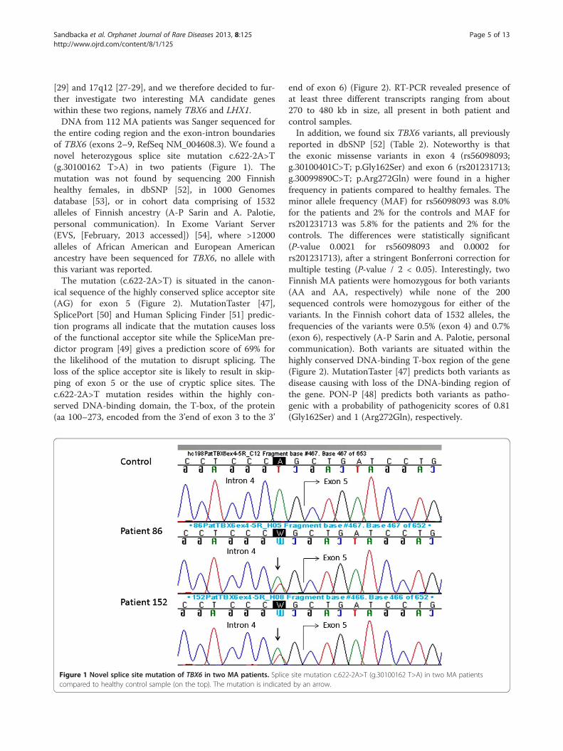
the entire coding region and the exon-intron boundariesof TBX6 (exons 2–9, RefSeq NM_004608.3). We found anovel heterozygous splice site mutation c.622-2A>T(g.30100162 T>A) in two patients (Figure 1). Themutation was not found by sequencing 200 Finnishhealthy females, in dbSNP [52], in 1000 Genomesdatabase [53], or in cohort data comprising of 1532alleles of Finnish ancestry (A-P Sarin and A. Palotie,personal communication). In Exome Variant Server(EVS, [February, 2013 accessed]) [54], where >12000alleles of African American and European Americanancestry have been sequenced for TBX6, no allele withthis variant was reported.The mutation (c.622-2A>T) is situated in the canon-
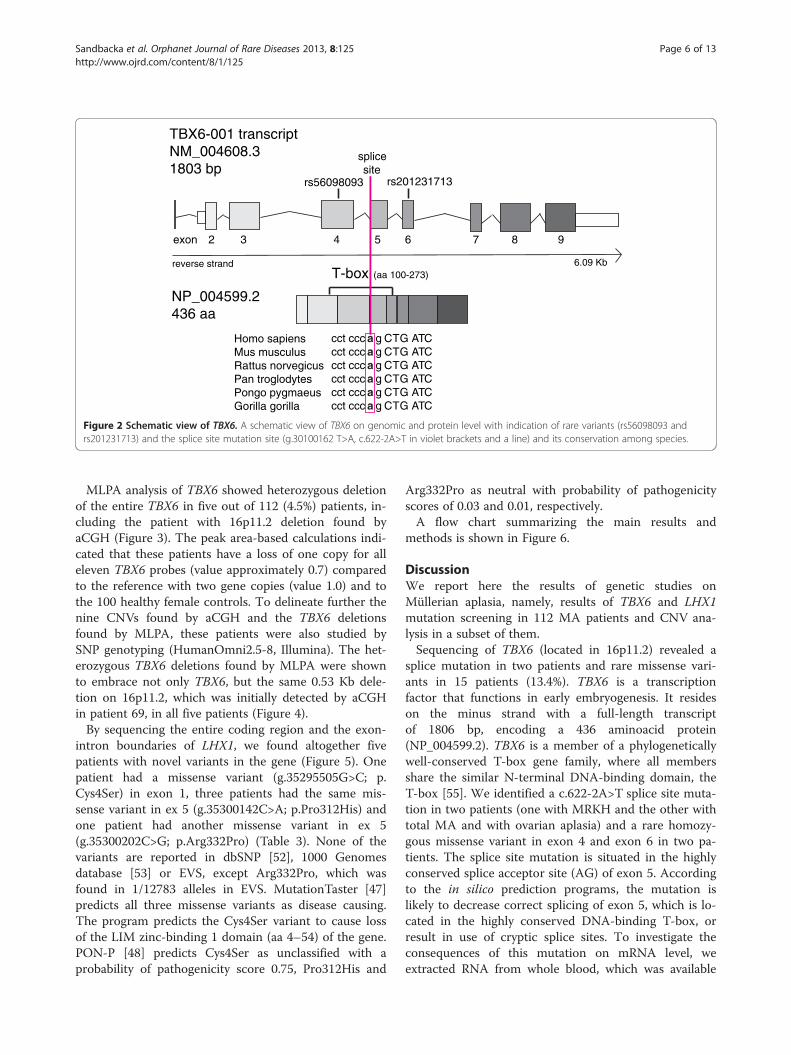
ical sequence of the highly conserved splice acceptor site(AG) for exon 5 (Figure 2). MutationTaster [47],SplicePort [50] and Human Splicing Finder [51] predic-tion programs all indicate that the mutation causes lossof the functional acceptor site while the SpliceMan pre-dictor program [49] gives a prediction score of 69% forthe likelihood of the mutation to disrupt splicing. Theloss of the splice acceptor site is likely to result in skip-ping of exon 5 or the use of cryptic splice sites. Thec.622-2A>T mutation resides within the highly con-served DNA-binding domain, the T-box, of the protein(aa 100–273, encoded from the 3’end of exon 3 to the 3’
Figure 1 Novel splice site mutation of TBX6 in two MA patients. Splicecompared to healthy control sample (on the top). The mutation is indicate
end of exon 6) (Figure 2). RT-PCR revealed presence ofat least three different transcripts ranging from about270 to 480 kb in size, all present in both patient andcontrol samples.In addition, we found six TBX6 variants, all previously
reported in dbSNP [52] (Table 2). Noteworthy is thatthe exonic missense variants in exon 4 (rs56098093;g.30100401C>T; p.Gly162Ser) and exon 6 (rs201231713;g.30099890C>T; p.Arg272Gln) were found in a higherfrequency in patients compared to healthy females. Theminor allele frequency (MAF) for rs56098093 was 8.0%for the patients and 2% for the controls and MAF forrs201231713 was 5.8% for the patients and 2% for thecontrols. The differences were statistically significant(P-value 0.0021 for rs56098093 and 0.0002 forrs201231713), after a stringent Bonferroni correction formultiple testing (P-value / 2 < 0.05). Interestingly, twoFinnish MA patients were homozygous for both variants(AA and AA, respectively) while none of the 200sequenced controls were homozygous for either of thevariants. In the Finnish cohort data of 1532 alleles, thefrequencies of the variants were 0.5% (exon 4) and 0.7%(exon 6), respectively (A-P Sarin and A. Palotie, personalcommunication). Both variants are situated within thehighly conserved DNA-binding T-box region of the gene(Figure 2). MutationTaster [47] predicts both variants asdisease causing with loss of the DNA-binding region ofthe gene. PON-P [48] predicts both variants as patho-genic with a probability of pathogenicity scores of 0.81(Gly162Ser) and 1 (Arg272Gln), respectively.
site mutation c.622-2A>T (g.30100162 T>A) in two MA patientsd by an arrow.
TBX6-001 transcriptNM_004608.31803 bp
NP_004599.2436 aa
98765432exon
reverse strandT-box (aa 100-273)
6.09 Kb
Homo sapiensMus musculusRattus norvegicusPan troglodytesPongo pygmaeusGorilla gorilla
splicesite
rs56098093 rs201231713
cct ccc a g CTG ATCcct ccc a g CTG ATCcct ccc a g CTG ATCcct ccc a g CTG ATCcct ccc a g CTG ATCcct ccc a g CTG ATC
Figure 2 Schematic view of TBX6. A schematic view of TBX6 on genomic and protein level with indication of rare variants (rs56098093 andrs201231713) and the splice site mutation site (g.30100162 T>A, c.622-2A>T in violet brackets and a line) and its conservation among species.
Sandbacka et al. Orphanet Journal of Rare Diseases 2013, 8:125 Page 6 of 13http://www.ojrd.com/content/8/1/125
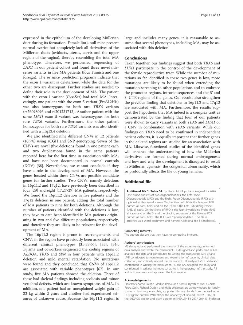
MLPA analysis of TBX6 showed heterozygous deletionof the entire TBX6 in five out of 112 (4.5%) patients, in-cluding the patient with 16p11.2 deletion found byaCGH (Figure 3). The peak area-based calculations indi-cated that these patients have a loss of one copy for alleleven TBX6 probes (value approximately 0.7) comparedto the reference with two gene copies (value 1.0) and tothe 100 healthy female controls. To delineate further thenine CNVs found by aCGH and the TBX6 deletionsfound by MLPA, these patients were also studied bySNP genotyping (HumanOmni2.5-8, Illumina). The het-erozygous TBX6 deletions found by MLPA were shownto embrace not only TBX6, but the same 0.53 Kb dele-tion on 16p11.2, which was initially detected by aCGHin patient 69, in all five patients (Figure 4).By sequencing the entire coding region and the exon-
intron boundaries of LHX1, we found altogether fivepatients with novel variants in the gene (Figure 5). Onepatient had a missense variant (g.35295505G>C; p.Cys4Ser) in exon 1, three patients had the same mis-sense variant in ex 5 (g.35300142C>A; p.Pro312His) andone patient had another missense variant in ex 5(g.35300202C>G; p.Arg332Pro) (Table 3). None of thevariants are reported in dbSNP [52], 1000 Genomesdatabase [53] or EVS, except Arg332Pro, which wasfound in 1/12783 alleles in EVS. MutationTaster [47]predicts all three missense variants as disease causing.The program predicts the Cys4Ser variant to cause lossof the LIM zinc-binding 1 domain (aa 4–54) of the gene.PON-P [48] predicts Cys4Ser as unclassified with aprobability of pathogenicity score 0.75, Pro312His and
Arg332Pro as neutral with probability of pathogenicityscores of 0.03 and 0.01, respectively.A flow chart summarizing the main results and
methods is shown in Figure 6.
DiscussionWe report here the results of genetic studies onMüllerian aplasia, namely, results of TBX6 and LHX1mutation screening in 112 MA patients and CNV ana-lysis in a subset of them.Sequencing of TBX6 (located in 16p11.2) revealed a
splice mutation in two patients and rare missense vari-ants in 15 patients (13.4%). TBX6 is a transcriptionfactor that functions in early embryogenesis. It resideson the minus strand with a full-length transcriptof 1806 bp, encoding a 436 aminoacid protein(NP_004599.2). TBX6 is a member of a phylogeneticallywell-conserved T-box gene family, where all membersshare the similar N-terminal DNA-binding domain, theT-box [55]. We identified a c.622-2A>T splice site muta-tion in two patients (one with MRKH and the other withtotal MA and with ovarian aplasia) and a rare homozy-gous missense variant in exon 4 and exon 6 in two pa-tients. The splice site mutation is situated in the highlyconserved splice acceptor site (AG) of exon 5. Accordingto the in silico prediction programs, the mutation islikely to decrease correct splicing of exon 5, which is lo-cated in the highly conserved DNA-binding T-box, orresult in use of cryptic splice sites. To investigate theconsequences of this mutation on mRNA level, weextracted RNA from whole blood, which was available

Table 2 Variants of TBX6 with allele frequencies
Varianta rs numberb Location in TBX6 Genotype of patientsc Genotype of controlsc
Predicted change N=112 N=200
g.30102391G>A rs112565029 intron 2 CC: 107 (95.5%) CC: 187 (93.5%)
c.118+6C>T CT: 5 (4.5%) CT: 13 (6.5%)
TT: 0 (0%) TT: 0 (0%)
g.30100402G>A rs147485102 exon 4 CC: 111 (99.1%) CC: 198 (99%)
p.Ser161= CT: 1 (0.9%) CT: 2 (1%)
TT: 0 (0%) TT: 0 (0%)
g.30100401C>T rs56098093 exon 4 GG: 97 (86.6%) GG: 192 (96%)
p.Gly162Ser GA: 12 (10.7%) GA: 8 (4%)
AA: 3 (2.7%) AA: 0 (0%)
g.30100162 T>A - intron 4 AA: 11 (98.2%) AA: 200 (100%)
c.622-2A>T AT: 2 (1.8%) AT: 0 (0%)
TT: 0 (0%) TT: 0 (0%)
g.30099890C>T rs201231713 exon 6 GG: 101 (90.2%) GG: 198 (99%)
p.Arg272Gln GA: 9 (8.0%) GA: 2 (1%)
AA: 2 (1.8%) AA: 0 (0%)
g.30098022G>A rs200310768 intron 7 CC: 109 (97.3%) CC: 194 (97%)
c.914-6C>T CT: 3 (2.7%) CT: 6 (3%)
TT: 0 (0%) TT: 0 (0%)
g.30097630C>T rs2289292 exon 9 GG: 32 (28.6%) GG: 67 (33.5%)
p.Pro409= GA: 55 (49.1%) GA: 95 (47.5%)
AA: 25 (22.3%) AA: 38 (19%)
The splice site mutation and the rare variants indicated in bold.avariations presented according to genomic reference sequence (g.) NC_000016.9, coding DNA reference sequence (c.) NM_004608.3 and/or protein referencesequence (p.) NP_004599.2, Genome Build 37.3, dbSNP [52].brs numbers for previously known SNPs according to dbSNP [52].cnumber of genotypes reported as: reference allele/reference allele; reference allele/alternative allele; alternative allele/alternative allele.
Sandbacka et al. Orphanet Journal of Rare Diseases 2013, 8:125 Page 7 of 13http://www.ojrd.com/content/8/1/125
from one of the patients with the mutation. Several PCRproducts were obtained, indicating that several mRNAtranscripts are present, but the sizes of the products didnot differ in patient and control samples. The reason forthis is most likely that very little or none of the mutatedtranscript is transcribed. Two patients were homozygousfor both the exon 4 and the exon 6 variant of TBX6, tenpatients were heterozygous for both, and three patientshad the exon 4 variant in a heterozygous state. Both var-iants are situated within the conserved T-box region andthe prediction programs indicate that the variants havean effect also on the protein level. We therefore believethat these variants together with other variations have arole in the multifactorial etiology of MA. However, to fi-nally determine the nature of the TBX6 splice site muta-tion and of the rare homozygous variants in ex 4 and 6of the gene, in vitro functional studies are needed.TBX6 has been suggested to associate with congenital
scoliosis in the Chinese Han population [56]. Interest-ingly, vertebral changes giving rise to e.g. scoliosis, are
commonly found in patients with MA. Recently, a mis-sense mutation in the last codon of the TBX6 transcriptwas observed in a Macedonian family with the auto-somal dominant form of spondylocostal dysostosis(SCD), characterized by segmentation defects of the ver-tebrae [57]. The affected individuals were all males andtherefore there is no data about the effect of the TBX6mutation on the development of the female urogenitaltract. The family had also experienced six infant deaths(one girl, five boys) of unknown cause without furtherphenotype data [57,58]. Mouse knock-out studies haveshown that Tbx6 is important for segmentation of thesomites in the paraxial mesoderm [59] and for left/rightpatterning [60]. White and coworkers have shown thatTbx6 interacts with the Notch-ligand delta-like 1 (Dll1)[36], a component in the Notch pathway. Also WNT sig-naling, acting upstream of Tbx6, has been shown toregulate Dll1 activity [61]. Both Notch and WNT signal-ling are import in the so called segmentation clock, cyc-lic expression of genes under which each somite is

Figure 3 Detection of TBX6 deletions by multiplex ligation probe amplification (MLPA). A ~30% reduction in peak size of the TBX6-specificprobes in patient (above) compared to control sample (below). Each exon of TBX6 is indicated by its corresponding number (in bold),prom = promotor, 3’UTR = 3’untranscribed region, D =a quality control fragment, and R=four reference probes.
Sandbacka et al. Orphanet Journal of Rare Diseases 2013, 8:125 Page 8 of 13http://www.ojrd.com/content/8/1/125
formed from the presomitic mesoderm [62]. It is con-ceivable that a similar segmentation clock could operatein the segmentation of Müllerian ducts to form uppervagina, uterus and Fallopian tubes.A spontaneous mouse model for Tbx6 is the homozy-
gous Tbx6rv (rib-vertebrae). It is caused by an insertion
Figure 4 SNP array genotyping confirming the 16p11.2 deletion. FiveHumanOmni2.5-8 v1.0, Illumina. The patient samples are here shown togetV2011.1, Illumina).
in the Tbx6 promotor upstream of the transcriptionalstart site resulting in a hypomorphic allele and reducedtranscript expression [63]. The mutation affects somiteformation and patterning featured as malformations ofthe axial skeleton and fusions of adjacent segments.Interestingly, the model also shows malformations in the
MA patients were confirmed with the 16p11.2 deletion usingher with a normal control sample (Genome Viewer, GenomeStudio

Figure 5 Three novel LHX1 variants found in five MA patients. Three novel LHX1 missense variants found in five MA patients. A) Theg.35295505G>C (p.Cys4Ser) variant in control and patient 223, B) the g.35300142C>A (p.Pro312His) in control and patients 162, 237, and 97 andC) the g.35300202C>G (p.Arg332Pro) in control and patient 110. The variations are indicated by arrows.
Sandbacka et al. Orphanet Journal of Rare Diseases 2013, 8:125 Page 9 of 13http://www.ojrd.com/content/8/1/125

Table 3 Variants of LHX1 with allele frequencies
Varianta rs numberb Location in LHX1 Genotype of patientsd Genotype of controlsd
Predicted change N=112 N= 180
g.35295505G>C - exon 1 GG: 111 (99.1%) GG: 180 (100%)
p.Cys4Ser GC: 1 (0.9%) GC: 0 (0%)
CC: 0 (0%) CC: 0 (0%)
g.35300142C>A - exon 5 CC: 109 (97.3%) CC: 180 (100%)
p.Pro312His CA: 3 (2.7%) CA: 0 (0%)
AA: 0 (0%) AA: 0 (0%)
g.35300202C>G TMP_ESP_17_35300202c exon 5 CC: 111 (99.1%) CC: 180 (100%)
p.Arg332Pro CG: 1 (0.9%) CG: 0 (0%)
GG: 0 (0%) GG: 0 (0%)avariations presented according to genomic reference sequence (g.) NC_000017.10, coding DNA reference sequence (c.) NM_005568.3 and/or protein referencesequence (p.) NP_005559.2, Genome Build 37.3, dbSNP [52].brs numbers for previously known SNPs according to dbSNP [52] or Exome Variant Server (EVS) [54].cone allele of 12783 reported in EVS [54].dnumber of genotypes reported as: reference allele/reference allele; reference allele/alternative allele; alternative allele/alternative allele.
Sandbacka et al. Orphanet Journal of Rare Diseases 2013, 8:125 Page 10 of 13http://www.ojrd.com/content/8/1/125
renal and urinary system e.g. with reported single kidneyand in the reproductive system with reduced female fer-tility. Unfortunately, no detailed information is availableon the status of the reproductive tract or on why the fe-male mice are reported as poor breeders [64]. However,the Tbx6rv has phenotypic similarities with MURCS, asthe phenotype includes skeletal and renal malformations.In addition, if the reduced fertility is caused by defects
DNA samples fromN=112
aCGHN=50
Patients with CNVsN=8a (16 %)
Patients with TBX6 deleti
N=5a (4.5%)
SNP genotypingN=12
Patients investigated for CNVs, N=54
Figure 6 Flow chart summarizing main results and methods. A flow chincluding number of patient samples investigated by each of the methods
in the reproductive tract, the mouse model would be ofinterest for functional studies in MA.Previous candidate gene studies of LHX1 residing in
the 17q12 deletion region revealed two MA patientswith heterozygous mutations [17,18]. The gene had beensuggested as a strong candidate for mutation screeningbased on studies in mice [19] showing that lim1, whichis 99.5% homologous to the human LHX1 gene [65], is
MA patients
Candidate gene screeningN=112
onsPatients with
TBX6 variantsnovel, N=2 (1.8 %)rare, N=15 (13.4 %)
Patients withLHX1 variants
novel,N=5 (4.5%)
a patient 69 included in both sample series
art summarizing the main results and methods used in the study,.
Sandbacka et al. Orphanet Journal of Rare Diseases 2013, 8:125 Page 11 of 13http://www.ojrd.com/content/8/1/125
expressed in the epithelium of the developing Müllerianduct during its formation. Female lim1-null mice presentnormal ovaries but completely lack all derivatives of theMüllerian ducts (oviducts, uterus, cervix and the upperregion of the vagina), thereby resembling the total MAphenotype. Therefore, we performed sequencing ofLHX1 in our patient cohort and found three novel mis-sense variants in five MA patients (four Finnish and oneforeign). The in silico prediction programs indicate thatthe exon 1 variant is deleterious, while the data for theother two are discrepant. Further studies are needed todefine their role in the development of MA. The patientwith the exon 1 variant (Cys4Ser) had total MA. Inter-estingly, one patient with the exon 5 variant (Pro312His)was also homozygous for both rare TBX6 variants(rs56098093 and rs201231713). Another patient with thesame LHX1 exon 5 variant was heterozygous for bothrare TBX6 variants. Furthermore, the other patienthomozygous for both rare TBX6 variants was also identi-fied with a 11q13.4 deletion.We also identified nine different CNVs in 12 patients
(10.7%) using aCGH and SNP genotyping. Seven of theCNVs are novel (five deletions found in one patient eachand two duplications found in the same patient)reported here for the first time in association with MA,and have not been documented in normal controls(DGV) [38]. Nevertheless, we cannot conclude if theyhave a role in the development of MA. However, thegenes located within these CNVs are possible candidategenes for further studies. Two CNVs, namely deletionsin 16p11.2 and 17q12, have previously been described infour [29] and eight [17,27-29] MA patients, respectively.We found the 16p11.2 deletion in five patients and the17q12 deletion in one patient, adding the total numberof MA patients to nine for both deletions. Although thenumber of patients identified with the deletions is low,they have to date been identified in MA patients origin-ating in two and five different populations, respectively,and therefore they are likely to be relevant for the devel-opment of MA.The 16p11.2 region is prone to rearrangements and
CNVs in the region have previously been associated withdifferent clinical phenotypes [31-33,66], [35], [34].Bijlsma and coworkers sequenced the coding regions ofALDOA, TBX6 and SPN in four patients with 16p11.2deletion and mild mental retardation. No mutationswere found and they concluded that CNVs of 16p11.2are associated with variable phenotypes [67]. In ourstudy, five MA patients showed the deletion. Three ofthese had skeletal findings including scoliosis and minorvertebral defects, which are known symptoms of MA. Inaddition, one patient had an unexplained weight gain of32 kg within 2 years and another had experienced sei-zures of unknown cause. Because the 16p11.2 region is
large and includes many genes, it is reasonable to as-sume that several phenotypes, including MA, may be as-sociated with this deletion.
ConclusionsTaken together, our findings suggest that both TBX6 andLHX1 participate in the control of the development ofthe female reproductive tract. While the number of mu-tations so far identified in these two genes is low, moremutations are likely to be found when extending themutation screening to other populations and to embracethe promotor regions, intronic sequences and the 5’ and3’ UTR regions of the genes. Our results also strengthenthe previous finding that deletions in 16p11.2 and 17q12are associated with MA. Furthermore, the results sup-port the hypothesis that MA indeed is a complex trait asdemonstrated by the finding that four of our patientswere shown to carry variants in both TBX6 and LHX1 ora CNV in combination with TBX6 variants. While ourfindings on TBX6 need to be confirmed in independentpatient cohorts, it is equally important that further genesin the deleted regions are studied for an association withMA. Likewise, functional studies of the identified geneswill enhance the understanding of how the Müllerianderivatives are formed during normal embryogenesisand how and why the development is disrupted to resultin Müllerian agenesis, the congenital abnormality, whichso profoundly affects the life of young females.
Additional file
Additional file 1: Table S1. Synthetic MLPA probes designed for TBX6.One probe consists of two oligonucleotides: the Left ProbeOligonucleotide (LPO) and the Right Probe Oligonucleotide (RPO) withoptional stuffers (small cases). On the 5’end of LPO is the Forward PCRprimer (all caps, bold) and on the 3’end is the Left Hybridizing Probe(LHS, all caps). On the 5’end of RPO is the Right Hybridizing Probe (RHS,all caps) and on the 3’ end the binding sequence of the Reverse PCRprimer (all caps, bold). The RPOs are 5’phosphorylated. (The file isattached as a Word-document and named: Additional file 1 Sandbacka).
Competing interestsThe authors declare that they have no competing interests.
Authors’ contributionsMS designed and performed the majority of the experiments, performeddata analysis and wrote the manuscript. EF designed and performed aCGH,analyzed the data and contributed to writing the manuscript. MH, VJ andLMP contributed to recruitment and examination of patients, clinical datacollection, and critically revised the manuscript. CR analyzed aCGH data andcontributed in writing the manuscript. HL and KA designed the study andcontributed in writing the manuscript. KA is the guarantor of the study. Allauthors have seen and approved the final version.
AcknowledgementsProfessors Aarno Palotie, Markus Perola and Samuli Ripatti as well as Antti-Pekka Sarin, Richard Durbin and Maija Wessman are acknowledged for kindlysharing cohort sequence data, supported through funds from the WellcomeTrust (grant number WT089062), the Academy of Finland (200923, 00213),the ENGAGE project and grant agreement HEALTH-F4-2007-201413. Professor

Sandbacka et al. Orphanet Journal of Rare Diseases 2013, 8:125 Page 12 of 13http://www.ojrd.com/content/8/1/125
Päivi Peltomäki and Annette Gylling are acknowledged for sharing theirinspiring thoughts. The genotyping of SNP markers was performed by theTechnology Centre, Institute for Molecular Medicine Finland (FIMM),University of Helsinki, Finland. The study has been funded by SamfundetFolkhälsan, Victoria Foundation, Finska Läkaresällskapet Foundation,Medicinska understödsföreningen Liv och Hälsa Foundation, University ofHelsinki and by Finnish State Funds.
Author details1Folkhälsan Institute of Genetics, Helsinki, Finland. 2Department of MedicalGenetics, Haartman Institute, University of Helsinki, Helsinki, Finland.3Department of Obstetrics and Gynecology, Helsinki University CentralHospital, Helsinki, Finland. 4Department of Genetics and Evolutionary Biology,Institute of Biosciences, University of São Paulo, São Paulo, Brazil.5Department of Obstetrics and Gynecology, University of Turku, Turku,Finland. 6Department of Obstetrics and Gynecology, Oulu University Hospital,Oulu, Finland. 7Department of Clinical Genetics, HUSLAB, Helsinki UniversityCentral Hospital, Helsinki, Finland.
Received: 25 April 2013 Accepted: 14 August 2013Published: 16 August 2013
References1. Griffin JE, Edwards C, Madden JD, Harrod MJ, Wilson JD: Congenital
absence of the vagina. The Mayer-Rokitansky-Kuster-Hauser syndrome.Ann Intern Med 1976, 2:224–236.
2. Aittomäki K, Eroila H, Kajanoja P: A population-based study of theincidence of Müllerian aplasia in Finland. Fertil Steril 2001, 3:624–625.
3. Kobayashi A, Behringer RR: Developmental genetics of the femalereproductive tract in mammals. Nat Rev Genet 2003, 12:969–980.
4. Spencer TE, Dunlap KA, Filant J: Comparative developmental biology ofthe uterus: insights into mechanisms and developmental disruption.Mol Cell Endocrinol 2012, 1–2:34–53.
5. Resendes BL, Sohn SH, Stelling JR, Tineo R, Davis AJ, Gray MR, Reindollar RH:Role for anti-Mullerian hormone in congenital absence of the uterus andvagina. Am J Med Genet 2001, 2:129–136.
6. Zenteno JC, Carranza-Lira S, Kofman-Alfaro S: Molecular analysis of theanti-Mullerian hormone, the anti-Mullerian hormone receptor, andgalactose-1-phosphate uridyl transferase genes in patients with theMayer-Rokitansky-Kuster-Hauser syndrome. Arch Gynecol Obstet 2004,4:270–273.
7. Oppelt P, Strissel PL, Kellermann A, Seeber S, Humeny A, Beckmann MW,Strick R: DNA sequence variations of the entire anti-Mullerian hormone(AMH) gene promoter and AMH protein expression in patients with theMayer-Rokitanski-Kuster-Hauser syndrome. Hum Reprod 2005, 1:149–157.
8. Timmreck LS, Pan HA, Reindollar RH, Gray MR: WNT7A mutations inpatients with Mullerian duct abnormalities. J Pediatr Adolesc Gynecol 2003,4:217–221.
9. Lalwani S, Wu HH, Reindollar RH, Gray MR: HOXA10 mutations incongenital absence of uterus and vagina. Fertil Steril 2008, 2:325–330.
10. Karnis MF, Stelling JR, Lalwani SI, Bhagavath B, Pan HA, Davis AJ, ReindollarRH, Gray MR: Mutation analysis of the HOXA13 gene in patients withcongenital absence of the uterus and vagina. J Soc Gynecol Invest 2000,7:172A.
11. Dang Y, Qin Y, Tang R, Mu Y, Li G, Xia M, Chen ZJ: Variants of the WNT7Agene in Chinese patients with mullerian duct abnormalities. Fertil Steril2012, 2:391–394.
12. Ekici AB, Strissel PL, Oppelt PG, Renner SP, Brucker S, Beckmann MW, StrickR: HOXA10 and HOXA13 sequence variations in human female genitalmalformations including congenital absence of the uterus and vagina.Gene 2013, 2:267–272.
13. Biason-Lauber A, Konrad D, Navratil F, Schoenle EJ: A WNT4 mutationassociated with Müllerian-duct regression and virilization in a 46,XX woman. N Engl J Med 2004, 8:792–798.
14. Biason-Lauber A, De Filippo G, Konrad D, Scarano G, Nazzaro A, Schoenle EJ:WNT4 deficiency–a clinical phenotype distinct from the classic Mayer-Rokitansky-Kuster-Hauser syndrome: a case report. Hum Reprod 2007,1:224–229.
15. Philibert P, Biason-Lauber A, Rouzier R, Pienkowski C, Paris F, Konrad D,Schoenle E, Sultan C: Identification and functional analysis of a newWNT4 gene mutation among 28 adolescent girls with primary
amenorrhea and mullerian duct abnormalities: a French collaborativestudy. J Clin Endocrinol Metab 2008, 3:895–900.
16. Philibert P, Biason-Lauber A, Gueorguieva I, Stuckens C, Pienkowski C,Lebon-Labich B, Paris F, Sultan C: Molecular analysis of WNT4 gene in fouradolescent girls with mullerian duct abnormality and hyperandrogenism(atypical Mayer-Rokitansky-Kuster-Hauser syndrome). Fertil Steril 2011,8:2683–2686.
17. Ledig S, Schippert C, Strick R, Beckmann MW, Oppelt PG, Wieacker P:Recurrent aberrations identified by array-CGH in patients with Mayer-Rokitansky-Kuster-Hauser syndrome. Fertil Steril 2011, 5:1589–1594.
18. Ledig S, Brucker S, Barresi G, Schomburg J, Rall K, Wieacker P: Frame shiftmutation of LHX1 is associated with Mayer-Rokitansky-Kuster-Hauser(MRKH) syndrome. Hum Reprod 2012, 9:2872–2875.
19. Kobayashi A, Shawlot W, Kania A, Behringer RR: Requirement of Lim1 forfemale reproductive tract development. Development 2004, 131:539–549.
20. Cheroki C, Krepischi-Santos AC, Rosenberg C, Jehee FS, Mingroni-Netto RC,Pavanello Filho I, Zanforlin Filho S, Kim CA, Bagnoli VR, Mendonca BB,Szuhai K, Otto PA: Report of a del22q11 in a patient with Mayer-Rokitansky-Küster-Hauser (MRKH) anomaly and exclusion of WNT-4, RAR-gamma, and RXR-alpha as major genes determining MRKH anomaly in astudy of 25 affected women. Am J Med Genet A 2006, 12:1339–1342.
21. Drummond JB, Rezende CF, Peixoto FC, Carvalho JS, Reis FM, De Marco L:Molecular analysis of the beta-catenin gene in patients with the Mayer-Rokitansky-Kuster-Hauser syndrome. J Assist Reprod Genet 2008,11–12:511–514.
22. Ma J, Qin Y, Liu W, Duan H, Xia M, Chen ZJ: Analysis of PBX1 mutations in192 Chinese women with Mullerian duct abnormalities. Fertil Steril 2011,8:2615–2617.
23. Wang P, Zhao H, Sun M, Li Y, Chen ZJ: PAX2 in 192 Chinese women withMullerian duct abnormalities: mutation analysis. Reprod Biomed Online2012, 2:219–222.
24. Ravel C, Bashamboo A, Bignon-Topalovic J, Siffroi JP, McElreavey K, Darai E:Polymorphisms in DLGH1 and LAMC1 in Mayer-Rokitansky-Kuster-Hausersyndrome. Reprod Biomed Online 2012, 4:462–465.
25. Bendavid C, Pasquier L, Watrin T, Morcel K, Lucas J, Gicquel I, Dubourg C,Henry C, David V, Odent S, Leveque J, Pellerin I, Guerrier D: Phenotypicvariability of a 4q34–>qter inherited deletion: MRKH syndrome in thedaughter, cardiac defect and Fallopian tube cancer in the mother.Eur J Med Genet 2007, 1:66–72.
26. Sundaram UT, McDonald-McGinn DM, Huff D, Emanuel BS, Zackai EH,Driscoll DA, Bodurtha J: Primary amenorrhea and absent uterus in the22q11.2 deletion syndrome. Am J Med Genet A 2007, 17:2016–2018.
27. Cheroki C, Krepischi-Santos AC, Szuhai K, Brenner V, Kim CA, Otto PA,Rosenberg C: Genomic imbalances associated with mullerian aplasia.J Med Genet 2008, 4:228–232.
28. Bernardini L, Gimelli S, Gervasini C, Carella M, Baban A, Frontino G,Barbano G, Divizia MT, Fedele L, Novelli A, Bena F, Lalatta F, MiozzoM, Dallapiccola B: Recurrent microdeletion at 17q12 as a cause ofMayer-Rokitansky-Kuster-Hauser (MRKH) syndrome: two casereports. Orphanet J Rare Dis 2009, 4:25.
29. Nik-Zainal S, Strick R, Storer M, Huang N, Rad R, Willatt L, Fitzgerald T,Martin V, Sandford R, Carter NP, Janecke AR, Renner SP, Oppelt PG,Oppelt P, Schulze C, Brucker S, Hurles M, Beckmann MW, Strissel PL,Shaw-Smith C: High incidence of recurrent copy number variants inpatients with isolated and syndromic Mullerian aplasia. J Med Genet2011, 3:197–204.
30. Edghill EL, Bingham C, Ellard S, Hattersley AT: Mutations in hepatocyte nuclearfactor-1beta and their related phenotypes. J Med Genet 2006, 1:84–90.
31. Kumar RA, KaraMohamed S, Sudi J, Conrad DF, Brune C, Badner JA,Gilliam TC, Nowak NJ, Cook EH Jr, Dobyns WB, Christian SL: Recurrent16p11.2 microdeletions in autism. Hum Mol Genet 2008, 4:628–638.
32. Weiss LA, Shen Y, Korn JM, Arking DE, Miller DT, Fossdal R, Saemundsen E,Stefansson H, Ferreira MA, Green T, Platt OS, Ruderfer DM, Walsh CA,Altshuler D, Chakravarti A, Tanzi RE, Stefansson K, Santangelo SL, Gusella JF,Sklar P, Wu BL, Daly MJ: Autism Consortium: Association betweenmicrodeletion and microduplication at 16p11.2 and autism. N Engl J Med2008, 7:667–675.
33. Rosenfeld JA, Coppinger J, Bejjani BA, Girirajan S, Eichler EE, Shaffer LG, BallifBC: Speech delays and behavioral problems are the predominantfeatures in individuals with developmental delays and 16p11.2microdeletions and microduplications. J Neurodev Disord 2010, 1:26–38.

Sandbacka et al. Orphanet Journal of Rare Diseases 2013, 8:125 Page 13 of 13http://www.ojrd.com/content/8/1/125
34. Walters RG, Jacquemont S, Valsesia A, de Smith AJ, Martinet D, Andersson J,Falchi M, Chen F, Andrieux J, Lobbens S, Delobel B, Stutzmann F, El-SayedMoustafa JS, Chevre JC, Lecoeur C, Vatin V, Bouquillon S, Buxton JL, BouteO, Holder-Espinasse M, Cuisset JM, Lemaitre MP, Ambresin AE, Brioschi A,Gaillard M, Giusti V, Fellmann F, Ferrarini A, Hadjikhani N, Campion D: Anew highly penetrant form of obesity due to deletions on chromosome16p11.2. Nature 2010, 7281:671–675.
35. McCarthy SE, Makarov V, Kirov G, Addington AM, McClellan J, Yoon S,Perkins DO, Dickel DE, Kusenda M, Krastoshevsky O, Krause V, Kumar RA,Grozeva D, Malhotra D, Walsh T, Zackai EH, Kaplan P, Ganesh J, Krantz ID,Spinner NB, Roccanova P, Bhandari A, Pavon K, Lakshmi B, Leotta A, KendallJ, Lee YH, Vacic V, Gary S, Iakoucheva LM, et al: Microduplications of16p11.2 are associated with schizophrenia. Nat Genet 2009, 11:1223–1227.
36. White PH, Farkas DR, McFadden EE, Chapman DL: Defective somitepatterning in mouse embryos with reduced levels of Tbx6.Development 2003, 8:1681–1690.
37. Sandbacka M, Halttunen M, Jokimaa V, Aittomaki K, Laivuori H: Evaluationof SHOX copy number variations in patients with Mullerian aplasia.Orphanet J Rare Dis 2011, 6:53.
38. Database of Genomic Variants. [http://projects.tcag.ca/variation/]39. Ensembl. [http://www.ensembl.org/index.html]40. CpG Plot. http://www.ebi.ac.uk/Tools/seqstats/emboss_cpgplot/41. CpG Island Searcher. [http://www.uscnorris.com/cpgislands2/cpg.aspx]42. UCSC Genome Browser. [http://www.genome.ucsc.edu/cgi-bin/hgGateway]43. RaW-Probe Version 0.15β. http://www.mlpa.com/WebForms/WebFormMain.
aspx?Tag=zjCZBtdOUyAt3KF3EwRZhAPz9QEm7akikAm7AOEGw1vtZvffaZPOiSig8uqel7Yd
44. Mfold. [http://mfold.rna.albany.edu/]45. ExonPrimer. [http://ihg.gsf.de/ihg/ExonPrimer.html]46. Primer3 v. 0.4.0. [http://frodo.wi.mit.edu/primer3/input.htm]47. Schwarz JM, Rodelsperger C, Schuelke M, Seelow D: MutationTaster
evaluates disease-causing potential of sequence alterations. Nat Methods2010, 8:575–576.
48. PON-P. [http://bioinf.uta.fi/PON-P/]49. SpliceMan. [http://fairbrother.biomed.brown.edu/spliceman/index.cgi]50. SplicePort. http://spliceport.cbcb.umd.edu/SplicingAnalyser.html51. Human Splicing Finder. [http://www.umd.be/HSF/]52. dbSNP. [http://www.ncbi.nlm.nih.gov/projects/SNP/].53. 1000 Genomes. [http://browser.1000genomes.org/index.html]54. Exome Variant Server. [http://evs.gs.washington.edu/EVS/]55. Yi CH, Terrett JA, Li QY, Ellington K, Packham EA, Armstrong-Buisseret L,
McClure P, Slingsby T, Brook JD: Identification, mapping, andphylogenomic analysis of four new human members of the T-box genefamily: EOMES, TBX6, TBX18, and TBX19. Genomics 1999, 1:10–20.
56. Fei Q, Wu Z, Wang H, Zhou X, Wang N, Ding Y, Wang Y, Qiu G: Theassociation analysis of TBX6 polymorphism with susceptibility tocongenital scoliosis in a Chinese Han population. Spine (Phila Pa.1976)2010, 9:983–988.
57. Sparrow DB, McInerney-Leo A, Gucev ZS, Gardiner B, Marshall M, Leo PJ,Chapman DL, Tasic V, Shishko A, Brown MA, Duncan EL, Dunwoodie SL:Autosomal dominant spondylocostal dysostosis is caused by mutation inTBX6. Hum Mol Genet 2013, 8:1625–1631.
58. Gucev ZS, Tasic V, Pop-Jordanova N, Sparrow DB, Dunwoodie SL, Ellard S,Young E, Turnpenny PD: Autosomal dominant spondylocostal dysostosisin three generations of a Macedonian family: Negative mutation analysisof DLL3, MESP2, HES7, and LFNG. Am J Med Genet A 2010, 6:1378–1382.
59. Chapman DL, Papaioannou VE: Three neural tubes in mouse embryoswith mutations in the T-box gene Tbx6. Nature 1998, 6668:695–697.
60. Hadjantonakis AK, Pisano E, Papaioannou VE: Tbx6 regulates left/rightpatterning in mouse embryos through effects on nodal cilia andperinodal signaling. PLoS One 2008, 6:e2511.
61. Hofmann M, Schuster-Gossler K, Watabe-Rudolph M, Aulehla A, HerrmannBG, Gossler A: WNT signaling, in synergy with T/TBX6, controls Notchsignaling by regulating Dll1 expression in the presomitic mesoderm ofmouse embryos. Genes Dev 2004, 22:2712–2717.
62. Oginuma M, Niwa Y, Chapman DL, Saga Y: Mesp2 and Tbx6 cooperativelycreate periodic patterns coupled with the clock machinery duringmouse somitogenesis. Development 2008, 15:2555–2562.
63. Watabe-Rudolph M, Schlautmann N, Papaioannou VE, Gossler A: The mouserib-vertebrae mutation is a hypomorphic Tbx6 allele. Mech Dev 2002,2:251–256.
64. Theiler K, Varnum DS: Development of rib-vertebrae: a new mutation inthe house mouse with accessory caudal duplications. Anat Embryol (Berl)1985, 1:111–116.
65. GeneCards. [http://www.genecards.org]66. Fernandez BA, Roberts W, Chung B, Weksberg R, Meyn S, Szatmari P,
Joseph-George AM, Mackay S, Whitten K, Noble B, Vardy C, Crosbie V,Luscombe S, Tucker E, Turner L, Marshall CR, Scherer SW: Phenotypicspectrum associated with de novo and inherited deletions andduplications at 16p11.2 in individuals ascertained for diagnosis of autismspectrum disorder. J Med Genet 2010, 3:195–203.
67. Bijlsma EK, Gijsbers AC, Schuurs-Hoeijmakers JH, van Haeringen A, van dePutte DE F, Anderlid BM, Lundin J, Lapunzina P, Perez Jurado LA, DelleChiaie B, Loeys B, Menten B, Oostra A, Verhelst H, Amor DJ, Bruno DL, vanEssen AJ, Hordijk R, Sikkema-Raddatz B, Verbruggen KT, Jongmans MC,Pfundt R, Reeser HM, Breuning MH, Ruivenkamp CA: Extending thephenotype of recurrent rearrangements of 16p11.2: deletions inmentally retarded patients without autism and in normal individuals.Eur J Med Genet 2009, 2–3:77–87.
doi:10.1186/1750-1172-8-125Cite this article as: Sandbacka et al.: TBX6, LHX1 and copy numbervariations in the complex genetics of Müllerian aplasia. Orphanet Journalof Rare Diseases 2013 8:125.
Submit your next manuscript to BioMed Centraland take full advantage of:
• Convenient online submission
• Thorough peer review
• No space constraints or color figure charges
• Immediate publication on acceptance
• Inclusion in PubMed, CAS, Scopus and Google Scholar
• Research which is freely available for redistribution
Submit your manuscript at www.biomedcentral.com/submit