research open access phase ii study of helical ... open access phase ii study of helical tomotherapy...
TRANSCRIPT

RESEARCH Open Access
Phase II study of helical tomotherapy in themultidisciplinary treatment of oligometastaticcolorectal cancerBenedikt Engels1*, Thierry Gevaert1, Hendrik Everaert2, Peter De Coninck1, Alexandra Sermeus3, Nicolas Christian1,Guy Storme1, Dirk Verellen1 and Mark De Ridder1
Abstract
Background: Complete metastasectomy provides a real chance for long-term survival in patients witholigometastatic colorectal cancer (CRC). For inoperable patients, we evaluated in this study intensity-modulated andimage-guided radiotherapy (IMRT-IGRT) by helical tomotherapy.
Methods: Twenty-four CRC patients with ≤ 5 metastases were enrolled, receiving a dose of 50 Gy in fractions of5 Gy. No limitations concerning dimension or localization of the metastases were imposed. Whole body PET-CTwas performed at baseline and 3 months after the initiation of RT to evaluate the metabolic response rateaccording to PET Response Criteria in Solid Tumors (PERCIST) version 1.0.
Results: A total of 53 metastases were treated. Seventeen patients (71%) received previously ≥ 1 line ofchemotherapy for metastatic disease, displaying residual (n = 7) or progressive (n = 10) metabolic activeoligometastatic disease at time of inclusion. Most common sites were the lung, liver and lymphnodes. One patient(4%) experienced grade 3 dysphagia. Twenty-two patients were evaluated by post-treatment PET-CT. Twelvepatients achieved a complete (n = 6) or partial (n = 6) metabolic response, resulting in an overall metabolicresponse rate of 55%. At a median follow-up of 10 months, 7 patients (29%) are in remission, of which 5 receivedprevious chemotherapy with residual oligometastatic disease at time of inclusion. The actuarial 1-year local control,progression-free survival, and overall survival were 54%, 14% and 78%.
Conclusions: Helical tomotherapy delivering 10 fractions of 5 Gy resulted in a metabolic response rate of 55%, andappeared to be attractive as consolidation of inoperable oligometastatic disease after effective chemotherapy.
Trial registration: Eudract 2008-008300-40; NCT00807313
Keywords: Metastatic colorectal cancer, Oligometastases, Helical tomotherapy, IMRT-IGRT
BackgroundNearly one fourth of the patients with newly diagnosedcolorectal cancer (CRC) present synchronous liver metas-tases and more than half of the resected CRC patientsdevelop distant recurrence during follow-up [1]. Thediagnosis of metastatic CRC (mCRC) does however notequal an acute fatal illness and might be classified moreas a chronic disease in patients with a clinical diseasestate between locoregionally confined and widely spread
metastatic disease, so called oligometastatic disease [2,3].It is well understood today that a complete metastasect-omy provides a real chance for long-term survival inthose patients, with documented 10-year overall survival(OS) rates in 1 out of 6 mCRC patients which underwenthepatic resection [4]. According to the data of the rando-mized EORTC intergroup Trial 40983, the perioperativeadministration of 5-fluorouracil (5-FU) and oxaliplatin(FOLFOX) has emerged as a new standard of care inmCRC patients with limited resectable liver metastases,by increasing the 3-year progression-free survival (PFS)with 8% (36% versus 28% without perioperative che-motherapy) [5]. Despite the improvements in surgical
* Correspondence: [email protected] of Radiation Oncology, UZ Brussel, Vrije Universiteit Brussel,Laarbeeklaan 101, B-1090 Brussels, BelgiumFull list of author information is available at the end of the article
Engels et al. Radiation Oncology 2012, 7:34http://www.ro-journal.com/content/7/1/34
© 2012 Engels et al; licensee BioMed Central Ltd. This is an Open Access article distributed under the terms of the Creative CommonsAttribution License (http://creativecommons.org/licenses/by/2.0), which permits unrestricted use, distribution, and reproduction inany medium, provided the original work is properly cited.

procedures and preoperative multi-agent chemotherapy,limitations imposed by localization, multifocal character,size, or comorbidities still exclude the major part ofmCRC patients from undergoing metastasectomy [6].Aiming at ablating metastases while preserving the sur-rounding healthy tissues, there has been an expansionover the past decade in the use of non-surgical local abla-tive alternatives such as radiofrequency ablation (RFA)and stereotactic body radiotherapy (SBRT), the latter atailored delivery of tumoricidal doses of radiation in aminimal number of fractions to small lesions by the com-bination of high conformal RT and rigorous localizationof the target by image-guided RT (IGRT) [7-10]. In theeradication of liver- and lungmetastases by SBRT, limitedtoxicity rates and sustained local control (LC) arereported by several authors up [7,8]. However, in orderto allow delivery of those cytotoxic doses, SBRT requirescarefully selection of the metastases on the base of theirlocalization and dimension. Our institution previouslyexplored in a pilot study the efficacy and toxicity of heli-cal tomotherapy, a technology combining rotationalintensity-modulated RT (IMRT) and IGRT by megavol-tage (MV) computed tomography (CT) scanning, inpatients with oligometastatic CRC who were not amen-able for metastasectomy and (further) systemic therapy[9]. In order to suit a variety of treatment sites includinglarge metastases and critically located lesions, we used amoderately hypofractionated regimen, delivering 10 frac-tions of 4 Gy. This schedule resulted in a complete meta-bolic response (CMR), the primary objective, in 5 out of23 enrolled oligometastatic CRC patients [9]. Besides,helical tomotherapy displayed a very safe toxicity profile,with grade 2 and 3 toxicity recorded in only 9% and 4%of the patients, respectively [9]. Taking into account therather disappointing 1-year LC rate of 54% in this pilotstudy, we now aimed for higher response and LC rates inthose patients. To do so, we investigated in this study theefficacy and toxicity of helical tomotherapy delivering50 Gy in daily fractions of 5 Gy in patients with inoper-able oligometastatic CRC. The primary objective was toevaluate the CMR rate 3 months after initiation of RT byperforming whole-body 18 F-fluorodeoxyglucose (FDG)-positron emission tomography (PET) at baseline and atevaluation. Secondary endpoints were toxicity, LC andPFS.
MethodsPatient populationPatients with a radically resected primary tumor with thehistological proof of a colorectal adenocarcinoma and attime of inclusion 1 to 5 metastases, showing increasedmetabolism on 18FDG-PET, were eligible for this study.No limitations were imposed on the localization or
dimension of the metastases. Patients were required tohave an Eastern Cooperative Oncology Group perfor-mance status of ≤ 2, to be > 18 years old and inoperableby the localization, number or dimension of the metas-tases, medically unfit to undergo resection or refusingsurgery. Patients were not permitted to receive che-motherapy within 1 month before initiation of RT.Patients who did not receive previous chemotherapy formetastatic disease had to be medically unfit to undergosystemic treatment or refusing chemotherapy. Patientswith Child B or C liver cirrhosis or a functional livervolume < 1000 cc in case of liver metastases and a lungdiffusion capacity for carbon monoxide (DLCO) of < 30%in case of lung metastases were excluded. Patients withan active second primary tumor were excluded. Allpatients signed study-specific informed consent. The pro-tocol was reviewed and approved by the Ethical Commit-tee and registered (inter)nationally (Eudract 2008-008300-40; NCT00807313).
Pre-treatment evaluation and radiotherapy techniquePre-treatment evaluation included a complete medicalhistory, physical examination, a pre-treatment freebreathing 18FDG-PET and computed tomography (CT)using a dedicated PET-CT camera (Gemini TF, PhilipsMedical Systems, OH, USA) and laboratory tests includ-ing carcinoembryonic antigen (CEA) with assessment ofthe Child-Pugh parameters and liver enzymes in patientswith liver metastases. RT was performed using theTomoTherapy Hi⋅Art II System (TomoTherapy Inc.,Madison, WI), which fully integrates IGRT by means ofMVCT scanning and IMRT by means of dynamic rota-tional therapy [10]. The gross tumor volume (GTV)included the visible gross tumor mass on CT. The GTVwas expanded by a 10, 10 and 12 mm for the anteropos-terior, laterolateral and craniocaudal direction, respec-tively, to create the planning target volume (PTV), fullyencompassing the 18FDG-PET-positive volume. Theplanning goals were to deliver at least 95% of the pre-scribed dose to at least 95% of the PTVs, while keepingthe maximum dose (Dmax) to the PTV below 105%. Thevolume of lung receiving more than 20 Gy (V20) was keptbelow 20% in case of lung irradiation. In patients withliver metastases, the liver volume receiving more than 22Gy (V22) and 30 Gy (V30) was kept to less than 50% and30%, respectively. A Dmax of 36 Gy (72% of the prescribeddose) was set to the spinal cord. In case of intersectionbetween the PTV and hollow viscous organs (smallbowel, large bowel, stomach or oesophagus) of ≥ 5 cc, aPTV subvolume was defined at this interface with a Dmax
of 40 Gy (80% of the prescribed dose) to this overlapvolume. The treatment was delivered daily in 10 frac-tions, excluding weekends. Before each treatment session,
Engels et al. Radiation Oncology 2012, 7:34http://www.ro-journal.com/content/7/1/34
Page 2 of 9

patients underwent scanning using the integrated MV-CT scan modality and were repositioned after co-regis-tration of these images with the planning kilovoltage(kV)-CT scan.
Toxicity monitoringToxicity was evaluated and scored according to theNational Cancer Institute Common Terminology Cri-teria for Adverse Events (NCI CTC AE) version 3.0,with toxicity occurring within 3 months after the initia-tion of RT classified as acute toxicity. Patients were con-tacted and/or invited for follow-up 3-monthly duringthe first year, 6-monthly thereafter.
Treatment evaluationResponse evaluation has been described extensively inthe previous pilot study in our institution [9]. Briefly, theprimary objective, CMR rate, was evaluated by comparingthe PET-CT at baseline with the PET-CT performed 3months after initiation of RT, according to the PETResponse Criteria in Solid Tumors (PERCIST) version1.0 [11]. A CMR in a patient was defined by a completeresolution of 18FDG uptake within all irradiated lesion(s),so that it is less than mean liver activity and indistin-guishable from surrounding background blood-poollevels, without new 18FDG-avid lesions in pattern typicalof CRC. For a partial metabolic response (PMR), a reduc-tion in SUVmax for patients with one lesion or a reduc-tion of the sum of the SUVmax data for patients with > 1lesion of minimum 30% was required, without theappearance of new 18FDG-avid lesions. Obvious progres-sion of any lesion (> 30% increase in SUVmax) or new18FDG-avid lesions negate a partial response and indicateprogressive metabolic disease (PMD). Stable metabolicdisease (SMD) is defined as not CMR, PMR, or PMD.During follow-up, the Response Evaluation Criteria in
Solid Tumors (RECIST) were used to evaluate response. Alocal recurrence was defined as the re-growth of tumorwithin or at the periphery of the irradiated volume. Theappearance of new lesions was considered as distantrecurrence.
StatisticsA Richard Simon two-stage optimal design was per-formed to obtain the sample size. Aiming at an overallacceptable and unacceptable CMR probability of 50%and 30%, respectively, with an a and b value of 0.10, thesample size for first and second stage were 7/22 and 17/46 evaluated patients, respectively. Actuarial LC, PFSand OS rates were estimated by Kaplan-Meier analysis,Log-rank testing was used to evaluate the associationbetween patient-related factors and treatment outcome.Laboratory tests were evaluated by t tests.
ResultsPatient characteristicsTwenty-four inoperable oligometastatic CRC patientswith a total number of 53 metastases were enrolledbetween March 2010 and July 2011. Patient characteris-tics are given in Table 1. Seventeen patients (71%)received previously ≥ 1 line of chemotherapy for meta-static disease, of which 7 and 10 patients presentingresidual and progressive metabolic active oligometastaticdisease at time of inclusion, respectively. Seven patients(29%) received no previous chemotherapy for treatmentof metastatic disease; 5 patients were medically unfit toundergo systemic therapy and 2 patients refusedchemotherapy.Seventeen patients (71%) received previous local ther-
apy for metastatic disease; radiotherapy (n = 7), RFA(n = 6) and/or metastasectomy (n = 8). Twelve patients(50%) presented with a GTV located in the vicinity ofhollow viscous organs (small/large bowel, stomach oresophagus).
Table 1 Patient characteristics (n = 24)
Variable Distribution No. of Patients %
Sex Male 14 58
Female 10 42
Age (years) Median 67 years
Range 45 - 91 years
Karnofsky Performance status Median 90
Range 50 - 100
Previous chemotherapy 0 7 29
(number of lines) 1 3 13
2 10 42
3 2 8
4 2 8
Previous local therapy No 7 29
for metastases Yes 17 71
Number of metastases 1 10 42
2 5 22
3 4 16
4 4 16
5 1 4
Gross tumor volume (cc) Median 7 cc
Range 1 - 100 cc
Number of involved sites 1 17 71
2 4 17
3 3 12
Localization Liver 7 29
Lymph node 9 38
Lung 13 54
Peritoneum 2 8
Follow-up (months) Median 10 months
Range 3 - 21 months
Engels et al. Radiation Oncology 2012, 7:34http://www.ro-journal.com/content/7/1/34
Page 3 of 9

ToxicityAll patients finished their RT course without interrup-tions of toxicity reasons. Grade 3 acute adverse eventswere observed in 1 patient (4%), which displayed grade3 dysphagia due to irradiation of an infracarinal lymphnode metastasis. No other grade ≥ 3 acute side effectsoccurred. Two patients (8%) and 1 patient (4%) experi-enced grade 2 dysphagia and diarrhea after irradiation ofmediastinal and pelvic lymph node metastases, respec-tively. Of the 14 patients that were irradiated for lungand/or mediastinal lymph node metastases, 5 patients(36%) and 4 patients (29%) displayed grade 1 pneumoni-tis (asymptomatic, radiographic findings only) and grade2 pneumonitis (symptomatic, not interfering with activ-ities of daily living), respectively. The recorded averageV20 of the lung for the 5 patients without pneumonitiswas 6.9% ± 4.1% compared to 15.7% ± 8.6% for patientswith grade ≥ 1 pneumonitis (n = 9) (p = 0.06). Averagefunctional liver volume (liver - GTV) for the patientsirradiated for liver metastases (n = 7) was 1412 cc ± 265cc. No radiation-induced liver disease (RILD) wasobserved in those 7 patients. With recorded average V22
and V30 of the liver of 27.0% ± 12.7% and 16.0% ± 7.5%,respectively, no violation of the liver dose-volume con-straints occurred. At a median follow up of 10 months(range, 3 - 21 months), 1 patient (4%) that was irradiatedon 4 lymph node metastases in the coeliac and liver hilarregion developed grade 2 pyloric ulcera 7 months afterthe end of RT. The dose to the nearby stomach and duo-denum was limited to maximal 4 Gy/fraction in thispatient. Endoscopic biopsy in this patient suggested thepresence of gastric antral vascular ectasia. No grade 3late adverse events occurred until now.
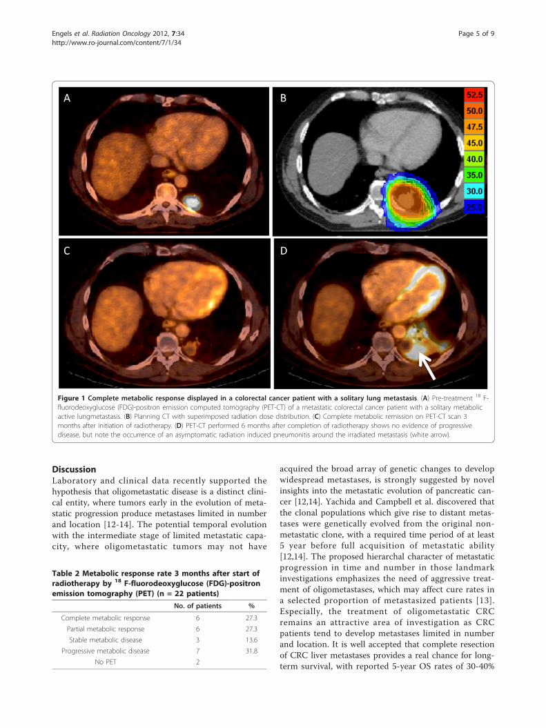
Response evaluationTwo out of 24 enrolled patients failed to receive a PET-CT3 months after the initiation of RT, 1 because of poor gen-eral condition and 1 being evaluated in another institutionafter the end of treatment. Six patients (27.3%) achieved acomplete metabolic response (CMR), 6 patients (27.3%) apartial metabolic response (PMR), resulting in an overallmetabolic response rate of 55%. The mean fractionalchange in SUVmax at evaluation as compared to baselinewas -56.1% (range, 43.5% - 70.2%) and -40.2% (range,33.3% - 45.1%) for complete and partial metabolic respon-ders, respectively. Figure 1 illustrates a CMR in a patienttreated for lungmetastasis. Metabolic response rates arelisted in Table 2. Seven patients (32%) displayed progres-sive metabolic disease (PMD), of which 6 patients atdistance and 1 patient both in- and outfield. Threepatients (13%) presented stable metabolic disease (SMD).Regarding the 17 patients who received previously ≥ 1 lineof chemotherapy, a metabolic response was recorded in86% (n = 5) of the patients with residual metabolic active
disease at time of inclusion, compared to a 40% (n = 4)metabolic response rate in the patients with progressivemetabolic active disease before RT. Lastly, 50% (n = 3) ofthe evaluated patients who did not receive previous che-motherapy for treatment of metastatic disease displayed ametabolic response 3 months after initiation of RT. Moni-toring of the tumor marker CEA at baseline showed amean value of 8.8 ± 8.7 ug/L for the whole patient group,with elevated CEA (> 3 ug/L) recorded in 71% (n = 14) ofthe patients. A significant decrease of the CEA level wasrecorded in patients with a CMR or PMR (n = 12), with amean value of 7.7 ± 8.6 ug/L pretreatment compared to4.6 ± 6.6 ug/L 3 months after initiation of RT (p = 0.01).Metabolic non-responders (SMD or PMD, n = 10)displayed a pretreatment CEA level of 10.5 ± 9.5 ug/L,compared to a posttreatment value of 27.5 ± 41.6 ug/L(p = 0.18).
Follow-upWith a median follow-up of 10 months (range, 3 - 21months), 7 patients (29%) are in remission in all irradiatedareas without evidence of distant recurrence. Five patientsdied, 4 because of progressive metastatic disease and 1because of non-cancer related cerebral bleeding. Seventeenpatients (71%) developed progressive disease, of which 10patients distant recurrence, 6 patients synchronous localand distant progression, and 1 patient with isolated localrecurrence, the latter underwent wedge resection of a soli-tary progressive lung metastasis. Among the other 16patients with progressive disease, 5 patients received bestsupportive care (BSC), 6 patients systemic therapy, 4patients an additional course of RT (2 patients because ofdistant relapse and 2 patients for local and distant relapse),1 patient underwent metastasectomy of a new livermetas-tasis. We report a 1-year actuarial LC, PFS and OS of 54%(95% C.I. 23-78%), 14% (95% C.I. 3-35%) and 78% (95% C.I. 52-91%), respectively. Log-rank testing found a statisti-cally significant benefit of the post-treatment outcome interms of PFS for patients with a metabolic response (CMR+ PMR) at 3 months after RT as compared with metabolicnon-responders (SMD + PMD) (p < 0.01). For the patientswho received previous chemotherapy (n = 17), log-ranktesting revealed that patients with residual disease pre-RTexhibited a trend toward superior PFS (p = 0.05) com-pared with patients presenting progressive disease at timeof inclusion, whereas LC and OS were not significantly dif-ferent (p = 0.63 and p = 0.18, respectively) [Figure 2]. Ofthe 7 patients which were in remission at a median follow-up of 10 months, 5 had residual oligometastatic metabolicactive disease after previous chemotherapy at time ofinclusion. Factors such as number of metastatic lesions,number of involved sites or number of previous lines ofchemotherapy were not correlated with post-treatmentoutcome.
Engels et al. Radiation Oncology 2012, 7:34http://www.ro-journal.com/content/7/1/34
Page 4 of 9
DiscussionLaboratory and clinical data recently supported thehypothesis that oligometastatic disease is a distinct clini-cal entity, where tumors early in the evolution of meta-static progression produce metastases limited in numberand location [12-14]. The potential temporal evolutionwith the intermediate stage of limited metastatic capa-city, where oligometastatic tumors may not have
acquired the broad array of genetic changes to developwidespread metastases, is strongly suggested by novelinsights into the metastatic evolution of pancreatic can-cer [12,14]. Yachida and Campbell et al. discovered thatthe clonal populations which give rise to distant metas-tases were genetically evolved from the original non-metastatic clone, with a required time period of at least5 year before full acquisition of metastatic ability[12,14]. The proposed hierarchal character of metastaticprogression in time and number in those landmarkinvestigations emphasizes the need of aggressive treat-ment of oligometastases, which may affect cure rates ina selected proportion of metastasized patients [13].Especially, the treatment of oligometastatic CRCremains an attractive area of investigation as CRCpatients tend to develop metastases limited in numberand location. It is well accepted that complete resectionof CRC liver metastases provides a real chance for long-term survival, with reported 5-year OS rates of 30-40%
Figure 1 Complete metabolic response displayed in a colorectal cancer patient with a solitary lung metastasis. (A) Pre-treatment 18 F-fluorodeoxyglucose (FDG)-positron emission computed tomography (PET-CT) of a metastatic colorectal cancer patient with a solitary metabolicactive lungmetastasis. (B) Planning CT with superimposed radiation dose distribution. (C) Complete metabolic remission on PET-CT scan 3months after initiation of radiotherapy. (D) PET-CT performed 6 months after completion of radiotherapy shows no evidence of progressivedisease, but note the occurrence of an asymptomatic radiation induced pneumonitis around the irradiated metastasis (white arrow).
Table 2 Metabolic response rate 3 months after start ofradiotherapy by 18 F-fluorodeoxyglucose (FDG)-positronemission tomography (PET) (n = 22 patients)
No. of patients %
Complete metabolic response 6 27.3
Partial metabolic response 6 27.3
Stable metabolic disease 3 13.6
Progressive metabolic disease 7 31.8
No PET 2
Engels et al. Radiation Oncology 2012, 7:34http://www.ro-journal.com/content/7/1/34
Page 5 of 9

[15,16]. Despite the improvements in surgical proce-dures and preoperative multi-agent chemotherapy, lim-itations imposed by localization, multifocal character,size, or comorbidities still exclude the major part ofmCRC patients from undergoing metastasectomy[17,18]. Recent evolutions in conformal RT and IGRThave driven the development of SBRT, of which thephysical properties allow sparing of the surroundingnormal tissues with consequently fewer side effects tobe expected [7,10]. Next, it enables delivery of tumorici-dal doses of radiation in a minimal number of fractionsto small target volumes. In the eradication of CRC liver-and/or lungmetastases, minimal toxicity and sustainedLC rates of 53%-100% are reported with the use ofSBRT [8,19,20]. However, SBRT requires a careful selec-tion of the metastases on the base of their localizationand dimension, as patients with metastases situated inthe proximity of hollow viscous organs such as smallbowel, esophagus or stomach could experience unaccep-table normal tissue toxicity with the delivery of ablativeradiation doses by SBRT [21]. In order to suit a variety
of treatment sites, we explored in a previous study inour institution the use of moderately hypofractionatedIMRT-IGRT (10 fractions of 4 Gy) by helical tomother-apy in oligometastatic CRC [9]. Taking into account thevery limited toxicity (4% grade 3 toxicity) and the rela-tively high local progression (22%) of irradiated metas-tases without development of new metastases in thistrial, the aim was on higher response rates and LC. Inthis report, we present the results of a prospective phaseII trial of helical tomotherapy delivering 50 Gy in dailyfractions of 5 Gy to inoperable oligometastatic CRCpatients. The primary objective was to evaluate theCMR rate by comparing 18FDG-PET 3 months afterinitiation of RT with 18FDG-PET at baseline. A samplesize for first stage of 7/22 was calculated based on aRichard Simon two-stage optimal design aiming at anoverall acceptable and unacceptable CMR probability of50% and 30%, respectively. After having evaluated 22patients by 18FDG-PET, a CMR was documented in 6patients. As 10 fractions of 5 Gy showed lower thanexpected activity in first stage (6/22), the second stage
Figure 2 Local control (LC), progression-free survival (PFS) and overall survival (OS) rates among the whole patient group, previoussystemically treated patients displaying residual metabolic active oligometastatic disease at time of inclusion (n = 7) and patientswith progressive metabolic active disease before RT (n = 10). Log-rank testing was used to evaluate the association between response onprevious chemotherapy and treatment outcome, with p values reported.
Engels et al. Radiation Oncology 2012, 7:34http://www.ro-journal.com/content/7/1/34
Page 6 of 9
of accrual is not carried out and the trial has been ter-minated. Indeed, the overall metabolic response rate of55% at 3 months and 1-year LC rate of 54% are similarto the rates observed after 10 fractions of 4 Gy in ourprevious study [9]. In comparison, Milano et al. reportedwith 50 Gy in 10 fractions in oligometastatic patients a2-year LC rate of 67% [22]. One should bear in mindthat LC rates of more than 95% with SBRT are reportedwith biologically effective doses (BED) of > 100 Gy,which can be only safely delivered in patients ideallywith maximal 3 metastases of less than 4 cm in dia-meter and far from hollow viscous organs. Although notsuperior to 10 fractions of 4 Gy, the delivery of 50 Gy(BED of 75 Gy assuming an a/b of 10 for tumorresponse, corresponding to a biologically equivalenttotal dose in 2-Gy fractions (EQD2Gy) of 62.5 Gy) withIMRT-IGRT by helical tomotherapy in a non-selectedpatient population resulted in a promising overallresponse rate of 55%, which is higher than the responserates achieved with second- and third-line systemictreatment regimens in mCRC, which are within therange of 9-37% [3,23].Of notice, we enrolled in our previous study mCRC
patients who already received previous systemic treatmentonly in the case when they presented progressive diseaseor cumulative toxicity limiting further continuation of sys-temic treatment [9]. Actually, not only patients with pro-gressive disease or cumulative toxicity limiting furthercontinuation of systemic treatment were enrolled in thecurrent study, but also mCRC patients with residual meta-bolic active oligometastatic disease after effective previoussystemic treatment (n = 7). The use of helical tomotherapyas consolidation appeared to be highly attractive in thissubgroup, reflected by a 86% metabolic response rate inthose patients, and a trend toward increased PFS (p =0.05) as compared to the patients with no response onprevious systemic treatment. At a median follow-up of10 months, 71% of those patients (n = 5) are still in remis-sion in all irradiated areas without evidence of distant pro-gression, whereas 4 of the 10 patients presentingprogressive disease after previous chemotherapy at time ofinclusion already died because of progressive metastaticdisease. To our knowledge, these findings for the first timeindicate a potential role for the use of RT as consolidationof previous systemically treated oligometastatic CRC. Thiscreates opportunities for future trials of SBRT that shouldtailor inoperable oligometastases according to their pre-vious response to systemic treatment, finally to resolve itsvalue in this patient population which is historically con-sidered to be incurable when treated with chemotherapyalone. Only in abstract form, Ruers et al. recently sug-gested as first a potential benefit for combining systemicand local treatment in mCRC by presenting the results of
a randomized phase II study which evaluated the benefitof RFA combined with chemotherapy compared to che-motherapy alone in 119 mCRC patients with unresectableliver metastases [24]. Although a statistically significantbenefit in median PFS has been reported for the RFA +chemotherapy arm (16.8 months versus 9.9 months forpatients receiving chemotherapy alone, p = 0.03), the fol-low-up and study design (primary endpoint: 30-monthsOS > 30%) do not allow a formal comparison between the2 treatment arms in terms of OS [24]. Lastly, in concor-dance with our previous experience, a metabolic response(CMR or PMR) 3 months after initiation of RT was alsofound to be predictive in terms of time to progression (p <0.01). Hence, 18FDG-PET should be offered complemen-tary to anatomical imaging for all oligometastatic CRCpatients undergoing a RT course.The delivery of 50 Gy in 10 fractions by the combination
of dose sculpting by IMRT with image-guidance techni-ques by the Tomotherapy Hi-Art II System appeared to bea safe regimen, with grade 3 acute and late toxicityrecorded in only 1 patient. The lowering of the maximaldose to 4 Gy/fraction on the stomach and duodenum didnot prevent the occurrence of grade 2 pyloric ulcera in thepatient treated because of 4 perihilar lymph node metas-tases. Taking into account the radiosensitive nature of thestomach and duodenum, patients with metastases locatedin the proximity of those organs should be excluded fromhigh-dose SBRT. The limited toxicity in the present studyin a patient population with critically located lesions sup-ports the further use of a moderately hypofractionated RTregimen, such as 10 × 5 Gy.Finally, from a technical point of view, the IGRT solu-
tion in the Tomotherapy Hi-Art II system allows onlypre-treatment management of tumor motion by volu-metric imaging (MV-CT) and thus requires the applica-tion of CTV to PTV margins in the order of 1 cm toaccount for intrafraction tumor motion, for example inthe lung and liver. Theoretically, real-time tracking ofthe metastases during treatment should allow a strongreduction of the CTV to PTV margin, and thus lesshealthy tissues need to be irradiated. This is especiallyattractive in performing dose escalation in criticallylocated lesions, as classical CTV to PTV margins resultin significant overlap between PTV and organs at risk,the latter limiting delivery of cytotoxic doses with regardto normal tissue toxicity. The VERO system is a novelplatform for image-guided SBRT designed to anticipatetumor motion during treatment by real-time tracking ofthe target [25]. Its dynamic capabilities have beenexplored currently in our department and a clinical trialinvestigating its value in the eradication of inoperableoligometastases will be initiated. In the context of doseescalation, one should also mention the potential of
Engels et al. Radiation Oncology 2012, 7:34http://www.ro-journal.com/content/7/1/34
Page 7 of 9

particle therapy, being proton therapy the example, inminimizing the irradiated volume of the surroundinghealthy tissues compared to 3D conformal RT andIMRT [26].
ConclusionsIn conclusion, 10 fractions of 5 Gy resulted in a promis-ing metabolic response rate of 55% and limited toxicity.Helical tomotherapy may further play a substantial rolein the multidisciplinary treatment of inoperable oligo-metastatic CRC, especially as consolidation in patientswith residual oligometastatic disease after being treatedsystemically.
AcknowledgementsThis research was funded by grants from the Foundation against Cancer,foundation of public interest (219.2008) and the Belgian Government(Nationaal Kankerplan).
Author details1Department of Radiation Oncology, UZ Brussel, Vrije Universiteit Brussel,Laarbeeklaan 101, B-1090 Brussels, Belgium. 2Department of NuclearMedicine, UZ Brussel, Vrije Universiteit Brussel, Laarbeeklaan 101, B-1090Brussels, Belgium. 3Department of Gastroenterology, UZ Brussel, VrijeUniversiteit Brussel, Laarbeeklaan 101, B-1090 Brussels, Belgium.
Authors’ contributionsConcept and design: BE, HE, GS, DV, MDR. Acquisition, analysis andinterpretation of data: BE, TG, HE, PDC, AS, NC, MDR. Drafting of themanuscript: BE, MDR. Reading and approval of final manuscript: all authors.
Competing interestsThe authors declare that they have no competing interests.
Received: 19 December 2011 Accepted: 16 March 2012Published: 16 March 2012
References1. Jemal A, Siegel R, Ward E, Hao Y, Xu J, Thun MJ: Cancer Statistics 2009. CA
Cancer J Clin 2009, 59:225.2. Hellmann S, Weichselbaum RR: Oligometastases. J Clin Oncol 1995, 13:8-10.3. Goldberg RM, Rothenberg ML, Van Cutsem E, Benson AB, Blanke CD,
Diasio RB, Grothey A, Lenz HJ, Meropol NJ, Ramanathan RK, Becerra CH,Wickham R, Armstrong D, Viele C: The continuum of care: a paradigm forthe management of metastatic colorectal cancer. Oncologist 2007,12:38-50.
4. Tomlinson JS, Jarnagin WR, DeMatteo RP, Fong Y, Kornprat P, Gonen M,Kemeny N, Brennan MF, Blumgart LH, D’Angelica M: Actual 10-yearsurvival after resection of colorectal liver metastases defines cure. J ClinOncol 2007, 25:4575-4580.
5. Nordlinger B, Sorbye H, Glimelius B, Poston GJ, Schlag PM, Rougier P,Bechstein WO, Primrose JN, Walpole ET, Finch-Jones M, Jaeck D, Mirza D,Parks RW, Collette L, Praet M, Bethe U, Van Cutsem E, Scheithauer W,Gruenberger T, EORTC Gastro-Intestinal Tract Cancer Group, CancerResearch UK, Arbeitsgruppe Lebermetastasen und -tumoren in derChirurgischen Arbeitsgemeinschaft Onkologie (ALM-CAO), AustralasianGastro-Intestinal Trials Group (AGITG), Fédération Francophone deCancérologie Digestive (FFCD): Perioperative chemotherapy withFOLFOX4 and surgery versus surgery alone for resectable livermetastases from colorectal cancer (EORTC Intergroup trial 40983): arandomised controlled trial. Lancet 2008, 371:1007-1016.
6. Nordlinger B, Van Cutsem E, Rougier P, Köhne CH, Ychou M, Sobrero A,Adam R, Arvidsson D, Carrato A, Georgoulias V, Giuliante F, Glimelius B,Golling M, Gruenberger T, Tabernero J, Wasan H, Poston G, EuropeanColorectal Metastases Treatment Group: Does chemotherapy prior to liverresection increase the potential for cure in patients with metastatic
colorectal cancer? A report from the European Colorectal MetastasesTreatment Group. Eur J Cancer 2007, 43:2037-2045.
7. Timmerman R, Paulus R, Galvin J, Michalski J, Straube W, Bradley J, Fakiris A,Bezjak A, Videtic G, Johnstone D, Fowler J, Gore E, Choy H: Stereotacticbody radiation therapy for inoperable early stage lung cancer. JAMA2010, 303:1070-1076.
8. Rusthoven KE, Kavanagh BD, Cardenes H, Stieber VW, Burri SH,Feigenberg SJ, Chidel MA, Pugh TJ, Franklin W, Kane M, Gaspar LE,Schefter TE: Multi-institutional phase I/II trial of stereotactic bodyradiation therapy for liver metastases. J Clin Oncol 2009, 27:1572-1578.
9. Engels B, Everaert H, Gevaert T, Duchateau M, Neyns B, Sermeus A,Tournel K, Verellen D, Storme G, De Ridder M: Phase II study of helicaltomotherapy for oligometastatic colorectal cancer. Ann Oncol 2011,22:362-368.
10. Verellen D, De Ridder M, Linthout N, Tournel K, Soete G, Storme G:Innovations in image-guided radiotherapy. Nat Rev Cancer 2007,7:949-960.
11. Wahl RL, Jacene H, Kasamon Y, Lodge MA: From RECIST to PERCIST:evolving considerations for PET response criteria in solid tumors. J NuclMed 2009, 50:122S-150S.
12. Campbell PJ, Yachida S, Mudie LJ, Stephens PJ, Pleasance ED, Stebbings LA,Morsberger LA, Latimer C, McLaren S, Lin ML, McBride DJ, Varela I, Nik-Zainal SA, Leroy C, Jia M, Menzies A, Butler AP, Teague JW, Griffin CA,Burton J, Swerdlow H, Quail MA, Stratton MR, Iacobuzio-Donahue C,Futreal PA: The patterns and dynamics of genomic instability inmetastatic pancreatic cancer. Nature 2010, 467:1109-1113.
13. Weichselbaum RR, Hellman S: Oligometastases revisited. Nat Rev ClinOncol 2011, 8:378-382.
14. Yachida S, Jones S, Bozic I, Antal T, Leary R, Fu B, Kamiyama M, Hruban RH,Eshleman JR, Nowak MA, Velculescu VE, Kinzler KW, Vogelstein B, Iacobuzio-Donahue CA: Distant metastasis occurs late during the genetic evolutionof pancreatic cancer. Nature 2010, 467:1114-1117.
15. Fong Y, Cohen AM, Fortner JG, Enker WE, Turnbull AD, Coit DG,Marrero AM, Prasad M, Blumgart LH, Brennan MF: Liver resection forcolorectal metastases. J Clin Oncol 1997, 15:938-946.
16. Scheele J, Stangl R, Altendorf-Hofmann A: Hepatic metastases fromcolorectal carcinoma: impact of surgical resection on the natural history.Br J Surg 1990, 77:1241-1246.
17. Bozetti F, Cozzaglio L, Baracchi P, Marubini E, Doci R, Bignami P, Gennari L:Comparing surgical resection of limited hepatic metastases fromcolorectal cancer to non-operative treatment. Eur J Surg Oncol 1993,19:162-167.
18. Selzner M, Hany TF, Widbrett P, McCormack L, Kadry Z, Clavien PA: Doesthe novel PET/CT imaging modality impact on the treatment of patientswith metastatic colorectal cancer of the liver? Ann Surg Oncol 2004,240:1027-1034.
19. Kim MS, Yoo SY, Cho CK, Yoo HJ, Choi CW, Seo YS, Kang JK, Lee DH,Hwang DY, Moon SM, Kim MS, Kang HJ, Kim YH: Stereotactic bodyradiation therapy using three fractions for isolated lung recurrence fromcolorectal cancer. Oncology 2009, 76:212-219.
20. van der Pool AEM, Mendez Romero A, Wunderink W, Heijmen BJ,Levendag PC, Verhoef C, Ijzermans JN: Stereotactic body radiation therapyfor colorectal liver metastases. Br J Surg 2010, 97:377-382.
21. Kavanagh BD, Timmerman R, Meyer JL: The expanding roles ofstereotactic body radiation therapy and oligofractionation: toward anew practice of radiotherapy. Front Radiat Ther Oncol 2011, 43:370-381.
22. Milano MT, Katz AW, Muhs AG, Philip A, Buchholz DJ, Schell MC, Okunieff P:A prospective pilot study of curative-intent stereotactic body radiationtherapy in patients with 5 or fewer oligometastatic lesions. Cancer 2008,112:650-658.
23. Peeters M, Price TJ, Cervantes A, Sobrero AF, Ducreux M, Hotko Y, André T,Chan E, Lordick F, Punt CJ, Strickland AH, Wilson G, Ciuleanu TE, Roman L,Van Cutsem E, Tzekova V, Collins S, Oliner KS, Rong A, Gansert J:Randomized phase III study of panitumumab with fluorouracil,leucovorin, and irinotecan (FOLFIRI) compared with FOLFIRI alone assecond-line treatment in patients metastatic colorectal cancer. J ClinOncol 2010, 28:4706-4713.
24. Ruers T, Punt CJ, van Coevorden F, Borel Rinkes I, Ledermann A, Poston GJ,Bechstein W, Lenz M, Mauer M, Nordlinger B: Final results of the EORTCintergroup randomized study 40004 (CLOCC) evaluating the benefit ofradiofrequency ablation (RFA) combined with chemotherapy for
Engels et al. Radiation Oncology 2012, 7:34http://www.ro-journal.com/content/7/1/34
Page 8 of 9

unresectable colorectal liver metastases (CRC LM). J Clin Oncol 2010,28:15S(suppl; abstr 3526).
25. Depuydt T, Verellen D, Haas O, Gevaert T, Linthout N, Ducheateau M,Tournel K, Reynders T, Leysen K, Hoogeman M, Storme G, De Ridder M:Geometric accuracy of a novel gimbals based radiation therapy tumortracking system. Radiother Oncol 2011, 98:365-372.
26. Roelofs E, Engelsman M, Rasch C, Persoon L, Qamhiyeh S, de Ruysscher D,Verhaegen F, Pijls-Johannesma M, Lambin P: Results of a multicentric insilico clinical trial (ROCOCO): comparing radiotherapy with photons andprotons for non-small cell lung cancer. J Thorac Oncol 2012, 7:165-176.
doi:10.1186/1748-717X-7-34Cite this article as: Engels et al.: Phase II study of helical tomotherapy inthe multidisciplinary treatment of oligometastatic colorectal cancer.Radiation Oncology 2012 7:34.
Submit your next manuscript to BioMed Centraland take full advantage of:
• Convenient online submission
• Thorough peer review
• No space constraints or color figure charges
• Immediate publication on acceptance
• Inclusion in PubMed, CAS, Scopus and Google Scholar
• Research which is freely available for redistribution
Submit your manuscript at www.biomedcentral.com/submit
Engels et al. Radiation Oncology 2012, 7:34http://www.ro-journal.com/content/7/1/34
Page 9 of 9