research open access network analysis of skin tumor
TRANSCRIPT

RESEARCH Open Access
Network analysis of skin tumor progressionidentifies a rewired genetic architecture affectinginflammation and tumor susceptibilityDavid A Quigley1, Minh D To1,2, Il Jin Kim1,2, Kevin K Lin1, Donna G Albertson1,3, Jonas Sjolund1,Jesús Pérez-Losada4, Allan Balmain1*
Abstract
Background: Germline polymorphisms can influence gene expression networks in normal mammalian tissues andcan affect disease susceptibility. We and others have shown that analysis of this genetic architecture can identifysingle genes and whole pathways that influence complex traits, including inflammation and cancer susceptibility.Whether germline variants affect gene expression in tumors that have undergone somatic alterations, and theextent to which these variants influence tumor progression, is unknown.
Results: Using an integrated linkage and genomic analysis of a mouse model of skin cancer that produces bothbenign tumors and malignant carcinomas, we document major changes in germline control of gene expressionduring skin tumor development resulting from cell selection, somatic genetic events, and changes in the tumormicroenvironment. The number of significant expression quantitative trait loci (eQTL) is progressively reduced inbenign and malignant skin tumors when compared to normal skin. However, novel tumor-specific eQTL aredetected for several genes associated with tumor susceptibility, including IL18 (Il18), Granzyme E (Gzme), Sproutyhomolog 2 (Spry2), and Mitogen-activated protein kinase kinase 4 (Map2k4).
Conclusions: We conclude that the genetic architecture is substantially altered in tumors, and that eQTL analysisof tumors can identify host factors that influence the tumor microenvironment, mitogen-activated protein (MAP)kinase signaling, and cancer susceptibility.
BackgroundCommon genetic variants have been shown to affectmany complex traits, including cancer susceptibility [1].However, factors responsible for most of the expectedheritable risk of cancer development have not yet beenidentified. Finding these alleles and isolating the causalpolymorphisms is challenging because the heritable com-ponent of susceptibility is influenced by many allelesexerting modest effects that may be pleiotropic, epistatic,or context-dependent [2,3]. Mouse models of cancerusing inbred strains of a defined genetic background donot recapitulate the genetic heterogeneity inherent inhuman populations. However, genetically heterogeneousmouse crosses permit analysis of the combinatorial
effects of host genetic background and somatic eventsduring tumor evolution, and these crosses have beenused to identify polymorphisms that influence tumorsusceptibility and progression [4-7]. Analysis of thegenetic architecture of gene expression in normal skinfrom a Mus spretus/Mus musculus backcross ([SPRET/EiX FVB/N] X FVB/N, hereafter FVBBX) identified expres-sion quantitative trait loci (eQTL) that influence bothstructural and functional phenotypes, including hair folli-cle development, inflammation and tumor susceptibility[8]. A systematic analysis of germline influence on geneexpression in benign and malignant skin tumors couldidentify novel alleles that influence tumorigenesis butare undetectable by analysis of normal tissue. Here wedemonstrate that somatic alterations during tumorprogression reduce the detectable influence of germlinepolymorphisms, but alleles that are not relevant innormal tissue are found to influence innate immune
* Correspondence: [email protected] Diller Family Comprehensive Cancer Center, University of CaliforniaSan Francisco, 1450 Third St, San Francisco, CA 94158, USAFull list of author information is available at the end of the article
Quigley et al. Genome Biology 2011, 12:R5http://genomebiology.com/2011/12/1/R5
© 2011 Quigley et al.; licensee BioMed Central Ltd. This is an open access article distributed under the terms of the Creative CommonsAttribution License (http://creativecommons.org/licenses/by/2.0), which permits unrestricted use, distribution, and reproduction inany medium, provided the original work is properly cited.
responses to skin tumors and are associated with tumorsusceptibility.
ResultsGermline control of gene expression is altered in tumorsSkin tumors were induced on a cohort of 71 FVBBX miceby treatment of dorsal back skin with dimethyl benzan-thracene (DMBA) and tetradecanoyl-phorbol acetate(TPA) (see experimental design in Figure S1 of Additionalfile 1). This treatment induced multiple benign papillomasas well as malignant carcinomas. Gene expression analysiswas performed on mRNA extracted from 68 of thesepapillomas: two papillomas from each of 31 FVBBX miceand a single papilloma from six additional FVBBX mice.Gene expression and DNA copy number analysis was per-formed on 60 carcinomas that developed on these animals.A second cohort of 28 FVBBX animals (the ‘confirmation’cohort) was subsequently generated and treated with thesame carcinogenesis protocol as the first set of mice inorder to confirm gene expression and eQTL results fromthe discovery cohort.Germline polymorphisms have been shown to influ-
ence gene expression in tissues from model organismsand humans [8-13], but it is not clear how this influenceis altered during tumor progression. If the germlineplays no significant role in tumor gene expression, wewould expect papilloma gene expression profiles fromthe same host to cluster near each other only by chance.Hierarchical clustering of gene expression profiles frompapillomas demonstrated that tumors from the samemouse are most similar to each other in 19 of the 31papilloma pairs (Figure 1a). The highly significant simi-larity of gene expression from same-host papillomassuggested that germline polymorphisms affect constitu-tive levels of gene expression in benign tumors (P <0.00001 by permutation; see Materials and methods).The contribution of genetic background to the benignand malignant tumor gene expression profiles was quan-tified by eQTL analysis. Our previous study of normalskin from the same animals identified almost 8,000candidate eQTL at ≤10% false discovery rate (FDR).We identified 3,408 candidate eQTL in the 68 papillo-mas and 912 candidate eQTL in the 60 carcinomas sig-nificant at ≤10% FDR (Figure 1b; carcinoma eQTL listedin Table S1 in Additional file 1). At ≤5% FDR we identi-fied 2,175 and 674 candidate eQTL in papillomas andcarcinomas, respectively; increasing statistical stringencyreduced the number of candidate eQTL but did notchange the subsequent results qualitatively, and wereport eQTL significant at the 10% FDR level.The striking reduction in eQTL detected in tumors,
particularly in malignant carcinomas, prompted us toinvestigate reasons why fewer genes are significantlyinfluenced by germline polymorphisms in carcinomas
than in normal skin. Of the 7,414 genes with significanteQTL in skin, only 237 are not expressed in tumors, socomplete absence of gene expression explains onlyabout 3% of the decrease. EQTLs affecting genes thatdid not undergo drastic changes (more than two stan-dard deviations from the mean fold-change) in theirexpression levels were more likely to be conservedbetween skin and carcinomas (P < 7.4e-06, Fisher exacttest). Conserved eQTL had significantly stronger statisti-cal significance in normal skin than non-conservedeQTL (P < 1e-16, Wilcoxon signed rank test). In normalskin we identified eQTL acting in cis (where the locus isphysically proximal to the gene it affects) and in trans(where the locus is distant from or on another chromo-some from the gene it affects) with approximately equalfrequencies. The most statistically significant eQTL inskin acted overwhelmingly in cis. The cis/trans propor-tion detected in tails was 0.8/1, while in papillomas itwas approximately 1.5/1, and in carcinomas it wasapproximately 5.75/1 (exact counts are listed in TableS2 in Additional file 1). We conclude that only verystrong eQTL effects carry through from normal skin toaffect the malignant carcinomas, and weaker trans-acting effects are rarely conserved.
Somatic events alter the genetic architecture of geneexpression in tumorsChanges in the wiring of signaling pathways throughepigenetic or genetic alterations may alter the influenceof germline polymorphisms on gene expression in trans-formed cells. We used array comparative genomic hybri-dization (aCGH) analysis to quantify alterations intumor DNA copy number. Tumors showed widespreadgenomic instability (Figure 2a). The most frequent targetof large-scale amplification in FVBBX carcinomas wasdistal chromosome 7, which showed copy number gainsin 45% (27 of 60) of carcinomas. Chromosome sevenhad a markedly smaller percentage of eQTL conservedbetween skin and carcinomas (2.2%) than other autoso-mal chromosomes (mean 10%, range 2.2% to -15%;Figure 2b). We identified a significant correlationbetween amplification of the most distal probe on chro-mosome 7 and fold-change increases of several geneslocated near the probe, including Ccnd1 (encodingCyclin D1; P = 3.0e-6, mean 10.5-fold up-regulation;Figure 2c). Cyclin D1 amplification or overexpression isan early event in numerous human tumors, and targetedover-expression of Ccnd1 drives several mouse modelsof carcinogenesis [14-16]. Although Ccnd1 had a signifi-cant cis-eQTL in skin (uncorrected P = 0.0001, permu-tation P = 0.009, q < 0.015), this cis-eQTL was notdetected in papillomas or carcinomas.DMBA induces a characteristic activating mutation in
Hras1 [17], which is also located on distal chromosome
Quigley et al. Genome Biology 2011, 12:R5http://genomebiology.com/2011/12/1/R5
Page 2 of 11

7 in the mouse. Hras1 also had a significant cis-eQTL inskin (uncorrected P = 8.7e-5, permutation P = 0.013, q <0.02) that was not detected in papillomas or carcinomas.Changes in Hras1 mutant gene copy number and/orloss of the normal wild-type allele play a role in tumorprogression, and trisomy of chromosome 7 is a commonearly event in both papillomas and carcinomas, leadingto increased copy number of the mutant Hras1 allele[18,19]. We conclude that gene copy number alterationson distal chromosome 7 have disrupted the normalgenetic control of expression of these target genes.
Genomic networks are rewired during tumorigenesisChanges in gene expression networks in tumors canresult from macroscopic alterations in cellular composi-tion during transformation, or from rewiring of signalingpathways. Coordinated alterations in gene expressionfrom normal to tumor can be visualized as a ‘progres-sion network’ by combining correlation and differentialexpression analysis (see Materials and methods; genesused to build this network and fold-change values arelisted in Table S3 in Additional file 1). This methodidentifies functionally related gene sets with significantly
(a)eQ
TL C
ount
Total cis trans
(b)
Skin
Papillomas
Carcinomas
8000
7000
6000
5000
4000
3000
2000
1000
0
Figure 1 The influence of germline polymorphisms on gene expression is present but reduced in tumors. (a) Hierarchical clustering oftotal gene expression from papilloma pairs indicates that germline polymorphisms continue to exert a major effect on gene expression at thebenign tumor stage. Bars indicate when both papillomas in a pair are most similar to each other. (b) Counts of total, cis-, and trans- eQTL inskin, papillomas, and carcinomas, showing that overall germline control of gene expression is strongly reduced, particularly for trans-eQTL, inmalignant carcinomas.
Quigley et al. Genome Biology 2011, 12:R5http://genomebiology.com/2011/12/1/R5
Page 3 of 11

correlated changes in expression between two states.The global network constructed in this way is shown inFigure 3 and demonstrates that pathways linked tomitosis, stress responses, and IL1-mediated signaling areseen as distinct network motifs that are up-regulated incarcinomas. Carcinomas result from clonal expansion ofinitiated epidermal cells, and this is reflected in thedown-regulation of motifs related to epithelial barrier,striated muscle, and hair follicles.We previously identified a hair follicle network in nor-
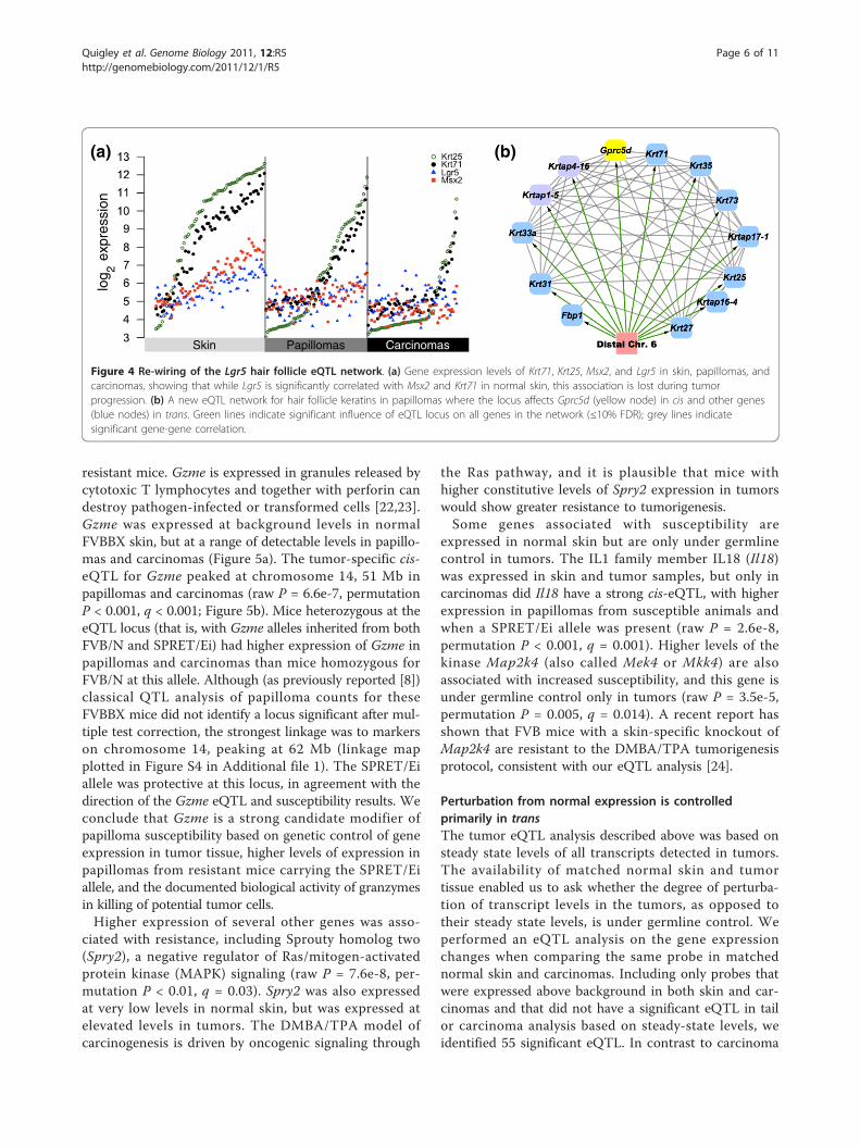
mal skin genetically linked to the G-protein coupledreceptor gene Lgr5, known to mark hair follicle stemcells [8,20]. Papillomas do not produce hair follicles,although they continue to express hair follicle keratins(Figure 4a; Figure S2 in Additional file 1). Although Lgr5is significantly expressed in papillomas and carcinomas,
it is not under the control of a cis-eQTL in tumors, andalso is not linked genetically to the hair follicle correla-tion network. A papilloma-specific eQTL networkincluding hair follicle keratins and keratin-associatedproteins was detected with a shared locus of control ondistal chromosome six (Figure 4b), a locus that was notsignificantly associated with these genes in normal tissue.The G-protein coupled receptor family member Gprc5dwas the only cis-eQTL in the new network (raw P =5.4e-4, permutation P = 0.02, q = 0.02; linkage mapplotted in Figure S3 in Additional file 1). Intriguingly,overexpression of Gprc5d promotes hair keratin geneexpression, and Gprc5d is expressed in whn (hairless)nude mice [21], compatible with a role that would onlybe revealed when normal hair follicle control has beendisrupted. These data suggest that the hair follicle stem
aCGH log2
ratio
Ccn
d1
fold
-ch
an
ge
(a)
(b) (c)
eQ
TL
co
unt
in skin
Chromosome
pe
rce
nt c
on
se
rve
d
Pe
rce
nt g
en
om
e a
lte
red
Figure 2 DNA copy number changes reduce germline influence. (a) Percentage of carcinomas with alterations across the mouse genome;amplifications (blue) plotted above zero, deletions (red) below zero. Chromosome 7 is most frequently amplified. (b) Counts of eQTL in skin onautosomal chromosomes (grey bars) compared to percentage of those eQTL conserved in carcinomas (black bars). Left-side scale indicates eQTLcounts, right-side scale indicates conservation percentage. Conservation percentage is lowest on chromosome 7. (c) Amplification of aCGHprobe MouseArray1M2_K17, at chromosome 7, 144.5 Mb, is significantly associated with increased expression of Cyclin D1 in carcinomascompared to matched normal skin. Amplification of this region of distal chromosome 7 accounts for loss of eQTL for Cyclin D1 and other genesin this region.
Quigley et al. Genome Biology 2011, 12:R5http://genomebiology.com/2011/12/1/R5
Page 4 of 11

cell network is significantly rewired during skin tumordevelopment, but the possible role of Lgr5 as a marker oftumor initiating cells remains to be determined. We con-clude that gene copy number changes, somatic muta-tions, and alterations in tissue composition in papillomasand carcinomas account for the loss of the Ccnd1, Hras1,and Lgr5 eQTL and likely are responsible for the loss ofmany other eQTL seen in normal skin.
Tumor-specific eQTL are associated with susceptibilityOf the 912 transcripts with significant eQTL in carci-noma, 210 did not have a significant eQTL in normalskin (carcinoma eQTL are listed in Table S1 in Addi-tional file 1). Of the 210 eQTL detected only in carcino-mas, in 45 cases the transcript was expressed only incarcinomas and not in normal tissue. This may be dueto activation of signaling pathways not expressed in
normal skin, or by infiltration of transformed epitheliumby cell populations from the microenvironment not nor-mally resident in the skin, particularly cells of the innateand adaptive immune systems. Loci that affect theexpression of transcripts in tumors but not normal skinmay affect tumor susceptibility, but these eQTL wouldnot be evident from analysis of normal tissue. To iden-tify genes with tumor-specific eQTL that were asso-ciated with susceptibility, we identified genes that weresignificantly differentially expressed when contrastingpapillomas from resistant and susceptible animals(FDR ≤5%). Genes were considered of interest if theyhad expression in papillomas significantly associatedwith susceptibility and also had a tumor-specific eQTL.Twenty-nine genes met these criteria (listed in Table 1).
Of these genes, the serine protease Granzyme E (Gzme)showed the largest induction in papillomas from
Hair follicle
Muscle
IL-1β Mitosis
Epithelial barrier
Proliferating epithelium
Stress response
Lipid synthesis
Figure 3 The progression network for squamous cell carcinomas. Gene pairs with significantly correlated expression change and change inexpression level >2 standard deviations from mean between skin and carcinomas are drawn as nodes. Red nodes indicate increased expressionin carcinomas and green nodes indicate decreased expression, with darker color indicating more extreme change. Grey lines connect genes withsignificant directly correlated change and blue lines indicate significant inverse correlation. The network demonstrates coordinated increases ingene expression motifs associated with mitosis, stress response, epidermal lineage proliferation, and IL1-mediated inflammatory responses.Concomitant decreases are seen in motifs linked to epithelial cell barrier function, hair follicles, lipid biosynthesis, and muscle cells due to majoralterations in cell populations in carcinomas compared to normal skin.
Quigley et al. Genome Biology 2011, 12:R5http://genomebiology.com/2011/12/1/R5
Page 5 of 11
resistant mice. Gzme is expressed in granules released bycytotoxic T lymphocytes and together with perforin candestroy pathogen-infected or transformed cells [22,23].Gzme was expressed at background levels in normalFVBBX skin, but at a range of detectable levels in papillo-mas and carcinomas (Figure 5a). The tumor-specific cis-eQTL for Gzme peaked at chromosome 14, 51 Mb inpapillomas and carcinomas (raw P = 6.6e-7, permutationP < 0.001, q < 0.001; Figure 5b). Mice heterozygous at theeQTL locus (that is, with Gzme alleles inherited from bothFVB/N and SPRET/Ei) had higher expression of Gzme inpapillomas and carcinomas than mice homozygous forFVB/N at this allele. Although (as previously reported [8])classical QTL analysis of papilloma counts for theseFVBBX mice did not identify a locus significant after mul-tiple test correction, the strongest linkage was to markerson chromosome 14, peaking at 62 Mb (linkage mapplotted in Figure S4 in Additional file 1). The SPRET/Eiallele was protective at this locus, in agreement with thedirection of the Gzme eQTL and susceptibility results. Weconclude that Gzme is a strong candidate modifier ofpapilloma susceptibility based on genetic control of geneexpression in tumor tissue, higher levels of expression inpapillomas from resistant mice carrying the SPRET/Eiallele, and the documented biological activity of granzymesin killing of potential tumor cells.Higher expression of several other genes was asso-
ciated with resistance, including Sprouty homolog two(Spry2), a negative regulator of Ras/mitogen-activatedprotein kinase (MAPK) signaling (raw P = 7.6e-8, per-mutation P < 0.01, q = 0.03). Spry2 was also expressedat very low levels in normal skin, but was expressed atelevated levels in tumors. The DMBA/TPA model ofcarcinogenesis is driven by oncogenic signaling through
the Ras pathway, and it is plausible that mice withhigher constitutive levels of Spry2 expression in tumorswould show greater resistance to tumorigenesis.Some genes associated with susceptibility are
expressed in normal skin but are only under germlinecontrol in tumors. The IL1 family member IL18 (Il18)was expressed in skin and tumor samples, but only incarcinomas did Il18 have a strong cis-eQTL, with higherexpression in papillomas from susceptible animals andwhen a SPRET/Ei allele was present (raw P = 2.6e-8,permutation P < 0.001, q = 0.001). Higher levels of thekinase Map2k4 (also called Mek4 or Mkk4) are alsoassociated with increased susceptibility, and this gene isunder germline control only in tumors (raw P = 3.5e-5,permutation P = 0.005, q = 0.014). A recent report hasshown that FVB mice with a skin-specific knockout ofMap2k4 are resistant to the DMBA/TPA tumorigenesisprotocol, consistent with our eQTL analysis [24].
Perturbation from normal expression is controlledprimarily in transThe tumor eQTL analysis described above was based onsteady state levels of all transcripts detected in tumors.The availability of matched normal skin and tumortissue enabled us to ask whether the degree of perturba-tion of transcript levels in the tumors, as opposed totheir steady state levels, is under germline control. Weperformed an eQTL analysis on the gene expressionchanges when comparing the same probe in matchednormal skin and carcinomas. Including only probes thatwere expressed above background in both skin and car-cinomas and that did not have a significant eQTL in tailor carcinoma analysis based on steady-state levels, weidentified 55 significant eQTL. In contrast to carcinoma
log
2expre
ssio
n
PapillomasSkin Carcinomas
(a) (b)
Figure 4 Re-wiring of the Lgr5 hair follicle eQTL network. (a) Gene expression levels of Krt71, Krt25, Msx2, and Lgr5 in skin, papillomas, andcarcinomas, showing that while Lgr5 is significantly correlated with Msx2 and Krt71 in normal skin, this association is lost during tumorprogression. (b) A new eQTL network for hair follicle keratins in papillomas where the locus affects Gprc5d (yellow node) in cis and other genes(blue nodes) in trans. Green lines indicate significant influence of eQTL locus on all genes in the network (≤10% FDR); grey lines indicatesignificant gene-gene correlation.
Quigley et al. Genome Biology 2011, 12:R5http://genomebiology.com/2011/12/1/R5
Page 6 of 11

Table 1 Genes with novel eQTL in tumors that are also associated with susceptibility
Symbol Probe Chr. Mb Fold change SAM q-value Higher in Higher genotype eQTL chr. eQTL Mb
Gzme 1421227_at 14 56.7 -16.67 <0.01 Resist. Het. 14 41
Gzme 1450171_x_at 14 56.7 -7.69 <0.01 Resist. Het. 14 41
Mnda 1452349_x_at 1 175.8 -2.94 4.31 Resist. Het. 1 169
2310005E10Rik 1453173_at 6 34.3 -2.27 3.02 Resist. Het. 6 32
Ddx6 1439122_at 9 44.4 -1.82 4.31 Resist. Hom. 9 34
Spry2 1421656_at 14 106.3 -1.39 3.02 Resist. Het. 14 94
Kctd3 1436811_at 1 190.8 1.16 4.31 Susc. Hom. 1 187
Map2k4 1451982_at 11 65.5 1.16 1.38 Susc. Hom. 11 101
Ssr1 1441327_a_at 13 38.1 1.18 3.02 Susc. Hom. 10 106
Ndst2 1417931_at 14 21.5 1.21 3.02 Susc. Hom. 14 19
Ppih 1429832_at 4 119.0 1.23 2.02 Susc. Hom. 5 44
1810063B07Rik 1427905_at 14 20.9 1.23 1.38 Susc. Hom. 14 23
Psme3 1418078_at 11 101.2 1.25 3.02 Susc. Hom. 4 75
Tardbp 1436318_at 4 148.0 1.25 2.02 Susc. Hom. 1 187
BC003266 1449189_at 4 126.9 1.25 <0.01 Susc. Hom. 4 121
Acbd6 1452601_a_at 1 157.4 1.26 2.02 Susc. Het. 9 116
2810457I06Rik 1436805_at 9 40.8 1.26 0.93 Susc. Het. 9 34
Dhdds 1450654_a_at 4 133.5 1.26 <0.01 Susc. Hom. 4 141
Nrd1 1424391_at 4 108.7 1.28 0.93 Susc. Hom. 10 118
Nme6 1448574_at 9 109.7 1.3 0.93 Susc. Het. 9 102
Sept8 1426802_at 11 53.3 1.35 2.02 Susc. Hom. 4 106
Hyls1 1431315_at 9 35.4 1.38 1.38 Susc. Het. 9 34
Pus3 1418491_a_at 9 35.4 1.42 0.93 Susc. Het. 9 34
C230096C10Rik 1436709_at 4 138.9 1.43 1.38 Susc. Hom. 4 141
Creg1 1415947_at 1 167.7 1.46 2.02 Susc. Hom. 1 160
Asah3l 1451355_at 4 86.5 1.51 0.33 Susc. Hom. 13 1
Rdh11 1449209_a_at 12 80.3 1.57 3.02 Susc. Het. 6 32
Mtap2 1434194_at 1 66.2 1.81 1.38 Susc. Hom. 10 102
Tslp 1450004_at 18 33.0 2.34 2.02 Susc. Het. 5 138
Il18 1417932_at 9 50.4 2.34 <0.01 Susc. Het. 9 34
Genes that satisfy two conditions: rewired or novel eQTL in tumors compared to normal skin and significant differential expression in papillomas when tumorsfrom resistant and susceptible mice are compared. ‘Chr.’ and ‘Mb’ indicate gene location; ‘Fold change’ indicates differential expression between resistant andsusceptible; ‘SAM q-value’ indicates differential expression significance; ‘Higher in’ indicates whether the gene was higher in resistant (Resist.) or susceptible(Susc.) animals; ‘Higher genotype’ indicates whether heterozygous (Het.) or homozygous (Hom.) alleles were associated with higher expression; ‘eQTL chr.’ and‘eQTL Mb’ indicate peak eQTL linkage.
0 10 20 30 40 50 60
6
5
4
3
2
1
0
centiMorgans (Chr. 14)
Gzm
e e
QT
L
LO
D
Gzm
elo
g2
expre
ssio
n
(a) (b)
PapillomasSkin Carcinomas
Figure 5 Granzyme E alleles are associated with su sceptibility. (a) Log2 expression of Gzme in skin, papillomas, and carcinomas. GzmemRNA is not detected in normal skin, and its level of expression is highest in papillomas from mice that are relatively resistant to papillomadevelopment. Papillomas from resistant animals are plotted as blue circles, susceptible animals as red circles. (b) Expression of Gzme inpapillomas and carcinomas is under germline genetic control. LOD plot for Gzme carcinoma eQTL significance on chromosome 14.
Quigley et al. Genome Biology 2011, 12:R5http://genomebiology.com/2011/12/1/R5
Page 7 of 11

eQTL, which acted almost exclusively in cis, 80% ofthese novel ‘perturbation eQTL’ acted in trans (44 of 55;listed in Table S4 in Additional file 1). A recent reportinvestigating gene expression changes in human celllines in response to ionizing radiation demonstrated thatloci associated with response overwhelmingly acted intrans [25]. It is possible, therefore, that major perturba-tions of gene expression as a result of DNA damage ortumor development are controlled indirectly throughthe influence of trans-acting regulatory factors (forexample, transcription factors) rather than throughwidespread influence on transcription levels of indivi-dual genes.
Confirmation of tumor eQTLOf the 912 transcripts with significant eQTL in the dis-covery carcinoma eQTL data set, 560 were significant inthe confirmation cohort at a 5% FDR. The number ofsamples in the confirmation cohort was relatively small(N = 28), and it is possible that more predicted eQTLwould have been confirmed with a more highly poweredstudy. These eQTL were mostly cis-eQTL and includedthe eQTL affecting Gzme and Il18 expression (Figure S5in Additional file 1; replication results listed in Table S1in Additional file 1).
DiscussionThe past few years have witnessed an explosion in gen-ome-wide association studies of cancer susceptibility inhuman populations. While these studies have revealedmany new genetic variants that influence cancer risk,each variant is predicted to have a very small effect onsusceptibility, and most heritable factors influencing riskremain to be discovered [26]. Some risk is conferred byrare variants with large effects, such as the BRCA1/BRCA2 mutations that increase breast cancer suscept-ibility. Rare variations cannot be detected by genome-wide association studies, which analyze only common(typically >5% minor allele frequency) alleles. Epistaticinteractions between common alleles may also contri-bute to cancer risk. The latter model is supported bystudies of mouse models of cancer susceptibility, whichhave demonstrated that common alleles interact in acomplex fashion to influence risk [27]. However, even inmouse models that combine defined inbred strains withdramatically different tumor susceptibilities under well-controlled environmental conditions, classical mappingstudies have not identified the majority of the risk fac-tors [28].The realization that cancer susceptibility is an emer-
gent property of the combinatorial effects of many genesnecessitated the development of more complex network-based approaches that integrate classical genetics withgene expression analysis in normal and transformed
tissues. We have previously used a systems geneticsapproach to analyze how gene expression networks innormal whole skin vary between animals that are suscep-tible or resistant to skin papilloma development. Thisapproach led to identification of pathways controllingmitosis, inflammation and tissue remodeling in normalskin that affect individual susceptibility [8]. In the presentstudy we have focused on analysis of the rewiring of thesenormal gene expression networks during development ofbenign and malignant tumors from the same heteroge-neous population of inter-specific backcross mice.Our data illuminate the dynamic changes in cell popu-
lations, both tumor-derived and host-derived, thataccompany the evolution of solid tumors. Genomic net-works in squamous cell carcinomas are profoundlyderegulated compared to normal epithelium and benignpapillomas, reflecting major changes in gross tissue orga-nization and signaling. Allelic variation continues toinfluence tumor gene expression, although this influenceis reduced by the somatic alterations accompanying pro-gression. The strongest reduction in tumors is seen ineQTL that act in trans, possibly due to genomic instabil-ity leading to alterations in transcription factor-mediatedcontrol of gene expression and the tissue-specific natureof trans-eQTL. eQTL under the control of cis-acting ele-ments in general have stronger effects than trans-eQTL,and they may be more robust in the face of somaticgenetic changes because the causal variant affects thegene directly. A recent study compared eQTL detected inhematopoietic cells at four stages of differentiation anddemonstrated that many eQTL are unique to each state,and trans-eQTL are less likely to be conserved betweendifferentiation states than cis-eQTL [29]. Trans-eQTLwere detected in all four states.We have also identified ‘perturbation eQTL’, which
measure the degree to which changes in levels of geneexpression between normal and transformed states areunder genetic control. These eQTL reflect genetic con-trol of the changes that occur in response to exogenousdamage. In contrast to the steady state eQTL that aremainly cis-acting, perturbation eQTL act primarily intrans, similar to a scenario recently described for humanlymphoblastoid cells subjected to ionizing radiation [25].The mechanistic basis for these observations remain tobe determined by isolation and analysis of the trans-act-ing factors responsible for these effects.Genetic and gene expression analyses of tumors
reveals features that cannot be detected by analysis ofnormal tissues, such as the cis-eQTL controlling expres-sion of Il18, Gzme, Map2k4, and Spry2 in tumors butnot normal skin. Il18 has an important and complexrole in inflammatory and immune responses; it has beenreported to have both tumor-promoting and anti-tumoractivities in different contexts [30]. It remains to be
Quigley et al. Genome Biology 2011, 12:R5http://genomebiology.com/2011/12/1/R5
Page 8 of 11

determined whether the gain of germline influence overIl18 expression reflects a change in cell populations or amodification in cell-autonomous signaling. The presenceof a tumor-specific eQTL for Il18 may reflect differencesin the relative proportions of epithelial and inflamma-tory cells in the tumors, or may be due to rewiring ofIl18 signaling during progression.Unlike Il18, Gzme expression is not detectable in nor-
mal skin, and appears in papillomas and carcinomasconcomitantly with the influx of innate immune cells.Mice with higher levels of Gzme within their papillomaswere relatively resistant to papilloma development, inagreement with a protective role for Gzme, and possiblyother granzymes within this gene cluster, in tumordevelopment. In contrast, mice with high levels of Il18in their papillomas were most susceptible to tumordevelopment. These data suggest that innate immunecell responses against tumors are stronger in animalsthat carry the SPRET/Ei allele at the Gzme locus, due toa polymorphism resulting in higher Gzme expression.This analysis also suggests opposing roles in tumor sus-ceptibility for Map2k4 and Spry2, genes that exert oppo-site effects on mitogen-activated protein kinase (MAPK)signaling.Tumor signaling can be rewired due to oncogenic
mutations or loss of tumor suppressor genes, possiblyrevealing activity of a germline polymorphism that isnot evident in normal tissue. The identification of sus-ceptibility genes by a combination of genetic and geneexpression analysis of tumors highlights the power ofthis approach to elucidate the genetic architecture ofcancer susceptibility. A combination of genetic and geneexpression analysis of human tumors will complementgenetic association methods and may identify additionalsusceptibility factors that cannot be detected using clas-sical methods.
Materials and methodsMouse models, gene expression, and aCGHFVBBX mice were generated and treated with DMBA/TPA as described in [8]. Gene expression was measuredwith the Affymetrix Mouse Genome 430 2.0 microarray,Affymetrix annotation release 30. Microarray probesetswhere all 11 probes did not hybridize to an annotatedRefseq gene were eliminated from analysis. Animals sen-sitive to papilloma tumorigenesis were defined as >7papillomas after 20 weeks of treatment (N = 22), resistantas <2 papillomas at that time point (N = 11). The confir-mation cohort of FVBBX mice was generated and treatedby the same protocol, with genotypes and gene expres-sion measured as described above. Genomic amplifica-tion/deletion was measured with a 2,504 probe aCGHsystem using log2 ± 0.3 cutoffs for amplification/deletion[31]. Percentage of the genome altered was calculated by
dividing each chromosome into 1,000,000 equally spacedbins and calling each bin amplified or deleted dependingon the status of the most physically proximal probe forwhich a measurement was available. Statistical analysiswas performed with the R package [32].
Statistical analysis of gene expressionPermutation analysis of hierarchical clustering was per-formed by first calculating the distance matrix for sam-ple gene expression using all present genes, countingcases where the closest papilloma to a given sample wasfrom the same mouse (Nobserved). We performed 10,000permutations of the sample labels and calculated Nperm
in the same manner, reporting the number of timesNperm ≥ Nobserved divided by 10,000. Differential expres-sion was analyzed with the Significance Analysis ofMicroarrays algorithm [33]. Correlation was defined assignificant at the 5% alpha level using the experiment-wise genome-wide error rate permutation method asdescribed in [34]. To calculate the tumor progressionnetwork, skin and carcinoma microarrays were normal-ized together and genotype-matched skin expressionwas subtracted from tumor expression. Mean fold-change values were approximately normally distributed.Highly significant change for progression networks wasdefined as >2 standard deviations from the global meanchange (N = 926). Significant correlation in fold-changewas assessed at the 5% genome-wide level using thegenome-wide error rate method as described in [34]. Allsignificantly correlated pairs of probes with highly signif-icant fold-change in expression and membership in anetwork clique of size 3 or greater were plotted. Corre-lation networks were drawn using Cytoscape [35].Microarray results have been deposited in the GeneExpression Omnibus under accession number [GEO:GSE21264].
eQTL analysisPairs of papillomas from the same animal were com-bined for eQTL analysis using the mean expression foreach probeset. eQTL were identified by linear regressionas previously described [8]. Briefly, corrected eQTLP-values were calculated by storing the lowest observedP-value pminimal-obs across all 230 SNPs and generating1,000 shuffled genotypes, calculating pminimal-perm foreach permutation, and reporting the rank of pminimal-obs
in the sorted set of pminimal-perm divided by 1,000. Thedistribution of corrected P-values was used as input toStorey’s QVALUE software [36] to calculate FDRq-values. Candidate cis-eQTL were defined as lociwithin 30 Mb of the gene they were predicted to affect(qualitatively similar results were obtained with windowsof 20 and 40 Mb). Interval mapping was performedwith R/QTL [37]. The 912 carcinoma eQTL from the
Quigley et al. Genome Biology 2011, 12:R5http://genomebiology.com/2011/12/1/R5
Page 9 of 11

discovery cohort were tested in the confirmation datasetby linear regression of the loci and gene expressionvalues. The distribution of 912 confirmation P-valueswas used with QVALUE to calculate q-values for confir-mation results.
Additional material
Additional file 1: Additional figures and tables. A schematic overviewof the experiment, additional detailed figures supporting the eQTLanalysis, a table listing eQTL detected in carcinomas, a table detailing cis-and trans-eQTL counts, a table listing genes altered more than twostandard deviations from the mean in carcinomas compared to matchednormal skin, and a table listing perturbation eQTL identified.
AbbreviationsaCGH: array comparative genomic hybridization; DMBA: dimethylbenzanthracene; eQTL: expression quantitative trait locus/loci; FDR: falsediscovery rate; FVBBX: [SPRET/Ei X FVB/N] X FVB/N; IL: interleukin; TPA:tetradecanoyl-phorbol acetate.
AcknowledgementsThis work was supported by the National Cancer Institute. AB acknowledgessupport from the Barbara Bass Bakar Chair of Cancer Genetics. MDT wassupported in part by a Sandler Foundation postdoctoral research fellowship.JS was supported by the Swedish Research Council and the TeggerFoundation. KKL was supported by an NIH Kirschstein-NRSA postdoctoralresearch fellowship. JPL is partially supported by Carlos III (FIS)/FEDER,MICIIN/plan-E 2009, JCyL (’Biomedicina y Educación’) and CSIC. The fundershad no role in study design, data collection and analysis, decision to publish,or preparation of the manuscript. We thank H Quigley, MH Barcellos-Hoff, RJAkhurst and members of the Balmain lab for critical reading of themanuscript, K Jen for assistance with histology, and JH Mao and H Nagasefor discussions in the early stages of this work.
Author details1Helen Diller Family Comprehensive Cancer Center, University of CaliforniaSan Francisco, 1450 Third St, San Francisco, CA 94158, USA. 2ThoracicOncology Program, Department of Surgery, University of California SanFrancisco, 1600 Divisadero St, San Francisco, CA 94143, USA. 3Department ofLaboratory Medicine, University of California San Francisco, 1521 ParnassusAve, Room C255, Box 0451, San Francisco, CA 94143, USA. 4Instituto deBiología Molecular y Celular del Cáncer, CSIC/Universidad de Salamanca,Campus M Unamuno s/n, 37007-Salamanca, Spain.
Authors’ contributionsThe study was conceived and supervised by AB. Bioinformatics analysis wasperformed by DAQ. RNA and DNA extraction was performed by IK, KKL, andMDT. JPL contributed mice and tumor samples and KKL performed arrayanalysis for the confirmation study. Taqman assays were performed by JS.Array CGH data were provided by DGA. The paper was written by DAQ andAB.
Received: 16 October 2010 Revised: 2 December 2010Accepted: 18 January 2011 Published: 18 January 2011
References1. McCarthy MI, Abecasis GR, Cardon LR, Goldstein DB, Little J, Ioannidis JPA,
Hirschhorn JN: Genome-wide association studies for complex traits:consensus, uncertainty and challenges. Nat Rev Genet 2008, 9:356-369.
2. Quigley D, Balmain A: Systems genetics analysis of cancer susceptibility:from mouse models to humans. Nat Rev Genet 2009, 10:651-657.
3. Flint J, Mackay TF: Genetic architecture of quantitative traits in mice, flies,and humans. Genome Res 2009, 19:723-733.
4. Nagase H, Bryson S, Cordell H, Kemp CJ, Fee F, Balmain A: Distinct geneticloci control development of benign and malignant skin tumours inmice. Nat Genet 1995, 10:424-429.
5. Ruivenkamp CA, van Wezel T, Zanon C, Stassen AP, Vlcek C, Csikos T,Klous AM, Tripodis N, Perrakis A, Boerrigter L, Groot PC, Lindeman J,Mooi WJ, Meijjer GA, Scholten G, Dauwerse H, Paces V, van Zandwijk N, vanOmmen GJ, Demant P: Ptprj is a candidate for the mouse colon-cancersusceptibility locus Scc1 and is frequently deleted in human cancers.Nat Genet 2002, 31:295-300.
6. Ewart-Toland A, Briassouli P, de Koning JP, Mao JH, Yuan J, Chan F,MacCarthy-Morrogh L, Ponder BA, Nagase H, Burn J, Ball S, Almeida M,Linardopoulos S, Balmain A: Identification of Stk6/STK15 as a candidatelow-penetrance tumor-susceptibility gene in mouse and human. NatGenet 2003, 34:403-412.
7. Park YG, Zhao X, Lesueur F, Lowy DR, Lancaster M, Pharoah P, Qian X,Hunter KW: Sipa1 is a candidate for underlying the metastasis efficiencymodifier locus Mtes1. Nat Genet 2005, 37:1055-1062.
8. Quigley D, To M, Pérez-Losada J, Pelorosso F, Mao J, Nagase H, Ginzinger D,Balmain A: Genetic architecture of mouse skin inflammation and tumoursusceptibility. Nature 2009, 458:505-508.
9. Kristensen VN, Edvardsen H, Tsalenko A, Nordgard SH, Sørlie T, Sharan R,Vailaya A, Ben-Dor A, Lønning PE, Lien S, Omholt S, Syvänen AC, Yakhini Z,Børresen-Dale AL: Genetic variation in putative regulatory loci controllinggene expression in breast cancer. Proc Natl Acad Sci USA 2006,103:7735-7740.
10. Chesler EJ, Lu L, Shou S, Qu Y, Gu J, Wang J, Hsu HC, Mountz JD,Baldwin NE, Langston MA, Threadgill DW, Manly KF, Williams RW: Complextrait analysis of gene expression uncovers polygenic and pleiotropicnetworks that modulate nervous system function. Nat Genet 2005,37:233-242.
11. Morley M, Molony CM, Weber TM, Devlin JL, Ewens KG, Spielman RS,Cheung VG: Genetic analysis of genome-wide variation in human geneexpression. Nature 2004, 430:743-747.
12. Brem RB, Yvert G, Clinton R, Kruglyak L: Genetic dissection oftranscriptional regulation in budding yeast. Science 2002, 296:752-755.
13. Schadt EE, Monks SA, Drake TA, Lusis AJ, Che N, Colinayo V, Ruff TG,Milligan SB, Lamb JR, Cavet G, Linsley PS, Mao M, Stoughton RB, Friend SH:Genetics of gene expression surveyed in maize, mouse and man. Nature2003, 422:297-302.
14. Wang TC, Cardiff RD, Zukerberg L, Lees E, Arnold A, Schmidt EV: Mammaryhyperplasia and carcinoma in MMTV-cyclin D1 transgenic mice. Nature1994, 369:669-671.
15. Diehl JA: Cycling to cancer with cyclin D1. Cancer Biol Ther 2002,1:226-231.
16. Opitz OG, Harada H, Suliman Y, Rhoades B, Sharpless NE, Kent R,Kopelovich L, Nakagawa H, Rustgi AK: A mouse model of human oral-esophageal cancer. J Clin Invest 2002, 110:761-769.
17. Quintanilla M, Brown K, Ramsden M, Balmain A: Carcinogen-specificmutation and amplification of Ha-ras during mouse skin carcinogenesis.Nature 1986, 322:78-80.
18. Aldaz CM, Trono D, Larcher F, Slaga TJ, Conti CJ: Sequential trisomizationof chromosomes 6 and 7 in mouse skin premalignant lesions. MolCarcinog 1989, 2:22-26.
19. Bremner R, Balmain A: Genetic changes in skin tumor progression:correlation between presence of a mutant ras gene and loss ofheterozygosity on mouse chromosome 7. Cell 1990, 61:407-417.
20. Jaks V, Barker N, Kasper M, van Es JH, Snippert HJ, Clevers H, Toftgård R:Lgr5 marks cycling, yet long-lived, hair follicle stem cells. Nat Genet 2008,40:1291-1299.
21. Inoue S, Nambu T, Shimomura T: The RAIG family member, GPRC5D, isassociated with hard-keratinized structures. J Invest Dermatol 2004,122:565-573.
22. Jenne DE, Tschopp J: Granzymes, a family of serine proteases releasedfrom granules of cytolytic T lymphocytes upon T cell receptorstimulation. Immunol Rev 1988, 103:53-71.
23. Cullen SP, Brunet M, Martin SJ: Granzymes in cancer and immunity. CellDeath Differ 2010, 17:616-623.
24. Finegan KG, Tournier C: The mitogen-activated protein kinase kinase4 has a pro-oncogenic role in skin cancer. Cancer Res 2010,70:5797-5806.
Quigley et al. Genome Biology 2011, 12:R5http://genomebiology.com/2011/12/1/R5
Page 10 of 11

25. Smirnov D, Morley M, Shin E, Spielman R, Cheung V: Genetic analysis ofradiation-induced changes in human gene expression. Nature 2009,459:587-591.
26. Manolio TA, Collins FS, Cox NJ, Goldstein DB, Hindorff LA, Hunter DJ,McCarthy MI, Ramos EM, Cardon LR, Chakravarti A, Cho JH, Guttmacher AE,Kong A, Kruglyak L, Mardis E, Rotimi CN, Slatkin M, Valle D, Whittemore AS,Boehnke M, Clark AG, Eichler EE, Gibson G, Haines JL, Mackay TF,McCarroll SA, Visscher PM: Finding the missing heritability of complexdiseases. Nature 2009, 461:747-753.
27. Nagase H, Mao JH, de Koning JP, Minami T, Balmain A: Epistaticinteractions between skin tumor modifier loci in interspecific (spretus/musculus) backcross mice. Cancer Res 2001, 61:1305-1308.
28. Mackay TFC, Stone EA, Ayroles JF: The genetics of quantitative traits:challenges and prospects. Nat Rev Genet 2009, 10:565-577.
29. Gerrits A, Li Y, Tesson BM, Bystrykh LV, Weersing E, Ausema A, Dontje B,Wang X, Breitling R, Jansen RC, de Haan G: Expression quantitative traitloci are highly sensitive to cellular differentiation state. PLoS Genet 2009,5:e1000692.
30. Vidal-Vanaclocha F, Mendoza L, Telleria N, Salado C, Valcarcel M, Gallot N,Carrascal T, Egilegor E, Beaskoetxea J, Dinarello CA: Clinical andexperimental approaches to the pathophysiology of interleukin-18 incancer progression. Cancer Metastasis Rev 2006, 25:417-434.
31. Snijders AM, Nowak NJ, Huey B, Fridlyand J, Law S, Conroy J, Tokuyasu T,Demir K, Chiu R, Mao JH, Jain AN, Jones SJ, Balmain A, Pinkel D,Albertson DG: Mapping segmental and sequence variations amonglaboratory mice using BAC array CGH. Genome Res 2005, 15:302-311.
32. R Core Development Team: R: A language and environment for statisticalcomputing Vienna, Austria: R Foundation for Statistical Computing; 2010.
33. Tusher VG, Tibshirani R, Chu G: Significance analysis of microarraysapplied to the ionizing radiation response. Proc Natl Acad Sci USA 2001,98:5116-5121.
34. Churchill GA, Doerge RW: Empirical threshold values for quantitative traitmapping. Genetics 1994, 138:963-971.
35. Shannon P, Markiel A, Ozier O, Baliga NS, Wang JT, Ramage D, Amin N,Schwikowski B, Ideker T: Cytoscape: a software environment forintegrated models of biomolecular interaction networks. Genome Res2003, 13:2498-2504.
36. Storey JD, Tibshirani R: Statistical significance for genomewide studies.Proc Natl Acad Sci USA 2003, 100:9440-9445.
37. Broman KW, Wu H, Sen S, Churchill GA: R/qtl: QTL mapping inexperimental crosses. Bioinformatics 2003, 19:889-890.
38. Quigley D, To M, Pérez-Losada J, Pelorosso F, Mao J, Nagase H, Ginzinger D,Balmain A: Genetic architecture of mouse skin inflammation and tumoursusceptibility. Nature 2009, 458:505-508.
39. Ciobanu DC, Lu L, Mozhui K, Wang X, Jagalur M, Morris JA, Taylor WL,Dietz K, Simon P, Williams RW: Detection, validation, and downstreamanalysis of allelic variation in gene expression. Genetics 2010, 184:119-128.
doi:10.1186/gb-2011-12-1-r5Cite this article as: Quigley et al.: Network analysis of skin tumorprogression identifies a rewired genetic architecture affectinginflammation and tumor susceptibility. Genome Biology 2011 12:R5.
Submit your next manuscript to BioMed Centraland take full advantage of:
• Convenient online submission
• Thorough peer review
• No space constraints or color figure charges
• Immediate publication on acceptance
• Inclusion in PubMed, CAS, Scopus and Google Scholar
• Research which is freely available for redistribution
Submit your manuscript at www.biomedcentral.com/submit
Quigley et al. Genome Biology 2011, 12:R5http://genomebiology.com/2011/12/1/R5
Page 11 of 11