research open access extremely low frequency
TRANSCRIPT

RESEARCH Open Access
Extremely low frequency electromagneticfields promote mesenchymal stem cellmigration by increasing intracellular Ca2+
and activating the FAK/Rho GTPasessignaling pathways in vitroYingchi Zhang, Jiyuan Yan, Haoran Xu, Yong Yang, Wenkai Li, Hua Wu* and Chaoxu Liu*
Abstract
Background: The ability of mesenchymal stem cells (MSCs) to migrate to the desired tissues or lesions is crucial forstem cell-based regenerative medicine and tissue engineering. Optimal therapeutics for promoting MSC migrationare expected to become an effective means for tissue regeneration. Electromagnetic fields (EMF), as a noninvasivetherapy, can cause a lot of biological changes in MSCs. However, whether EMF can promote MSC migration has notyet been reported.
Methods: We evaluated the effects of EMF on cell migration in human bone marrow-derived MSCs. With the useof Helmholtz coils and an EMF stimulator, 7.5, 15, 30, 50, and 70 Hz/1 mT EMF was generated. Additionally, weemployed the L-type calcium channel blocker verapamil and the focal adhesion kinase (FAK) inhibitor PF-573228 toinvestigate the role of intracellular calcium content, cell adhesion proteins, and the Rho GTPase protein family(RhoA, Rac1, and Cdc42) in EMF-mediated MSC migration. Cell adhesion proteins (FAK, talin, and vinculin) weredetected by Western blot analysis. The Rho GTPase protein family activities were assessed by G-LISA, and F-actinlevels, which reflect actin cytoskeletal organization, were detected using immunofluorescence.
Results: All the 7.5, 15, 30, 50, and 70 Hz/1 mT EMF promoted MSC migration. EMF increased MSC migration in anintracellular calcium-dependent manner. Notably, EMF-enhanced migration was mediated by FAK activation, whichwas critical for the formation of focal contacts, as evidenced by increased talin and vinculin expression. Moreover,RhoA, Rac1, and Cdc42 were activated by FAK to increase cytoskeletal organization, thus promoting cell contraction.
Conclusions: EMF promoted MSC migration by increasing intracellular calcium and activating the FAK/Rho GTPasesignaling pathways. This study provides insights into the mechanisms of MSC migration and will enable the rationaldesign of targeted therapies to improve MSC engraftment.
Keywords: Electromagnetic fields, Cell migration, Intracellular Ca2+ , Focal adhesion kinase, Rho GTPase proteinfamily
* Correspondence: [email protected]; [email protected] Zhang and Jiyuan Yan contributed equally to this work and co-firstauthors.Hua Wu and Chaoxu Liu contributed equally to this work and co-corresponding authors.Department of Orthopedics, Tongji Hospital, Tongji Medical College,Huazhong University of Science and Technology, Jiefang Avenue 1095,Wuhan 430030, China
© The Author(s). 2018 Open Access This article is distributed under the terms of the Creative Commons Attribution 4.0International License (http://creativecommons.org/licenses/by/4.0/), which permits unrestricted use, distribution, andreproduction in any medium, provided you give appropriate credit to the original author(s) and the source, provide a link tothe Creative Commons license, and indicate if changes were made. The Creative Commons Public Domain Dedication waiver(http://creativecommons.org/publicdomain/zero/1.0/) applies to the data made available in this article, unless otherwise stated.
Zhang et al. Stem Cell Research & Therapy (2018) 9:143 https://doi.org/10.1186/s13287-018-0883-4

BackgroundMesenchymal stem cells (MSC) are present in the con-nective tissue that surrounds other tissues and organs,and exhibit the capability of differentiation into multiplecell types, including osteoblast, adipocyte, chondrocyte,and potentially muscle cells, myocytes, neurons, and glialcells [1–6]. MSCs can be easily isolated from several adulttissues, readily expanded in vitro, and exhibit robustimmunomodulatory properties. All these highly desirableattributes make MSCs a stem cell source for the develop-ment of regenerative medicines. Indeed, a huge number ofpreclinical studies have demonstrated promising thera-peutic applications of MSCs in tissue engineering andcell-based therapy to repair and replace damaged or lostcells and tissues due to a wide variety of injury or disease,including autoimmune disorders [1–6]. The migrating orhoming ability of stem cells to the desired tissues or le-sions is not only crucial for normal tissue morphogenesis,homeostasis, and repair, but also for development of stemcell-based regenerative medicines [7–9].Cell migration is a complex and highly coordinated
process. Adhesive cells often migrate in the so-calledmesenchymal mode, in which the migrating cell under-goes rear-to-front polarization, protrusion and adhesionformation, and rear retraction. All these major steps incell migration are orchestrated by numerous scaffold,adaptor, and adhesion proteins (e.g., actin, myosin, integ-rin, paxillin, and tensin) in concerted actions that areregulated by various signaling molecules, including pro-tein kinase C (PKC), mitogen-activated protein kinases(MAPK; c-Jun N-terminal kinase (JNK), extracellularsignal-regulated kinase (ERK), and p38), Rho GTPase,Rho kinase, and focal adhesion kinase (FAK) [10–13].Increasing MSC migration for injury or diseases might
be a novel way to improve the efficiency of MSC engraft-ment in clinical applications. The term ‘electromagneticfields’ (EMF) indicates a combination of electric and mag-netic fields that are able to give rise to each other undercertain conditions. From their time of discovery, EMFhave attracted the attention of scientists as a potentialtherapeutic and diagnostic modality, in particular relatedto the application of nonionizing EMF for induction ofvarious biological effects on cells. It has already beenshown that EMF can cause changes in cell proliferation,differentiation, cell cycle, apoptosis, DNA replication andexpression, cytokine expression, and more [14–22]. How-ever, whether EMF can promote MSC migration has notyet been reported.In the present study, we demonstrated that 50 Hz/1 mT
EMF promoted MSC migration. We also found an intracel-lular calcium (Ca2+) increase following the EMF exposure,which activated FAK/Rho GTPase migratory signaling. Wehypothesize that EMF treatment is an effective approach topromote the migration of MSCs to the site of injury or
lesions and this has broad application prospects forregenerative medicine.
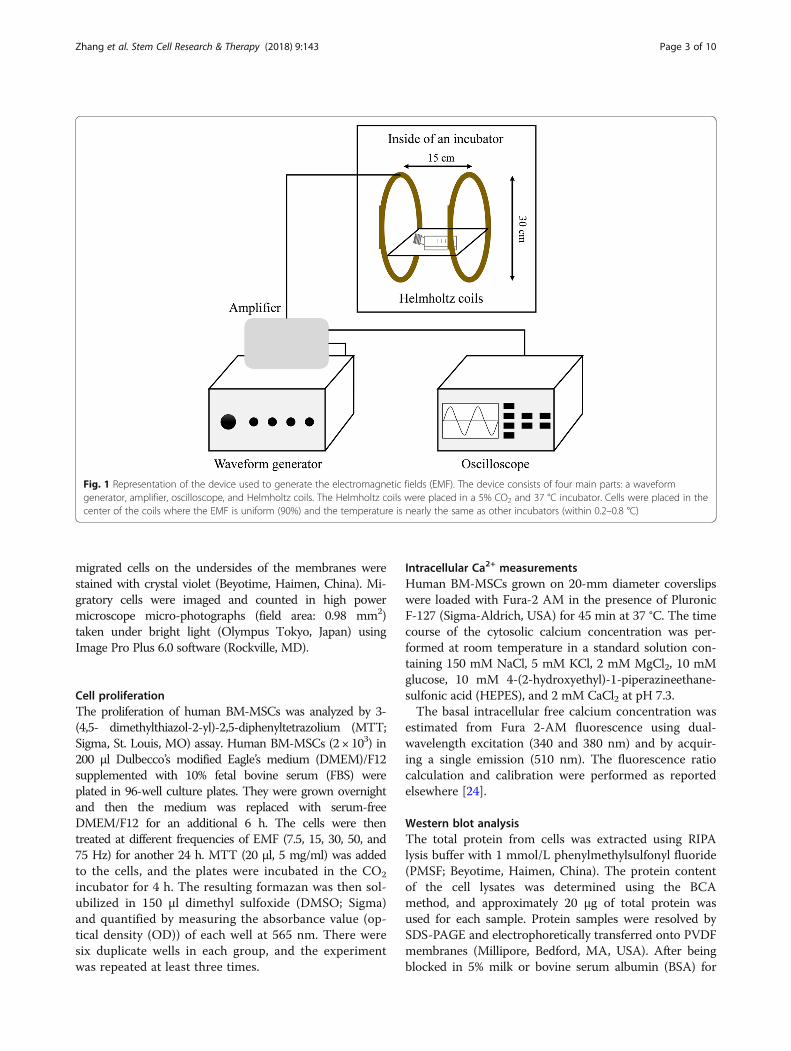
MethodsEMF deviceThe EMF-producing device was designed and manufac-tured by the Naval University of Engineering of China(Wuhan, China). It comprises a waveform generator,amplifier, oscilloscope, and Helmholtz coils. The wave-form generator creates the signals which, after beingamplified, are output to the coils. The Helmholtz coilsproducing the EMF are wound with 0.8-mm diametercoated copper wire. The coils are 30 cm in diameter,15 cm apart, and are placed perpendicular to the hori-zontal plane in a CO2 incubator (Thermo Scientific,Wilmington, DE; 5% CO2, 37 °C, and 100% humidity)(Fig. 1). The device can produce a magnetic flux densityrange of 0.0–5.0 mT and a frequency range of 0–100 Hz.A sinusoidal electromagnetic field was introduced as thisshowed a satisfying effect in our previous study [23].The magnetic field amplitude of the activated coils wasmeasured using a gauss meter (GM55A; TinDun Industry,Shanghai, China). The uniformity of EMF was approxi-mately 90% in the 7 cm of the spherical region (from thecoil center to the origin).
Human MSC culture and stimulationHuman bone marrow-derived MSCs (BM-MSCs) werepurchased from the Cell Bank of Chinese Academy ofSciences (Shanghai, China). The cells were identified bydetecting cell surface markers and the MSC multipotentpotential for differentiation toward the adipogenic,osteogenic, and chondrogenic lineages (Additional file 1:Figure S1). Cells were passaged every 3 days using 0.25%trypsin (Gibco, USA) when they reached approximately90% confluence and were used for the experimental pro-tocols between passages 3 and 5. For the EMF treat-ment, MSCs were serum starved for 6–8 h, and EMFexposure was applied for 24 h. To inhibit the L-type cal-cium channels or activities of FAK, cells were treatedwith 10 μg/ml of the L-type calcium channel blocker ver-apamil (Sigma-Aldrich, St. Louis, MO) or 5 μg/ml FAKinhibitor (PF-573228; Sigma-Aldrich, St. Louis, MO).
Transwell migration assayModified Boyden chamber assays were conductedusing 24-well Transwell polyester membrane filterinserts with 8-μm pores and 0.33 cm2 surface area(Corning Inc., Corning, NY, USA) at a density of500,000 cells/ml per transwell (upper chamber). TheL-type calcium channel blocker verapamil (10 μM)and/or PF-573228 were used in the bottom chambersof the transwells. After culturing for 24 h, the cellsfrom the upper chambers were removed, and the
Zhang et al. Stem Cell Research & Therapy (2018) 9:143 Page 2 of 10
migrated cells on the undersides of the membranes werestained with crystal violet (Beyotime, Haimen, China). Mi-gratory cells were imaged and counted in high powermicroscope micro-photographs (field area: 0.98 mm2)taken under bright light (Olympus Tokyo, Japan) usingImage Pro Plus 6.0 software (Rockville, MD).
Cell proliferationThe proliferation of human BM-MSCs was analyzed by 3-(4,5- dimethylthiazol-2-yl)-2,5-diphenyltetrazolium (MTT;Sigma, St. Louis, MO) assay. Human BM-MSCs (2 × 10³) in200 μl Dulbecco’s modified Eagle’s medium (DMEM)/F12supplemented with 10% fetal bovine serum (FBS) wereplated in 96-well culture plates. They were grown overnightand then the medium was replaced with serum-freeDMEM/F12 for an additional 6 h. The cells were thentreated at different frequencies of EMF (7.5, 15, 30, 50, and75 Hz) for another 24 h. MTT (20 μl, 5 mg/ml) was addedto the cells, and the plates were incubated in the CO2
incubator for 4 h. The resulting formazan was then sol-ubilized in 150 μl dimethyl sulfoxide (DMSO; Sigma)and quantified by measuring the absorbance value (op-tical density (OD)) of each well at 565 nm. There weresix duplicate wells in each group, and the experimentwas repeated at least three times.
Intracellular Ca2+ measurementsHuman BM-MSCs grown on 20-mm diameter coverslipswere loaded with Fura-2 AM in the presence of PluronicF-127 (Sigma-Aldrich, USA) for 45 min at 37 °C. The timecourse of the cytosolic calcium concentration was per-formed at room temperature in a standard solution con-taining 150 mM NaCl, 5 mM KCl, 2 mM MgCl2, 10 mMglucose, 10 mM 4-(2-hydroxyethyl)-1-piperazineethane-sulfonic acid (HEPES), and 2 mM CaCl2 at pH 7.3.The basal intracellular free calcium concentration was
estimated from Fura 2-AM fluorescence using dual-wavelength excitation (340 and 380 nm) and by acquir-ing a single emission (510 nm). The fluorescence ratiocalculation and calibration were performed as reportedelsewhere [24].
Western blot analysisThe total protein from cells was extracted using RIPAlysis buffer with 1 mmol/L phenylmethylsulfonyl fluoride(PMSF; Beyotime, Haimen, China). The protein contentof the cell lysates was determined using the BCAmethod, and approximately 20 μg of total protein wasused for each sample. Protein samples were resolved bySDS-PAGE and electrophoretically transferred onto PVDFmembranes (Millipore, Bedford, MA, USA). After beingblocked in 5% milk or bovine serum albumin (BSA) for
Fig. 1 Representation of the device used to generate the electromagnetic fields (EMF). The device consists of four main parts: a waveformgenerator, amplifier, oscilloscope, and Helmholtz coils. The Helmholtz coils were placed in a 5% CO2 and 37 °C incubator. Cells were placed in thecenter of the coils where the EMF is uniform (90%) and the temperature is nearly the same as other incubators (within 0.2–0.8 °C)
Zhang et al. Stem Cell Research & Therapy (2018) 9:143 Page 3 of 10
2 h, the membranes were incubated with FAK, talin, vinculin,and GAPDH primary antibodies (Cell Signaling Technology,Danvers, MA, USA) at 4 °C overnight. The primary anti-bodies were detected with their corresponding horseradishperoxidase-conjugated secondary antibodies (Boster Biotech-nology, Wuhan, China). Immunoreactive bands were ob-tained using a chemiluminescence imaging system (ChemiQ4800 mini; Ouxiang, Shanghai, China). Protein levels weredetermined by normalizing to GAPDH.
Rho GTPase activity assayGTP-bound RhoA, Rac1, and Cdc42 were measured usingcorresponding G-LISA Activation Assay Kits (Cytoskel-eton). After stimulation, cells were washed twice with coldphosphate-buffered saline (PBS) and lysed using the lysisbuffer provided with the kits for 15 min on ice. The lysateswere centrifuged at 10,000 × g for 1 min at 4 °C. Superna-tants were aliquoted, snap-frozen in liquid nitrogen, andstored at −80 °C, as indicated by the manufacturer’s proto-col. Protein concentrations were determined, and RhoGTPase activity was assessed according to the manufac-turer’s instructions.
F-actin staining by fluorescence microscopyCells were grown on glass coverslips until they were ap-proximately 50% confluent and washed with PBS at 37 °C, followed by fixation with 4% paraformaldehyde inPBS for 10 min at room temperature. Cells were thenwashed and permeabilized with 0.5% Triton-X in PBSfor 5 min. After washing, phalloidin-conjugated rhoda-mine (Beyotime, Haimen, China) was added at 100 nMin 200 μl PBS. After a 30-min incubation in the dark,slides were washed and stained with 4′,6-diamidino-2-phenylindole (DAPI; Beyotime, Haimen, China). Aftermounting with antifade mounting media, samples wereexamined with a microscope (Olympus) equipped withfluorescent illumination and a digital charge-coupled-device (CCD) camera. All micro-photographs were ob-tained under the same microscope settings.
Statistical analysisAll values are expressed as mean values ± standard devi-ation. Data obtained at each time point were analyzed byone-way analysis of variance (ANOVA). Once a signifi-cant difference was detected, Bonferroni’s post-hoc ana-lysis was used to determine the significance betweenevery two groups. Statistical analysis was performedusing the Statistical Package for Social Sciences (SPSS15.0 for Windows; SPSS, Chicago, IL). A significancelevel of 95% with a P value of 0.05 was used in all statis-tical tests performed.
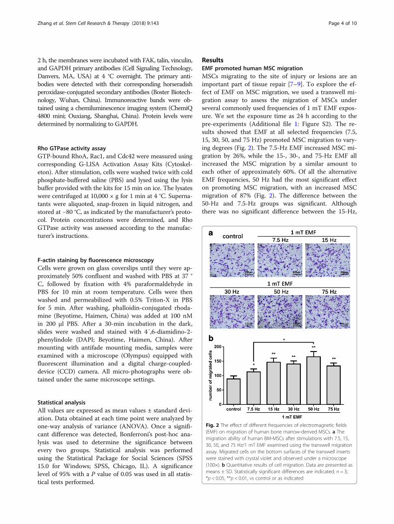
ResultsEMF promoted human MSC migrationMSCs migrating to the site of injury or lesions are animportant part of tissue repair [7–9]. To explore the ef-fect of EMF on MSC migration, we used a transwell mi-gration assay to assess the migration of MSCs underseveral commonly used frequencies of 1 mT EMF expos-ure. We set the exposure time as 24 h according to thepre-experiments (Additional file 1: Figure S2). The re-sults showed that EMF at all selected frequencies (7.5,15, 30, 50, and 75 Hz) promoted MSC migration to vary-ing degrees (Fig. 2). The 7.5-Hz EMF increased MSC mi-gration by 26%, while the 15-, 30-, and 75-Hz EMF allincreased the MSC migration by a similar amount toeach other of approximately 60%. Of all the alternativeEMF frequencies, 50 Hz had the most significant effecton promoting MSC migration, with an increased MSCmigration of 87% (Fig. 2). The difference between the50-Hz and 7.5-Hz groups was significant. Althoughthere was no significant difference between the 15-Hz,
Fig. 2 The effect of different frequencies of electromagnetic fields(EMF) on migration of human bone marrow-derived MSCs. a Themigration ability of human BM-MSCs after stimulations with 7.5, 15,30, 50, and 75 Hz/1 mT EMF examined using the transwell migrationassay. Migrated cells on the bottom surfaces of the transwell insertswere stained with crystal violet and observed under a microscope(100×). b Quantitative results of cell migration. Data are presented asmeans ± SD. Statistically significant differences are indicated; n = 3;*p < 0.05, **p < 0.01, vs control or as indicated
Zhang et al. Stem Cell Research & Therapy (2018) 9:143 Page 4 of 10

30-Hz, 50-Hz, and 75-Hz groups, the average migratedcell number in the 50-Hz group was the highest of allthe treated groups (Fig. 2b). Therefore, 50 Hz/1 mTEMF was used for further research.
EMF-promoted MSC migration is not mediated throughcell proliferationTo verify whether EMF-promoted MSC migration re-sulted from the proliferative effects of EMF, we per-formed MTT assays to measure MSC proliferation afterstimulations with the commonly used frequencies (7.5,15, 30, 50, and 75 Hz) of EMF for 24 h. The resultsshowed that EMF at all selected frequencies had no ef-fect on MSC proliferation (Fig. 3), which suggests thatthe EMF-promoted MSC migration was not mediated byproliferation.
Increased intracellular Ca2+ is critical for MSC migration inresponse to EMFCytosolic Ca2+ is a primary second messenger in thecontrol and regulation of a wide range of cell functionsincluding cell migration [25–28]. To explain why EMFpromotes MSC migration, we examined the effect ofEMF on intracellular Ca2+ content in MSCs. After 24 hof 50 Hz/1 mT EMF exposure, the intracellular Ca2+
increased by about 30%. Following treatment with the L-type calcium channel blocker verapamil (10 μM), EMFexposure did not significantly increase intracellular Ca2+
in MSCs (Fig. 4a). These results suggest that EMF mayincrease intracellular Ca2+ levels by activating L-typecalcium channels.Furthermore, we examined the effect of blocking calcium
channels on MSC migration under EMF stimulation. EMFstimulation did not significantly enhance MSC migration inthe presence of verapamil (Fig. 4b). These results indicate
that the mechanism by which EMF promote MSC migra-tion may be related to the increase in intracellular Ca2+.
Increased intracellular Ca2+ after EMF exposure activatedFAK and enhanced formation of focal contact in MSCsStudies have suggested that FAK and its downstreamproteins talin and vinculin are important for the forma-tion of focal contacts which mediate cell adhesion [29,30]. We therefore assessed the key adhesion proteinsFAK, talin, and vinculin, by Western blot analysis afterEMF stimulation following treatments with or withoutthe calcium channel blocker verapamil. We found that
Fig. 3 The effect of electromagnetic fields (EMF) on the proliferationof MSCs. MSCs were stimulated with different frequencies of EMF(7.5, 15, 30, 50, and 75 Hz/1 mT) for 24 h. Cells cultured undernormal conditions served as the baseline. The proliferation rate ofMSCs following stimulation was evaluated using the MTT assay. Dataare presented as means ± SD. n = 3. OD, optical density
Fig. 4 The effect of the L-type calcium channel blocker verapamil onelectromagnetic field (EMF)-mediated migration of MSCs. aIntracellular Ca2+ content in MSCs after 50 Hz/1 mT EMF exposurewith or without verapamil (10 μM) treatment examined using theFura 2-AM fluorescence assay. b Migration ability of MSCs after 50 Hz/1mT EMF exposure with or without verapamil (10 μM) treatmentexamined using the transwell migration assay. Migrated cells on thebottom surfaces of the transwell inserts were stained with crystal violetand observed under a microscope (100×). Quantitative results of cellmigration. Data are presented as means ± SD. Statistically significantdifferences are indicated; n = 3; *p < 0.05, **p < 0.01, vs control;#p < 0.05, ##p < 0.01, vs the EMF group
Zhang et al. Stem Cell Research & Therapy (2018) 9:143 Page 5 of 10

50 Hz/1 mT EMF increased the expression of FAK, talin,and vinculin, which are important proteins that formstructural focal contracts. However, when stimulatedwith EMF in the presence of verapamil, MSCs showedsignificant decreases in the expression of FAK, talin, andvinculin compared with the EMF group without verap-amil (Fig. 5). These data suggested that FAK was animportant downstream molecule and critical for the for-mation of focal contacts in the EMF-increased migrationof MSCs.
Rho GTPases are activated by FAK after EMF exposureFAK also influences the activities of the Rho-familyGTPases, which participate in the dynamic remodelingof the actin cytoskeleton that drives cell migration [31–33]. To study the effect of EMF-mediated FAK on theactivities of the Rho-family GTPases, the FAK inhibitorPF-573228 was used. We detected the activities of RhoA,Rac1, and Cdc42 in MSCs treated with verapamil or/andPF-573228 in the presence of EMF exposure. The results
indicated that 50 Hz/1 mT EMF could significantly in-crease the activation of RhoA, Rac1, and Cdc42 whilethis change could be blocked by verapamil or PF-573228. A combined treatment with verapamil and PF-573228 did not lead to any additional inhibition (Fig. 6).These results suggested that the EMF-dependent intracellu-lar Ca2+ accumulation activated the Rho-family GTPasesvia FAK activation.
FAK-mediated Rho GTPase activation induces F-actinorganization in MSCs after EMF exposureF-actin cytoskeleton networks, controlled by RhoGTPases, can regulate cellular shape changes and forcethe migration of MSCs [31, 32]. Therefore, we observedthe actin structure by staining cells with rhodamine phal-loidin as a probe for filamentous actin after treatmentswith verapamil and/or PF-573228 in presence of EMF ex-posure. We observed that MSCs stimulated by 50 Hz/1mT EMF displayed more noticeable stress fibers. Mean-while, verapamil and/or PF-573229 treatments in the
Fig. 5 Focal adhesion kinase (FAK) contributed to the formation of focal contacts in electromagnetic field (EMF)-mediated migration of MSCs.a–d Immunoblot analysis of the total cell lysates performed using antibodies against FAK, talin, and vinculin in MSCs after 50 Hz/1 mT EMFexposure with or without verapamil (10 μM) treatment. Data are presented as means ± SD. Statistically significant differences are indicated; n = 3;**p < 0.01, vs control; ##p < 0.01, vs the EMF group)
Zhang et al. Stem Cell Research & Therapy (2018) 9:143 Page 6 of 10

presence of EMF decreased F-actin organization success-fully in comparison with the single EMF group (Fig. 7).These results suggested that the EMF-dependent intracel-lular Ca2+ increase induced FAK to activate RhoA, Rac1,and Cdc42, which control F-actin organization in MSCs.
DiscussionThe use of MSC transplantation to enhance therapeuticeffects have been reported previously [34]. However, theinvasive character of local transplantation might not befeasible for widespread clinical application. Thus, a suc-cessful systemic transplantation and the migratory abilityof MSCs toward sites of injury are essential to enhance
the healing process [35]. Finding a new treatment topromote MSC migration can bring new ideas to regen-erative medicine and tissue engineering. EMF therapyhas been proven to be an effective noninvasive approachin treating a wide range of bone diseases, such as freshand nonunion fractures [36, 37], osteoarthritis [38], andosteoporosis [39–41], both experimentally and clinicallyover the past decades. Our previous studies confirmedthat EMF could significantly enhance the osteoblast dif-ferentiation of MSCs [23, 42–44]. However, whetherEMF can promote the migratory ability of MSCs has notyet been reported. In our study, EMF significantly en-hanced the migration of MSCs (Fig. 8). In addition, the
Fig. 6 Role of intracellular Ca2+ and FAK on Rho GTPase activity after electromagnetic field (EMF) stimulation. A colorimetric ELISA-based assaywas used to estimate a RhoA, b Rac1, and c Cdc42 activity levels in cell lysates. Date are presented as means ± SD. Statistically significant differ-ences are indicated; n = 3; **p < 0.01, ***p < 0.001, vs control; ##p < 0.01, vs the EMF group. OD, optical density
Fig. 7 FAK-mediated Rho GTPase activation induces F-actin organization after electromagnetic field (EMF) exposure. Immunofluorescence analysisof F-actin polymerization was performed using rhodamine phalloidin (F-actin; green). Nuclei were stained with DAPI (blue). An overlay of the twofluorescent signals is shown
Zhang et al. Stem Cell Research & Therapy (2018) 9:143 Page 7 of 10

EMF-promoted MSC migration is not mediated throughcell proliferation. Therefore, EMF may be an effectiveadjunctive treatment in regenerative medicine. However,the pro-migration effect of EMF on the MSCs was de-tected in the absence of any inflammatory factors re-leased at the injury sites. Interestingly, the extent ofMSC migration increased when the EMF frequency wasincreased from 7.5 Hz to 50 Hz, but the effect wasweaker when the pulsed electromagnetic field frequencywas increased from 50 Hz to 75 Hz. Our observationsmimic results of Luo et al. study, which reported that50Hz was the most effective frequency in regulating theaction of MSCs [45]. However, the mechanisms associ-ated with those variations remain unknown.The potential mechanism of MSC migration facilitated
by EMF is worthy of being investigated for EMF applica-tion. Several studies have revealed that EMF induced an in-creased intracellular Ca2+ concentration through activatingvoltage-gated calcium channels (VGCCs) [46, 47]. The L-type calcium channel blocker verapamil can lower or blockchanges in response to EMF [46, 48]. As the ubiquitoussecond messenger, cytosolic Ca2+ plays an important role inregulating many cell functions, including cell migration,and is cyto-responsive to diverse physical, chemical, andbiological clues from the surrounding environment [25–28]. This motivated our investigation as to whether EMFpromotes MSC migration through increasing intracellularCa2+ and initiating specific downstream signaling. Ourresults demonstrated that EMF increased MSC migrationas well as intracellular Ca2+, while inhibition of the EMF-dependent intracellular Ca2+ increase by the calciumchannel blocker verapamil significantly weakened thepro-migration effect of EMF. These results indicated thatincreased intracellular Ca2+ plays an important role inEMF-stimulated MSC migration.
Cell migration is a complex and highly coordinatedprocess. Adhesive cells often migrate in the so-called mes-enchymal mode, in which the migrating cells undergorear-to-front polarization, protrusion and adhesion forma-tion, and rear retraction. All these major steps in cell mi-gration are orchestrated by numerous scaffold, adaptor,and adhesion proteins (e.g., actin, myosin, integrin, paxil-lin, and tensin) in concerted actions that are regulated byvarious signaling molecules, including adhesion FAK, RhoGTPase, and Rho kinase [10–13]. FAK and the other regu-lators of adhesion turnover at the front appear to work atthe rear as well. In addition, intracellular Ca2+ levels areimplicated in the disassembly of adhesions at the rear.Potential targets for intracellular Ca2+ are the calcium-regulated phosphatase calcineurin and the calcium-activated protease calpain, which has the potential tocleave several focal adhesion proteins, including FAK,talin, and vinculin [49, 50]. We further investigated poten-tial pathways downstream of increased intracellular Ca2+
that were responsible for EMF-increased MSC migration.FAK functions as an adaptor protein to recruit other focalcontact proteins or their regulators, which affects the as-sembly or disassembly of focal contacts. Talin and vincu-lin, in the regulation of FAK, are important proteins thatform structural focal contacts [31–33]. In this study, wefound that EMF enhanced the expression of FAK, talin,and vinculin in human BM-MSCs. Moreover, blocking thecalcium channel with verapamil effectively reduced EMF-induced talin and vinculin FAK expression to baselinelevels, thus indicating that FAK was activated by increasedintracellular Ca2+ in EMF-induced MSC migration.FAK not only affects the assembly or disassembly of
focal contacts but also influences the activity of Rho-family GTPases. The regulation of the Rho family ofsmall GTPases, which includes RhoA, Rac1, and Cdc42,
Fig. 8 Schematic of electromagnetic field (EMF)-increased mesenchymal stem cell (MSC) migration by signaling through increased intracellularCa2+ and focal adhesion kinase (FAK)/Rho GTPase pathways. EMF increases intracellular Ca2+. The intracellular Ca2+ accumulation then activatesFAK, leading to the formation of focal contacts and Rho GTPase mediating the organization of the cytoskeleton, which together increases themigration of MSCs
Zhang et al. Stem Cell Research & Therapy (2018) 9:143 Page 8 of 10
is essential for controlling the dynamics of the actincytoskeleton and actin-associated adhesions during po-larized cell migration [31, 32]. Therefore, with the use ofthe FAK inhibitor PF-573228, another important findingin our study showed that EMF not only enhanced theexpression of focal adhesion proteins which facilitate celladhesion, but also increased the activity of RhoA, Rac1,and Cdc42, which subsequently increased the formationof the F-actin network. Additionally, these changes couldbe reversed by calcium channel or FAK blockade. Theseresults suggested that Rho GTPases and F-actin forma-tion were downstream of FAK signaling in response tothe EMF-induced intracellular Ca2+ increase.
ConclusionsThe present study firstly demonstrated that EMF increasedintracellular Ca2+. Afterwards, the intracellular Ca2+
accumulation activated FAK, leading to the formation offocal contacts and Rho GTPases mediating organization ofthe cytoskeleton, which synergistically contributed to theincreased migration of MSCs. More work is required tobuild an injury model and verify whether EMF can promoteMSC migration to injury sites. These findings confirmed ourhypothesis that EMF were capable of promoting MSCmigration, which might be a promising approach to improvethe therapeutic effect for many diseases.
Additional file
Additional file 1: Human MSC culture and stimulation. (DOCX 4878 kb)
AbbreviationEMF: Electromagnetic fields; FAK: Focal adhesion kinase; MSC: Mesenchymalstem cell
AcknowledgementsThe authors thank Zhihua Zhao for technical assistance and Wenchun Zhaofor help in producing the EMF signal generator.
FundingThis study was supported by the National Natural Science Foundation ofChina (grant nos. 51537004 and 31300799) and the Independent InnovativeResearch Funds of HUST (2014QN094).
Availability of data and materialsThe datasets used and/or analyzed during the current study are availablefrom the corresponding author on reasonable request.
Authors’ contributionsHW and CL designed the experimental research. YZ, JY, HX, WL, and YYperformed the experiments. CL analyzed and interpreted the data. YZ and JYwere major contributors in writing the manuscript. All authors read andapproved the final manuscript.
Competing interestsThe authors declare that they have no competing interests.
Publisher’s NoteSpringer Nature remains neutral with regard to jurisdictional claims inpublished maps and institutional affiliations.
Received: 27 December 2017 Revised: 3 April 2018Accepted: 20 April 2018
References1. Karantalis V, Hare JM. Use of mesenchymal stem cells for therapy of cardiac
disease. Circ Res. 2015;116:1413–30.2. Jiang LH, Hao Y, Mousawi F, Peng H, Yang X. Expression of P2 purinergic
receptors in mesenchymal stem cells and their roles in extracellularnucleotide regulation of cell functions. J Cell Physiol. 2017;232:287–97.
3. Uccelli A, Moretta L, Pistoia V. Mesenchymal stem cells in health anddisease. Nat Rev Immunol. 2008;8:726–36.
4. Bianco P. “Mesenchymal” stem cells. Annu Rev Cell Dev Biol. 2014;30:677–704.
5. Hwang SJ, Cho TH, Lee B, Kim IS. Bone-healing capacity of conditionedmedium derived from three-dimensionally cultivated human mesenchymalstem cells and electrical stimulation on collagen sponge. J Biomed MaterRes A. 2017;
6. Kim SK, Lee J, Song M, Kim M, Hwang SJ, Jang H, Park Y. Combination ofthree angiogenic growth factors has synergistic effects on sprouting ofendothelial cell/mesenchymal stem cell-based spheroids in a 3D matrix. JBiomed Mater Res B Appl Biomater. 2016;104:1535–43.
7. Jiang LH, Mousawi F, Yang X, Roger S. ATP-induced Ca(2+)-signallingmechanisms in the regulation of mesenchymal stem cell migration. CellMol Life Sci. 2017;74:3697–710.
8. Cheng Z, Ou L, Zhou X, Li F, Jia X, Zhang Y, Liu X, Li Y, Ward CA, Melo LG,Kong D. Targeted migration of mesenchymal stem cells modified withCXCR4 gene to infarcted myocardium improves cardiac performance. MolTher. 2008;16:571–9.
9. Ferrari D, Gulinelli S, Salvestrini V, Lucchetti G, Zini R, Manfredini R, Caione L,Piacibello W, Ciciarello M, Rossi L, Idzko M, Ferrari S, Di Virgilio F, Lemoli RM.Purinergic stimulation of human mesenchymal stem cells potentiates theirchemotactic response to CXCL12 and increases the homing capacity andproduction of proinflammatory cytokines. Exp Hematol. 2011;39:360–74. 374e361-365
10. Ridley AJ, Schwartz MA, Burridge K, Firtel RA, Ginsberg MH, Borisy G, ParsonsJT, Horwitz AR. Cell migration: integrating signals from front to back.Science. 2003;302:1704–9.
11. Amano M, Nakayama M, Kaibuchi K. Rho-kinase/ROCK: a key regulator ofthe cytoskeleton and cell polarity. Cytoskeleton. 2010;67:545–54.
12. Fogh BS, Multhaupt HA, Couchman JR. Protein kinase C, focaladhesions and the regulation of cell migration. J Histochem Cytochem.2014;62:172–84.
13. Huang C, Jacobson K, Schaller MD. MAP kinases and cell migration. J CellSci. 2004;117:4619–28.
14. Geng DY, Li CH, Wan XW, Xu GZ. Biochemical kinetics of cell proliferationregulated by extremely low frequency electromagnetic field. Biomed MaterEng. 2014;24:1391–7.
15. Cheng K, Zou C. Electromagnetic field effect on separation of nucleotidesequences and unwinding of a double helix during DNA replication. MedHypotheses. 2006;66:148–53.
16. Jasti AC, Wetzel BJ, Aviles H, Vesper DN, Nindl G, Johnson MT. Effect of awound healing electromagnetic field on inflammatory cytokine geneexpression in rats. Biomed Sci Instrum. 2001;37:209–14.
17. Kaszuba-Zwoinska J, Chorobik P, Juszczak K, Zaraska W, Thor PJ. Pulsedelectromagnetic field affects intrinsic and endoplasmatic reticulumapoptosis induction pathways in MonoMac6 cell line culture. J PhysiolPharmacol. 2012;63:537–45.
18. Li K, Ma S, Li Y, Ding G, Teng Z, Liu J, Ren D, Guo Y, Ma L, Guo G. Effects ofPEMF exposure at different pulses on osteogenesis of MC3T3-E1 cells. ArchOral Biol. 2014;59:921–7.
19. Li K, Ma S, Ren D, Li Y, Ding G, Liu J, Guo Y, Guo G. Effects ofelectromagnetic pulse on serum element levels in rat. Biol Trace Elem Res.2014;158:81–6.
20. Lei T, Liang Z, Li F, Tang C, Xie K, Wang P, Dong X, Shan S, Jiang M, Xu Q,Luo E, Shen G. Pulsed electromagnetic fields (PEMF) attenuate changes invertebral bone mass, architecture and strength in ovariectomized mice.Bone. 2017;108:10–9.
21. Wang J, An Y, Li F, Li D, Jing D, Guo T, Luo E, Ma C. The effects of pulsedelectromagnetic field on the functions of osteoblasts on implant surfaceswith different topographies. Acta Biomater. 2014;10:975–85.
Zhang et al. Stem Cell Research & Therapy (2018) 9:143 Page 9 of 10

22. Yang HJ, Kim RY, Hwang SJ. Pulsed electromagnetic fields enhance bonemorphogenetic protein-2 dependent-bone regeneration. Tissue Eng A.2015;21:2629–37.
23. Liu C, Yu J, Yang Y, Tang X, Zhao D, Zhao W, Wu H. Effect of 1 mTsinusoidal electromagnetic fields on proliferation and osteogenicdifferentiation of rat bone marrow mesenchymal stromal cells.Bioelectromagnetics. 2013;34:453–64.
24. Zocchi E, Daga A, Usai C, Franco L, Guida L, Bruzzone S, Costa A, MarchettiC, De Flora A. Expression of CD38 increases intracellular calciumconcentration and reduces doubling time in HeLa and 3T3 cells. J BiolChem. 1998;273:8017–24.
25. Melchionda M, Pittman JK, Mayor R, Patel S. Ca2+/H+ exchange by acidicorganelles regulates cell migration in vivo. J Cell Biol. 2016;212:803–13.
26. Clapham DE. Calcium signaling. Cell. 2007;131:1047–58.27. Chun J, Prince A. Ca2+ signaling in airway epithelial cells facilitates
leukocyte recruitment and transepithelial migration. J Leukoc Biol. 2009;86:1135–44.
28. Wei C, Wang X, Zheng M, Cheng H. Calcium gradients underlying cellmigration. Curr Opin Cell Biol. 2012;24:254–61.
29. Tomakidi P, Schulz S, Proksch S, Weber W, Steinberg T. Focal adhesionkinase (FAK) perspectives in mechanobiology: implications for cellbehaviour. Cell Tissue Res. 2014;357:515–26.
30. Mitra SK, Hanson DA, Schlaepfer DD. Focal adhesion kinase: in commandand control of cell motility. Nat Rev Mol Cell Biol. 2005;6:56–68.
31. Raftopoulou M, Hall A. Cell migration: Rho GTPases lead the way. Dev Biol.2004;265:23–32.
32. Xu XP, He HL, Hu SL, Han JB, Huang LL, Xu JY, Xie JF, Liu AR, Yang Y, QiuHB. Ang II-AT2R increases mesenchymal stem cell migration by signalingthrough the FAK and RhoA/Cdc42 pathways in vitro. Stem Cell Res Ther.2017;8:164.
33. Jaffe AB, Hall A. Rho GTPases: biochemistry and biology. Annu Rev Cell DevBiol. 2005;21:247–69.
34. Sohni A, Verfaillie CM. Mesenchymal stem cells migration homing andtracking. Stem Cells Int. 2013;2013:130763.
35. Nitzsche F, Muller C, Lukomska B, Jolkkonen J, Deten A, Boltze J. Concisereview: MSC adhesion cascade-insights into homing and transendothelialmigration. Stem Cells. 2017;35:1446–60.
36. Griffin XL, Costa ML, Parsons N, Smith N. Electromagnetic field stimulationfor treating delayed union or non-union of long bone fractures in adults.Cochrane Database Syst Rev. 2011;4:CD008471.
37. Atalay Y, Gunes N, Guner MD, Akpolat V, Celik MS, Guner R. Pentoxifyllineand electromagnetic field improved bone fracture healing in rats. Drug DesDevel Ther. 2015;9:5195–201.
38. Bagnato GL, Miceli G, Marino N, Sciortino D, Bagnato GF. Pulsedelectromagnetic fields in knee osteoarthritis: a double blind, placebo-controlled, randomized clinical trial. Rheumatology. 2016;55:755–62.
39. Liu C, Zhang Y, Fu T, Liu Y, Wei S, Yang Y, Zhao D, Zhao W, Song M, Tang X,Wu H. Effects of electromagnetic fields on bone loss in hyperthyroidism ratmodel. Bioelectromagnetics. 2017;38:137–50.
40. Androjna C, Fort B, Zborowski M, Midura RJ. Pulsed electromagnetic fieldtreatment enhances healing callus biomechanical properties in an animalmodel of osteoporotic fracture. Bioelectromagnetics. 2014;35:396–405.
41. Wang R, Wu H, Yang Y, Song M. Effects of electromagnetic fields on osteoporosis: asystematic literature review. Electromagn Biol Med. 2016;35:384–90.
42. Song M, Zhao D, Wei S, Liu C, Liu Y, Wang B, Zhao W, Yang K, Yang Y, WuH. The effect of electromagnetic fields on the proliferation and theosteogenic or adipogenic differentiation of mesenchymal stem cellsmodulated by dexamethasone. Bioelectromagnetics. 2014;35:479–90.
43. Yu JZ, Wu H, Yang Y, Liu CX, Liu Y, Song MY. Osteogenic differentiation ofbone mesenchymal stem cells regulated by osteoblasts under EMFexposure in a co-culture system. J Huazhong Univ Sci Technolog. 2014;34:247–53.
44. Yong Y, Ming ZD, Feng L, Chun ZW, Hua W. Electromagnetic fields promoteosteogenesis of rat mesenchymal stem cells through the PKA and ERK1/2pathways. J Tissue Eng Regen Med. 2016;10:E537–45.
45. Luo F, Hou T, Zhang Z, Xie Z, Wu X, Xu J. Effects of pulsed electromagneticfield frequencies on the osteogenic differentiation of human mesenchymalstem cells. Orthopedics. 2012;35:e526–31.
46. Pall ML. Electromagnetic fields act via activation of voltage-gatedcalcium channels to produce beneficial or adverse effects. J Cell MolMed. 2013;17:958–65.
47. Walleczek J. Electromagnetic field effects on cells of the immune system:the role of calcium signaling. FASEB J. 1992;6:3177–85.
48. Papatheofanis FJ. Use of calcium channel antagonists as magnetoprotectiveagents. Radiat Res. 1990;122:24–8.
49. Hendey B, Klee CB, Maxfield FR. Inhibition of neutrophil chemokinesis onvitronectin by inhibitors of calcineurin. Science. 1992;258:296–9.
50. Glading A, Lauffenburger DA, Wells A. Cutting to the chase: calpainproteases in cell motility. Trends Cell Biol. 2002;12:46–54.
Zhang et al. Stem Cell Research & Therapy (2018) 9:143 Page 10 of 10