research open access biotransformation of ginsenoside rb1 ... · research open access...
TRANSCRIPT

Shen et al. Chinese Medicine 2013, 8:22http://www.cmjournal.org/content/8/1/22
RESEARCH Open Access
Biotransformation of ginsenoside Rb1 via thegypenoside pathway by human gut bacteriaHong Shen1,2†, Weng-Im Leung1†, Jian-Qing Ruan1, Song-Lin Li2, Jacky Pui-Cheong Lei3, Yi-Tao Wang1 and Ru Yan1*
Abstract
Background: Bacterial conversion of ginsenosides is crucial for the health-promoting effects of ginsenosides.Previous studies on the biotransformation of ginsenoside Rb1 (Rb1) by gut bacteria have focused on theginsenoside Rd (Rd) pathway (Rb1→ Rd→ ginsenoside F2 (F2)→ compound K (Cpd K)). This study aims toexamine the gypenoside pathway in human gut bacteria in vitro.
Methods: The metabolic pathways of ginsenoside Rb1 and its metabolites ginsenoside Rd and gypenoside XVII inhuman gut bacteria were investigated by incubating the compounds anaerobically with pooled or individual gutbacteria samples from healthy volunteers. Ginsenoside Rb1, the metabolites generated by human gut bacteria, anddegraded products in simulated gastric fluid (SGF) were qualitatively analyzed using an LC/MSD Trap system in thenegative ion mode and quantitatively determined by HPLC-UV analysis.
Results: When incubated anaerobically with pooled gut bacteria, Rb1 generated five metabolites, namely Rd, F2,Cpd K, and the rare gypenosides XVII (G-XVII) and LXXV (G-LXXV). The gypenoside pathway (Rb1→ G-XVII→ G-LXXV→ Cpd K) was rapid, intermediate, and minor, and finally converted Rb1 to Cpd K via G-XVII→ F2(major)/G-LXXV (minor). Both the Rd and gypenoside pathways exhibited great inter-individual variations in age-andsex-independent manners (P > 0.05). Rb1 was highly acid-labile and degraded rapidly to form F2, ginsenoside Rg3,ginsenoside Rh2, and Cpd K, but did not generate the gypenosides in SGF. The formation of the gypenosides mightbe explained by the involvement of a gut bacteria-mediated enzymatic process.
Conclusions: Rb1 was metabolized to G-XVII, F2 (major) or G-LXXL (minor), and finally Cpd K by human gut bacteriain vitro.
BackgroundGinsenosides are the major bioactive components inPanax, including Panax ginseng, Panax quinquefolium,and Panax notoginseng [1], which are the most popularherbs used for medicinal and nutritional purposes inChina, Japan, Korea, and some Western countries. Twodammarane-type triterpene O-glycosides, namely 20(S)-protopanaxadiol (PPD) and 20(S)-protopanaxatriol, are themajor ginsenosides in different parts of Panax, includingthe flower bud, berry, crown, rootlet, side root, seed bud,seed, stem, and leaf [2,3], and their differently processedherbal products [4]. As one of the most important PPD-type ginsenosides, ginsenoside Rb1 (Rb1) has exhibited
* Correspondence: [email protected]†Equal contributors1State Key Laboratory of Quality Research in Chinese Medicine, Institute ofChinese Medical Sciences, University of Macau, Macao, ChinaFull list of author information is available at the end of the article
© 2013 Shen et al.; licensee BioMed Central LtCommons Attribution License (http://creativecreproduction in any medium, provided the or
various pharmacological activities, including neuroprotec-tive, antitumor, cardiovascular-protective, and anti-agingeffects, in many in vitro and in vivo models [5-7].The intestinal absorption of glycosides in plants or
foods is poor, while their metabolites generated by intes-tinal microflora are more permeable and bioactive [8-11].The oral bioavailability of PPD-type ginsenosides is gener-ally low (<10%) [12]. The bacterial metabolites are themain forms transported across the epithelial membraneand are most likely to be the real in vivo active forms [13].This finding has led to research on microbial metabolismand the pharmacological activities of the resultant metabo-lites of ginsenosides including Rb1 [14-16]. Hasegawa et al.[17] proposed metabolism of Rb1 via the ginsenoside Rd(Rd) pathway by human intestinal bacteria in vitro, whichwas initiated at the C-20 glucose (Rb1→ Rd→ ginsenosideF2 (F2)→Compound K (Cpd K)), and the gypenosideXVII (G-XVII) pathway, which was initiated by removal of
d. This is an open access article distributed under the terms of the Creativeommons.org/licenses/by/2.0), which permits unrestricted use, distribution, andiginal work is properly cited.

Shen et al. Chinese Medicine 2013, 8:22 Page 2 of 11http://www.cmjournal.org/content/8/1/22
the C-3 glucose (Rb1→G-XVII→ gypenoside LXXV (G-LXXV)→Cpd K). However, the gypenoside pathway ofRb1 was only speculated from the results of TLC, and thedata were not provided by the authors [18,19].The formation of G-XVII from Rb1 has been investi-
gated in several metabolic studies with individual fungalstrains [20-22]. Moreover, G-XVII was detected in vivoin rats receiving oral administration of Rb1 [23,24]. Al-though there are no reports on the formation of G-LXXVin rats, the presence of G-XVII suggests the existence ofboth the gypenoside and Rd pathways of Rb1 metabolismin rats. In plasma and urine samples from healthy subjectswho received oral administration of a ginseng extract, onlyRd and Cpd K were detected [25,26]. The gypenosidepathway of Rb1 metabolism in the human body remainsto be confirmed.Ginsenoside Rd and Cpd K have been shown to pos-
sess potent pharmacological activities [27], contributingto the beneficial effects of Rb1. The available, albeit limited,reports on the bioactivities of gypenosides have describedthat the gypenosides from Gynostemma pentaphyllumhave anticancer, antioxidative, and antihyperlipidemic ef-fects [28,29]. It is important to know whether gypenosidescan be formed from ginsenoside Rb1 by bacterial biotrans-formation in the human gut lumen.Gypenosides could be generated from ginsenoside Rb1
via a non-enzymatic process in vivo, such as degradationin the acidic environment of the stomach [30], otherthan biotransformation by gut bacteria. Several studieshave reported the degradation of several ginsenosides,including Rb1, in acidic environment [31,32]. However,the aspect of whether Rb1 is degraded to form gypeno-sides remains unclear.This study aims to examine the gypenoside pathway in
human gut bacteria in vitro. The biotransformation ofRb1 by human gut bacteria was characterized by high-performance liquid chromatography with a UV detector(HPLC-UV) and HPLC-mass spectrometry (MS) analyses.The gypenoside pathway of Rb1 was investigated andcompared with the Rd pathway by incubating Rb1, Rd,and G-XVII separately with pooled human gut bacterialsamples. The inter-individual variations in the G-XVII andRd pathways were also characterized with gut bacterialsamples from 58 individuals of both sexes and differentages. The stabilities of Rb1 and Rd in simulated gastricfluid (SGF) were examined to rule out the generation ofthese gypenosides under the acidic gastric environment.
MethodsChemicals, reagents, and materialsGinsenoside Rb1, Rd, F2, Rg3, Cpd K, Rh2, G-XVII, andRg1 were supplied by Chengdu Must Biotechnology Co.Ltd. (Chengdu, China). The purity was >98% (HPLC-UV). The BBL™ Brain Heart Infusion (BHI) medium,
GasPak™ EZ anaerobe container system with an indica-tor, and GasPak™ EZ large incubation container werepurchased from Becton Dickinson (Franklin Lakes, NJ,USA). L-cystine was purchased from Research OrganicsInc. (Cleveland, OH, USA). Dimethyl sulfoxide (DMSO),bovine hemin, and vitamin K1 were supplied by Sigma-Aldrich (St. Louis, MO, USA). Methanol, 1-butanol, andacetonitrile were HPLC-grade and purchased from Merck(Darmstadt, Germany). Dulbecco’s phosphate-buffered sa-line (PBS) was provided by Life Technologies (Carlsbad,CA, USA). Hydrochloric acid and sodium carbonate(analytical grade) were purchased from Sigma-Aldrich(St. Louis, MO, USA). Deionized water was preparedin-house using a Milli-Q purification system (Millipore,Bedford, MA, USA).
Instrumentation and analytical conditionsChromatographic separation was performed on an Agi-lent 1200 Series HPLC apparatus (Agilent Technologies,Santa Clara, CA, USA) equipped with a vacuum degasser,a binary pump, and an autosampler, and connected to adiode array detector (DAD). Samples were loaded onto anAlltima C18 column (250 × 4.6 mm, 5 μm). The columntemperature was maintained at 25°C. The mobile phaseconsisted of water (A) and acetonitrile (B) and flowed at1.5 mL/min. The following gradient elution program wasused for samples obtained from bacterial incubations: 0-10 min, 19-20% B; 10-28 min, 20-30% B; 28-40 min, 30-60% B; 40-48 min, 60-100% B. The elution program wasmodified when the incubations of ginsenosides with SGFwere analyzed as follows: 0-10 min, 19-20% B; 10-28 min,20-30% B; 28-35 min, 30-60% B; 35-45 min, 60-100% B.Rb1 and its bacterial metabolites were monitored at203.4 nm and their UV absorbance was recorded over200-400 nm. The injection volume was 5 μL.MS analysis was performed on an liquid chromato-
graph/mass selective detector (LC/MSD) Trap system(Agilent Technologies, Palo Alto, CA, USA) equippedwith an ion-trap mass spectrometer with an electrosprayionization (ESI) interface. Agilent ChemStation for LC3D System software (Rev. B. 01. 03-SR2) (Agilent Tech-nologies, Santa Clara, CA, USA) was used for instru-ment control, data acquisition, and data management.Except for replacement of the water with 50 mM am-monium acetate in water, the mobile phase compositionand gradient elution program were the same as thosefor the above HPLC analysis method for human gut bac-teria samples. The mass spectrometer was operated inthe negative ion mode with the following conditions:drying gas (N2), 8 L/min; temperature, 325°C; nebulizerpressure, 30 psi; scan range, m/z 100-1400. The ESI-MS/MS conditions were as follows: negative ion mode;separation width, 4; fragment amplification, 1.0 V; scanrange, m/z 100-1400.

Shen et al. Chinese Medicine 2013, 8:22 Page 3 of 11http://www.cmjournal.org/content/8/1/22
Preparation of human gut bacteriaThe medium was prepared according to our previous re-port [33]. Briefly, 100 mL of autoclaved BHI medium(3.7 g/100 mL) was supplemented with 0.05 mg of vitaminK1, 0.5 mg of bovine hemin, and 50 mg of L-cystine. Thefecal samples (healthy Chinese, 18-92 years old) were pro-vided by Kiang Wu Hospital and the experimental proto-col is approved by Kiang Wu Hospital and University ofMacau before conduct. Human gut bacteria suspensionswere prepared at 4°C according to our previous report[33] with minor modifications. Fecal samples were freshlycollected and pooled at 1 g each for identification of Rb1metabolic pathways, or individually processed at 5 g eachfor characterization of individual variations in Rb1 metab-olism. The individual or pooled fecal samples were mixedwith 20 mL of BHI medium and the resulting suspensionswere centrifuged at 200 × g for 5 min. The supernatantswere then centrifuged at 5000 × g for 30 min. The humangut bacteria suspensions were obtained by resuspension ofthe bacterial precipitates in 5 mL of BHI medium.
Biotransformation of Rb1 by human gut bacteriaBiotransformation assays of Rb1 were conducted withpooled or individual human gut bacteria samples toexamine the existence of the gypenoside pathway andcompare it with the Rd pathway. A high concentrationof Rb1 was used to capture the minor formation andquantitative variations in the metabolites formed fromthe minor gypenoside pathway based on the great indi-vidual variations in gut bacteria compositions and meta-bolic capabilities [34]. The anaerobic incubation systemcontained 250 μL of gut bacteria suspension, 100 μL ofRb1 in DMSO (final concentration, 100 mM Rb1), and4.75 mL of BHI medium. The incubation system wasmaintained in an anaerobic state at 37°C using the Gas-Pak™ EZ anaerobe container system.The metabolic pathway and time-course of Rb1 me-
tabolism by human gut bacteria were characterized infive independent experiments using pooled samples. Ineach experiment, the reaction of Rb1 with a human gutbacteria suspension was conducted in duplicate. Controlreactions were conducted in parallel by adding 250 μL ofPBS (pH 7.0) instead of gut bacteria suspension. The reac-tions were terminated at 0, 2, 4, 12, 18, 24, 36, and 48 h,respectively, by adding 15 mL of 1-butanol followed byimmediate centrifugation at 5000 × g for 30 min to removethe bacteria. The resulting supernatants were mixed with100 μL of ginsenoside Rg1 (final concentration, 100 mM)as an internal standard (IS), and each mixture was ex-tracted twice with water-saturated 1-butanol. The organiclayers were combined and dried by rotary evaporation(BUCHI, Flavil, Switzerland). The residue was reconsti-tuted in 0.2 mL of methanol, and an aliquot (5 μL) wassubjected to HPLC-UV or HPLC-MS analysis.
Inter-individual variations in the gypenoside and Rdpathways were investigated by incubating Rb1 with gutbacteria suspensions from different individuals under thesame conditions described above, and the incubatedsamples collected at 18 h were processed as describedabove. Samples were analyzed by HPLC-UV, and thepeak area ratio data of each analyte were collected forcomparison.
Biotransformation of Rd and G-XVII by human gutbacteriaThe metabolism of the two positional isomers Rd andG-XVII by human gut bacteria was investigated by incu-bation with the same human gut bacteria sample in par-allel to identify the pathways between Rd/G-XVII andCpd K. The anaerobic incubation system contained20 μL of gut bacteria suspension, 10 μL of ginsenosideRd or G-XVII (final concentration, 100 μM Rd or G-XVII), and 170 μL of BHI medium. The concentrationsof Rd and G-XVII were determined based on the highamounts of all metabolites determined from a prelimin-ary study. Each reaction was conducted in triplicate.Control reactions without gut bacteria were conductedin parallel.Reactions were terminated at 0, 2, 4, 6, 8, 12, 18, 24,
36, and 48 h, respectively, by adding 1.2 mL of 1-butanolfollowed by centrifugation at 5000 × g for 30 min to re-move the bacteria. After adding 10 μL of ginsenosideRg1 (final concentration, 100 μM Rg1) as an internalstandard, the resulting mixture was processed in thesame manner described above before being subjected toHPLC-UV analysis.
Stabilities of Rb1 and Rd in SGFSGF was prepared by diluting 3.84 mL of hydrochloricacid to 1000 mL with deionized water (pH 1.2) accord-ing to the Chinese Pharmacopoeia [35]. Ten microlitersof ginsenoside Rb1 or Rd in DMSO (final concentration,71.4 μM) was mixed with 200 μL of SGF, and kept at37°C for different time intervals (0, 20, 40, 60, 90, 150,180 and 300 min) before adding 70 μL of 0.1 mol/LNa2CO3 to examine the acidic stability. Each reactionwas conducted in triplicate. The mixture was shaken for1 min by vortexing and centrifuged at 14,000 × g for5 min. The upper layer was decanted and an aliquot(20 μL) was subjected to HPLC-UV analysis.
Calibration curves of Rb1 and its metabolites Rd, F2,G-XVII, and Cpd K in human gut bacteriaSerial concentrations of Rb1, Rd, G-XVII, F2, and Cpd Kwere prepared in DMSO, and mixed with a pooled hu-man gut bacteria suspension and BHI medium as de-scribed in the section entitled Biotransformation of Rb1by Human Gut Bacteria. The resulting mixtures were

Shen et al. Chinese Medicine 2013, 8:22 Page 4 of 11http://www.cmjournal.org/content/8/1/22
immediately extracted with 1-butanol and centrifuged(5000 × g, 30 min). The organic layers were processed inthe same manner described above and then subjected toHPLC-UV analysis. Calibration curves were constructedby plotting the peak area ratio of each analyte to the IS(Y) as a function of the analyte concentration (X) in thereaction system.For measurements of the intra-day precision and accur-
acy, three concentration levels for each standard (low,medium, and high) were prepared as quality control (QC)samples and analyzed on the same day. The inter-day
Figure 1 HPLC-UV chromatograms of (A) mixed ginsenoside standardand intestinal bacteria from (C) a 65-year-old male, (D) a 44-year-oldgypenoside XVII; M3, ginsenoside F2; M4, gypenoside LXXV; M5, compound
precision and accuracy were determined with the samethree QC samples on three consecutive days.
Calibration curves of ginsenosides in SGFA mixed stock solution of Rb1, Rd, G-XVII, F2, Rg3,Cpd K, and Rh2 was prepared in DMSO. A series ofworking solutions were prepared with DMSO using dilu-tion factors of 2.5, 5, 10, 20, 40, and 80 from the stocksolution. An aliquot (10 μL) of each working solutionwas spiked with 200 μL of SGF and processed with70 μL of 0.1 mol/L Na2CO3 as described above. The
s, and incubated samples of Rb1 with (B) pooled gut bacteriamale, and (E) a 54-year-old female. M1, ginsenoside Rd; M2,K.

Shen et al. Chinese Medicine 2013, 8:22 Page 5 of 11http://www.cmjournal.org/content/8/1/22
sample (20 μL) was injected into the HPLC instrument. Acalibration curve was obtained by plotting the peak areaof each analyte (Y) against its concentration in SGF (X).
Data analysisData were presented as the means ± standard deviation(SD) of triplicate analyses (experiments with pooledhuman gut bacteria or SGF) or individual measurements(experiments on inter-individual variations). Sex and agedifferences in ginsenoside Rb1 metabolism were com-pared by Student’s t-test. Values of P < 0.05 were consid-ered statistically significant.
ResultsMetabolism of ginsenoside Rb1 by pooled human gutbacteriaFecal samples from 50 healthy volunteers (25 males and25 females, range, 45.7-73.7 years) were collected andpooled at 10 individuals each to prepare five human gutbacteria pools. When Rb1 was incubated with pooledhuman gut bacteria, five peaks (M1-M5) were observedat 35.1, 35.6, 38.2, 39.7, and 44.4 min, respectively, whichwere absent in control reactions (Figure 1). Under the de-veloped analytical conditions, Rb1 and its metaboliteswere well-separated from the endogenous interferences ofthe in vitro anaerobic incubation system. The retentiontimes and characteristic CID fragment ions of the ginse-noside standards and Rb1 metabolites are summarized inTable 1.Rb1 was eluted at 33.2 min and showed its pseudo-
molecular ion at m/z 1107 ([M-H]-) and chloride adduction at m/z 1143 ([M + Cl]-), corresponding to a molecu-lar weight of 1108 Daltons. The fragment ions at m/z945 ([M-H2O-H]-), m/z 783 ([M-2H2O-H]-), and m/z621 ([M-3H2O-H]-) in the CID spectrum correspondedto loss of one, two, and three glucose molecules fromRb1, respectively.
Table 1 HPLC-ESI-MS and corresponding CID data of ginsenohuman gut bacteria
Retention time (min) MS (m/z)
[M-H]- [M + Cl]- [M + AcO]-
20.9 799 835 859 637[
33.2 1107 1143 - 94
35.1 945 981 -
35.6 945 981 -
38.2 783 819 843
39.7 783 819 843
40.3 783 819 -
45.4 621 657 681
44.4 621 657 681
The retention times and mass spectral profiles of M1(35.1 min), M3 (38.2 min), and M5 (44.4 min) were identi-cal to those of Rd, F2, and Cpd K, respectively. The reten-tion time and mass spectral profile of M2 (35.6 min) wereidentical to those of G-XVII, which was formed from Rb1by hydrolytic removal of one glucose molecule from theC-3 position of Rb1. Thus, M1–M3 and M5 were unam-biguously identified as ginsenosides Rd, G-XVII, F2, andCpd K, respectively.M4 was eluted at 39.7 min. The MS spectral profile of
M4 was similar to those of M3 (ginsenoside F2) and gin-senoside Rg3 (Table 1), supporting that M4 contained aginsenoside glycoside with two glucoses. However, theretention time of M4 was different from that of eitherginsenoside Rg3 (40.3 min, with two glucoses at the C-3position) or F2 (38.2 min, with one glucose at the C-3position and another at the C-20 position) (Figure 1).Thus, M4 was tentatively identified as G-LXXV, a me-tabolite formed from Rb1 with two glucose moleculesremaining at the C-20 position.Taken together, Rb1 biotransformation by human gut
bacteria in vitro resulted in the formations of gypeno-sides as well as metabolites of the Rd pathway.
Time-course of Rb1 metabolism by human gut bacteriaThe calibration curves of Rb1, Rd, F2, G-XVII, and CpdK in the in vitro anaerobic incubation system showedgood linearity (r2 > 0.998, P < 0.001) over the concentra-tion ranges tested (Table 2). The overall intra-day andinter-day variations were less than 6%, and recovery washigher than 85% (except for ~60% for 10 μM Cpd K)(Additional file 1: Table S1).When Rb1 was incubated with the pooled human gut
bacteria (Figure 2A), Rb1 was rapidly eliminated by thehuman gut bacteria and less than 20% of the Rb1 remainedintact at 12 h. The metabolites from the Rd pathway, Rdand F2, appeared in the incubations in turn, with Rd reach-ing its maximum at 12 h (representing ~50% of the initial
side standards and the metabolites of ginsenoside Rb1 by
CID (m/z) Identity
M-H-Glc]-, 619[M-H-Glc-H2O]-, 475[M-H-2Glc]- Ginsenoside Rg1 (IS)
5[M-H-Glc]-, 783[M-H-2Glc]-, 621[M-H-3Glc]- Ginsenoside Rb1
783[M-H-Glc]-, 621[M-H-2Glc]-, Ginsenoside Rd (M1)
783[M-H-Glc]-, 621[M-H-2Glc]-, Gypenoside XVII (M2)
621[M-H-Glc]-, 459[M-H-2Glc]- Ginsenoside F2 (M3)
621[M-H-Glc]-, 459[M-H-2Glc]- Gypenoside LXXV (M4)
621[M-H-Glc]-, 459[M-H-2Glc]- Ginsenoside Rg3
459[M-H-Glc]- Ginsenoside Rh2
459[M-H-Glc]- Compound K (M5)
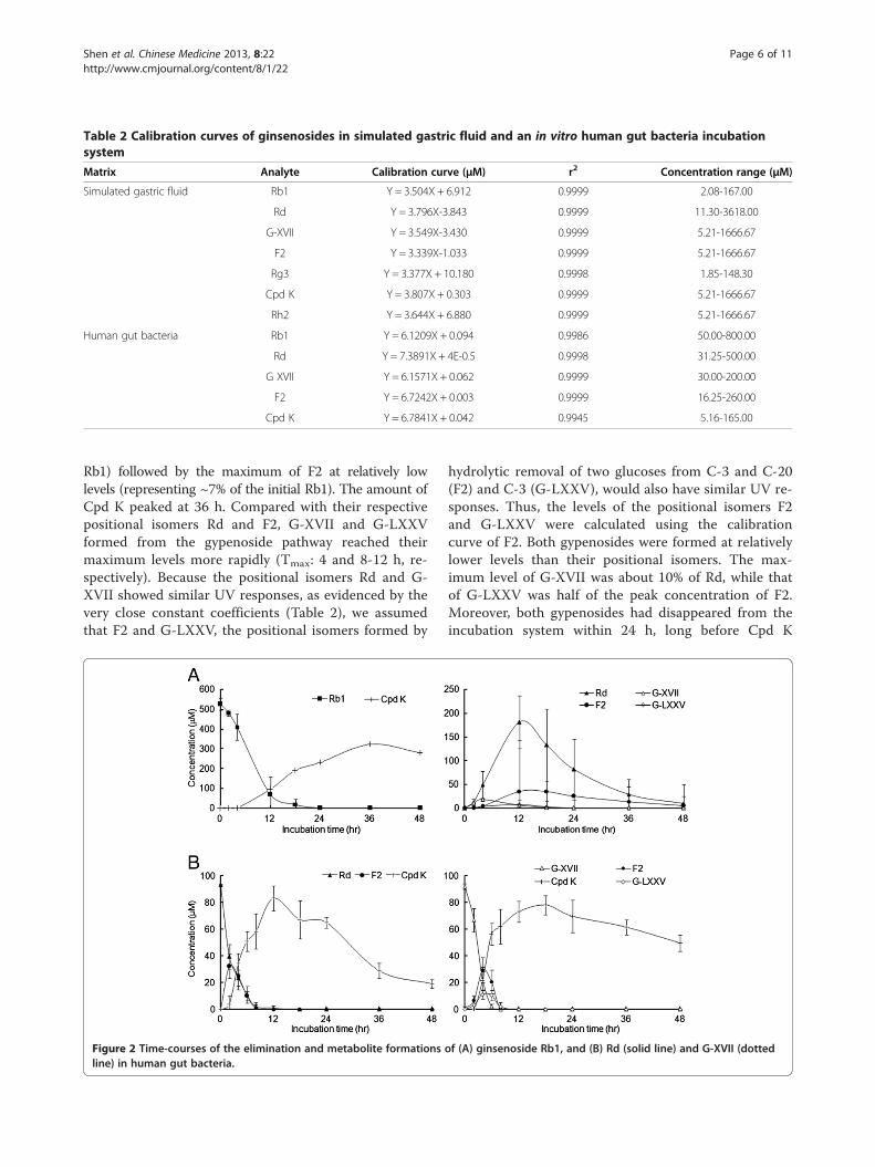
Table 2 Calibration curves of ginsenosides in simulated gastric fluid and an in vitro human gut bacteria incubationsystem
Matrix Analyte Calibration curve (μΜ) r2 Concentration range (μΜ)
Simulated gastric fluid Rb1 Y = 3.504X + 6.912 0.9999 2.08-167.00
Rd Y = 3.796X-3.843 0.9999 11.30-3618.00
G-XVII Y = 3.549X-3.430 0.9999 5.21-1666.67
F2 Y = 3.339X-1.033 0.9999 5.21-1666.67
Rg3 Y = 3.377X + 10.180 0.9998 1.85-148.30
Cpd K Y = 3.807X + 0.303 0.9999 5.21-1666.67
Rh2 Y = 3.644X + 6.880 0.9999 5.21-1666.67
Human gut bacteria Rb1 Y = 6.1209X + 0.094 0.9986 50.00-800.00
Rd Y = 7.3891X + 4E-0.5 0.9998 31.25-500.00
G XVII Y = 6.1571X + 0.062 0.9999 30.00-200.00
F2 Y = 6.7242X + 0.003 0.9999 16.25-260.00
Cpd K Y = 6.7841X + 0.042 0.9945 5.16-165.00
Shen et al. Chinese Medicine 2013, 8:22 Page 6 of 11http://www.cmjournal.org/content/8/1/22
Rb1) followed by the maximum of F2 at relatively lowlevels (representing ~7% of the initial Rb1). The amount ofCpd K peaked at 36 h. Compared with their respectivepositional isomers Rd and F2, G-XVII and G-LXXVformed from the gypenoside pathway reached theirmaximum levels more rapidly (Tmax: 4 and 8-12 h, re-spectively). Because the positional isomers Rd and G-XVII showed similar UV responses, as evidenced by thevery close constant coefficients (Table 2), we assumedthat F2 and G-LXXV, the positional isomers formed by
Figure 2 Time-courses of the elimination and metabolite formationsline) in human gut bacteria.
hydrolytic removal of two glucoses from C-3 and C-20(F2) and C-3 (G-LXXV), would also have similar UV re-sponses. Thus, the levels of the positional isomers F2and G-LXXV were calculated using the calibrationcurve of F2. Both gypenosides were formed at relativelylower levels than their positional isomers. The max-imum level of G-XVII was about 10% of Rd, while thatof G-LXXV was half of the peak concentration of F2.Moreover, both gypenosides had disappeared from theincubation system within 24 h, long before Cpd K
of (A) ginsenoside Rb1, and (B) Rd (solid line) and G-XVII (dotted

Shen et al. Chinese Medicine 2013, 8:22 Page 7 of 11http://www.cmjournal.org/content/8/1/22
reached its maximum level at 36 h, while Rd and F2were still detectable at 48 h. Thus, the gypenoside path-way should be the minor pathway of Rb1 biotransform-ation by human gut bacteria.
Biotransformation of Rd and G-XVII by human gutbacteriaRd and G-XVII were depleted within 8 h by human gutbacteria, and generated F2 and Cpd K when incubatedseparately. The generations of F2 and Cpd K from Rdpeaked at 2 and 12 h, respectively, while those generatedfrom G-XVII reached their maximum levels at 4 and18 h, respectively. The maximum levels of F2 and Cpd Kformed from Rd were comparable to those from G-XVII(about 30 and 80 μM, reflecting 30% and 80% of theoriginal amounts of the respective parent compounds)(Figure 2B).
Figure 3 Ginsenoside Rb1 metabolism and its metabolite formation bdifferent ages (B) and sexes (C).
G-LXXV was also generated by human gut bacteria asevidenced by the retention time and mass spectral pro-file. F2 presented as a major metabolite and the Cmax
was two times higher that of G-LXXV (Figure 2B). There-fore, both the G-XVII→ F2 and G-XVII→G-LXXV path-ways of G-XVII metabolism existed in the human gutbacteria, with the former as the major pathway in vitro.
Rb1 biotransformation by individual human gut bacterialsamplesWhen Rb1 was separately incubated with gut bacterialsamples from 58 individuals aged 18-92 years (30 femalesand 28 males; range, 39.6-75.3 years), great inter-individualvariations were observed in terms of the remaining Rb1and the amounts of metabolites formed via the Rd andgypenoside pathways (Figure 3A). However, when com-pared by age, there were no significant differences in Rb1
y gut bacteria samples from 58 healthy individuals (A) of
Shen et al. Chinese Medicine 2013, 8:22 Page 8 of 11http://www.cmjournal.org/content/8/1/22
elimination and each pathway among individuals aged18-45 years (20 individuals; range, 29.2-47 years), 46-70 years (22 individuals; range, 52.7-65.9 years), and 71-92 years (16 individuals; range, 72.6-85.4 years) (Figure 3B).No sex differences (P values: 0.3 ~ 0.5) were observed inRb1 metabolism and each pathway by human gut bacteria(Figure 3C).
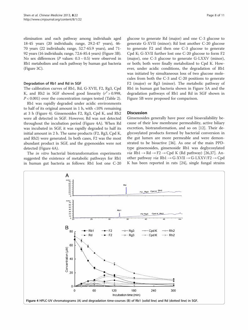
Degradation of Rb1 and Rd in SGFThe calibration curves of Rb1, Rd, G-XVII, F2, Rg3, CpdK, and Rh2 in SGF showed good linearity (r2 > 0.998,P < 0.001) over the concentration ranges tested (Table 2).Rb1 was rapidly degraded under acidic environments
to half of its original amount in 1 h, with <10% remainingat 3 h (Figure 4). Ginsenosides F2, Rg3, Cpd K, and Rh2were all detected in SGF. However, Rd was not detectedthroughout the incubation period (Figure 4A). When Rdwas incubated in SGF, it was rapidly degraded to half itsinitial amount in 2 h. The same products (F2, Rg3, Cpd K,and Rh2) were generated. In both cases, F2 was the mostabundant product in SGF, and the gypenosides were notdetected (Figure 4A).The in vitro bacterial biotransformation experiments
suggested the existence of metabolic pathways for Rb1in human gut bacteria as follows: Rb1 lost one C-20
Figure 4 HPLC-UV chromatograms (A) and degradation time-courses
glucose to generate Rd (major) and one C-3 glucose togenerate G-XVII (minor); Rd lost another C-20 glucoseto generate F2 and then one C-3 glucose to generateCpd K; G-XVII further lost one C-20 glucose to form F2(major), one C-3 glucose to generate G-LXXV (minor),or both; both were finally metabolized to Cpd K. How-ever, under acidic conditions, the degradation of Rb1was initiated by simultaneous loss of two glucose mole-cules from both the C-3 and C-20 positions to generateF2 (major) or Rg3 (minor). The metabolic pathway ofRb1 in human gut bacteria shown in Figure 5A and thedegradation pathways of Rb1 and Rd in SGF shown inFigure 5B were proposed for comparison.
DiscussionGinsenosides generally have poor oral bioavailability be-cause of their low membrane permeability, active biliaryexcretion, biotransformation, and so on [12]. Their de-glycosylated products formed by bacterial conversion inthe gut lumen are more permeable and were demon-strated to be bioactive [36]. As one of the main PPD-type ginsenosides, ginsenoside Rb1 was deglycosylatedvia Rb1→ Rd→ F2→ Cpd K (Rd pathway) [26,37]. An-other pathway via Rb1→G-XVII→G-LXXV/F2→ CpdK has been reported in rats [24], single fungal strains
(B) of Rb1 (solid line) and Rd (dotted line) in SGF.

Figure 5 Pathways of (A) Rb1 biotransformation by human intestinal microflora (→ major pathway; ⤏ minor pathway) and (B) Rb1degradation in SGF.
Shen et al. Chinese Medicine 2013, 8:22 Page 9 of 11http://www.cmjournal.org/content/8/1/22
[20-22], and bacteria such as Fusobacterium sp. and Bifi-dobacterium longum sp. [38,39].In this study with application of both LC-MS and LC-
DAD techniques, G-XVII and three metabolites (Rd, F2,and Cpd K) were identified from the Rd pathway and G-LXXV was tentatively assigned. The existence of both theRd and gypenoside pathways of Rb1 metabolism in humangut bacteria was confirmed. The conversion of Rb1 toCpd K via both G-XVII→G-LXXV and G-XVII→ F2 wasalso evidenced, for the first time, by the observation thatG-XVII produced G-LXXV, F2, and Cpd K, while Rd onlygenerated F2 and Cpd K, in human gut bacteria in vitro.The gypenoside pathway was demonstrated to be the
minor pathway of Rb1 biotransformation in human gutbacteria by comparing the maximum levels of Rd and F2with those of their respective positional isomers G-XVIIand G-LXXV determined from a time-course study. More-over, the maximum levels of the gypenoside products werereached more rapidly than the products of the Rd pathway,indicating that the gypenoside pathway was a transientintermediate process. In contrast, the conversion rate ofRd to Cpd K was faster than that of G-XVII to Cpd K, as
judged by the times required to reach the maximum levelof Cpd K when Rd and G-XVII were separately incubatedwith human gut bacteria. These findings suggested that theremoval of the remote glucose molecule at the C-20 pos-ition by human gut bacteria was faster than that of theglucose molecule from the C-3 position, while furthercleavage of the second glucose from the C-3 position is amuch more difficult process, leading to the formation ofF2 much more rapidly than the formation of G-LXXVfrom G-XVII. Eubacterium sp., Streptococcus sp., and Bifi-dobacterium sp., which were more potent in hydrolyzinggentiobiose than sophorose, metabolized Rb1 to Cpd K viaRd rather than G-XVII [39]. However, Fusobacterium K-60,which hydrolyzed sophorose more potently than gentio-biose, metabolized Rb1 to Cpd K via G-XVII. Bifidobac-teria are probiotics and belong to the predominant gutbacteria, while streptococci and fusobacteria are patho-genic bacteria and are in the minority in healthy humans[40]. This may explain why the Rd pathway was themajor pathway, while the gypenoside pathway was theminor pathway of Rb1 in human gut bacteria fromhealthy volunteers.

Shen et al. Chinese Medicine 2013, 8:22 Page 10 of 11http://www.cmjournal.org/content/8/1/22
In this study, rapid degradation of Rb1 was observedin SGF and its products including F2, Rg3, Cpd K, andRh2 were detected, consistent with the findings ob-tained from previous in vitro [41] and in vivo [42] stud-ies. Rd yielded the same four products in SGF. However,gypenosides were not detected in SGF in both cases, in-dicating that when Rb1 is taken orally, gypenosides mayonly be formed through bacterial biotransformation inthe gut lumen.Great inter-individual variations in Rb1 metabolism by
human gut bacteria were observed with both the gypeno-side and Rd pathways owing to the great individual varia-tions in the species and amounts of the gut bacteria. Thesevariations showed no differences between sexes and ages.Thus far, only the Rd pathway has been reported
in vivo in the human body [25,26]. The minor amountof gypenosides formed by the human gut bacteria mayprovide beneficial effects for the human body. Thus, fur-ther studies are warranted to evaluate the contributionsof these gypenosides to the health-promoting effects ofginsenoside Rb1 in vivo.
ConclusionsRb1 was metabolized to G-XVII, F2 (major) or G-LXXL(minor), and finally Cpd K by human gut bacteria in vitro.
Additional file
Additional file 1: Table S1. Intraday and interday variability for theassays of ginsenoside Rb1, Rd, F2 and compound Kin incubates withhuman gut bacteria.
AbbreviationsDMSO: Dimethyl sulfoxide; PBS: Phosphate-buffered saline; PPD: 20(S)-protopanaxadiol; G-XVII: Gypenoside XVII; G-LXXV: Gypenoside LXXV; CpdK: Compound K; HPLC-UV: High-performance liquid chromatography with aUV detector; MS: Mass spectrometry; ESI: Electrospray ionization; BHI: Brainheart infusion; SGF: Simulated gastric fluid; QC: Quality control; DAD: Diodearray detector; LC/MSD: Liquid chromatograph/mass selective detector;IS: Internal standard; SD: Standard deviation.
Competing interestsThe authors declare that they have no competing interests.
Authors’ contributionsRY conceived the study. JPCL, YTW, and RY designed the study. WIL carriedout the ginsenoside Rb1 biotransformation by pooled and individual humangut bacteria. HS carried out the experiments on metabolism by human gutbacteria and acidic stability. JQR collected the human fecal samples andprepared bacterial suspensions. SLL and JPCL performed the statisticalanalysis. WIL, HS, and RY wrote the manuscript. All authors read andapproved the final manuscript.
AcknowledgementsThis work was financially supported by the Science and TechnologyDevelopment Fund of Macao SAR (Ref. No. 043/2011/A2), the ResearchCommittee of University of Macau (Ref. No. MYRG162(Y3-L2)-ICMS11-YR), andthe National Natural Science Foundation of China (Grant Nos. 81274068 &81202920).
Author details1State Key Laboratory of Quality Research in Chinese Medicine, Institute ofChinese Medical Sciences, University of Macau, Macao, China. 2Departmentof Pharmaceutical Analysis & Metabolomics, Jiangsu Provincial Academy ofTraditional Chinese Medicine, Nanjing, Jiangsu, China. 3Clinical Laboratory,Kiang Wu Hospital, Estrada do Repouso, Macao, China.
Received: 12 July 2013 Accepted: 21 November 2013Published: 23 November 2013
References1. Yang WZ, Ye M, Qiao X, Liu CF, Miao WJ, Bo T, Tao HY, Guo DA: A strategy
for efficient discovery of new natural compounds by integratingorthogonal column chromatography and liquid chromatography/massspectrometry analysis: its application in Panax ginseng, Panaxquinquefolium and Panax notoginseng to characterize 437 potentialnew ginsenosides. Anal Chim Acta 2012, 739:56–66.
2. Shi W, Wang YT, Li J, Zhang HQ, Ding L: Investigation of ginsenosides indifferent parts and ages of Panax ginseng. Food Chem 2007, 102:664–668.
3. Wang CZ, Wu JA, McEntee E, Yuan CS: Saponins composition in AmericanGinseng leaf and berry assayed by high-performance liquid chromatog-raphy. J Agric Food Chem 2006, 6:2261–2266.
4. Xie YY, Luo D, Cheng YJ, Ma JF, Wang YM, Liang QL, Luo GA: Steaming-induced chemical transformations and holistic quality assessment of redGinseng derived from Panax ginseng by means of HPLC-ESI-MS/MSn-based multicomponent quantification fingerprint. J Agric Food Chem2012, 33:8213–8224.
5. Jiang Z, Wang Y, Zhang X, Peng T, Lu Y, Leng J, Xie Q: Preventive andtherapeutic effects of ginsenoside rb1 for neural injury during cerebralinfarction in rats. Am J Chin Med 2013, 2:341–352.
6. Shen L, Xiong Y, Wang DQ, Howles P, Basford JE, Wang J, Xiong YQ, Hui DY,Woods SC, Liu M: Ginsenoside Rb1 reduces fatty liver by activating AMP-activated protein kinase in obese rats. J Lipid Res 2013, 5:1430–1438.
7. Li QY, Chen L, Fu WH, Li ZD, Wang B, Shi XJ, Zhong MK: Ginsenoside Rb1inhibits proliferation and inflammatory responses in rat aortic smoothmuscle cells. J Agric Food Chem 2011, 11:6312–6318.
8. VanDuynhoven J, Vaughan EE, Jacobs DM, Kemperman RA, Van Velzen EJ,Gross G, Roger LC, Possemiers S, Smilde AK, Doré J, Westerhuis JA, Van deWiele T: Metabolic fate of polyphenols in the human superorganism.Proc Natl Acad Sci U S A 2011, 108:4531–4538.
9. Hidalgo M, Oruna-Concha MJ, Kolida S, Walton GE, Kallithraka S, Spencer JP,de Pascual-Teresa S: Metabolism of anthocyanins by human gut micro-flora and their influence on gut bacterial growth. J Agric Food Chem 2012,15:3882–3890.
10. Sánchez-Patán F, Cueva C, Monagas M, Walton GE, Gibson GR, Quintanilla-López JE, Lebrón-Aguilar R, Martín-Álvarez PJ, Moreno-Arribas MV, BartoloméB: In vitro fermentation of a red wine extract by human gut microbiota:changes in microbial groups and formation of phenolic metabolites.J Agric Food Chem 2012, 9:2136–2147.
11. Selma MV, Espín JC, Tomás-Barberán FA: Interaction between phenolicsand gut microbiota: role in human health. J Agric Food Chem 2009,15:6485–6501.
12. Liu H, Yang J, Du F, Gao X, Ma X, Huang Y, Xu F, Niu W, Wang F, Mao Y,Sun Y, Lu T, Liu C, Zhang B, Li C: Absorption and disposition ofginsenosides after oral administration of Panax notoginseng extract torats. Drug Metab Dispos 2009, 37:2290–2298.
13. Wakabayashi C, Hasegawa H, Murata J, Saiki I: In vivo antimetastaticaction of ginseng protopanaxadiol saponins is based on their intestinalbacterial metabolites after oral administration. Oncol Res 1997,9:411–417.
14. Lee GW, Yoo MH, Shin KC, Kim KR, Kim YS, Lee KW, Oh DK: β-Glucosidasefrom Penicillium aculeatum hydrolyzes exo-, 3-O-, and 6-O-β-glucosidesbut not 20-O-β-glucoside and other glycosides of ginsenosides.Appl Microbiol Biotechnol 2013. DOI: 10.1007/s00253-013-4828-7.
15. Lee IK, Kang KA, Lim CM, Kim KC, Kim HS, Kim DH, Kim BJ, Chang WY, ChoiJH, Hyun JW: Compound K, a metabolite of ginseng saponin, inducesmitochondria-dependent and caspase-dependent apoptosis via the gen-eration of reactive oxygen species in human colon cancer cells. Int J MolSci 2011, 11:4916–4931.
16. Niu T, Smith DL, Yang Z, Gao S, Yin T, Jiang ZH, You M, Gibbs RA, PetrosinoJF, Hu M: Bioactivity and bioavailability of ginsenosides are dependent

Shen et al. Chinese Medicine 2013, 8:22 Page 11 of 11http://www.cmjournal.org/content/8/1/22
on the glycosidase activities of the A/J mouse intestinal microbiomedefined by pyrosequencing. Pharm Res 2013, 30:836–846.
17. Hasegawa H: Proof of the mysterious efficacy of ginseng: basic andclinical trials: metabolic activation of ginsenoside: deglycosylation byintestinal bacteria and esterification with fatty acid. J Pharmacol Sci 2004,95:153–157.
18. Hasegawa H, Sung JH, Matsumiya S, Uchiyama M: Main ginseng saponinmetabolites formed by intestinal bacteria. Planta Med 1996, 62:453–457.
19. Hasegawa H, Sung JH, Benno Y: Role of human intestinal prevotella orisin hydrolyzing ginseng saponins. Planta Med 1997, 63:436–440.
20. Chen GT, Song Y, Lu ZQ, Zhang JQ, Huang HL, Wu LJ, Guo DA: Microbialtransformation of ginsenoside Rb1 by Acremonium strictum.Appl Microbiol Biotechnol 2008, 77:1345–1350.
21. Hou JG, Xue JJ, Sun MQ, Wang CY, Liu L, Zhang DL, Lee MR, Gu LJ, WangCL, Wang YB, Zheng Y, Li W, Sung CK: Highly selective microbialtransformation of major ginsenoside Rb1 to gypenoside LXXV by Esteyavermicola CNU120806. J Appl Microbiol 2012, 113:807–814.
22. Zhao X, Gao J, Song C, Fang Q, Wang N, Zhao T, Liu D, Zhou Y: Fungalsensitivity to and enzymatic deglycosylation of ginsenosides.Phytochemistry 2012, 78:65–71.
23. Karikura M, Miyase T, Tanizawa H, Taniyama T, Takino Y: Studies onabsorption, distribution, excretion and metabolism of ginseng saponins: VII:comparison of the decomposition modes of ginsenoside-Rb1 and -Rb2 inthe digestive tract of rats. Biol Pharm Bull 1991, 39:2357–2361.
24. Chen G, Yang M, Song Y, Lu Z, Zhang J, Huang H, Guan S, Wu L, Guo DA:Comparative analysis on microbial and rat metabolism of ginsenosideRb1 by high-performance liquid chromatography coupled with tandemmass spectrometry. Biomed Chromatogr 2008, 22:779–785.
25. Lee J, Lee E, Kim D, Lee J, Yoo J, Koh B: Studies on absorption, distributionand metabolism of ginseng in humans after oral administration.J Ethnopharmacol 2009, 122:143–148.
26. Tawab MA, Bahr U, Karas M, Wurglics M, Schubert-Zsilavecz M: Degradationof ginsenosides in humans after oral administration. Drug Metab Dispos2003, 31:1065–1071.
27. Ye R, Kong X, Yang Q, Zhang Y, Han J, Zhao G: Ginsenoside Rd attenuatesredox imbalance and improves stroke outcome after focal cerebralischemia in aged mice. Neuropharmacology 2011, 61:815–824.
28. Qin R, Zhang J, Li C, Zhang X, Xiong A, Huang F, Yin Z, Li K, Qin W, Chen M,Zhang S, Liang L, Zhang H, Nie H, Ye W: Protective effects of gypenosidesagainst fatty liver disease induced by high fat and cholesterol diet andalcohol in rats. Arch Pharm Res 2012, 35:1241–1250.
29. Zhang GL, Deng JP, Wang BH, Zhao ZW, Li J, Gao L, Liu BL, Xiong JR, Guo XD,Yan ZQ, Gao GD: Gypenosides improve cognitive impairment induced bychronic cerebral hypoperfusion in rats by suppressing oxidative stress andastrocytic activation. Behav Pharmacol 2011, 22:633–644.
30. Pereira JM, Mejia-Ariza R, Ilevbare GA, McGettigan HE, Sriranganathan N,Taylor LS, Davis RM, Edgar KJ: Interplay of degradation, dissolution andstabilization of clarithromycin and its amorphous solid dispersions.Mol Pharm 2013. DOI: 10.1021/mp400441d.
31. Pietta P, Mauri P, Rava A: Hydrolysis of ginsenosides in artificial gastricfluid monitored by HPLC. J Chromatogr 1986, 362:291–297.
32. Miyamoto E, Odashima S, Kitagawa I, Tsuji A: Stability kinetics ofginsenosides in aqueous solution. J Pharm Sci 1984, 73:409–410.
33. Zhou RN, Song YL, Ruan JQ, Wang YT, Yan R: Pharmacokinetic evidenceon contribution of intestinal bacterial conversion to beneficial actions ofastragaloside IV, a marker compound of Astragali Radix, in traditionaloral use of the herb. Drug Metab Pharmacok 2012, 27:586–597.
34. Kim KA, Jung IH, Park SH, Ahn YT, Huh CS, Kim DH: Comparative analysisof the gut microbiota in people with different levels of ginsenoside Rb1degradation to compound K. PLoS One 2013, 8:e62409.
35. Pharmacopoeia Commission of China: Appendix. In Chinese PharmacopoeiaII. Beijing: China Medical Science Press; 2010:85.
36. Hasegawa H, Uchiyama M: Antimetastatic efficacy of orally administeredginsenoside Rb1 in dependence on intestinal bacterial hydrolyzingpotential and significance of treatment with an activebacterial metabolite. Planta Med 1998, 64:696–700.
37. Qian TX, Jiang ZH, Cai ZW: High-performance liquid chromatographycoupled with tandem mass spectrometry applied for metabolic study ofginsenoside Rb1 on rat. Anal Biochem 2006, 352:87–96.
38. Jung IH, Lee JH, Hyun YJ, Kim DH: Metabolism of ginsenoside Rb1 byhuman intestinal microflora and cloning of its metabolizing β-D-
glucosidase from Bifidobacterium longum H-1. Biol Pharm Bull 2012,35:573–581.
39. Bae EA, Park SY, Kim DH: Constitutive beta-glucosidases hydrolyzing gin-senoside Rb1 and Rb2 from human intestinal bacteria. Biol Pharm Bull2000, 23:1481–1485.
40. Claesson MJ, Cusack S, O’Sullivan O, Greene-Diniz R, De Weerd H, FlanneryE, Marchesi JR, Falush D, Dinan T, Fitzgerald G, Stanton C, Van Sinderen D,O’Connor M, Harnedy N, O’Connor K, Henry C, O’Mahony D, Fitzgerald AP,Shanahan F, Twomey C, Hill C, Ross RP, O’Toole PW: Composition, variabil-ity, and temporal stability of the intestinal microbiota of the elderly. ProcNatl Acad Sci U S A 2011, 108:4586–4591.
41. Zhang X, Song F, Cui M, Liu Z, Liu S: Investigation of the hydrolysis ofginsenosides by high performance liquid chromatography-electosprayionization mass spectrometry. Planta Med 2007, 73:1225–1229.
42. Qian TX, Cai ZW: Biotransformation of ginsenosides Rb1, Rg3 and Rh2 inrat gastrointestinal tracts. Chin Med 2010, 5:19.
doi:10.1186/1749-8546-8-22Cite this article as: Shen et al.: Biotransformation of ginsenoside Rb1 viathe gypenoside pathway by human gut bacteria. Chinese Medicine2013 8:22.
Submit your next manuscript to BioMed Centraland take full advantage of:
• Convenient online submission
• Thorough peer review
• No space constraints or color figure charges
• Immediate publication on acceptance
• Inclusion in PubMed, CAS, Scopus and Google Scholar
• Research which is freely available for redistribution
Submit your manuscript at www.biomedcentral.com/submit