research article subtractive cell-selex selection of...
TRANSCRIPT

Research ArticleSubtractive Cell-SELEX Selection of DNA AptamersBinding Specifically and Selectively to HepatocellularCarcinoma Cells with High Metastatic Potential
Hao Chen1 Chun-Hui Yuan2 Yi-Fei Yang2 Chang-Qing Yin1 Qing Guan2
Fu-Bing Wang1 and Jian-Cheng Tu1
1Department of Laboratory Medicine and Center for Gene Diagnosis Zhongnan Hospital of Wuhan University169 Donghu Road Wuchang District Wuhan 430071 China2Department of Immunology School of Basic Medical Sciences Wuhan University 185 Donghu Road Wuchang DistrictWuhan 430071 China
Correspondence should be addressed to Fu-Bing Wang wfb20042002sinacom and Jian-Cheng Tu jian 1999yahoocom
Received 17 November 2015 Revised 25 February 2016 Accepted 7 March 2016
Academic Editor Melchiorre Cervello
Copyright copy 2016 Hao Chen et al This is an open access article distributed under the Creative Commons Attribution Licensewhich permits unrestricted use distribution and reproduction in any medium provided the original work is properly cited
Relapse andmetastasis are two key risk factors of hepatocellular carcinoma (HCC) prognosis thus it is emergent to develop an earlyand accurate detection method for prognostic evaluation of HCC after surgery In this study we sought to acquire oligonucleotideDNA aptamers that specifically bind to HCC cells with highmetastatic potential TwoHCC cell lines derived from the same geneticbackground but with differentmetastatic potential were employedMHCC97L (lowmetastatic properties) as subtractive targets andHCCLM9 (highmetastatic properties) as screening targets Tomimic a fluid combining environment initial DNA aptamers librarywas firstly labelled with magnetic nanoparticles using biotin-streptavidin system and then applied for aptamers selectionThrough10-round selectionwith subtractive Cell-SELEX six aptamers LY-1 LY-13 LY-46 LY-32 LY-2745 and LY-743 display high affinityto HCCLM9 cells and do not bind to MHCC97L cells as well as other tumor cell lines including breast cancer lung cancer colonadenocarcinoma gastric cancer and cervical cancer suggesting high specificity for HCCLM9 cells Thus the aptamers generatedhere will provide solid basis for identifying new diagnostic targets to detect HCC metastasis and also may provide valuable cluesfor developing new targeted therapeutics
1 Introduction
Hepatocellular carcinoma (HCC) is the second most com-mon cause of cancer-related death worldwide estimated tobe responsible for nearly 746000 deaths in 2012 (91 of thetotal) and is a formidable public health challenge of Chinawhere 50 of the estimated 782000 new cancer cases world-wide occurred [1 2] In recent decades great advancementshave been achieved in the development of therapeutics forHCC besides hepatic resection as a mainstay of HCC treat-ment local ablative therapies have greatly improved patientsurvival when HCC is diagnosed at early stages and of themradiofrequency ablation (RFA) is considered the referencestandard [3ndash5] However according to the data presentedby WHO in 2012 (httpglobocaniarcfrDefaultaspx) the
prognosis for hepatocellular carcinoma is still very poor(overall ratio of mortality to incidence is 095) [2 6 7]
As the two pivotal prognostic factors of HCC postopera-tively relapse andmetastasis significantly shorted the survivaltime of surgically treated patients [8ndash10] Currently regularreexamination of serum alpha fetoprotein (AFP) level orcontrast enhanced ultrasound (CEUS) still represents thetwo preferred diagnostic strategies in clinical examination todetect postoperatively relapse and metastasis [11] Howeverwith regard to early diagnosis of HCC the positive rate ofAFP is only 60ndash80 and often resulted in a false-positiveresult during pregnancy as well as for active liver diseaseembryonic tumor and certain gastrointestinal tumors [12]CEUS has been applied for more than ten years and hasproved to be of great value in the management of HCC [13]
Hindawi Publishing CorporationBioMed Research InternationalVolume 2016 Article ID 5735869 9 pageshttpdxdoiorg10115520165735869
2 BioMed Research International
In most of the cases HCC always shows earlier enhancementthan the surrounding liver tissue the detection rate in lesionslarger than 21 cm is up to 92ndash100 [14 15] However whenlesions are less than 10 cm the detection rate is lower than67 and apparently CEUS has a relatively low ability todetermine the smaller lesions of HCC in an early stage [16]Thus the identification of new tumor biomarkers involved inmetastasis and recurrence is urgent in surveillance for HCC
Since potential biomarkers can encompass various typesof molecules ranging from glycolipids to proteins thus thestrategy of Systematic Evolution of Ligands through Expo-nential Enrichment (SELEX) is ideally suited for the creationof biomarker as aptamers generated by SELEX are capableof selective binding to any class of molecules [17] Aptamersare synthetic single-stranded oligonucleotides DNA or RNAthat could fold into unique structures including hairpin fakefestival convex ring and G-tetramer to bind specifically totheir target molecules [18] Compared with antibodies theypossess several key advantages smallermolecular weight (theaveragemolecular weight of aDNA aptamer is about 25 kDa)without immunogenicity greater specificity and affinity andbeing easier to be economically produced and modified withmultiple chemical molecules [18 19] Thus aptamers havebeen widely used in cell imaging [20] clinical diagnosis andtargeted therapeutics [21ndash23]
Cell-SELEX derives from traditional SELEX process anduses whole living cells as target [24] With the help of thistechnology aptamers can be obtained even without priorknowledge of potential target molecules of cancer cells [25]More importantly Cell-SELEX-based selection of aptamersagainst cancer cells has been reported in different cancersincluding leukemia lung cancer colon cancer glioma andovarian cancer as well as in HCC [25ndash28] However noinformation was given on the ability of aptamer to differen-tiate tumor cells with metastatic potential in HCC In thepresent study two HCC cell lines derived from the samegenetic background but with different metastatic potentialwere employed MHCC97L (low metastatic properties) ascounterparts and HCCLM9 (high metastatic properties) asscreening targets Initial DNA aptamers library was labelledwith magnetic nanoparticles and then applied for aptamersselection in a fluid compartment Six aptamers selected by theCell-SELEXdisplay high affinity toHCCLM9 cells and do notbind toMHCC97L cells and other tumor cell lines suggestingspecificity for HCCLM9 cells Thus the aptamers generatedhere will provide solid basis for identifying new diagnostictargets to detect HCC metastasis
2 Materials and Methods
21 Cell Lines and Reagents MHCC97L cell and HCCLM9cell were obtained from research center of Zhongnan Hos-pital Wuhan University as we previously described [29]and cultured in RPMI1640 (Gibco) containing 10 FBS(Gibco) and 100 unitsmL penicillin-streptomycin (Bey-otime Shanghai China) Other cell lines were maintained atour laboratory Salmon sperm DNA yeast tRNA and BSAwere purchased from Roche (F Hoffmann-La Roche LtdUSA) Streptavidin-coated magnetic nanoparticles M-280
(Dynabeads) were used for modifying biotin-labelled single-stranded DNA (ssDNA) aptamers pool
22 Random DNA Library and Primers An initial ssDNAaptamer library (51015840-ATCCAGAGTGACGCAGCA-N40-T-GGACACGGT GGCTTAGT-31015840) consisting of 40-base ran-domized sequences was synthesized (Invitrogen ShanghaiChina) where N represents a randomized nucleotide ofeither A G C or T Forward primer 1 (51015840-ATCCAG-AGTGACGCAGCA-31015840) and biotin-labelled reverse primer2 (51015840-ACTAA GCCACCGTGTCCA-31015840) were used for PCRamplification of the DNA library or to separate the single-strandedDNAby streptavidin-coatedmagnetic particlesTheFAM-labelled forward primer 1 (SBS Genetech Co Ltd)was used to monitor progress of selection by flow cytometry(Beckman Coulter USA)
23 Subtractive Cell-SELEX Procedure Subtractive Cell-SELEX procedure was performed as we described previously[29] Briefly initial ssDNA aptamers library (8 nmol) wasfirst dissolved in 1mL precooled binding buffer (PBS 1MMgCl
201mgmL yeast tRNA 1mgmL BSA 01mgmL and
Salmon sperm DNA 100 120583gmL) and then incubated withadherent HCCLM9 cells for 1 h in an orbital shaker Afterincubation the cells were washed 3 times with binding bufferto remove unbound ssDNA aptamers Adherent HCCLM9cells were scraped off and resuspended in 500120583L DNase-free deionized water Cell suspension was then heated at100∘C for 5min and centrifuged at 12000 g for 5min Super-natant containing eluted ssDNA aptamers was collected andthen amplified by PCR using biotin-labelled primer Thebiotin-labelled ssDNA aptamers were then incubated withstreptavidin-coated magnetic nanoparticles (Dynabeads M-280 Streptavidin Invitrogen) and used for the next round ofselection
From the fourth round of selection the selected ssDNAaptamers pool was firstly incubated with subtractiveMHCC97L cells to perform subtractive selection and thusfiltered out ssDNA aptamers that may bind to subtractiveMHCC97L cells The unbounded aptamers pool wasspecifically targeted to HCCLM9 cells and then incubatedwith HCCLM9 cells for positive selection Furthermorethe target cell number and the concentration of ssDNAaptamers pool were gradually reduced with the selectiveround proceeding to 10 cycles
24 The Binding Ability of Each Round of Enriched ssDNAAptamers Pool FAM-labelled ssDNA aptamers pools of 3 57 and 9 rounds were incubated with target cells HCCLM9or subtractive cells MHCC97L (1 times 106 for each) in 500120583Lbinding buffer on ice for 30min Cells werewashed twice afterincubation and the fluorescence intensity was determinedby flow cytometry The FAM-labelled control aptamer NK8(bound toMycobacterium tuberculosis) was used as a negativecontrol [29 30]
25 Clones Selection and Sequencing The ssDNA aptamerspool of 9 rounds was amplified by PCR to obtain
BioMed Research International 3
double-stranded DNAs (dsDNAs) using unmodifiedprimers and then cloned into a T vector (Invitrogen) Therecombinant plasmidwas then transformed intoE coliDH5120572and randomly selected 50 clones using blue-white selectionThe selected 50 clones were sequenced by Invitrogen CoLtd (Shanghai China) and designated as LY1 to LY50
26 Sequence Alignments and Secondary Structures Analysis ofAptamers DNAMAN software version 60 (Lynnon BiosoftCA USA) was used for sequence analysis and alignmentsRNA structure (version 45 University of Rochester MedicalCenter) and MEME online analysis software version 4102(httpmeme-suiteorgtoolsmeme) were used to estimatethe secondary structures of sequenced aptamers
27 Fluorescence Imaging of the Selected Aptamers Bound toHCCLM9 Cells The FAM-labelled aptamers were synthe-sized by SBSGenetechCo Ltd (Shanghai China) HCCLM9cells were cultured in chamber slides overnight The FAM-labelled individual aptamer was incubated with cell mono-layer in chamber slides in binding buffer on 4∘C for 30minAfter washing 3 times with PBS the cells were imaged withOlympus BX51 fluorescence microscope (Olympus TokyoJapan)
28 Determination of the Dissociation Constants (119870119889) of
Individual ssDNA Aptamers To determine the 119870119889value of
the six selected aptamers HCCLM9 cells (1 times 106) were incu-bated with various concentrations of FAM-labelled aptamersin binding buffer at 4∘C for 30min and the fluorescenceintensity was determined by flow cytometry The meanfluorescence intensity (MFI) of negative control aptamerNK8was subtracted from that of each aptamer with the targetcells to determine the specific binding of each aptamerThenthe equilibrium dissociation constants (119870
119889) of each aptamer
were determined by nonlinear regression for one-site bindingaccording to the equation 119884 = 119861max times 119883(119870119889 + 119883) usingGraphPad Prism version 50 (GraphPad Software Inc)
29 Specificity Analysis of Selected Aptamers To determinethe cell specificity of the selected aptamers human cancercell lines including HCC cell lines MHCC97L HepG2 andHuh-7 breast cancer cell line MDA-MB-231 lung cancer cellline H1299 colon adenocarcinoma cell line SW48 gastriccancer cell line MGC803 and cervical cancer cell line HeLawere used to test the specific binding affinity with fluorescentmicrocopy assay
3 Results
31 Aptamers Selection and Binding Affinity Analysis Sub-tractive Cell-SELEX was performed using HCC cell lineHCCLM9 cells (with high metastatic properties) as the targetand MHCC97L cells (low metastatic properties) as subtrac-tive target respectively for the selection ofmetastatic-specificaptamers HCCLM9 andMHCC97Lwere differentiated fromMHCC97 cells as shown in our previous studies [29 31]In order to get aptamers with high specificity 10 rounds of
selection were performed And with increasing rounds ofenrichment the number of target cells ssDNAconcentrationand the incubation time were gradually decreased whilethe number of washing times was increased to reinforce theselective pressure In the selection procedure we amplifiedthe aptamer pools of 2 4 6 8 9 and 10 cycles andanalyzed them with agarose gel electrophoresis With theselection cycles increased the gray value of correspondingpool was gradually increased in the same PCR cycle andwas the highest in 9 cycles In 10 cycles of selection thecorresponding gray value was decreased compared with 9cycles thus we speculated that aptamers of 9 cycles wereoptimal and included ssDNA aptamers that specifically bindto HCCLM9 cells Simultaneously we compared minimumPCR amplification cycles of each round pool the minimumPCR amplification cycles in 9 rounds were 8 significantlylower than other rounds these results further confirmed that9 rounds of enrichment were optimal
Next we labelled these selected rounds of aptamers withFAM and then incubated them with HCCLM9 cells andMHCC97L cells to examine the specific binding ability usingflow cytometry Relative fluorescence intensity was calculatedas (119865aptamer minus 119865cell)(119865control minus 119865cell) With increasing selectiverounds the relative fluorescence intensity of aptamer poolbound on HCCLM9 cells gradually increased and reachedthe highest value in 9 rounds while the relative fluorescenceintensity of MHCC97L cells showed no discernible change(Figure 1) Therefore it was evident that the pool of ssDNAaptamers in 9 cycles has preferential and specific binding toHCCLM9 cells
32 Selection Sequencing and Structure Prediction ofAptamers Then the ssDNA aptamers pool in 9 cycleswas amplified into double strand and cloned into T vectorand transformed into E coli 5120572 Next fifty clones wererandomly selected using blue-white selection and subjectedto sequencing analysis Among these 50 clones 10 cloneswere discarded with no or multiple sequences detected 23clones yielding the same sequence are marked as LY-1 9clones yielding the same sequence are marked as LY-13 2clones yielding the same sequence are marked as LY-743another 2 clones yielding the same sequence are marked asLY-2745 and the rest 2 clones each yielding one sequenceare marked as LY-32 and LY-46 (Figure 2(a))
According to the sequencing result we further analyzedthe sequence homology and structure prediction of thesesix selected aptamers The greatest percentage of homologyexisted between LY-32 and LY-46 the second was LY-1 andLY-13 (Figure 2(b)) Aptamers that are contained withinhomologous groups are likely to have been strongly prefer-entially enriched during the selection process All of the sixaptamers showed a tendency towards G-richness T-richnessor both (Figure 2(a))The secondary-structural analysis of thesix chosen aptamers was shown in Figure 2(c)
33 Specific Binding Affinity of the Six Selected AptamersTo further confirm the specific binding affinity of thesesix selected aptamers to HCCLM9 cells we firstly labelled
4 BioMed Research International
Control R2 R4
R6 R8 R9 R10
MHCC97LHCCLM9
MHCC97LHCCLM9
MHCC97LHCCLM9
MHCC97LHCCLM9
0
100
200
300
400
Cou
nt
0
100
200
300
400
Cou
nt
0
100
200
300
400
Cou
nt
104
105
106
107
103
FAM-aptamers library10
410
510
610
710
3
FAM-aptamers library10
410
510
610
710
3
FAM-aptamers library
0
100
200
300
400
Cou
nt
104
105
106
107
103
FAM-aptamers library10
410
510
610
710
3
FAM-aptamers library
0
100
200
300
400
Cou
nt
0
100
200
300
400
Cou
nt10
410
510
610
710
3
FAM-aptamers library10
410
510
610
710
3
FAM-aptamers library
0
100
200
300
400
Cou
nt
(a)
MHCC97LHCCLM9
0
25
50
75
100
Relat
ive fl
uore
scen
ce in
tens
ity
Control R10R8 R9R4R2 R6Aptamer
(b)
Figure 1 (a) The specific binding ability of aptamers library in selected rounds with HCCLM9 (target cells) and MHCC97L (subtractivecells) was analyzed by flow cytometry With increasing rounds of enrichment significant increases in fluorescence intensity were detectedon HCCLM9 cells but not on MHCC97L and reached the peak at the ninth round (b) Fluorescence shift was calculated using the equation(119865aptamer minus 119865cell)(119865library minus 119865cell) where 119865aptamer 119865library and 119865cell refer to the fluorescence of the selected aptamers pool initial library and thecell background respectively
aptamers with FAM at the 51015840 end And then their bindingaffinity to HCCLM9 cells was evaluated with fluorescentmicrocopy assayThepercentage of the cells with fluorescenceabove the set threshold was used to evaluate the binding
capacity of the aptamer to the cells [32] All six aptamers weresignificantly bound to the target HCCLM9 cells comparedto the ssDNA library particularly LY-1 and LY-13 had thehighest binding capacity (Figure 3(a)) Moreover all six
BioMed Research International 5
73LY-174LY-1375LY-74375LY-274575LY-3275LY-46
Consensus a
A
A
A
A
A
A
t
T
T
T
T
T
T
c
C
C
C
C
C
C
c
C
C
C
C
C
C
a
A
A
A
A
A
A
g
G
G
G
G
G
G
a
A
A
A
A
A
A
g
G
G
G
G
G
G
t
T
T
T
T
T
T
g
G
G
G
G
G
G
a
A
A
A
A
A
A
c
C
C
C
C
C
C
g
G
G
G
G
G
G
c
C
C
C
C
C
C
a
A
A
A
A
A
A
g
G
G
G
G
G
G
c
C
C
C
C
C
C
A
A
A
A
A
T
G
C
T
T
A
C
A
G
G
G
C
G
A
A
C
G
G
G
G
T
G
G
G
G
G
G
G
T
G
A
A
G
T
G
G
T
T
C
T
G
G
T
T
T
A
G
G
G
G
G
G
T
T
G
G
G
G
T
T
G
G
G
G
T
T
T
T
G
T
T
T
G
G
T
G
C
C
T
G
T
C
G
G
T
C
G
A
G
G
A
G
G
G
T
T
G
G
G
G
C
C
G
G
A
G
G
G
C
C
T
G
G
G
T
A
G
G
T
T
G
T
G
G
T
G
G
G
C
G
G
T
T
T
G
T
T
T
C
T
G
C
C
A
T
C
A
G
T
T
T
C
T
T
T
T
A
G
G
G
G
G
A
C
T
T
G
T
T
T
G
G
T
T
C
G
G
G
G
T
G
G
G
G
T
T
G
G
A
G
T
G
G
G
G
T
T
T
T
T
G
C
T
G
C
C
G
G
G
T
G
G
C
T
G
T
G
G
T
C
T
T
G
G
T
G
G
T
A
C
C
C
T
G
C
T
G
G
t
T
T
T
T
T
T
g
G
G
G
G
G
G
g
G
G
G
G
G
G
a
A
A
A
A
A
A
c
C
C
C
C
C
C
a
A
A
A
A
A
A
c
C
C
C
C
C
C
g
G
G
G
G
G
G
g
G
G
G
G
G
G
t
T
T
T
T
T
T
g
G
G
G
G
G
G
g
G
G
G
G
G
G
c
C
C
C
C
C
C
t
T
T
T
T
T
T
t
T
T
T
T
T
T
a
A
A
A
A
A
A
g
G
G
G
G
G
G
(a)
LY-1 LY-13 LY-743 LY-2745 LY-32 LY-46LY-1 100LY-13 8360 100LY-743 6720 6720 100LY-2745 7460 7460 7310 100LY-32 6870 6870 6270 6870 100LY-46 6570 6270 6420 6870 8660 100
Homology matrix of 6 sequences
(b)
G
T TT
T T
A
A C
G
GG
G A
A
CC
C
C CC 50
70
40
30
G G GGGGGG
GA
A
C
T
T T
T
TT
TT
TT
T
T
T
T
T
60
T
GG
GA
A
AA A
A
AA
GG
GG
GG G
G
GG
70G
GG
GG
10
5020
AC AC
C
CC
C C
CC
C
CC
GG
GGCG
GG
60
10A
A
A
A
A
A
A
AA
A
G
G
G
G
G G G
G G
G G
G
G G G G GGG
G
GG
G G G
G
G
GG
C
CC
C C
C
C
C C
C
C
C
T
T
T
T T TT
T
TT T
T T TT
TT
T
50
T70
40
30GGG
G G G G
GC
CC
C
C
C
CC
C
C
A
AA A
AA
A
AA
A
AT T
TT T
T
TT T T
T
T
T T
TT
TT
T
T
TT
TTTT
G GG
GG
GG
G
G G G G
GG G
G
G
G
GG
20 40
10 50
70
6060
T
T
T T30
T
T20
CC
CC
10
C
G
G
G
G G G G G
G G G
G GG
GG
GGG
G
T
T T T
T
T
T TT
TT
T
T
TT
T
A
A 40
40
30
50C
CC
C
C C
C C
C
CC
C
C
T
T T
T
T
T
T
T
T T60
70
50T
GGG
G G
G GG
GG
G
G
G GG G G
G G GG
G GG G
G G
G
GG
GG
G
G
G
AA
A
A
AA
A
AA
A
A10
A
30
20 4060
20
10
70
A
A
A
G
GG
GG
G
G
G
G
G GG
G G G
G
G
G
G
GG
G
GA
AA
AA
CC
C
TT
T TT
T
GG
G G
G
GG
G
T
T
T T
TT
TT
T
TTA
A
A
A
A
AA
C C CC
CC
C
C
CC
C
C
AA
A
C
GGG
20
30
G
LY-1 LY-13 LY-743
LY-2745 LY-32 LY-46
T T TAG
(c)
Figure 2 The homology analysis and secondary structure prediction of six aptamers (a) Sequence alignment of six aptamers all of thesix aptamers showed a tendency towards G-richness T-richness or both (b) Homology analysis of six aptamers the greatest percentageof homology existed between LY-32 and LY-46 the second was LY-1 and LY-13 (c) Secondary structure prediction of six aptamers by RNAstructure software
aptamers displayed high binding affinity to HCCLM9 cellswith 119870
119889values in the tiny range from 1673 to 3697 nM
(Figure 3(b) Table 1) and LY-1 exhibited the highest bindingaffinity with a 119870
119889value of 1673 plusmn 302 nM
Next to test the binding specificity of these six aptamersdifferent HCC cells (MHCC97L Huh-7 and HepG2) humanlung cancer cell line H1299 human colon adenocarcinomacell line SW48 human gastric cancer cell line MGC803
human cervical cancer cell line HeLa human breast cancercell line MDA-MB-231 and human peripheral WBC wereused As concluded in Table 2 all six aptamers exhibited spe-cific binding affinity to HCCLM9 cells to different extent LY-1 and LY-13 also recognized other HCC cell lines includingMHCC97L Huh-7 and HepG2 cells while the rest aptamersonly recognized one kind of the other HCC cell linesbeside HCCLM9 cells and control aptamer NK8 showed no
6 BioMed Research International
ssDNA library
LY-1
LY-46LY-743LY-32
LY-2745LY-13
(a)
0
5
10
15
Mea
n flu
ores
cenc
e int
ensit
y
200 400 600 8000Aptamer LY-1 concentration (nM)
600 800200 4000Aptamer LY-46 concentration (nM)
0
5
10
15
Mea
n flu
ores
cenc
e int
ensit
y
2625 plusmn 398nM1673 plusmn 302nM Kd =Kd =
(b)
Figure 3The specific binding affinity of these six selected aptamers to HCCLM9 cells (a)The fluorescence imaging of six selected aptamersand initial ssDNA library control bound to HCCLM9 cells Compared with the control group all six selected aptamers were obviously boundto the membrane of target HCCLM9 cells particularly LY-1 and LY-13 had the highest binding capacity (b) Binding curve of aptamers LY-1and LY-46 with target HCCLM9 cells Cells were incubated with increasing concentrations of FAM-labelled aptamers and the fluorescenceintensity (MFI) was detected by flow cytometry After subtracting the MFI of negative aptamer control (NK8) the MFI of FAM-aptamerbound target cells was used to calculate the119870
119889value of each aptamer
detectable binding affinity to any kinds of cells Howeverthese six aptamers exhibited no detectable recognition of anyother tumor cell lines and normal white blood cells (WBC)revealing that the six aptamers specifically targetedmetastaticHCC cells with high affinity especially LY-1 and LY-13
4 Discussion
When developing new molecular probes for cancers Cell-SELEX has advantages over traditional protein-SELEX as itcould be fulfilled just depending on expressive difference ofcell surface molecules between target cells and control cellseven without the need to figure out the detailed molecularcharacteristics of target cells [32] More importantly surfacemolecules of target cells can maintain native folding and
Table 1 119870119889s of six selected aptamers of the HCCLM9 cells
Aptamer 119870119889value (nM)
LY-1 1673 plusmn 302LY-13 1856 plusmn 283LY-2745 3036 plusmn 345LY-32 2457 plusmn 444LY-743 3697 plusmn 463LY-46 2625 plusmn 398
glycosylation states and an improved specificity can furtherbe achieved by performing an additional subtractive step [25]The targetmolecules of aptamers selected by subtractive Cell-SELEX may be the different compositions between tumor
BioMed Research International 7
Table 2 The binding specificity of six aptamers to different cell lines
Cell lines AptamersLY-1 LY-13 LY-2745 LY-32 LY-743 LY-46 Control
HCCLM9 ++++ ++++ ++ ++ + + minus
MHCC97L + + minus + minus minus minus
Huh7 + + minus minus + minus minus
HepG2 + + + minus minus + minus
H1299 minus minus minus minus minus minus minus
SW480 minus minus minus minus minus minus minus
MGC803 minus minus minus minus minus minus minus
HeLa minus minus minus minus minus minus minus
MDA-MB-231 minus minus minus minus minus minus minus
WBC minus minus minus minus minus minus minus
cells and normal counterparts [33 34] Thus studies focusedon these different compositions may provide valuable cluesfor the development of new tumor-specific biomarkers ortherapeutic targets
In recent years subtractive Cell-SELEX has been widelyapplied in identifying new tumor biomarker In gastriccancer Cao et al [35] successfully selected ssDNA aptamercy-apt 20 by using human gastric cancer AGS cells as targetcells and normal gastric epithelium GES-1 cells as subtractivecells In clinical reexamination cy-apt 20 further showedgreat diagnostic and therapeutic potential of gastric cancerShangguan et al [36] developed 6 Cell-SELEX-generatedaptamers against neoplastic cells among these aptamerssgd5 recognized only its target Toledo cells while sgc3sgd2 sgd3 sgc4 and sgc8 selected from a T-cell acuteleukemia (T-ALL) cell line CCRF-CEM cells identified allof the cultured T-ALL cell lines Furthermore sgc8 sgc3and sgd3 showed good selectivity toward T-ALL cells andneoplastic cells in patient samples while almost not bindingto normal hematopoietic cells or lymphoma and myelomacells Importantly by using differential Cell-SELEX Cerchiaet al developed five aptamers (GL36 GL35 GL44 GL43 andGL21) which specifically distinguished U87MG glioma cellsfrom the less malignant cell line T98G and other nonrelatedcancer cell types [28]
Metastasis is themajor obstacle that resulted in the failuretreatment and recurrence of HCC thus to identify newtumor biomarkers with great potential to reflect HCC-relatedmetastasis has been a hot research point in recent years[37ndash39] Gkretsi and Bogdanos [8] firstly demonstrated thatmigfilin promoted HCC metastasis in vitro and may serveas a potential therapeutic target Aptamer is a new kind ofbiomarkers which has been widely used in diagnosis of mul-tiple cancers [40ndash42] Kashefi-Kheyrabadi et al [43] and Sunet al [44 45] coupled aptamer TLS11a with electrochemicalbiosensor which effectively differentiated HCC HepG2 cellsfrom lung cancer or prostate cancer cells and provided anew target for early diagnosis While most aptamers usedHepG2 cells as selective target and normal liver cells assubtractive cells thus these aptamers could be only used forearly diagnosis and showed no help in indicating metastasisof HCC [25] Herein we used high metastatic HCCLM9
cells as the target cells and the less metastatic MHCC97Lcells as the negative cells to perform subtractive Cell-SELEXunder strict selection conditions and thereby generated apanel of six aptamers that can specifically bind to HCCcells with high metastatic potential It is worth noting thatHCCLM9 cell linewas derived fromMHCC97L cells but withhighmetastatic potential the different compositions betweenthese two cell lines may only relate to metastasis [46 47]and thus aptamers generated from these two cell lines mayspecifically recognize metastatic-related surface moleculesexpressed on HCC cells and could be potential biomarkersto predict metastasis of HCC
In aptamers selection procedure we randomly selected 50clones of the nine rounds of ssDNA aptamers pool Aptamerssequencing and alignments analysis revealed that five mutualmotifs existed in these selected aptamers and continuous G-rich motif abundantly existed in our six selected aptamerswhich showed high binding affinity to HCCLM9 cells Thisresult suggested that aptamers in our initial ssDNA pool werespecifically enriched by subtractive Cell-SELEX technique[48] These six aptamers will further be subdivided into fourfamilies according to mutual motifs existing in secondarystructure analysis and continuous G-rich motif form stem-loops in these aptamers Next we designated these aptamersas LY-1 LY-13 LY-46 LY-32 LY-2745 and LY-743 andfurther labelled these six aptamers with FAM to examinethe specific binding ability of individual aptamer to lessmetastatic HCC cells (MHCC97L HepG2 and Huh-7) ordifferent tumor cell lines including breast cancer lung cancercolon adenocarcinoma gastric cancer and cervical cancerThough the 119870
119889values of six selected aptamers may be
higher than the aptamer reported by other researchers [3249] the six selected aptamers showed high specificity andsensitivity as potential molecular probes And no matterwhether aptamers aremodified with fluorophore ormagneticnanoparticle the specific binding ability of aptamers to highmetastatic HCC cells was not changed this may hint thatthese aptamers could be further conjugated with new typesof luminescent or imaging materials and thus be developedinto promising molecular probes for early prediction of HCCmetastasis
8 BioMed Research International
Competing Interests
The authors declare that there are no competing interestsregarding the publication of this paper
Authorsrsquo Contributions
Hao Chen and Chun-Hui Yuan contributed equally to thiswork
Acknowledgments
This work was supported by National Natural Science Foun-dation of China (no 81371897) and research grants funded byIndependent Research Fund Program of Wuhan University(no 2042014kf0241) This work was also funded by ldquo351Talent Project (Luojia Young Scholars)rdquo of Wuhan Universityand National Program on Key Basic Research Project (973Program no 2012CB720600-05)
References
[1] J Ferlay I Soerjomataram R Dikshit et al ldquoCancer incidenceand mortality worldwide sources methods and major patternsin GLOBOCAN 2012rdquo International Journal of Cancer vol 136no 5 pp E359ndashE386 2015
[2] M J Song and S H Bae ldquoNewer treatments for advancedhepatocellular carcinomardquo Korean Journal of Internal Medicinevol 29 no 2 pp 149ndash155 2014
[3] A Cucchetti F Piscaglia M Cescon G Ercolani and A DPinna ldquoSystematic review of surgical resection vs radiofre-quency ablation for hepatocellular carcinomardquoWorld Journal ofGastroenterology vol 19 no 26 pp 4106ndash4118 2013
[4] T Kamiyama K Nakanishi H Yokoo et al ldquoPerioperativemanagement of hepatic resection toward zero mortality andmorbidity analysis of 793 consecutive cases in a single institu-tionrdquo Journal of the American College of Surgeons vol 211 no 4pp 443ndash449 2010
[5] A Cucchetti M Zanello M Cescon et al ldquoImproved diag-nostic imaging and interventional therapies prolong survivalafter resection for hepatocellular carcinoma in cirrhosis theuniversity of Bologna experience over 10 yearsrdquo Annals ofSurgical Oncology vol 18 no 6 pp 1630ndash1637 2011
[6] Y-P Wang and D-X Tang ldquoExpression of yes-associated pro-tein in liver cancer and its correlation with clinicopathologicalfeatures and prognosis of liver cancer patientsrdquo InternationalJournal of Clinical and Experimental Medicine vol 8 no 1 pp1080ndash1086 2015
[7] M Lempinen I Lyytinen A Nordin et al ldquoPrognostic value ofserum MMP-8 -9 and TIMP-1 in patients with hepatocellularcarcinomardquoAnnals of Medicine vol 45 no 7 pp 482ndash487 2013
[8] V Gkretsi and D P Bogdanos ldquoExperimental evidence ofMigfilin as a new therapeutic target of hepatocellular carcinomametastasisrdquo Experimental Cell Research vol 334 no 2 pp 219ndash227 2015
[9] L-Y Yang W-Q Lu G-W Huang andWWang ldquoCorrelationbetween CD105 expression and postoperative recurrence andmetastasis of hepatocellular carcinomardquo BMC Cancer vol 6article 110 2006
[10] D-P Ou L-Y Yang G-W Huang Y-M Tao X Ding and Z-G Chang ldquoClinical analysis of the risk factors for recurrence
of HCC and its relationship with HBVrdquo World Journal ofGastroenterology vol 11 no 14 pp 2061ndash2066 2005
[11] L Wang M Yao Z Dong Y Zhang and D Yao ldquoCirculatingspecific biomarkers in diagnosis of hepatocellular carcinomaand its metastasis monitoringrdquo Tumor Biology vol 35 no 1 pp9ndash20 2014
[12] Y J ZhaoQ Ju andGC Li ldquoTumormarkers for hepatocellularcarcinomardquo Molecular and Clinical Oncology vol 1 no 4 pp593ndash598 2013
[13] S G Zheng H X Xu and L N Liu ldquoManagement of hepato-cellular carcinoma the role of contrast-enhanced ultrasoundrdquoWorld Journal of Radiology vol 6 no 1 pp 7ndash14 2014
[14] H-X Xu X-Y Xie M-D Lu et al ldquoContrast-enhancedsonography in the diagnosis of small hepatocellular carcinomale 2 cmrdquo Journal of Clinical Ultrasound vol 36 no 5 pp 257ndash266 2008
[15] L Bolondi S Gaiani N Celli et al ldquoCharacterization of smallnodules in cirrhosis by assessment of vascularity the problemof hypovascular hepatocellular carcinomardquoHepatology vol 42no 1 pp 27ndash34 2005
[16] MH Chen Y Dai K Yan et al ldquoThe role of contrast-enhancedultrasound on the diagnosis of small hepatocellular carcinoma(lt = 3cm) in patients with cirrhosisrdquoHepatology Research vol35 no 4 pp 281ndash288 2006
[17] J A Phillips D Lopez-Colon Z Zhu Y Xu and W TanldquoApplications of aptamers in cancer cell biologyrdquo AnalyticaChimica Acta vol 621 no 2 pp 101ndash108 2008
[18] R D Walters D T McSwiggen J A Goodrich and J F KugelldquoSelection and characterization of a DNA aptamer that candiscriminate between cJuncJun and cJuncFosrdquo PLoSONE vol9 no 6 Article ID e101015 2014
[19] S J Lee B Adler S Ekstrom et al ldquoAptamerISET-MS anew affinity-based MALDI technique for improved detectionof biomarkersrdquo Analytical Chemistry vol 86 no 15 pp 7627ndash7634 2014
[20] M Chen S Bi X Jia and P He ldquoAptamer-conjugated bio-bar-code Au-Fe
3O4nanoparticles as amplification station for
electrochemiluminescence detection of tumor cellsrdquo AnalyticaChimica Acta vol 837 pp 44ndash51 2014
[21] Z Han X Wang C Heng et al ldquoSynergistically enhancedphotocatalytic and chemotherapeutic effects of aptamer-func-tionalized ZnO nanoparticles towards cancer cellsrdquo PhysicalChemistry Chemical Physics vol 17 no 33 pp 21576ndash215822015
[22] M Yang G Jiang W Li et al ldquoDeveloping aptamer probesfor acute myelogenous leukemia detection and surface proteinbiomarker discoveryrdquo Journal of Hematology and Oncology vol7 article 5 2014
[23] X-L Tang Y-X Zhou S-M Wu Q Pan B Xia and X-LZhang ldquoCFP10 and ESAT6 aptamers as effective Mycobacterialantigen diagnostic reagentsrdquo Journal of Infection vol 69 no 6pp 569ndash580 2014
[24] K-T Guo A Paul C Schichor G Ziemer and H P WendelldquoCell-SELEX novel perspectives of aptamer-based therapeu-ticsrdquo International Journal of Molecular Sciences vol 9 no 4pp 668ndash678 2008
[25] K Ninomiya K Kaneda S Kawashima Y Miyachi C Oginoand N Shimizu ldquoCell-SELEX based selection and characteri-zation of DNA aptamer recognizing human hepatocarcinomardquoBioorganic and Medicinal Chemistry Letters vol 23 no 6 pp1797ndash1802 2013
BioMed Research International 9
[26] D Shangguan Y Li Z Tang et al ldquoAptamers evolved from livecells as effective molecular probes for cancer studyrdquo Proceedingsof the National Academy of Sciences of the United States ofAmerica vol 103 no 32 pp 11838ndash11843 2006
[27] H W Chen C D Medley K Sefah et al ldquoMolecular recogni-tion of small-cell lung cancer cells using aptamersrdquo ChemMed-Chem vol 3 no 6 pp 991ndash1001 2008
[28] L Cerchia C L Esposito A H Jacobs B Tavitian and Vde Franciscis ldquoDifferential SELEX in human glioma cell linesrdquoPLoS ONE vol 4 no 11 Article ID e7971 2009
[29] F-B Wang Y Rong M Fang et al ldquoRecognition and captureof metastatic hepatocellular carcinoma cells using aptamer-conjugated quantumdots andmagnetic particlesrdquo Biomaterialsvol 34 no 15 pp 3816ndash3827 2013
[30] F Chen J Zhou F Luo A-B Mohammed and X-L ZhangldquoAptamer from whole-bacterium SELEX as new therapeuticreagent against virulent Mycobacterium tuberculosisrdquo Bio-chemical and Biophysical Research Communications vol 357 no3 pp 743ndash748 2007
[31] Y Li Z Y Tang S L Ye et al ldquoEstablishment of a hepatocel-lular carcinoma cell line with unique metastatic characteristicsthrough in vivo selection and screening for metastasis-relatedgenes through cDNA microarrayrdquo Journal of Cancer Researchand Clinical Oncology vol 129 no 1 pp 43ndash51 2003
[32] W-M Li T Bing J-Y Wei Z-Z Chen D-H Shangguanand J Fang ldquoCell-SELEX-based selection of aptamers thatrecognize distinct targets on metastatic colorectal cancer cellsrdquoBiomaterials vol 35 no 25 pp 6998ndash7007 2014
[33] L-Y Hung C-H Wang K-F Hsu C-Y Chou and G-BLee ldquoAn on-chip Cell-SELEX process for automatic selectionof high-affinity aptamers specific to different histologicallyclassified ovarian cancer cellsrdquo Lab on a Chip vol 14 no 20pp 4017ndash4028 2014
[34] J C Graham and H Zarbl ldquoUse of cell-SELEX to generateDNA aptamers as molecular probes of HPV-associated cervicalcancer cellsrdquo PLoS ONE vol 7 no 4 Article ID e36103 2012
[35] H-Y Cao A-H Yuan W Chen X-S Shi and Y Miao ldquoADNA aptamer with high affinity and specificity for molecularrecognition and targeting therapy of gastric cancerrdquo BMCcancer vol 14 article 699 2014
[36] D Shangguan Z C Cao Y Li and W Tan ldquoAptamers evolvedfrom cultured cancer cells reveal molecular differences ofcancer cells in patient samplesrdquo Clinical Chemistry vol 53 no6 pp 1153ndash1155 2007
[37] S K Yoon N K Lim S-A Ha et al ldquoThe human cervicalcancer oncogene protein is a biomarker for human hepatocellu-lar carcinomardquo Cancer Research vol 64 no 15 pp 5434ndash54412004
[38] H-W L Hann J Lee A Bussard et al ldquoPreneoplastic markersof hepatitis B virus-associated hepatocellular carcinomardquo Can-cer Research vol 64 no 20 pp 7329ndash7335 2004
[39] J A Marrero P R Romano O Nikolaeva et al ldquoGP73a resident Golgi glycoprotein is a novel serum marker forhepatocellular carcinomardquo Journal of Hepatology vol 43 no 6pp 1007ndash1012 2005
[40] L Meng L Yang X Zhao et al ldquoTargeted delivery ofchemotherapy agents using a liver cancer-specific aptamerrdquoPloS ONE vol 7 no 4 Article ID e33434 2012
[41] X Wu Z Zhao H Bai et al ldquoDNA aptamer selected againstpancreatic ductal adenocarcinoma for in vivo imaging andclinical tissue recognitionrdquo Theranostics vol 5 no 9 pp 985ndash994 2015
[42] H Shi W Cui X He et al ldquoWhole Cell-SELEX aptamers forhighly specific fluorescence molecular imaging of carcinomasin vivordquo PLoS ONE vol 8 no 8 Article ID e70476 2013
[43] L Kashefi-Kheyrabadi M A Mehrgardi E Wiechec A P FTurner and A Tiwari ldquoUltrasensitive detection of human liverhepatocellular carcinoma cells using a label-free aptasensorrdquoAnalytical Chemistry vol 86 no 10 pp 4956ndash4960 2014
[44] D Sun J Lu Z Chen Y Yu and M Mo ldquoA repeatable assem-bling and disassembling electrochemical aptamer cytosensorfor ultrasensitive and highly selective detection of human livercancer cellsrdquoAnalytica ChimicaActa vol 885 pp 166ndash173 2015
[45] D Sun J Lu Y Zhong et al ldquoSensitive electrochemical aptamercytosensor for highly specific detection of cancer cells basedon the hybrid nanoelectrocatalysts and enzyme for signalamplificationrdquo Biosensors and Bioelectronics vol 75 pp 301ndash307 2015
[46] F V Chisari P Filippi J Buras et al ldquoStructural and patho-logical effects of synthesis of hepatitis B virus large envelopepolypeptide in transgenic micerdquo Proceedings of the NationalAcademy of Sciences of the United States of America vol 84 no19 pp 6909ndash6913 1987
[47] M Fang C-W Peng S-P Liu J-P Yuan and Y Li ldquoIn vitroinvasive pattern of hepatocellular carcinoma cell line HCCLM9based on three-dimensional cell culture and quantum dotsmolecular imagingrdquo Journal of Huazhong University of Scienceand Technology [Medical Sciences] vol 33 no 4 pp 520ndash5242013
[48] X Fang and W Tan ldquoAptamers generated from cell-SELEX formolecular medicine a chemical biology approachrdquo Accounts ofChemical Research vol 43 no 1 pp 48ndash57 2010
[49] X L Tang Y Hua Q Guan and C H Yuan ldquoImproveddetection of deeply invasive candidiasis with DNA aptamersspecific binding to (1 rarr 3)-120573-D-glucans from Candida albi-cansrdquo European Journal of Clinical Microbiology amp InfectiousDiseases vol 35 no 4 pp 587ndash595 2016
Submit your manuscripts athttpwwwhindawicom
ScientificaHindawi Publishing Corporationhttpwwwhindawicom Volume 2014
CorrosionInternational Journal of
Hindawi Publishing Corporationhttpwwwhindawicom Volume 2014
Polymer ScienceInternational Journal of
Hindawi Publishing Corporationhttpwwwhindawicom Volume 2014
Hindawi Publishing Corporationhttpwwwhindawicom Volume 2014
CeramicsJournal of
Hindawi Publishing Corporationhttpwwwhindawicom Volume 2014
CompositesJournal of
NanoparticlesJournal of
Hindawi Publishing Corporationhttpwwwhindawicom Volume 2014
Hindawi Publishing Corporationhttpwwwhindawicom Volume 2014
International Journal of
Biomaterials
Hindawi Publishing Corporationhttpwwwhindawicom Volume 2014
NanoscienceJournal of
TextilesHindawi Publishing Corporation httpwwwhindawicom Volume 2014
Journal of
NanotechnologyHindawi Publishing Corporationhttpwwwhindawicom Volume 2014
Journal of
CrystallographyJournal of
Hindawi Publishing Corporationhttpwwwhindawicom Volume 2014
The Scientific World JournalHindawi Publishing Corporation httpwwwhindawicom Volume 2014
Hindawi Publishing Corporationhttpwwwhindawicom Volume 2014
CoatingsJournal of
Advances in
Materials Science and EngineeringHindawi Publishing Corporationhttpwwwhindawicom Volume 2014
Smart Materials Research
Hindawi Publishing Corporationhttpwwwhindawicom Volume 2014
Hindawi Publishing Corporationhttpwwwhindawicom Volume 2014
MetallurgyJournal of
Hindawi Publishing Corporationhttpwwwhindawicom Volume 2014
BioMed Research International
MaterialsJournal of
Hindawi Publishing Corporationhttpwwwhindawicom Volume 2014
Nano
materials
Hindawi Publishing Corporationhttpwwwhindawicom Volume 2014
Journal ofNanomaterials
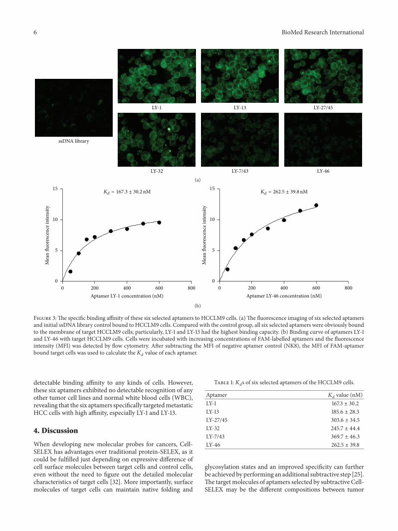