research article pegylated graphene oxide for tumor ... · demonstrated that go-peg exhibited no...
TRANSCRIPT
For reprint orders, please contact: [email protected]
1247Nanomedicine (Lond.) (2015) 10(8), 1247–1262 ISSN 1743-5889
part of
Research Article
10.2217/NNM.14.233 © 2015 Future Medicine Ltd
Nanomedicine (Lond.)
Research Article10
8
2015
Aim: The graphene oxide (GO) sheet has been considered one of the most promising carbon derivatives in the field of material science for the past few years and has shown excellent tumor-targeting ability, biocompatibility and low toxicity. We have endeavored to conjugate paclitaxel (PTX) to GO molecule and investigate its anticancer efficacy. Materials & Methods: We conjugated the anticancer drug PTX to aminated PEG chains on GO sheets through covalent bonds to get GO-PEG-PTX complexes. The tissue distribution and anticancer efficacy of GO-PEG-PTX were then investigated using a B16 melanoma cancer-bearing C57 mice model. Results: The GO-PEG-PTX complexes exhibited excellent water solubility and biocompatibility. Compared with the traditional formulation of PTX (Taxol®), GO-PEG-PTX has shown prolonged blood circulation time as well as high tumor-targeting and -suppressing efficacy. Conclusion: PEGylated graphene oxide is an excellent nanocarrier for paclitaxel for cancer targeting.
Keywords: cancer therapy • drug delivery • graphene oxide • paclitaxel
Graphene has a hexagonal packed struc-ture formed by a 2D single layer of carbon atoms [1,2]. Owing to its unique properties, graphene and its derivatives have attracted tremendous attention in the fields of elec-tronics, energy, materials and biomedical applications [3–6]. Although the naked GO did not exhibit any obvious in vitro toxicity nor in vivo inflammation at low concentra-tion [7,8], proper surface functionalization is essential to design graphene-based nanocar-rier systems with high solubility in the physi-ological fluids and acceptable biocompatibil-ity for biomedical application in physiological environments. Various types of hydrophilic polymers have been utilized to functionalize graphene oxide (GO) using covalent or non-covalent bonding [9]. Functionalized GO has been used for drug and gene delivery [9–21], as well as in biosensor platforms, photother-mal therapy and in tumor and cell imag-ing [11,22–32]. Numerous types of polymers and molecules are possible to use for modify-ing the surfaces of GO via hydrogen bond-
ing or π–π interactions [9,33]. Many aromatic chemotherapy drug molecules can be conju-gated onto the surface of GO through physi-cal adsorption, resulting in changing GO from poorly soluble sheets to water-soluble conjugates [10–12,34]. Dai et al. first loaded a water insoluble anticancer analog of camp-tothecin (CPT) onto GO surface through noncovalent conjugation via π–π interac-tion. The GO loaded with SN38 exhibited acceptable biocompatibility and excellent solubility both in aqueous and physiologi-cal solutions [10]. Several research groups have studied targeted delivery of doxorubicin (DOX) and CPT attached to surface modi-fied GO, which exhibited superior anticancer efficacy [12,16,19,21,34]. Despite its lower optical properties, the 2D shape and ultra-small sizes of GO may offer many advantages compar-ing with other carbon-based nanomaterials such as single-walled nanotubes (SWNTs). For example, the in vitro toxicity of graphene towards human cells appears to be lower than that of SWNTs. Moreover, the photothermal
PEGylated graphene oxide for tumor-targeted delivery of paclitaxel
Hongyang Xu1, Minmin Fan1, Abdelbary MA Elhissi2, Zhirong Zhang1, Ka-Wai Wan3, Waqar Ahmed3, David A Phoenix4 & Xun Sun*,1
1Key Laboratory of Drug Targeting & Drug Delivery Systems, Ministry of Education, West China School of Pharmacy, Sichuan University, Chengdu 610041, PR China 2College of Pharmacy, Qatar University, Doha, Qatar 3Institute of Nanotechnology & Bioengineering, School of Pharmacy & Biomedical Sciences, University of Central Lancashire, Preston, UK 4Office of the Vice Chancellor, London South Bank University, 103 Borough Road, London, SE1 0AA, UK
*Author for correspondence: Tel.: +86 28 85502307 Fax: +86 28 85501615 [email protected]
1248 Nanomedicine (Lond.) (2015) 10(8) future science group
Research Article Xu, Fan, Elhissi, Zhang et al.
anticancer efficacy of graphene is superior to that of SWNT because of the better dispersion and smaller size of graphene nanoparticles.
Liu et al. have first reported that GO modified by coat-ing its surfaces with PEG (GO-PEG) can become highly aqueous soluble and stable in physiological fluids such as serum [10]. These findings can make the in vivo intra-venous application of GO feasible. Moreover, GO-PEG has shown significant passive tumor-targeting ability, which might be ascribed to the enhanced permeability and retention effect of tumor tissue [35]. The long-term in vivo biodistribution and toxicity investigations have demonstrated that GO-PEG exhibited no obvious tissue toxicity, and no organ damage or inflammation symp-toms [35,36], which revealed the superior characteristics of GO-PEG compared with uncoated GO [37]. Accord-ing to Liu et al., following intravenous injection of GO-PEG, the formulation has accumulated in the reticulo-endothelial systems (RES) such as liver and spleen and were gradually cleared out via renal excretion and biliary pathway into feces over time [36,38,39].
Paclitaxel (PTX) is a widely used chemotherapeutic drug that promotes tubulin polymerization and forma-tion of extraordinarily dysfunctional and stable micro-tubules, disrupting the normal tubule dynamics [40]. However, its poor aqueous solubility is a serious limita-tion that prevents proper formulation and clinical appli-cation of the drug [41]. In order to overcome low aqueous solubility of PTX, formulations based on Cremophor EL (e.g., Taxol®) have been prepared and used via slow intravenous infusion following dilution with NaCl (0.9%) or dextrose (5%) solutions. However, the sol-vent system mainly Cremophor EL in Taxol causes seri-ous toxicological effects [42] such as neurotoxicity and nephrotoxicity, which may significantly reduce the over-all therapeutic benefit of PTX. Therefore, Cremophor EL-free delivery systems of PTX have been proposed, including those based on polymeric nanoparticles, poly-meric micelles, liposomes and many prodrug formula-tions [43–47]. Owing to the advantages and unique per-formance of GO as a targeting system, formulations of PTX loaded onto modified GO are worth exploring.
Unlike DOX and CPT, PTX has no extend π-structure larger than one aromatic ring [48]; hence, it is difficult to load PTX onto GO by physical adsorption. Even though some paper has reported the successful noncovalent con-jugation of PTX and GO [49], the loading capacity may be optimized using other methods. Thus, we have inves-tigated the possibility of loading PTX onto GO-PEG through covalent conjugation, aiming to improve the drug physiological solubility and enhance the formula-tion compatibility and cancer targeting ability. In this work we used GO-PEG as a drug carrier to load PTX via an ester bond, which is cleavable after in vivo injection.
The covalent conjugation has been reported to be suc-cessful at attaching PTX to SWNTs and α,β-Poly(N-2-hydroxyethyl)-DL-aspartamide (PHEA) [41,50]. The influence of GO-PEG-PTX conjugation on drug solu-bility in water and mouse serum was investigated. More-over, further in vivo investigations were carried out to evaluate the anticancer efficacy of GO-PEG-PTX, the blood circulation time, drug bioavailability, tissue distri-bution and tumor-uptake rate. The in vivo biodistribu-tion of GO-PEG-PTX was also investigated and com-pared with that of Taxol. To the best of our knowledge, this is the first study that demonstrated the feasibility of using PEGylated GO as nanocarriers for PTX and inves-tigated the anticancer activity of the resultant complex compared with Taxol.
Experimental section MaterialsGO was purchased from Tianjin Plannano Technology Co., Ltd. Chloroacetic acid was purchased from Fuchen Chemical Reagents Factory (Tianjin, China). N-(3-di-methylamino propyl-N′-ethylcar-bodiimide) hydro-chloride (EDC-HCl), N-hydroxysuc-cinimide (NHS), Succinic anhydride and pyridine were purchased from Kelong Chemical Reagents Factory (Chengdu, China). PTX and docetaxel were purchased from Hao-xuan Biological Technology Co., Ltd (Xi’an, China). Ami-nated PEG (NH
2-PEG-NH
2, Mw = 4000) was pur-
chased from Kaizheng Biological Technology Co., Ltd, (Beijing, China). Cholesterol esterase was purchased from Shifeng Biological Technology CO., Ltd (Shang-hai, China). DMEM cell culture medium was bought from Thermo Fisher Scientific (MA, USA). fetal bovine serum was supplied by Fumeng Gene Co., Ltd (Shang-hai, China). 3-(4,5-dimethylthiazol-2-yl)-2,5-dipheny-ltetrazolium bromide (MTT) was supplied by Sigma (CA, USA). BCA protein assay reagent kit was pur-chased from Thermo Fisher Scientific.
Cell lines & animalsA549 and B16 melanoma cancer cell lines were pur-chased from the Type Culture Collection of the Chinese Academy of Sciences (Shanghai, China). C57 mouse and Wistar rats were provided by Chengdu Dashuo Biotechnology Co., Ltd. All procedures with ani-mals were conducted in accordance with institutional animal care and use guidelines.
Carboxylation of GOTo carboxylate GO sheet, GO (10 mg) was added to water and sonicated to form an aqueous suspension. NaOH (0.12 g/ml) and chloroacetic acid (0.5 g) were then added, and the resultant mixture was sonicated using probe sonicator for 3 h to form carboxyl group on
www.futuremedicine.com 1249future science group
PEGylated graphene oxide for tumor-targeted delivery of paclitaxel Research Article
the surface of GO. GO-COOH suspension was neu-tralized and purified by centrifugation at 10,000 rpm. The supernatant was discarded and the residue was washed with water twice.
Modification of GO with aminated PEGCarboxylated GO was diluted by water (10 ml) and then bath sonicated with amino-terminated PEG4000 (100 mg) for 30 min, followed by addition of N-(3-dimethylaminopropyl-N′-ethylcarbodiiminde) hydro-chloride (EDC-HCl) to reach a concentration of 40 mmol/l. Then the mixture was allowed to react overnight. The final reactants were dialyzed in dialysis bags (molecular weight cutoff = 100,000) for 4 days to obtain the PEGylated GO.
Fourier transform infrared (IR) absorbance spec-trum, atomic force microscopy and UV-vis spectrum were used to identify whether the carboxylation and PEGylation were successful.
Synthesis of 2′-O-succinyl-PTX derivativePTX (200 mg) and succinic anhydride (0.3 g) were added to 5 ml of anhydrous pyridine. The solution was stirred for 24 h at room temperature. The progress of reaction was detected by thin-layer chromatography (CH
3OH/CHCl
2 1:20 v/v). After that, 6 ml of water
was added and stirred for 1 h at 60°C. The solvent (pyri-dine and water) was then evaporated under vacuum at 55°C using a rotary evaporator (BUCHI Rotavapor R-3). The resultant modified PTX was retrieved via extraction using ethyl acetate as solvent. Purification of the desired compounds was carried out by column chromatography on silica gel (the elution phase was CH
3OH/CHCl
2
1:50 v/v). 1H-NMR (CDCl3) and LC-MS were employed
to confirm the chemical structures.
Conjugation between GO-PEG & 2′-O-succinyl-PTXTo synthesize GO-PEG-PTX, 2′-O-succinyl-PTX (20 mg) and GO-PEG aqueous solution (∼0.4 mg/ml) were dissolved in DMSO-water component solvent (1:1 v/v) followed by addition of EDC-HCl (50 mg) and NHS (50 mg). The resultant solution was stirred at 25°C for 6 h, followed by 2 days of dialysis in DMSO and another 2 days in water. HPLC was used for identification of the conjugation of GO-PEG-PTX.
HPLC analysisHPLC assay methods were established for the detec-tion of PTX concentration in cellular uptake, pharma-cokinetics and biodistribution studies. Analysis was performed using Shimadzu instruments (Chiyoda-Ku, Kyoto, Japan) consisting of a 50 μl injector loop, a CTO-10A column thermostat, two LC-10AT pumps
and an Diamonsil C18 reverse phase column. The col-umn effluent was monitored at 227 nm with a flow rate of 1 ml/min at 35°C. The mobile phase was composed of acetonitrile and water (45: 55 v/v). The total run time for each sample was 15 min.
In vitro release behavior of PTX from GO-PEG-PTXGO-PEG-PTX was dissolved in phosphate-buffered saline (PBS; pH 7.4), C57 mouse serum or cholesterol esterase solution and incubated for 48 h at 37°C. The released PTX was separated via ultrafiltration using 100 kDa MWCO filters, and the retained PTX was detected by HPLC at 4, 12, 24 and 48 h incubation.
Cell culture & in vitro toxicityA549 and B16 melanoma cancer cell lines were cultured in DMEM with high glucose supplemented with 10% fatal bovine serum, 100 μg/ml of streptomycin and 100 μg/ml of penicillin. Cells were placed in a humidi-fied atmosphere containing 5% CO
2 at 37°C. The cell
medium was changed every other day. For the in vitro cell toxicity study, cells were seeded onto a 96-well bot-tom plate and incubated at 37°C overnight. The cells were then incubated with different concentrations of GO-PEG, GO-PEG-PTX or Taxol (all dissolved in DMEM with high glucose) and blank DMEM with high glucose. After 24 h of incubation, the relative cell viability was measured by MTT assay with Fluoroskan Ascent FL microplate fluorometer and luminometer (Thermo Scientific).
Cellular uptake assayBoth A549 and B16 cells were seeded in 6-well plates. The cells were then exposed to GO-PEG-PTX or Taxol at a range of concentrations (10, 25, 50, 100 μg/ml) for 2 h at 37°C. Cold PBS (20°C pH 7.4) was used to wash the cells in order to remove the drug molecules that were not taken up by those cells. The cells were then digested and collected as the cel-lular uptake sample, which were centrifuged at 5000 rpm for 5 min. The cell residue was lysed with radio-immunoprecipitation assay buffer in order to release the intracellular drug. Methanol and 20% of trichlo-roacetic acid were added to the cell digests, followed by centrifugation at 12,000 rpm for 10 min. Super-natants were collected and analyzed by HPLC. The intracellular concentrations of PTX were investigated by HPLC assay using the method described earlier. Twenty microliters of the cell lysate from each sample was taken to determine the total cell protein content using reagent kit (Pierce, USA). The uptake rate was expressed as the amount of PTX associated with a unit weight of cellular protein.
1250 Nanomedicine (Lond.) (2015) 10(8)
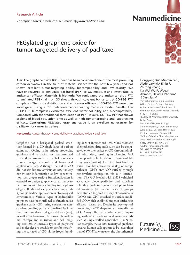
Figure 1. Characterization of graphene oxide-PEG. (A) Atomic force microscopy showed that all shards in the vision were below 50 nm. (B) Fourier transform infrared spectra of GO and GO-PEG. (C) UV-vis spectra of GO and GO-PEG at the concentration of 0.05 mg/ml. GO: Graphene oxide.
140
120
100
80
60
40
20
0 0
0.5
Ab
sorb
ance
(a.
u.)
1.0
1.5
4000 3500 3000 2500 2000Wavenumber cm-1
1500 1000 500 500 600 700 800 900
GO
GO-4
GO-PEG
GO
GO-PEG
400
Tra
nsm
itta
nce
(%
)
1.00
0.500.50
0.00
0.00
1.65(nm)
1.06(nm)
0.001.25 x 1.25 μm 1.25 x 1.25 (μm) z 0.00 - 1.65 (nm)500.00 nm
1.00
1.001
0.500.50
5)
1.000
GO-PEG
A
B C
future science group
Research Article Xu, Fan, Elhissi, Zhang et al.
Pharmacokinetic studyFemale Wistar rats (220 ± 20 g) were randomly divided into two groups. The rats received GO-PEG-PTX (GO-PEG-PTX group) or Taxol (Taxol group) via tail vein at a dose equivalent to 2 mg/kg of PTX (n = 5). At 5 min, 10 min, 15 min, 30 min, 1 h, 2 h, 4 h, 8 h, 12 h and 24 h intervals after injection, blood samples were taken and the plasma was separated by centrifugation at 5000 rpm for 5 min. Docetaxel were added to each sample as the internal standard and methanol was used to precipitate the protein followed by centrifugation at 13,000 rpm for 10 min. Supernatants were collected and analyzed by HPLC following the method described above.
Animal model & drug efficacy studyMurine B16 melanoma cancer model was established by subcutaneous injection of about 1.5 × 10⁶ cells in PBS under the right arm of female C57 mice. The mice were used for experiments 14 days after injection (the tumor volume was about 100 mm3). For the treat-ment, GO-PEG-PTX or Taxol with a dose equivalent to 4 mg/kg of PTX, GO-PEG and the same volume of saline were injected via caudal vein for three-times (0, 3, 6 days after injection). The tumor size was measured
by a calipher every other day and was calculated as V = (tumor length) × (tumor width)2/2. Relative tumor volumes were calculated as V/V
0 (V
0 was the
tumor volume before mice were injected with the for-mulation). The survival rates of each group were then calculated to construct the survival rate curve.
Biodistribtion studyFemale C57 mice bearing B16 tumor (tumor size was about 200 mm3) were intravenously injected with GO-PEG-PTX or Taxol with a dose equivalent to 50 mg/kg of PTX. The mice were sacrificed at 30 min, 1 h, 4 h, 8 h, 12 h or 24 h after injection. Samples of blood, liver, spleen, lung, kidney and tumor were then collected. The samples of the animals’ blood were collected in heparinized tubes. Tissues were isolated, washed with saline and homogenized with twofold volume of 0.9% sodium chloride (g/ml). The blood samples and tissues homogenates were processed and measured by HPLC as described in the pharmacokinetic study.
In vivo toxicity studyHealthy female C57 mice (18∼20 g) were randomly assigned into four groups for drug administration. GO-
www.futuremedicine.com 1251
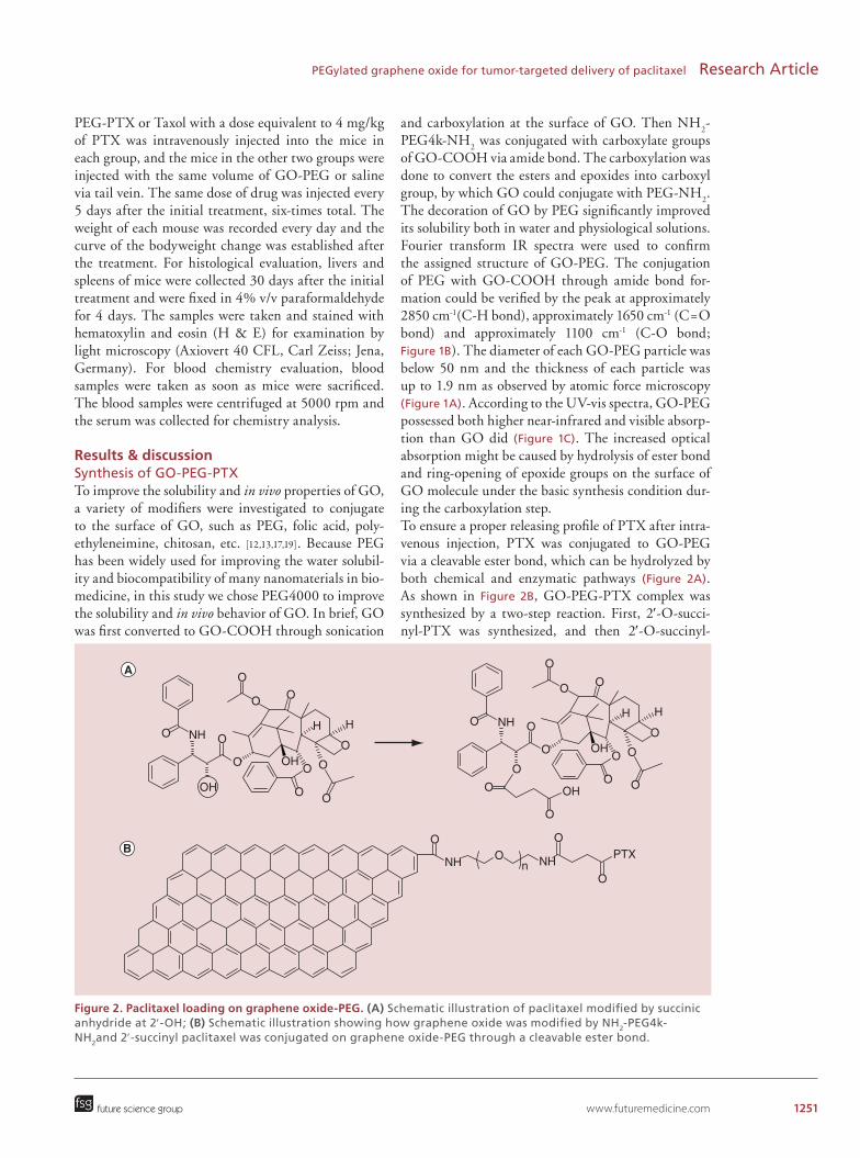
Figure 2. Paclitaxel loading on graphene oxide-PEG. (A) Schematic illustration of paclitaxel modified by succinic anhydride at 2′-OH; (B) Schematic illustration showing how graphene oxide was modified by NH2-PEG4k-NH2and 2′-succinyl paclitaxel was conjugated on graphene oxide-PEG through a cleavable ester bond.
O
O
O
HH
OO
O
O OH O
OO
OO NH
OHO
O
OO
OPTX
ONH NHn
O
O
O
HH
OO
O
O OH O
OOH
OO NH
future science group
PEGylated graphene oxide for tumor-targeted delivery of paclitaxel Research Article
PEG-PTX or Taxol with a dose equivalent to 4 mg/kg of PTX was intravenously injected into the mice in each group, and the mice in the other two groups were injected with the same volume of GO-PEG or saline via tail vein. The same dose of drug was injected every 5 days after the initial treatment, six-times total. The weight of each mouse was recorded every day and the curve of the bodyweight change was established after the treatment. For histological evaluation, livers and spleens of mice were collected 30 days after the initial treatment and were fixed in 4% v/v paraformaldehyde for 4 days. The samples were taken and stained with hematoxylin and eosin (H & E) for examination by light microscopy (Axiovert 40 CFL, Carl Zeiss; Jena, Germany). For blood chemistry evaluation, blood samples were taken as soon as mice were sacrificed. The blood samples were centrifuged at 5000 rpm and the serum was collected for chemistry analysis.
Results & discussion Synthesis of GO-PEG-PTXTo improve the solubility and in vivo properties of GO, a variety of modifiers were investigated to conjugate to the surface of GO, such as PEG, folic acid, poly-ethyleneimine, chitosan, etc. [12,13,17,19]. Because PEG has been widely used for improving the water solubil-ity and biocompatibility of many nanomaterials in bio-medicine, in this study we chose PEG4000 to improve the solubility and in vivo behavior of GO. In brief, GO was first converted to GO-COOH through sonication
and carboxylation at the surface of GO. Then NH2-
PEG4k-NH2 was conjugated with carboxylate groups
of GO-COOH via amide bond. The carboxylation was done to convert the esters and epoxides into carboxyl group, by which GO could conjugate with PEG-NH
2.
The decoration of GO by PEG significantly improved its solubility both in water and physiological solutions. Fourier transform IR spectra were used to confirm the assigned structure of GO-PEG. The conjugation of PEG with GO-COOH through amide bond for-mation could be verified by the peak at approximately 2850 cm-1(C-H bond), approximately 1650 cm-1 (C=O bond) and approximately 1100 cm-1 (C-O bond; Figure 1B). The diameter of each GO-PEG particle was below 50 nm and the thickness of each particle was up to 1.9 nm as observed by atomic force microscopy (Figure 1A). According to the UV-vis spectra, GO-PEG possessed both higher near-infrared and visible absorp-tion than GO did (Figure 1C). The increased optical absorption might be caused by hydrolysis of ester bond and ring-opening of epoxide groups on the surface of GO molecule under the basic synthesis condition dur-ing the carboxylation step.To ensure a proper releasing profile of PTX after intra-venous injection, PTX was conjugated to GO-PEG via a cleavable ester bond, which can be hydrolyzed by both chemical and enzymatic pathways (Figure 2A). As shown in Figure 2B, GO-PEG-PTX complex was synthesized by a two-step reaction. First, 2′-O-succi-nyl-PTX was synthesized, and then 2′-O-succinyl-

1252 Nanomedicine (Lond.) (2015) 10(8)
Figure 3. The drug release profile using graphene oxide-PEG-paclitaxel in phospate-buffered saline solution, C57 mouse serum, esterase enzyme solution and heat denatured enzyme solution after 48 h of incubation with phospate-buffered saline and serum, respectively, at 37°C. The retained paclitaxel was collected by filtration and detected by HPLC.
120
100
80
60
40
20
00 10 20 30 40 50
Time (h)
Co
nju
gat
ed P
TX
(%
)PBS
Serum
Esterase enzyme
Denatured enzyme
future science group
Research Article Xu, Fan, Elhissi, Zhang et al.
PTX was conjugated to GO-PEG. The synthesis of 2′-O-succinyl-PTX was carried out according to the previously reported methods [41,50–52]. As described in Figure 2A, the PTX molecule was first linked by its 2′-hydroxyl terminus with succinic anhydride. Thin-layer chromatography analysis was used for detecting whether free PTX was fully converted to the resultant. The unreacted succinic anhydride turned into water soluble succinic acid after stirring at 60°C for 1 h. The desired compound was obtained by purification using a silica gel column. After that, modified PTX was cou-pled to the terminal amine group of the PEG previously being attached on the surface of GO molecule through amide bond [50]. As a standard reaction of forming amide bond, 2′-O-succiyl-PTX and GO-PEG were dis-solved in water/DMSO (1:1) mixed solvent and EDC-HCL and NHS were added as catalysts. After dialysis in DMSO and water respectively, the purified GO-PEG-PTX was analyzed by HPLC method to confirm the successful synthesis. According to the HPLC result, approximately 0.51 mg of PTX was conjugated on 1 mg of GO-PEG. Different from blank PTX, GO-PEG-PTX exhibited improved solubility both in water and in saline. In vitro release study at 37°C indicated that GO-PEG-PTX displayed excellent stability in PBS solution, in which only very low proportion of PTX was released from GO-PEG-PTX complex within 48 h (Figure 3). The release rate of PTX in C57 mice serum was faster than that in PBS (about 30% of total PTX was released within 24 h). What is more, the release rate of PTX in esterase enzyme solution was much faster than those
in PBS and serum. While in denatured enzyme solu-tion, the release rate was similar with that in PBS. So the cleavage of ester bond in serum could be ascribed to the existence of esterase enzyme. Importantly, the relatively moderate release rate might ensure that PTX loaded onto GO-PEG would reach the target tissue and then release PTX to exert therapeutic effect.
In vitro investigation of GO-PEG-PTXCytotoxicity of GO-PEG-PTX was investigated using MTT assay with A549 and B16 cancer cell lines. As indicated in Figure 4, GO-PEG-PTX exhib-ited approximately similar cytotoxic efficacy to that exhibited by Taxol at relatively high concentrations. No obvious cytotoxicity of plain GO-PEG was found during the experiments, even at high concentrations. These results indicated that loading PTX in GO-PEG did not significantly interfere or reduce cytotoxicity of PTX against cancer cells.
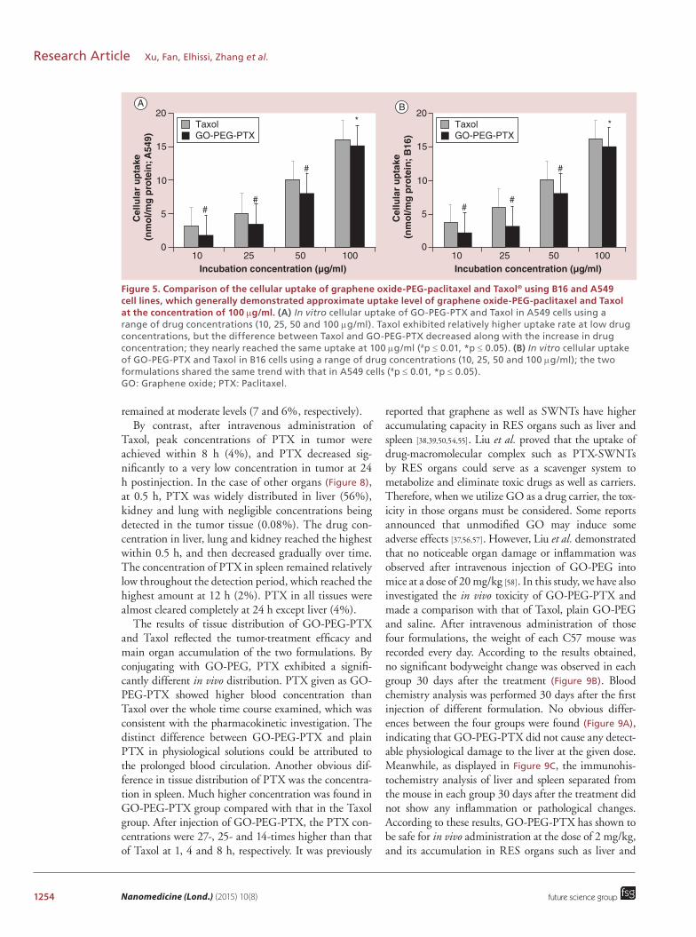
We then further investigated the cellular uptake of GO-PEG-PTX using A549 and B16 cell lines. Some previous research investigations have employed fluo-rescent or radioactive materials to tag GO molecules in order to monitor their dynamics following cellar uptake [34,50]. In this study, we used HPLC to investi-gate the cellular uptake rate because the linear chain of NH
2-PEG4k-NH
2 did not have much room for con-
jugating the fluorescent material, especially when two terminals of PEG were occupied by both GO and PTX. HPLC method was established to monitor the intracel-lular drug contents. It was shown in Figure 5 that the

www.futuremedicine.com 1253
Figure 4. Comparison of cell survival curves for A549 and B16 cells incubated with graphene oxide-PEG, graphene oxide-PEG-paclitaxel or Taxol® for 48 h. The cytotoxicity of graphene oxide-PEG-paclitaxel approximated to that of Taxol at high drug concentrations. #p ≤ 0.01; *p ≤ 0.05.
120
100
Rel
ativ
e ce
ll vi
abili
ty
(%;
B16
) 80
60
40
20
00 20 40 60 80 100 120
Drug concentration (μg/ml)
120
100
Rel
ativ
e ce
ll vi
abili
ty
(%;
A54
9) 80
60
40
20
00 20 40 60 80 100 120
Drug concentration (μg/ml)
#
##
* *
#
## #
*
Taxol
GO-PEG-PTX
GO-PEG
Taxol®
GO-PEG-PTX
GO-PEG
A B
future science group
PEGylated graphene oxide for tumor-targeted delivery of paclitaxel Research Article
intracellular concentration of drug using GO-PEG-PTX was slightly lower than that of Taxol at low doses. However, when the drug concentration was increased the intracellular drug content almost reached the same level of that seen by Taxol. Both A549 and B16 cell lines shared the same tendency. The cellular uptake results and the cytotoxicity investigation suggested that the in vitro anti-cancer ability of GO-PEG-PTX was compa-rable to that of Taxol at the relatively high concentra-tions used. However, the in vitro anticancer ability of GO-PEG-PTX was lower than that of Taxol at the rela-tively low doses. The previous study of Liu et al. showed that cytotoxicity of PTX conjugated to SWNTs was relatively lower than that of Taxol at certain drug dose range (10∼100 μg/ml) [50]. The low cellular uptake rate of the PTX on GO-PEG might be attributed to con-jugation between PTX and GO-PEG. This was con-firmed previously by Chang et al. who have reported that GO alone could hardly be taken up by can-cer cells [7]. However, GO-PEG could enter the cells through energy required endocytosis mechanism [10,38]. Thus when conjugated with GO-PEG, PTX was able to enter the cancer cells along with GO-PEG-PTX com-plex through endocytosis. Although the conjugation with GO hindered the immediate uptake of PTX by the cells, the long blood circulation time offered by con-jugating the drug to GO-PEG after in vivo treatment was highly advantageous at enhancing the anticancer efficacy of PTX. Using this strategy, PTX adminis-tration as GO-PEG-PTX may result in higher drug bioavailability and enhanced antitumor effect.
Pharmacokinectic investigation of GO-PEG-PTXIn order to understand the pharmacokinetics of GO-PEG-PTX, we used HPLC assay to measure PTX con-centration in plasma at different time intervals after intravenous injection of the formulations into Wistar
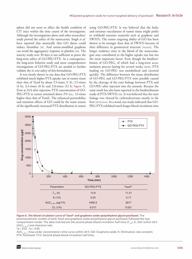
rats via tail vein (2 mg/kg). Blood was collected at dif-ferent time points after injection. The time–concentra-tion curves of GO-PTX-PEG and Taxol both exhib-ited a standard two-compartment model. As shown in Figure 6, the elimination speed of PTX was relatively rapid both in GO-PEG-PTX and Taxol group. Com-pared with Taxol, longer second phase blood circulation half-time, higher bioavailability and lower clearance rate were observed for GO-PEG-PTX. The underlying reason was that some time is needed for the cleavage of ester linkage between PTX and GO-PEG to occur [53].
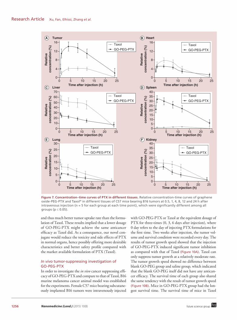
Biodistribution & in vivo toxicity investigations of GO-PEG-PTXTo further evaluate the tumor targeting ability of GO-PEG-PTX, we studied tissue distribution of GO-PEG-PTX and Taxol in B16 melanoma cancer bearing C57 mice. The tissue distribution of GO-PEG-PTX and Taxol were measured at 30 min, 1 h, 4 h, 8 h, 12 h and 24 h after injecting GO-PEG-PTX or Taxol via tail veins of B16 tumor-bearing mice. At predetermined time points, the blood samples were collected and the animals were sacrificed. Tissues including hearts, livers, spleens, lungs and kidneys were isolated immediately. Plasma or tissue homogenates were extracted by metha-nol, and then quantitative measurement of PTX was done by HPLC. As shown in Figure 7, after intravenous administration of GO-PEG-PTX, peak concentrations of PTX in tumor were achieved within 4 h (14.2%), which was significantly higher than that of Taxol group. The concentration of PTX remained relatively high even at 24 h (2%) after administration. For other tis-sues (Figure 8), the liver and kidney reached the highest concentration within 0.5 h (28 and 16%), decreasing gradually over time. The highest concentration in spleen and lung were achieved within 1 h (32 and 6%, respec-tively). After 24 h of treatment, PTX in liver and spleen
1254 Nanomedicine (Lond.) (2015) 10(8)
Figure 5. Comparison of the cellular uptake of graphene oxide-PEG-paclitaxel and Taxol® using B16 and A549 cell lines, which generally demonstrated approximate uptake level of graphene oxide-PEG-paclitaxel and Taxol at the concentration of 100 μg/ml. (A) In vitro cellular uptake of GO-PEG-PTX and Taxol in A549 cells using a range of drug concentrations (10, 25, 50 and 100 μg/ml). Taxol exhibited relatively higher uptake rate at low drug concentrations, but the difference between Taxol and GO-PEG-PTX decreased along with the increase in drug concentration; they nearly reached the same uptake at 100 μg/ml (#p ≤ 0.01, *p ≤ 0.05). (B) In vitro cellular uptake of GO-PEG-PTX and Taxol in B16 cells using a range of drug concentrations (10, 25, 50 and 100 μg/ml); the two formulations shared the same trend with that in A549 cells (#p ≤ 0.01, *p ≤ 0.05). GO: Graphene oxide; PTX: Paclitaxel.
20
15
5
0
10
10 25 50 100
Taxol *
#
##
GO-PEG-PTX
Incubation concentration (μg/ml)
Cel
lula
r u
pta
ke
(nm
ol/m
g p
rote
in;
B16
)
20
15
5
0
10
10 25 50 100Incubation concentration (μg/ml)
Cel
lula
r u
pta
ke
(nm
ol/m
g p
rote
in; A
549)
*
#
##
TaxolGO-PEG-PTX
A B
future science group
Research Article Xu, Fan, Elhissi, Zhang et al.
remained at moderate levels (7 and 6%, respectively).By contrast, after intravenous administration of
Taxol, peak concentrations of PTX in tumor were achieved within 8 h (4%), and PTX decreased sig-nificantly to a very low concentration in tumor at 24 h postinjection. In the case of other organs (Figure 8), at 0.5 h, PTX was widely distributed in liver (56%), kidney and lung with negligible concentrations being detected in the tumor tissue (0.08%). The drug con-centration in liver, lung and kidney reached the highest within 0.5 h, and then decreased gradually over time. The concentration of PTX in spleen remained relatively low throughout the detection period, which reached the highest amount at 12 h (2%). PTX in all tissues were almost cleared completely at 24 h except liver (4%).
The results of tissue distribution of GO-PEG-PTX and Taxol reflected the tumor-treatment efficacy and main organ accumulation of the two formulations. By conjugating with GO-PEG, PTX exhibited a signifi-cantly different in vivo distribution. PTX given as GO-PEG-PTX showed higher blood concentration than Taxol over the whole time course examined, which was consistent with the pharmacokinetic investigation. The distinct difference between GO-PEG-PTX and plain PTX in physiological solutions could be attributed to the prolonged blood circulation. Another obvious dif-ference in tissue distribution of PTX was the concentra-tion in spleen. Much higher concentration was found in GO-PEG-PTX group compared with that in the Taxol group. After injection of GO-PEG-PTX, the PTX con-centrations were 27-, 25- and 14-times higher than that of Taxol at 1, 4 and 8 h, respectively. It was previously
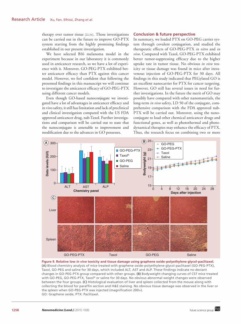
reported that graphene as well as SWNTs have higher accumulating capacity in RES organs such as liver and spleen [38,39,50,54,55]. Liu et al. proved that the uptake of drug-macromolecular complex such as PTX-SWNTs by RES organs could serve as a scavenger system to metabolize and eliminate toxic drugs as well as carriers. Therefore, when we utilize GO as a drug carrier, the tox-icity in those organs must be considered. Some reports announced that unmodified GO may induce some adverse effects [37,56,57]. However, Liu et al. demonstrated that no noticeable organ damage or inflammation was observed after intravenous injection of GO-PEG into mice at a dose of 20 mg/kg [58]. In this study, we have also investigated the in vivo toxicity of GO-PEG-PTX and made a comparison with that of Taxol, plain GO-PEG and saline. After intravenous administration of those four formulations, the weight of each C57 mouse was recorded every day. According to the results obtained, no significant bodyweight change was observed in each group 30 days after the treatment (Figure 9B). Blood chemistry analysis was performed 30 days after the first injection of different formulation. No obvious differ-ences between the four groups were found (Figure 9A), indicating that GO-PEG-PTX did not cause any detect-able physiological damage to the liver at the given dose. Meanwhile, as displayed in Figure 9C, the immunohis-tochemistry analysis of liver and spleen separated from the mouse in each group 30 days after the treatment did not show any inflammation or pathological changes. According to these results, GO-PEG-PTX has shown to be safe for in vivo administration at the dose of 2 mg/kg, and its accumulation in RES organs such as liver and
www.futuremedicine.com 1255
Figure 6. The blood circulation curve of Taxol® and graphene oxide-polyethylene glycol-paclitaxel. The pharmacokinetic models of both Taxol and graphene oxide-polyethylene glycol-paclitaxel followed the two compartment model. The data inserted are the second-phase blood circulation half-time (T1/2), K, AUC within 24 h (AUC0–24 h) and clearance rate. #p ≤ 0.01, *p ≤ 0.05. AUC0–24 h: Area under concentration–time curve within 24 h; GO: Graphene oxide; K: Elimination rate constant; PTX: Paclitaxel; T1/2: Second-phase blood circulation half-time.
Parameters GO-PEG-PTX Taxol®
T1/2 (h) 13.9 11.51
K (1/h) 0.23 0.17
AUC0–24 h (μg/1/h) 4409.3 2617
CL (1/h) 0.017 0.031
3500
2500
1500
500
00 200 400 600
* * *#
800 1000 1200 1400Time (min)
Dru
g c
on
cen
trat
ion
(µ
g/l)
1000
2000
3000PTX
GO-PEG-PTX
future science group
PEGylated graphene oxide for tumor-targeted delivery of paclitaxel Research Article
spleen did not seem to affect the health condition of C57 mice within the time course of the investigation. Although the investigations above and other researchers’ study proved the safety of the nanocarrier, Singh et al. have reported that atomically thin GO sheets could induce thrombus [59]. And amine-modified graphene can avoid the aggregatory response in platelets [60]. The toxicity study over 30 days is not sufficient to prove the long-term safety of GO-PEG-PTX. As a consequence, the long-term behavior study and more comprehensive investigations of GO-PEG-PTX are needed to further validate the in vivo safety of this formulation.
It was clearly shown in our data that GO-PEG-PTX exhibited much higher PTX uptake rate in tumor tissue than that of Taxol by about 5.5-times (1 h), 3.5-times (4 h), 3.3-times (8 h) and 2.8-times (12 h; Figure 7). Even at 24 h after injection, PTX concentration of GO-PEG-PTX in tumor remained above 2% (i.e., 12-times higher than that of Taxol). The enhanced permeability and retention effects of GO could be the main reason of the significantly increased PTX distribution in tumor
using GO-PEG-PTX. It was believed that the leaky and tortuous vasculatures of tumor tissue might prefer to withhold nanosize materials such as graphene and SWNTs. The tumor targeting ability of GO has been shown to be stronger than that of SWNTs because of their difference in geometrical structure [54,61,62]. The longer residence time in the blood of the nanoconju-gate may contributed to the higher uptake rate but not the most important factor. Even though the biodistri-bution of GO-PEG, of which had a long-term accu-mulation process lasting for several weeks [38,50], PTX loading on GO-PEG was metabolized and excreted quickly. The difference between the tissue distribution of GO-PEG and GO-PEG-PTX were possibly caused by the cleavage of the ester linkage between PTX and GO-PEG after injection into the animals. Because the same result has also been reported in the biodistribution study of PTX-SWNTs [50]. It was believed that the ester linkage was cleaved by carboxylesterases mostly in the liver [50,53,63,64]. In a word, our study indicated that GO-PEG-PTX exhibited much longer blood circulation time
1256 Nanomedicine (Lond.) (2015) 10(8)
Figure 7. Concentration–time curves of PTX in different tissues. Relative concentration-time curves of graphene oxide-PEG-PTX and Taxol® in different tissues of C57 mice bearing B16 tumors at 0.5, 1, 4, 8, 12 and 24 h after intravenous injection (n = 5 for each group at each time point), which were significantly different among all groups (p ≤ 0.05).
16Taxol
GO-PEG-PTX
Tumor
Liver
Lung Kidney
Heart
Spleen
12
8
Rel
ativ
e co
nce
ntr
atio
n (
%)
Rel
ativ
e co
nce
ntr
atio
n (
%)
Rel
ativ
e co
nce
ntr
atio
n (
%)
Rel
ativ
e co
nce
ntr
atio
n (
%)
Rel
ativ
e co
nce
ntr
atio
n (
%)
Rel
ativ
e co
nce
ntr
atio
n (
%)
4
00 5 10
Time after injection (h)15 2520
0 0
0
510
20
30
40
15
25
35
10
20
30
40
50
60
70
0 5 10Time after injection (h)
15 2520
16
12
8
4
00 5 10
Time after injection (h)15 2520
0 5
510
20
30
40
15
25
35
10Time after injection (h)
15 2520
0 5 10Time after injection (h)
15 25200
5
10
20
30
15
25
0 5 10Time after injection (h)
15 2520
Taxol
GO-PEG-PTX
Taxol
GO-PEG-PTX
Taxol
GO-PEG-PTX
Taxol
GO-PEG-PTX
Taxol
GO-PEG-PTX
A B
C D
FE
future science group
Research Article Xu, Fan, Elhissi, Zhang et al.
and thus much better tumor uptake rate than the formu-lation of Taxol. These results implied that a lower dosage of GO-PEG-PTX might achieve the same anticancer efficacy as Taxol did. As a consequence, our novel con-jugate would reduce the toxicity and side effects of PTX in normal organs, hence possibly offering more desirable characteristics and better safety profile compared with the market available formulation of PTX (Taxol).
In vivo tumor-suppressing investigation of GO-PEG-PTXIn order to investigate the in vivo cancer suppressing effi-cacy of GO-PEG-PTX and compare to that of Taxol, B16 murine melanoma cancer animal model was established for the experiments. Female C57 mice bearing subcutane-ously implanted B16 tumors were intravenously injected
with GO-PEG-PTX or Taxol at the equivalent dosage of PTX for three-times (0, 3, 6 days after injection), where 0 day refers to the day of injecting PTX formulations for the first time. Two weeks after injection, the tumor vol-ume and survival condition were recorded every day. The results of tumor growth speed showed that the injection of GO-PEG-PTX induced significant tumor inhibition as compared with that of Taxol (Figure 10A). Taxol can only suppress tumor growth at a relatively moderate rate. The tumor growth speed showed no difference between blank GO-PEG group and saline group, which indicated that the blank GO-PEG itself did not have any antican-cer efficacy. The survival time of each group also shared the same tendency with the result of tumor growth speed (Figure 10B). Mice in GO-PEG-PTX group had the lon-gest survival time. The survival time of mice in Taxol

www.futuremedicine.com 1257
Figure 8. Tissue distribution of graphene oxide-PEG-paclitaxel and Taxol®. Tissue distribution of graphene oxide-polyethylene glycol-paclitaxel and Taxol in C57 mice bearing B16 tumors at 0.5, 1, 4, 8, 12 and 24 h after intravenous injection (n = 5 for each group at each time interval), which were significantly different among all groups (p ≤ 0.05). GO: Graphene oxide; PTX: Paclitaxel.
10
8
6
4
2
0
Dis
trib
uti
on
(%
)
Heart Liver Spleen Lung Kidney Blood Tumor
0Heart Liver Spleen Lung Kidney Blood Tumor
20
30
40
50
60
10
0
Dis
trib
uti
on
(%
)
20
30
40
50
60
10Dis
trib
uti
on
(%
)
0Heart Liver Spleen Lung Kidney Blood Tumor
20
30
40
50
60
10Dis
trib
uti
on
(%
)
Heart Liver Spleen Lung Kidney Blood Tumor
20
30
40
50
60
70
100
Dis
trib
uti
on
(%
)
Heart Liver Spleen Lung Kidney Blood Tumor
Dis
trib
uti
on
(%
)
Heart Liver Spleen Lung Kidney Blood Tumor
Taxol
24 h
12 h
8 h
4 h
1 h
0.5 h
GO-PEG-PTX
0
10
20
30
40
50
60
70A B
C D
FE
TaxolGO-PEG-PTX
TaxolGO-PEG-PTX
Taxol®
GO-PEG-PTX
TaxolGO-PEG-PTX
TaxolGO-PEG-PTX
future science group
PEGylated graphene oxide for tumor-targeted delivery of paclitaxel Research Article
group was less than that of GO-PEG-PTX group, but was longer than those of GO-PEG and saline groups.
It was shown that PTX loading on GO-PEG pos-sessed significantly improved anticancer efficacy com-pared with Taxol, which was proved by slowing down the tumor growth and the prolonging survival time. The significantly higher tumor-suppressing rate of GO-PEG-PTX might be ascribed to its much higher tumor uptake compared with that of Taxol, which was veri-fied in our biodistribution study. No enhanced tumor-suppressing effect or longer survival time of mice after blank GO-PEG treatment was observed, indicating that the GO-PEG material only played a role of drug carrier without eliciting any noticeable anticancer effi-cacy. However, Arya et al. have reported that carbon nanostructures such as GO and SWNTs can enhance the sensitivity of lung cancer cells to PTX when car-bon nanostructures and PTX were incubated with cells
together. Their in vitro study indicated that GO and SWNTs had the ability to generate reactive oxygen spe-cies, which was crucial for PTX induced cell death, as potential cotherapeutics for PTX [65]. We believe that further in vivo investigations are needed to illuminate the anticancer ability of blank GO-PEG using different cancer models, and more prolonged course of therapy and different experimental conditions.
To the best of our knowledge, using GO as a car-rier for PTX through covalent conjugation to achieve in vivo therapeutic effects is a novel approach to improve the physiological solubility, bioavailability and tumor targeting ability of PTX. The unique features of the surface of GO facilitates the loading of other anticancer drugs or photosensitizers [10,14,66], which might cause additive or synergistic effects along with the anti cancer agent, and the GO-drug complex can also absorb near infrared light to achieve photothermal
1258 Nanomedicine (Lond.) (2015) 10(8)
Figure 9. Relative low in vivo toxicity and tissue damage using graphene oxide-polyethylene glycol-paclitaxel. (A) Blood chemistry analysis of mice treated with graphene oxide-polyethylene glycol-paclitaxel (GO-PEG-PTX), Taxol, GO-PEG and saline for 30 days, which included ALT, AST and ALP. These findings indicate no deviant changes in GO-PEG-PTX group compared with other groups. (B) Bodyweight changing curves of C57 mice treated with GO-PEG, GO-PEG-PTX, Taxol® or saline for 30 days. No obvious abnormal weight changes were observed between the four groups. (C) Histological evaluation of liver and spleen collected from the mouse along with collecting the blood for paraffin section and H&E staining. No obvious tissue damage was observed in the liver or the spleen when GO-PEG-PTX was injected (magnification 200×). GO: Graphene oxide; PTX: Paclitaxel.
GO-PEG-PTX Taxol GO-PEG Saline
Liver
Spleen
25
23
21
19
17
150
0ALT AST ALP
Chemistry panel
100
200
300
4 8 12 16 20 24 28
GO-PEG
GO-PEG
GO-PEG-PTXGO-PEG-PTX
TaxolTaxol® Saline
Saline
Days after injection
Bo
dyw
eig
ht
(g)
Dat
a le
vels
(IU
/l)
A B
C
future science group
Research Article Xu, Fan, Elhissi, Zhang et al.
therapy over tumor tissue [22,34]. Those investigations can be carried out in the future to improve GO-PTX system starting from the highly promising findings established in our present investigation.
We have selected B16 melanoma model in the experiment because in our laboratory it is commonly used in anticancer research, so we have a lot of experi-ence with it. Moreover, GO-PEG-PTX exhibited bet-ter anticancer efficacy than PTX against this cancer model. However, we feel confident that following the presented findings in this manuscript we will continue to investigate the anticancer efficacy of GO-PEG-PTX using different cancer models.
Even though GO-based nanoconjugate we investi-gated have a lot of advantages in anticancer efficacy and in vivo safety, it still has limitation and lack of preclinical and clinical investigation compared with the US FDA approved anticancer drug, nab-Taxol. Further investiga-tions and comparison will be carried out to state that the nanoconjugate is amenable to improvement and modification due to the advances in GO possesses.
Conclusion & future perspectiveIn summary, we loaded PTX on GO-PEG carrier sys-tem through covalent conjugation, and studied the therapeutic effects of GO-PEG-PTX in vitro and in vivo. Compared with Taxol, GO-PEG-PTX exhibited better tumor-suppressing efficacy due to the higher uptake rate in tumor tissue. No obvious in vivo tox-icity or tissue damage was found in mice after intra-venous injection of GO-PEG-PTX for 30 days. All findings in this study indicated that PEGylated GO is an excellent nanocarrier for PTX for cancer targeting. However, GO still has several issues in need for fur-ther investigations. In the future the merit of GO may possibly have compared with other nano materials, the long-term in vivo safety, LD 50 of the conjugate, com-prehensive comparison with the FDA approved nab-PTX will be carried out. Moreover, using the nano-conjugate to load other chemical anticancer drugs and functional genes, as well as photothermal and photo-dynamical therapies may enhance the efficacy of PTX. Thus, the research focus on combining two or more

www.futuremedicine.com 1259
Figure 10. The tumor-suppressing efficacy of graphene oxide-polyethylene glycol-paclitaxel after intravenous injecting into the cancer bearing mice; each group consisted of ten mice. (A) The changing rate of relative tumor volume on tumor-bearing mice after injecting the same PTX dose using GO-PEG-PTX or Taxol®, along with the same volume of GO-PEG and saline (#p ≤ 0.01, *p ≤ 0.05; GO-PEG-PTX comparing to Taxol). (B) The survival time of tumor-bearing mice after injecting GO-PEG-PTX, Taxol, GO-PEG or saline. GO: Graphene oxide; PTX: Paclitaxel
14
14 16 18 20 22 24 26 28
####
######**
Saline
Taxol
GO-PEG
GO-PEG-PTX
Saline
Taxol
GO-PEG
GO-PEG-PTX
12
12
10
10
8
8
6
6
4
4
2
20
0
100
80
60
40
20
00 5 10 15 20 25 30 35 40 45
Days after injection
Days after injection
Rel
ativ
e tu
mo
r vo
lum
e (V
/V0)
Su
rviv
al r
ate
(%)
A
B
future science group
PEGylated graphene oxide for tumor-targeted delivery of paclitaxel Research Article
different anticancer strategies to achieve synergistic effect of GO may have a good perspective. Being a promising nanocarrier for PTX, GO has a brilliant future in anticancer application. We also believe GO can attract more novel researches in cancer therapy biomedical imaging and biological sensing because of its unique properties.
Financial & competing interests disclosureThe authors are grateful for the financial support from the
University of Central Lancashire, United Kingdom and UClan
Biomedical Technology (Shenzhen) Ltd. The authors have no
other relevant affiliations or financialinvolvement with any
organization or entity with a financial interest in or financial
conflict with the subject matter or materials discussed in the
manuscript apart from those disclosed.
No writing assistance was utilized in the production of this
manuscript.
Ethical conduct of research The authors state that they have obtained appropriate insti
tutional review board approval or have followed the princi ples
outlined in the Declaration of Helsinki for all human or animal
experimental investigations. In addition, for investi gations in
volving human subjects, informed consent has been obtained
from the participants involved.
1260 Nanomedicine (Lond.) (2015) 10(8)
Executive summary
Merits of PEGylated graphene oxide applied in anticancer therapy field• Owing to the good water solubility, highly dispersed ability, high drug-loading efficacy and passive
tumor-targeting ability, graphene oxide (GO) can be used as a carrier to deliver certain drugs into tumor tissues.
• PEGylation of GO can improve its solubility in saline and biocompatibility. The obtained GO-PEG can be utilized for biomedical applications.
Demand for improvement using paclitaxel in anticancer chemotherapy strategy• The side effects, toxicity and severe anaphylaxis of paclitaxel (PTX) injections used in clinic limit their
application.• PTX was covalently conjugated onto GO-PEG to optimize the solubility and biocompatibility. GO-PEG-PTX can
delivery PTX molecules to tumor tissues resulting the improved therapeutic effect.In vivo investigation • GO-PEG-PTX exhibited prolonged blood circulation time, much higher tumor distribution and better
anticancer efficacy compared with Taxol®.• GO-PEG-PTX did not show any obvious or severe in vivo toxicity in the current investigation.Conclusion & future perspective• PEGylated graphene oxide is an excellent nanocarrier to load PTX for cancer targeting. • More detailed in vivo toxicity investigation will be carried out to improve its safety. • Further investigations in the future will include other anticancer drugs and differently surface-engineered
graphene oxide.
future science group
Research Article Xu, Fan, Elhissi, Zhang et al.
ReferencesPapers of special note have been highlighted as: •• of considerable interest
1 Geim AK, Novoselov KS. The rise of graphene. Nat. Mater. 6(3), 183–191 (2007).
2 Hass J, De Heer W, Conrad E. The growth and morphology of epitaxial multilayer graphene. J. Phys. Condens. Matter 20(32), 323202 (2008).
3 Huang X, Yin Z, Wu S et al. Graphene-based materials: synthesis, characterization, properties, and applications. Small 7(14), 1876–1902 (2011).
4 Wang Y, Li Z, Wang J, Li J, Lin Y. Graphene and graphene oxide: biofunctionalization and applications in biotechnology. Trends Biotechnol. 29(5), 205–212 (2011).
5 Li X, Wang X, Zhang L, Lee S, Dai H. Chemically derived, ultrasmooth graphene nanoribbon semiconductors. Science 319(5867), 1229–1232 (2008).
6 Loh KP, Bao Q, Eda G, Chhowalla M. Graphene oxide as a chemically tunable platform for optical applications. Nat. Chem. 2(12), 1015–1024 (2010).
7 Chang Y, Yang ST, Liu JH et al. In vitro toxicity evaluation of graphene oxide on A549 cells. Toxicol. Lett. 200(3), 201–210 (2011).
8 Ali‐Boucetta H, Bitounis D, Raveendran‐Nair R, Servant A, Van Den Bossche J, Kostarelos K. Purified graphene oxide dispersions lack in vitro cytotoxicity and in vivo pathogenicity. Adv. Healthc. Mater. 2(3), 433–441 (2013).
9 Feng L, Liu Z. Graphene in biomedicine: opportunities and challenges. Nanomedicine 6(2), 317–324 (2011).
•• Firstpaperreportedthatmodifiedgraphenemoleculecanloadwater-insolubleanticancerdrugsthroughnoncovalentconjugation.
10 Liu Z, Robinson JT, Sun X, Dai H. PEGylated nanographene oxide for delivery of water-insoluble cancer drugs. J. Am. Chem. Soc. 130(33), 10876–10877 (2008).
11 Sun X, Liu Z, Welsher K et al. Nano-graphene oxide for cellular imaging and drug delivery. Nano Res. 1(3), 203–212 (2008).
12 Zhang L, Xia J, Zhao Q, Liu L, Zhang Z. Functional graphene oxide as a nanocarrier for controlled loading and targeted delivery of mixed anticancer drugs. Small 6(4), 537–544 (2010).
13 Feng L, Zhang S, Liu Z. Graphene based gene transfection. Nanoscale 3(3), 1252–1257 (2011).
14 Tian B, Wang C, Zhang S, Feng L, Liu Z. Photothermally enhanced photodynamic therapy delivered by nano-graphene oxide. ACS Nano 5(9), 7000–7009 (2011).
15 Yang X, Zhang X, Ma Y, Huang Y, Wang Y, Chen Y. Superparamagnetic graphene oxide–Fe3O4 nanoparticles hybrid for controlled targeted drug carriers. J. Mater. Chem. 19(18), 2710 (2009).
16 Yang X, Zhang X, Liu Z, Ma Y, Huang Y, Chen Y. High-efficiency loading and controlled release of doxorubicin hydrochloride on graphene oxide. J. Phys. Chem. C 112(45), 17554–17558 (2008).
17 Rana VK, Choi M-C, Kong J-Y et al. Synthesis and Drug-delivery behavior of chitosan-functionalized graphene oxide hybrid nanosheets. Macromol. Mater. Eng. 296(2), 131–140 (2011).
18 Yang X, Wang Y, Huang X et al. Multi-functionalized graphene oxide based anticancer drug-carrier with dual-targeting function and pH-sensitivity. J. Mater. Chem. 21(10), 3448 (2011).
19 Bao H, Pan Y, Ping Y et al. Chitosan-functionalized graphene oxide as a nanocarrier for drug and gene delivery. Small 7(11), 1569–1578 (2011).
20 Liu K, Zhang JJ, Cheng FF, Zheng TT, Wang C, Zhu JJ. Green and facile synthesis of highly biocompatible graphene

www.futuremedicine.com 1261future science group
PEGylated graphene oxide for tumor-targeted delivery of paclitaxel Research Article
nanosheets and its application for cellular imaging and drug delivery. J. Mater. Chem. 21(32), 12034–12040 (2011).
21 Pan Y, Bao H, Sahoo NG, Wu T, Li L. Water-soluble poly(N-isopropylacrylamide)-graphene sheets synthesized via click chemistry for drug delivery. Adv. Funct. Mater. 21(14), 2754–2763 (2011).
22 Yang K, Zhang S, Zhang G, Sun X, Lee ST, Liu Z. Graphene in mice: ultrahigh in vivo tumor uptake and efficient photothermal therapy. Nano Lett. 10(9), 3318–3323 (2010).
23 Yang K, Hu L, Ma X et al. Multimodal imaging guided photothermal therapy using functionalized graphene nanosheets anchored with magnetic nanoparticles. Adv. Mater. 24(14), 1868–1872 (2012).
24 Wang Y, Li Z, Hu D, Lin C-T, Li J, Lin Y. Aptamer/graphene oxide nanocomplex for in situ molecular probing in living cells. J. Am. Chem. Soc. 132(27), 9274–9276 (2010).
25 Gollavelli G, Ling Y-C. Multi-functional graphene as an in vitro and in vivo imaging probe. Biomaterials 33(8), 2532–2545 (2012).
26 Tang LaL, Wang J, Loh KP. Graphene-based SELDI probe with ultrahigh extraction and sensitivity for DNA oligomer. J. Am. Chem. Soc. 132(32), 10976–10977 (2010).
27 He S, Song B, Li D et al. A graphene nanoprobe for rapid, sensitive, and multicolor fluorescent DNA analysis. Adv. Funct. Mater. 20(3), 453–459 (2010).
28 Zhao XH, Ma QJ, Wu XX, Zhu X. Graphene oxide-based biosensor for sensitive fluorescence detection of DNA based on exonuclease III-aided signal amplification. Anal. Chim. Acta 727, 67–70 (2012).
29 Dong H, Zhang J, Ju H et al. Highly sensitive multiple microRNA detection based on fluorescence quenching of graphene oxide and isothermal strand-displacement polymerase reaction. Anal. Chem. 84(10), 4587–4593 (2012).
30 Zhu L, Luo L, Wang Z. DNA electrochemical biosensor based on thionine-graphene nanocomposite. Biosens. Bioelectron. 35(1), 507–511 (2012).
31 Akhavan O, Ghaderi E, Rahighi R. Toward single-DNA electrochemical biosensing by graphene nanowalls. ACS Nano 6(4), 2904–2916 (2012).
32 Jung JH, Cheon DS, Liu F, Lee KB, Seo TS. A Graphene Oxide Based Immuno‐biosensor for Pathogen Detection. Angew. Chem. Int. Ed. 49(33), 5708–5711 (2010).
33 Loh KP, Bao Q, Ang PK, Yang J. The chemistry of graphene. J. Mater. Chem. 20(12), 2277–2289 (2010).
34 Zhang W, Guo Z, Huang D, Liu Z, Guo X, Zhong H. Synergistic effect of chemo-photothermal therapy using PEGylated graphene oxide. Biomaterials 32(33), 8555–8561 (2011).
35 Yang K, Wan J, Zhang S, Tian B, Zhang Y, Liu Z. The influence of surface chemistry and size of nanoscale graphene oxide on photothermal therapy of cancer using ultra-low laser power. Biomaterials 33(7), 2206–2214 (2012).
36 Zhang S, Yang K, Feng L, Liu Z. In vitro and in vivo behaviors of dextran functionalized graphene. Carbon 49(12), 4040–4049 (2011).
37 Wang K, Ruan J, Song H et al. Biocompatibility of graphene oxide. Nanoscale Res. Lett. 6(8), (2011).
•• Symstematicallyinvestigatesthein vivopharmacokinetics,long-termbiodistributionandtoxicityofgrapheneoxide-PEG.
38 Yang K, Wan J, Zhang S, Zhang Y, Lee ST, Liu Z. In vivo pharmacokinetics, long-term biodistribution, and toxicology of PEGylated graphene in mice. ACS Nano 5(1), 516–522 (2010).
39 Liu Z, Davis C, Cai W, He L, Chen X, Dai H. Circulation and long-term fate of functionalized, biocompatible single-walled carbon nanotubes in mice probed by Raman spectroscopy. Proc. Natl Acad. Sci. 105(5), 1410–1415 (2008).
40 Wall ME, Wani MC. Camptothecin and taxol: from discovery to clinic. J. Ethnopharmacol. 51(1), 239–254 (1996).
•• Demonstratespaclitaxelloadingontosingle-wallednanotubesthroughcovalentchemicalbondtoimproveitssolubility,biocompatibilityandtumortargetingability.
41 Cavallaro G, Licciardi M, Caliceti P, Salmaso S, Giammona G. Synthesis, physico-chemical and biological characterization of a paclitaxel macromolecular prodrug. Eur. J. Pharm. Biopharm. V58(1), 151–159 (2004).
42 Allwood M, Martin H. The extraction of diethylhexylphthalate (DEHP) from polyvinyl chloride components of intravenous infusion containers and administration sets by paclitaxel injection. Int. J. Pharm. 127(1), 65–71 (1996).
43 Crosasso P, Ceruti M, Brusa P, Arpicco S, Dosio F, Cattel L. Preparation, characterization and properties of sterically stabilized paclitaxel-containing liposomes. J. Control. Release 63(1), 19–30 (2000).
44 Shuai X, Merdan T, Schaper AK, Xi F, Kissel T. Core-cross-linked polymeric micelles as paclitaxel carriers. Bioconjug. Chem. 15(3), 441–448 (2004).
45 Dong Y, Feng SS. In vitro and in vivo evaluation of methoxy polyethylene glycol–polylactide (MPEG–PLA) nanoparticles for small-molecule drug chemotherapy. Biomaterials 28(28), 4154–4160 (2007).
46 Skwarczynski M, Hayashi Y, Kiso Y. Paclitaxel prodrugs: toward smarter delivery of anticancer agents. J. Med. Chem. 49(25), 7253–7269 (2006).
47 Davis ME. Nanoparticle therapeutics: an emerging treatment modality for cancer. Nat. Rev. Drug Discov. 7(9), 771–782 (2008).
48 Lay CL, Liu HQ, Tan HR, Liu Y. Delivery of paclitaxel by physically loading onto poly(ethylene glycol) (PEG)-graft-carbon nanotubes for potent cancer therapeutics. Nanotechnology 21(6), 065101 (2010).
49 Xin J, Zhang R, Hou W. Assembly of gold nanoparticles on like-charge graphene oxide for fast release of hydrophobic molecules. RSC Adv. 4(12), 5834–5837 (2014).
50 Liu Z, Chen K, Davis C et al. Drug delivery with carbon nanotubes for in vivo cancer treatment. Cancer Res. 68(16), 6652–6660 (2008).
1262 Nanomedicine (Lond.) (2015) 10(8)
51 Deutsch H, Glinski J, Hernandez M et al. Synthesis of congeners and prodrugs. 3. Water-soluble prodrugs of taxol with potent antitumor activity. J. Med. Chem. 32(4), 788–792 (1989).
52 Dosio F, Brusa P, Crosasso P, Arpicco S, Cattel L. Preparation, characterization and properties in vitro and in vivo of a paclitaxel-albumin conjugate. J. Control. Release 47(3), 293–304 (1997).
53 Satoh T, Hosokawa M. The mammalian carboxylesterases: from molecules to functions. Annu. Rev. Pharmacol. Toxicol. 38(1), 257–288 (1998).
54 Liu Z, Cai W, He L et al. In vivo biodistribution and highly efficient tumour targeting of carbon nanotubes in mice. Nat. Nanotechnol. 2(1), 47–52 (2006).
55 Yang K, Gong H, Shi X, Wan J, Zhang Y, Liu Z. In vivo biodistribution and toxicology of functionalized nano-graphene oxide in mice after oral and intraperitoneal administration. Biomaterials 34(11), 2787–2795 (2013).
56 Zhang X, Yin J, Peng C et al. Distribution and biocompatibility studies of graphene oxide in mice after intravenous administration. Carbon 49(3), 986–995 (2011).
57 Hu W, Peng C, Lv M et al. Protein corona-mediated mitigation of cytotoxicity of graphene oxide. ACS Nano 5(5), 3693–3700 (2011).
58 Yang K, Li Y, Tan X, Peng R, Liu Z. Behavior and toxicity of graphene and its functionalized derivatives in biological systems. Small 9(9-10), 1492–1503 (2013).
59 Singh SK, Singh MK, Nayak MK et al. Thrombus inducing property of atomically thin graphene oxide sheets. ACS Nano 5(6), 4987–4996 (2011).
60 Singh SK, Singh MK, Kulkarni PP, Sonkar VK, Grácio JJ, Dash D. Amine-modified graphene: thrombo-protective safer alternative to graphene oxide for biomedical applications. ACS Nano 6(3), 2731–2740 (2012).
61 Liu Z, Fan AC, Rakhra K et al. Supramolecular stacking of doxorubicin on carbon nanotubes for in vivo cancer therapy. Angew. Chem. Int. Ed. 48(41), 7668–7672 (2009).
62 Liu Y, Wu DC, Zhang WD et al. Polyethylenimine‐grafted multiwalled carbon nanotubes for secure noncovalent immobilization and efficient delivery of DNA. Angew. Chem. 117(30), 4860–4863 (2005).
63 Guengerich F, Peterson L, Böcker R. Cytochrome P-450-catalyzed hydroxylation and carboxylic acid ester cleavage of Hantzsch pyridine esters. J. Biol. Chem. 263(17), 8176–8183 (1988).
64 Morgan EW, Yan B, Greenway D, Petersen DR, Parkinson A. Purification and characterization of two rat-liver microsomal carboxylesterases (hydrolase A and B). Archiv. Biochem. Biophys. 315(2), 495–512 (1994).
65 Arya N, Arora A, Vasu KS, Sood AK, Katti DS. Combination of single walled carbon nanotubes/graphene oxide with paclitaxel: a reactive oxygen species mediated synergism for treatment of lung cancer. Nanoscale 5(7), 2818–2829 (2013).
66 Zhou L, Wang W, Tang J, Zhou JH, Jiang HJ, Shen J. Graphene oxide noncovalent photosensitizer and its anticancer activity in vitro. Chemistry 17(43), 12084–12091 (2011).
future science group
Research Article Xu, Fan, Elhissi, Zhang et al.