research article open access ultrasound screening for
TRANSCRIPT
RESEARCH ARTICLE Open Access
Ultrasound screening for asymptomatic carotidstenosis in subjects with calcifications in the areaof the carotid arteries on panoramic radiographs:a cross-sectional studyElias P Johansson1*, Jan Ahlqvist2, Maria Garoff2, Kjell Karp3, Eva Levring Jäghagen2 and Per Wester1
Abstract
Background: Directed ultrasonic screening for carotid stenosis is cost-effective in populations with > 5%prevalence of the diagnosis. Occasionally, calcifications in the area of the carotid arteries are incidentally detectedon odontological panoramic radiographs. We aimed to determine if directed screening for carotid stenosis withultrasound is indicated in individuals with such calcifications.
Methods: This was a cross-sectional study. Carotid ultrasound examinations were performed on consecutivepersons, with findings of calcifications in the area of the carotid arteries on panoramic radiography that wereotherwise eligible for asymptomatic carotid endarterectomy.
Results: Calcification in the area of the carotid arteries was seen in 176 of 1182 persons undergoing panoramicradiography. Of these, 117 fulfilled the inclusion criterion and were examined with carotid ultrasound. Eightpersons (6.8%; 95% CI 2.2-11.5%) had a carotid stenosis - not significant over the 5% pre-specified threshold (p =0.232, Binomial test). However, there was a significant sex difference (p = 0.008), as all stenoses were found in men.Among men, 12.5% (95%CI 4.2-20.8%) had carotid stenosis - significantly over the 5% pre-specified threshold (p =0.014, Binomial test).
Conclusions: The incidental finding of calcification in the area of the carotid arteries on panoramic radiographsshould be followed up with carotid screening in men that are otherwise eligible for asymptomatic carotidendarterectomy.Trial Registration: The study was registered at http://www.clinicaltrials.gov; NCT00514644
BackgroundIn patients with asymptomatic carotid stenosis, carotidendarterectomy (CEA) reduces the net risk of strokeand perioperative events at 5 [1] and 10 years follow-up[2]. Patients ≥ 75 years of age do not benefit fromasymptomatic CEA [2]. The benefit of asymptomaticCEA has come into question since a lower risk of strokewithout CEA has been shown in recent observationalstudies compared to the randomized studies [3]. Thiscan, at least in part, be explained by that lipid lowering
medications were less commonly used during the timeperiod of these randomized trials [3]. Current guidelinessuggest that asymptomatic CEA should only be per-formed when the perioperative risk is low [4-6]. Oneongoing randomized study will determine if patientswith statin and other cardiovascular preventive treat-ment benefit from asymptomatic CEA [7]. There is lim-ited but promising evidence that improved patientselection to asymptomatic CEA can be achieved by pla-que characteristics, microemboli detection and cerebro-vascular reactivity testing [8-10].In a systematic review, the prevalence of asymptomatic
carotid stenosis was 7.5% in men aged ≥ 80 years, 5.0% inwomen aged ≥ 80 years, 2.3% in men aged 60-69 years
* Correspondence: [email protected] of Public Health and Clinical Medicine, Umeå University, Umeå,SwedenFull list of author information is available at the end of the article
Johansson et al. BMC Cardiovascular Disorders 2011, 11:44http://www.biomedcentral.com/1471-2261/11/44
© 2011 Johansson et al; licensee BioMed Central Ltd. This is an Open Access article distributed under the terms of the CreativeCommons Attribution License (http://creativecommons.org/licenses/by/2.0), which permits unrestricted use, distribution, andreproduction in any medium, provided the original work is properly cited.

and 2.0% in women aged 60-69 years [11]. Asymptomaticcarotid stenosis can be detected incidentally - e.g. detec-tion of a contralateral lesion when examining patientsafter a transient ischemic attack (TIA) or stroke - or bydirected screening. Directed screening for asymptomaticcarotid stenosis is suggested to be cost-effective in popu-lations with > 5% prevalence and low perioperative risk,and in populations with > 20% prevalence of carotidstenosis and moderate perioperative risk [12].In practice, panoramic radiographs are performed
prior to routine dental care, implant placement, trauma,and local cancer treatment. In 3.5-4.2% of these persons,a calcification in the area of the carotid artery isdetected [13,14] (see Figure 1). A calcification in thearea of the carotid arteries might indicate a carotid ste-nosis, since the calcification could be part of an athero-sclerotic plaque that reduces the lumen more than 50%.Some carotid stenoses contain calcifications while othersdo not. In two previous studies, 85 individuals with cal-cifications in the area of the carotid artery were exam-ined with carotid ultrasound; 50-99% carotid stenosiswas detected in 26% of the corresponding carotidarteries [13,14].The aim of this study is to determine if screening with
carotid ultrasound is indicated in persons, otherwise eli-gible for asymptomatic CEA, with calcification in thearea of the carotid arteries incidentally detected onpanoramic examinations. Screening is considered indi-cated if the prevalence of stenosis exceeds 5%.
MethodsStudy groupWe interpreted 1182 consecutive panoramic examina-tions for calcifications in the area of the carotid arteriesin a prospective manner. Persons < 18 or ≥ 75 years arenot eligible for asymptomatic CEA and were notincluded [1,2]. Examinations were performed at thedepartment of Oral and Maxillofacial Radiology, Umeå,
Sweden, between August 1st 2007 and February 26th
2009. Age, sex, and indication for the examination wererecorded for all persons.When calcification in the area of the carotid arteries
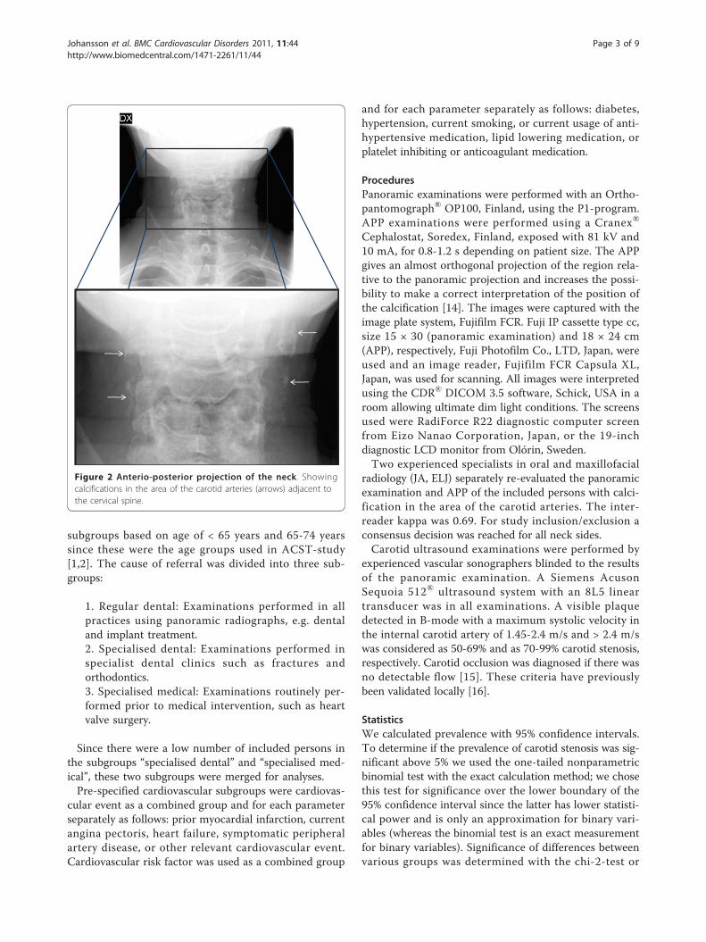
was detected on a panoramic radiograph the radio-graphic examination was extended with an anterio-pos-terior projection (APP) of the neck. If the APPconfirmed the calcification the individual was includedin the study if they were otherwise eligible for asympto-matic CEA. We excluded persons with cancer or otherserious co-morbidities whom were not eligible forasymptomatic CEA due to a short life expectancy and/or increased perioperative risk. We excluded all personswith a previous stroke or TIA since we aimed to studypersons without any pervious cerebrovascular event.Refer to Figure 1 and 2 for an example of a calcificationin the carotid arteries detected on a panoramic radio-graph and confirmed on an APP. Medical records werereviewed of all participants, see Figure 3 for trial profile.
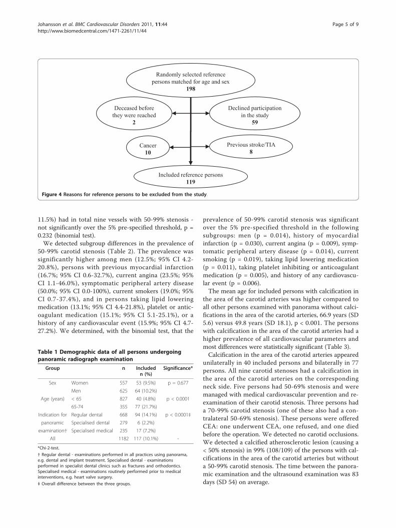
Reference groupWe selected a sex- and age-matched reference group inorder to determine if persons with calcification in thearea of the carotid arteries have an average or aboveaverage burden of atherosclerotic disease. The referenceparticipants were recruited among the persons in whomthe panoramic examination did not reveal any calcifica-tion in the area of the carotid arteries (Figure 3). Onehundred ninety-eight reference persons were randomlyselected. Their medical background was assessed viaquestionnaire; 79 were excluded based on questionnaireresults (Figure 4). The reference population did notundergo carotid ultrasound.
SubgroupsPre-specified subgroups were based on sex, age, and thecause of referral for the panoramic radiograph examina-tion. With all persons ≥ 75 years excluded, we chose
HBHB
M M CSCS
Figure 1 Panoramic image. Showing bilateral calcification in the area of the carotid arteries (arrows). (M = mandible, CS = cervical spine, HB =hyoid bone).
Johansson et al. BMC Cardiovascular Disorders 2011, 11:44http://www.biomedcentral.com/1471-2261/11/44
Page 2 of 9
subgroups based on age of < 65 years and 65-74 yearssince these were the age groups used in ACST-study[1,2]. The cause of referral was divided into three sub-groups:
1. Regular dental: Examinations performed in allpractices using panoramic radiographs, e.g. dentaland implant treatment.2. Specialised dental: Examinations performed inspecialist dental clinics such as fractures andorthodontics.3. Specialised medical: Examinations routinely per-formed prior to medical intervention, such as heartvalve surgery.
Since there were a low number of included persons inthe subgroups “specialised dental” and “specialised med-ical”, these two subgroups were merged for analyses.Pre-specified cardiovascular subgroups were cardiovas-
cular event as a combined group and for each parameterseparately as follows: prior myocardial infarction, currentangina pectoris, heart failure, symptomatic peripheralartery disease, or other relevant cardiovascular event.Cardiovascular risk factor was used as a combined group
and for each parameter separately as follows: diabetes,hypertension, current smoking, or current usage of anti-hypertensive medication, lipid lowering medication, orplatelet inhibiting or anticoagulant medication.
ProceduresPanoramic examinations were performed with an Ortho-pantomograph® OP100, Finland, using the P1-program.APP examinations were performed using a Cranex®
Cephalostat, Soredex, Finland, exposed with 81 kV and10 mA, for 0.8-1.2 s depending on patient size. The APPgives an almost orthogonal projection of the region rela-tive to the panoramic projection and increases the possi-bility to make a correct interpretation of the position ofthe calcification [14]. The images were captured with theimage plate system, Fujifilm FCR. Fuji IP cassette type cc,size 15 × 30 (panoramic examination) and 18 × 24 cm(APP), respectively, Fuji Photofilm Co., LTD, Japan, wereused and an image reader, Fujifilm FCR Capsula XL,Japan, was used for scanning. All images were interpretedusing the CDR® DICOM 3.5 software, Schick, USA in aroom allowing ultimate dim light conditions. The screensused were RadiForce R22 diagnostic computer screenfrom Eizo Nanao Corporation, Japan, or the 19-inchdiagnostic LCD monitor from Olórin, Sweden.Two experienced specialists in oral and maxillofacial
radiology (JA, ELJ) separately re-evaluated the panoramicexamination and APP of the included persons with calci-fication in the area of the carotid arteries. The inter-reader kappa was 0.69. For study inclusion/exclusion aconsensus decision was reached for all neck sides.Carotid ultrasound examinations were performed by
experienced vascular sonographers blinded to the resultsof the panoramic examination. A Siemens AcusonSequoia 512® ultrasound system with an 8L5 lineartransducer was in all examinations. A visible plaquedetected in B-mode with a maximum systolic velocity inthe internal carotid artery of 1.45-2.4 m/s and > 2.4 m/swas considered as 50-69% and as 70-99% carotid stenosis,respectively. Carotid occlusion was diagnosed if there wasno detectable flow [15]. These criteria have previouslybeen validated locally [16].
StatisticsWe calculated prevalence with 95% confidence intervals.To determine if the prevalence of carotid stenosis was sig-nificant above 5% we used the one-tailed nonparametricbinomial test with the exact calculation method; we chosethis test for significance over the lower boundary of the95% confidence interval since the latter has lower statisti-cal power and is only an approximation for binary vari-ables (whereas the binomial test is an exact measurementfor binary variables). Significance of differences betweenvarious groups was determined with the chi-2-test or
Figure 2 Anterio-posterior projection of the neck. Showingcalcifications in the area of the carotid arteries (arrows) adjacent tothe cervical spine.
Johansson et al. BMC Cardiovascular Disorders 2011, 11:44http://www.biomedcentral.com/1471-2261/11/44
Page 3 of 9

t-test; in all analyses of the prevalence of carotid stenosis,the presented subgroups were pre-specified. We pre-selected a p-value < 0.05 as cut-off for all relevant calcula-tions. SPSS version 17.0 was used for all calculations.The sample size was determined by an interim analy-
sis after inclusion of 100 persons that had undergonecarotid ultrasound examination. After the interim analy-sis the study inclusion was closed. The sample size ofreference persons was aimed to include at least as manyreference participants as there were non-referenceparticipants.
Ethical considerationsThe Regional Ethical Review Board in Umeå approvedthis study. The study was registered at http://www.
clinicaltrials.gov, NCT00514644 before it was launched.All included and reference persons provided informedconsent.
ResultsCalcification in the area of the carotid arteries, seen inpanoramic radiographs and confirmed with an APP,was detected in 178 persons. We excluded 61 personswith calcification in the area of the carotid arteriessince they either were not eligible for asymptomaticCEA, had a history of stroke or TIA, or were not ableto participate in the study (Figure 3). See Table 1 forbaseline characteristics.Eight of the included persons with a calcification in
the area of the carotid arteries (6.8%, 8/117; 95%CI 2.2-
Other serious co-morbidity8
No calcification in the area of the carotid arteries on panoramic examination + all duplicates
982
Examined with panoramic radiograph, 18-74 years of age
1182
Calcification not verified on APP
22Calcification
confirmed178
Cancer24
APP* performed for confirmation
200
Included in study117
Previous stroke/TIA15
Failed to provide informed consent
7
Examined during a temporary visit
1
Missed†6
Reference persons were selected from this population
*Anterio-posterior projection.† Some of the examinations with a finding of a calcification in the area of the carotid arteries were notinterpreted by JA or ELJ the same day as the examination, in most these cases the person was reachedfor the purpose of this study; these six missed persons were for various reasons not reached.
Figure 3 Trial profile.
Johansson et al. BMC Cardiovascular Disorders 2011, 11:44http://www.biomedcentral.com/1471-2261/11/44
Page 4 of 9
11.5%) had in total nine vessels with 50-99% stenosis -not significantly over the 5% pre-specified threshold, p =0.232 (binomial test).We detected subgroup differences in the prevalence of
50-99% carotid stenosis (Table 2). The prevalence wassignificantly higher among men (12.5%; 95% CI 4.2-20.8%), persons with previous myocardial infarction(16.7%; 95% CI 0.6-32.7%), current angina (23.5%; 95%CI 1.1-46.0%), symptomatic peripheral artery disease(50.0%; 95% CI 0.0-100%), current smokers (19.0%; 95%CI 0.7-37.4%), and in persons taking lipid loweringmedication (13.1%; 95% CI 4.4-21.8%), platelet or antic-oagulant medication (15.1%; 95% CI 5.1-25.1%), or ahistory of any cardiovascular event (15.9%; 95% CI 4.7-27.2%). We determined, with the binomial test, that the
prevalence of 50-99% carotid stenosis was significantover the 5% pre-specified threshold in the followingsubgroups: men (p = 0.014), history of myocardialinfarction (p = 0.030), current angina (p = 0.009), symp-tomatic peripheral artery disease (p = 0.014), currentsmoking (p = 0.019), taking lipid lowering medication(p = 0.011), taking platelet inhibiting or anticoagulantmedication (p = 0.005), and history of any cardiovascu-lar event (p = 0.006).The mean age for included persons with calcification in
the area of the carotid arteries was higher compared toall other persons examined with panorama without calci-fications in the area of the carotid arteries, 66.9 years (SD5.6) versus 49.8 years (SD 18.1), p < 0.001. The personswith calcification in the area of the carotid arteries had ahigher prevalence of all cardiovascular parameters andmost differences were statistically significant (Table 3).Calcification in the area of the carotid arteries appeared
unilaterally in 40 included persons and bilaterally in 77persons. All nine carotid stenoses had a calcification inthe area of the carotid arteries on the correspondingneck side. Five persons had 50-69% stenosis and weremanaged with medical cardiovascular prevention and re-examination of their carotid stenosis. Three persons hada 70-99% carotid stenosis (one of these also had a con-tralateral 50-69% stenosis). These persons were offeredCEA: one underwent CEA, one refused, and one diedbefore the operation. We detected no carotid occlusions.We detected a calcified atherosclerotic lesion (causing a< 50% stenosis) in 99% (108/109) of the persons with cal-cifications in the area of the carotid arteries but withouta 50-99% carotid stenosis. The time between the panora-mic examination and the ultrasound examination was 83days (SD 54) on average.
Declined participation in the study
59
Randomly selected reference persons matched for age and sex
198
Deceased before they were reached
2
Previous stroke/TIA8
Cancer10
Included reference persons119
Figure 4 Reasons for reference persons to be excluded from the study.
Table 1 Demographic data of all persons undergoingpanoramic radiograph examination
Group n Includedn (%)
Significance*
Sex Women 557 53 (9.5%) p = 0.677
Men 625 64 (10.2%)
Age (years) < 65 827 40 (4.8%) p < 0.0001
65-74 355 77 (21.7%)
Indication for Regular dental 668 94 (14.1%) p < 0.0001‡
panoramic Specialised dental 279 6 (2.2%)
examination† Specialised medical 235 17 (7.2%)
All 1182 117 (10.1%) -
*Chi-2-test.
† Regular dental - examinations performed in all practices using panorama,e.g. dental and implant treatment. Specialised dental - examinationsperformed in specialist dental clinics such as fractures and orthodontics.Specialised medical - examinations routinely performed prior to medicalinterventions, e.g. heart valve surgery.
‡ Overall difference between the three groups.
Johansson et al. BMC Cardiovascular Disorders 2011, 11:44http://www.biomedcentral.com/1471-2261/11/44
Page 5 of 9
DiscussionThe main finding of this study was the high prevalenceof significant (50-99%) carotid stenosis in men with inci-dentally detected calcification in the area of the carotidartery seen on panoramic examinations. Thus, in thissubpopulation, directed screening for asymptomatic car-otid stenosis is indicated [12].We missed six out of 123 eligible persons in the study.
Thus the inclusion rate of the intended population wasgood. The kappa values for the existence of a calcifica-tion in the area of the carotid arteries on each side ofthe neck showed good agreement between observers.We detected a different rate of calcifications in the
area of the carotid arteries compared with previous stu-dies [13,14]. This difference has at least three possible
explanations: 1) Not all panoramic examinations includethe area of the carotid arteries, thus calcifications in thevessels can be missed. 2) For inclusion in the study, werequired that the calcification in the area of the carotidarteries seen on panoramic radiograph should be con-firmed on APP. A calcification can be undetectable inthe APP examination if the spine superimposes the cal-cification. This may have reduced the number ofdetected calcifications in the area of the carotid arteriesin this study. 3) There are several known risk factors forarterial calcification such as age, dietary factors, andsmoking habits that differ among geographic areas [17].Thus, it is possible that the prevalence of calcificationsin the area of the carotid arteries varies between differ-ent studies populations.
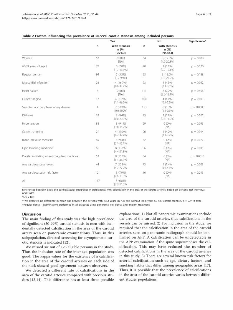
Table 2 Factors influencing the prevalence of 50-99% carotid stenosis among included persons
Yes No Significance*
n With stenosisn (%)
[95%CI]
n With stenosisn (%)
[95%CI]
Women 53 0 (0%)[NA]
64 8 (12.5%)[4.2-20.8%]
p = 0.008
65-74 years of age† 77 6 (7.8%)[1.7-13.9%]
40 2 (5.0%)[0.0-12.1%]
p = 0.570
Regular dental‡ 94 5 (5.3%)[0.7-9.9%]
23 3 (13.0%)[0.0-27.9%]
p = 0.188
Myocardial infarction 24 4 (16.7%)[0.6-32.7%]
93 4 (4.3%)[0.1-8.5%]
p = 0.032
Heart Failure 6 0 (0%)[NA]
111 8 (7.2%)[2.3-12.1%]
p = 0.496
Current angina 17 4 (23.5%)[1.1-46.0%]
100 4 (4.0%)[0.1-7.9%]
p = 0.003
Symptomatic peripheral artery disease 4 2 (50.0%)[0.0-100%]
113 6 (5.3%)[1.1-9.5%]
p = 0.0005
Diabetes 32 3 (9.4%)[0.0-20.1%]
85 5 (5.9%)[0.8-11.0%]
p = 0.505
Hypertension 88 8 (9.1%)[3.0-15.2%]
29 0 (0%)[NA]
p = 0.093
Current smoking 21 4 (19.0%)[0.7-37.4%]
96 4 (4.2%)[0.1-8.2%]
p = 0.014
Blood pressure medicine 85 8 (9.4%)[3.1-15.7%]
32 0 (0%)[NA]
p = 0.072
Lipid lowering medicine 61 8 (13.1%)[4.4-21.8%]
56 0 (0%)[NA]
p = 0.005
Platelet inhibiting or anticoagulant medicine 53 8 (15.1%)[5.1-25.1%]
64 0 (0%)[NA]
p = 0.0013
Any cardiovascular event 44 7 (15.9%)[4.7-27.2%]
73 1 (1.4%)[0.0-4.1%]
p = 0.003
Any cardiovascular risk factor 101 8 (7.9%)[2.6-13.3%]
16 0 (0%)[NA]
p = 0.243
All 117 8 (6.8%)[2.2-11.5%]
- - -
Differences between basic and cardiovascular subgroups in participants with calcification in the area of the carotid arteries. Based on persons, not individualneck-sides.
*Chi-2-test
† We detected no difference in mean age between the persons with (68.4 years SD 4.5) and without (66.8 years SD 5.6) carotid stenosis, p = 0.44 (t-test)
‡Regular dental - examinations performed in all practices using panorama, e.g. dental and implant treatment.
Johansson et al. BMC Cardiovascular Disorders 2011, 11:44http://www.biomedcentral.com/1471-2261/11/44
Page 6 of 9

We detected differences in the proportion of includedpersons between the three referral subgroups. The ‘regu-lar dental’ subgroup had the highest rate of inclusion inthe study; persons in the ‘specialised dental’ subgroupwere younger (mean age 36.5 years, SD 17.3) and shouldtherefore have a lower degree of atherosclerosis. Thirtyof 47 persons with calcification in the area of the carotidarteries in the ‘specialised medical’ group were excluded;in most cases, these persons were referred for thepanoramic radiographs due to a diagnosis of cancer orserious co-morbidity and were excluded since thesediagnoses made them ineligible for asymptomatic CEA.We included a sex- and age-matched reference group in
order to determine if the persons with calcification in thearea of the carotid arteries have an average or above aver-age burden of atherosclerotic disease. The reference groupwas representative for age, sex, and cause of referral forthe panoramic examination compared to study personswith calcifications in the area of the carotid arteries. Basedon the differences seen in Table 3 we believe that personswith calcification in the area of the carotid arteries have anabove average burden of atherosclerotic disease. In gen-eral, arterial calcification is more prevalent in persons withcardiovascular disease [17]; thus, this finding was expected.It is uncertain what the lowest degree of carotid stenosis
is that entails benefit with asymptomatic CEA. The resultsfrom the ACST study suggest that asymptomatic CEA isof similar benefit for persons with 70-99% and with 50-69% carotid stenosis [2]. Ongoing studies might clarify thebenefit vs. risk ratio of carotid surgery or stenting in
various degrees of asymptomatic carotid stenoses [7]. Dueto the present uncertainty in clinical indication, we havepresented data for 50-99% carotid stenosis.Overall, we detected a lower prevalence of carotid ste-
nosis in persons with calcification in the area of the car-otid arteries compared with previous studies [13,14].One reason for this is that we only examined personseligible for asymptomatic CEA, with age below 75 years[1,2]. Persons > 75 years of age were examined in pre-viously published studies [13,14] and the prevalence ofcarotid stenosis increases with increasing age [11]. Inthe largest of the previous studies, 94% of included per-sons were men [13]; we found men to have a higherprevalence of carotid stenosis.We found several significant subgroup heterogeneities
for the prevalence of 50-99% carotid stenosis. These find-ing were expected since they mark atherosclerotic diseasein other parts of the body. These clinical features aremost certainly not independent of each other, for exam-ple: persons with vascular events are prescribed the med-ications analysed. A multivariate analysis to test forindependence was not performed since the total numberof outcomes - i.e. 50-99% carotid stenosis - was few (n =8). We advocate that until such a multivariate analysiscan be performed in a larger study, only male sex shouldbe used as basis for selection to carotid screening withultrasound since: (1) it is one of few factors available todentists; (2) it was one of few factors that was clinicallyuseful, i.e. in addition to be significant, when positive, itincluded all findings of carotid stenosis.
Table 3 Comparisons between included persons and reference persons
Included personsn (%)
Reference personsn (%)
Significance*
Women 53 (45.3%) 56 (47.1%) p = 0.786
65-74 years 77 (65.8%) 79 (66.4%) p = 0.926
Indication - Regular dental† 94 (80.3%) 100 (84.0%) p = 0.458
Myocardial infarction 24 (20.5%) 7 (5.9%) p = 0.0009
Heart Failure 6 (5.1%) 3 (2.5%) p = 0.296
Current angina 17 (14.5%) 2 (1.7%) p = 0.0003
Symptomatic peripheral artery disease 4 (3.4%) 1 (0.8%) p = 0.169
Diabetes 32 (27.4%) 10 (8.4%) p = 0.00014
Hypertension 88 (75.2%) 54 (45.4%) p < 0.0001
Current smoking 21 (17.9%) 8 (6.7%) p = 0.009
Blood pressure medicine 85 (72.6%) 56 (47.1%) p < 0.0001
Lipid lowering medicine 61 (52.1%) 27 (22.7%) p < 0.0001
Platelet inhibiting or anticoagulant medicine 53 (45.3%) 34 (28.6%) p = 0.008
Any cardiovascular event 44 (37.6%) 12 (10.1%) p < 0.0001
Any cardiovascular risk factor 101 (86.3%) 78 (65.5%) p = 0.00019
All 117 119 -
Differences in the rate of basic and cardiovascular subgroup findings between included persons with a calcification in the area of the carotid arteries andreference persons.
*Chi-2-test
† Regular dental - examinations performed in all practices using panorama, e.g. dental and implant treatment.
Johansson et al. BMC Cardiovascular Disorders 2011, 11:44http://www.biomedcentral.com/1471-2261/11/44
Page 7 of 9
Age did not affect the prevalence of carotid stenosis inthe included population. This could be a false negativefinding since the prevalence of carotid stenosis increaseswith increasing age [11]. However, until shown other-wise, age should not be used as a criterion to go aheadwith or abstain from carotid ultrasound screening inpersons < 75 years with calcifications in the area of thecarotid arteries.We have not analysed the appearance (intensity, size,
and/or shape) of the calcification in the area of the caro-tid arteries on the panoramic images. These factorsmight be useful to further select persons for carotidscreening.Our further clinical experience confirms the results
presented here: between the study’s stop date in Febru-ary 2009 and February 2011, we have found 65 addi-tional men with calcification in the area of the carotidarteries on panoramic examinations. Carotid ultrasoundexaminations revealed that six of these individuals had50-99% carotid stenosis and two have undergone CEA.The carotid ultrasound examinations also revealed ananeurysm (12 mm in diameter) of the internal carotidartery in one person and a carotid occlusion in one per-son. Our collected experience is that, of 129 men withcalcification in the area of the carotid arteries found onpanoramic radiographs, 14 (10.9%, 95%CI 5.4-16.3%)had a 50-99% carotid stenosis and 15 (11.6%, 95%CI6.0-17.2%) had a 50-100% carotid stenosis; significantlyover the 5% pre-specified threshold, p = 0.005 and p =0.002 respectively (Binomial test).The findings of this study must be interpreted with
caution since we did not find a prevalence of carotid ste-nosis > 5% in the whole study group, but only in sub-groups [18]. However, previous similar studies onpanoramic examination report the prevalence of 50-99%carotid stenosis to be > 5% [13,14] of persons with calcifi-cations in the area of the carotid arteries; the reproduci-bility of this finding strengthens this study’s finding [18].Carotid ultrasound was not performed on persons in thereference group. There were too few cases to perform amultivariate subgroup analysis. We did not analyse onthe appearance of the calcification in the area of the caro-tid arteries. This warrants further studies, which we planto conduct.The high prevalence of carotid stenosis in the persons
with calcifications in the area of the carotid arteries wasprobably influenced by both the revelation of a localatherosclerotic process and by the above average burdenof generalised atherosclerotic disease. To what extenteach factor contributed to this finding is unknown.We only included persons that were eligible for
asymptomatic CEA; this mirrors clinical practice. Con-trary to the largest previous study, men and womenwere included in almost equal proportions; however,
only men were found to have carotid stenosis. Thus,this study is externally valid for both sexes. There wereno significant differences in the prevalence of 50-99%carotid stenosis between the subgroups based on reasonfor referral for the panoramic examination. The resultsare valid for dentists in general practice and for centresthat perform examinations corresponding to the sub-groups ‘regular dental’ or ‘specialised dental’.The design of this study intended to mirror a clinical
practice based from the ACST study [2]; the conclusionwas based on a threshold for what prevalence of carotidstenosis is required for carotid screening that was calcu-lated for the same clinical practice [12]. Nowadays, statintreatment is more common than at the time of the ACSTstudy; Recent, ongoing, and coming trials will determineif asymptomatic CEA is still indicated in patients withstatin and other cardiovascular preventive treatment, andin whom asymptomatic CEA is of benefit [7-10]. Thus, itis likely that the clinical practice for persons with asymp-tomatic carotid stenosis will change within a few years. Ifso, the threshold for what prevalence of carotid stenosisis required for carotid ultrasound screening must berecalculated based on that clinical practice. Our resultsshould then be compared to this new threshold and theconclusions revised if necessary.
ConclusionsCarotid screening with ultrasound should be performedin men with the incidental finding of calcification in thearea of the carotid arteries seen on panoramic examina-tions, confirmed with an anterio-posterior examination,if they are otherwise eligible for asymptomatic carotidendarterectomy.
List of abbreviationsAPP: Anterio-Posterior Projection; CEA: Carotid EndArterectomy; SD: StandardDeviation; TIA: Transient Ischemic Attack.
Acknowledgements and fundingThe authors acknowledge the assistance of the staff in the department ofOral and Maxillofacial Radiology for their continuous work to make the dataavailable during the study, the help of Fredrik Hermansson in gathering dataon the reference population, and the advice from Marie Eriksson, statistician.This study was supported by the Swedish Heart and Lung Foundation, theSwedish Stroke Foundation, the Northern Swedish Stroke Fund, the Countyof Västerbotten, and the medical faculty of Umeå University.
Author details1Department of Public Health and Clinical Medicine, Umeå University, Umeå,Sweden. 2Department of Odontology, Umeå University, Umeå, Sweden.3Clinical physiology, Department of Surgical and Perioperative Sciences,Umeå University, Umeå, Sweden.
Authors’ contributionsEPJ assisted in the study design, gathered most of the data, analyzed all thedata, and wrote the first draft of the manuscript. JA came up with thegeneral study design, gathered some of the data, made constructivecomments to the manuscript, and served as mentor for MG. MG gatheredsome of the data and wrote minor parts of the manuscript. KK was
Johansson et al. BMC Cardiovascular Disorders 2011, 11:44http://www.biomedcentral.com/1471-2261/11/44
Page 8 of 9
responsible for the carotid ultrasound examinations and made constructivecomments to the manuscript. ELJ assisted in the study design, gatheredsome of the data, made constructive comments to the manuscript, andserved as mentor for MG. PW came up with the general study design, madeconstructive comments to the manuscript, and served as mentor for EPJ. Allauthors have read and approved the final version of the manuscript.
Competing interestsThe authors declare that they have no competing interests.
Received: 10 March 2011 Accepted: 13 July 2011Published: 13 July 2011
References1. Chambers BR, Donnan GA: Carotid endarterectomy for asymptomatic
carotid stenosis. Cochrane Database of Systematic Reviews 2005, 4:CD001923.
2. Halliday A, Harrison M, Hayter E, Kong X, Mansfield A, Marro J, Pan H,Peto R, Potter J, Rahimi K, Rau A, Robertson S, Streifler J, Thomas D, onbehalf of the Asymptomatic Carotid Surgery Trial (ACST) CollaborativeGroup: 10-year stroke prevention after successful carotid endarterectomyfor asymptomatic stenosis (ACST-1): a multicentre randomised trial.Lancet 2010, 376:1074-1084.
3. Abbott AL: Medical (Nonsurgical) Intervention Alone Is Now Best forPrevention of Stroke Associated With Asymptomatic Severe CarotidStenosis Stroke. Stroke 2009, 40:e573-e583.
4. The ESO Guidelines. [http://www.eso-stroke.org/recommendations.php].5. Brott TG, Halperin JL, Abbara S, Bacharach JM, Barr JD, Bush RL, Cates CU,
Creager MA, Fowler SB, Friday G, Hertzberg VS, McIff EB, Moore WS,Panagos PD, Riles TS, Rosenwasser RH, Taylor AJ: 2011 ASA/ACCF/AHA/AANN/AANS/ACR/ASNR/CNS/SAIP/SCAI/SIR/SNIS/SVM/SVS Guideline onthe Management of Patients With Extracranial Carotid and VertebralArtery Disease. JACC 2011, 57:e16-94.
6. The Swedish National Board of Health and Welfare ("Socialstyrelsen”)Stroke Guidelines from 2005. [http://www.socialstyrelsen.se/Lists/Artikelkatalog/Attachments/8934/2007-102-10_200710210.pdf].
7. Reiff T, Stingele R, Eckstein HH, Fraedrich G, Jansen O, Mudra H,Mansmann U, Hacke W, Ringleb P, SPACE 2-Study Group: Stent-protectedangioplasty in asymptomatic carotid artery stenosis vs. endarterectomy:SPACE2 - a three-arm randomised-controlled clinical trial. Int J Stroke2009, 4:294-299.
8. Nicolaides AN, Kyriacou E, Griffin M, Sabetai M, Thomas DJ, Tegos T,Geroulakos G, Labropoulos N, Doré CJ, Morris TP, Naylor R, Abbott AL, forthe Asymptomatic Carotid Stenosis and Risk of Stroke (ACSRS) Study Group:Asymptomatic internal carotid artery stenosis and cerebrovascular riskstratification. J Vasc Surg 2010, 52:1486-1496.
9. Markus HS, King A, Shipley M, Topakian R, Cullinane M, Reihill S,Bornstein NM, Schaafsma A: Asymptomatic embolisation for prediction ofstroke in the Asymptomatic Carotid Emboli Study (ACES): a prospectiveobservational study. Lancet Neurol 2010, 9:663-671.
10. King a, Serena J, Bornstein NM, Markus HS, ACES Investigators: DoesImpaired Cerebrovascular Reactivity Predict Stroke Risk in AsymptomaticCarotid Stenosis? A Prospective Substudy of the Asymptomatic CarotidEmboli Study. Stroke 2011, 42:1550-1555.
11. de Weerd M, Greving JP, Hedblad B, Lorenz MW, Mathiesen EB, O’Leary DH,Rosvall M, Sitzer M, Buskens E, Bots ML: Prevalence of AsymptomaticCarotid Artery Stenosis in the General Population An IndividualParticipant Data Meta-Analysis. Stroke 2010, 41:1294-1297.
12. Qureshi AI, Alexandrov AV, Tegeler CH, Hobson RW II, Baker JD, Hopkins LN:Guidelines for Screening of Extracranial Carotid Artery Disease: AStatement for Healthcare Professionals from the MultidisciplinaryPractice Guidelines Committee of the American Society ofNeuroimaging; Cosponsored by the Society of Vascular andInterventional Neurology. J Neuroimaging 2007, 17:19-47.
13. Friedlander AH, Garrett NR, Chin EE, Baker JD: Ultrasonographicconfirmation of carotid artery atheromas diagnosed via panoramicradiography. J Am Dent Assoc 2005, 136:635-640.
14. Almog DM, Horev T, Illig KA, Green RM, Carter LC: Correlating carotidartery stenosis detected by panoramic radiography with clinicallyrelevant carotid artery stenosis determined by duplex ultrasound. OralSurg Oral Med Oral Pathol Oral Radiol Endod 2002, 94:768-773.
15. Hansen F, Bergqvist D, Lindblad B, Lindh M, Mätzsch T, Länne T: Accuracyof Duplex Sonography berfore Carotid Endarterectomy - A Comparisonwith Angiography. Eur J Vasc Endovasc Surg 1996, 12:331-336.
16. Ågren Wilsson A, Backman C, Fagerlund M, Malm J: Quality assurance ofultrasound prior to carotid surgery must be effected locally.Läkartidningen 2000, 97:2313-2316.
17. Nicoll R, Henein M: Extensive coronary calcification: a clinicallyunrecognised condition. Curr Vasc Pharmacol 2010, 8:701-705.
18. Rothwell PM: Subgroup analysis in randomized controlled trials:importance, indications and interpretation. Lancet 2005, 365:176-186.
Pre-publication historyThe pre-publication history for this paper can be accessed here:http://www.biomedcentral.com/1471-2261/11/44/prepub
doi:10.1186/1471-2261-11-44Cite this article as: Johansson et al.: Ultrasound screening forasymptomatic carotid stenosis in subjects with calcifications in the areaof the carotid arteries on panoramic radiographs: a cross-sectionalstudy. BMC Cardiovascular Disorders 2011 11:44.
Submit your next manuscript to BioMed Centraland take full advantage of:
• Convenient online submission
• Thorough peer review
• No space constraints or color figure charges
• Immediate publication on acceptance
• Inclusion in PubMed, CAS, Scopus and Google Scholar
• Research which is freely available for redistribution
Submit your manuscript at www.biomedcentral.com/submit
Johansson et al. BMC Cardiovascular Disorders 2011, 11:44http://www.biomedcentral.com/1471-2261/11/44
Page 9 of 9