research article open access the arthritis severity … · 36% (7,925) of the genes in the...
TRANSCRIPT

Brenner and Gulko BMC Genomics 2012, 13:710http://www.biomedcentral.com/1471-2164/13/710
RESEARCH ARTICLE Open Access
The arthritis severity locus Cia5a regulates theexpression of inflammatory mediators includingSyk pathway genes and proteases inpristane-induced arthritisMax Brenner1 and Pércio S Gulko1,2*
Abstract
Background: Cia5a is a locus on rat chromosome 10 that regulates disease severity and joint damage in twomodels of rheumatoid arthritis, collagen- and pristane-induced arthritis (PIA). In this study, we aimed to identifycellular and molecular processes regulated by Cia5a using microarray-based gene expression analysis ofsynovial tissues from MHC identical DA (severe erosive disease) and DA.F344(Cia5a) congenics (mild non-erosivedisease) rats.
Results: Synovial tissues from six DA and eight DA.F344(Cia5a) rats were analyzed 21 days after the induction of PIAusing the Illumina RatRef-12 BeadChip (21,922 genes) and selected data confirmed with qPCR. There was asignificantly increased expression of pro-inflammatory mediators such as Il1b (5-fold), Il18 (3.9-fold), Cxcl1 (10-fold),Cxcl13 (7.5-fold) and Ccl7 (7.9-fold), and proteases like Mmp3 (23-fold), Mmp9 (32-fold), Mmp14 (4.4-fold) andcathepsins in synovial tissues from DA, with reciprocally reduced levels in congenics. mRNA levels of 47 membersof the Spleen Tyrosine Kinase (Syk) pathway were significantly increased in DA synovial tissues compared withDA.F344(Cia5a), and included Syk (5.4-fold), Syk-activating receptors and interacting proteins, and genes regulatedby Syk such as NFkB, and NAPDH oxidase complex genes. Nuclear receptors (NR) such as Rxrg, Pparg and Rev-erbawere increased in the protected congenics, and so was the anti-inflammatory NR-target gene Scd1 (54-foldincrease). Tnn (72-fold decrease) was the gene most significantly increased in DA.
Conclusions: Analyses of gene expression in synovial tissues revealed that the arthritis severity locus Cia5aregulates the expression of key mediators of inflammation and joint damage, as well as the expression of membersof the Syk pathway. This expression pattern correlates with disease severity and joint damage and along with thegene accounting for Cia5a could become a useful biomarker to identify patients at increased risk for severe anderosive disease. The identification of the gene accounting for Cia5a has the potential to generate a new andimportant target for therapy and prognosis.
Keywords: Rheumatoid arthritis, Articular damage, Autoimmune
* Correspondence: [email protected] of Experimental Rheumatology, Center for Genomics andHuman Genetics, The Feinstein Institute for Medical Research, 350Community Drive Room 1240, Manhasset 11030, NY, USA2The Elmezzi Graduate School for Molecular Medicine, Manhasset, USA
© 2012 Brenner and Gulko; licensee BioMed Central Ltd. This is an Open Access article distributed under the terms of theCreative Commons Attribution License (http://creativecommons.org/licenses/by/2.0), which permits unrestricted use,distribution, and reproduction in any medium, provided the original work is properly cited.

Brenner and Gulko BMC Genomics 2012, 13:710 Page 2 of 16http://www.biomedcentral.com/1471-2164/13/710
BackgroundRheumatoid arthritis (RA) is a common, chronic and po-tentially debilitating form of autoimmune erosive arth-ritis. Advances in the understanding of RA pathogenesishave led to the development of new and better treat-ments [1-3]. Yet, sustained remission is still rarelyachieved [4], and more effective therapies are needed.The identification of genes implicated in the regulation
of arthritis severity and articular damage has the poten-tial to generate new and potentially better targets fortherapies aimed at preserving joint architecture andfunction, and reducing the risk of developing joint de-formities. Yet, little is known about those genes [5], andthe large cohorts of RA patients used in genome-wideassociation studies for susceptibility were not designedto address disease severity and articular damage.We have previously identified several disease severity
and articular damage quantitative trait loci (QTL) in ratmodels of RA [6-10]. Using a combination of positionalcloning and functional studies that include transcriptomeanalyses of synovial cells and synovial tissues we are be-ginning to understand the molecular processes regulatingarthritis severity and joint damage in pristane- andcollagen-induced arthritis (PIA and CIA) [10-14]. Similarstrategies have been successfully used to identify otherautoimmunity genes in rodent models [15,16].Cia5a is a 20.6Mb QTL on rat chromosome 10 that
regulates arthritis severity, cartilage and bone damage,synovial hyperplasia and inflammation in both PIA andCIA [9,10]. In the present study we used synovial tissuesfrom arthritis-protected DA.F344(Cia5a) congenics andfrom arthritis-susceptible and MHC-identical DA rats ina microarray analysis of gene expression. We determinedthat the Cia5a locus regulates the expression of severalgenes central to RA pathogenesis and joint damage, suchas cytokines Il1b and Il18, chemokines, proteases, med-iators of the synthesis of reactive oxygen species andprostaglandins, and genes involved in Toll-like receptorsignaling. Additionally, the expression of 47 members ofthe Syk kinase pathway genes, including NFκB geneswere significantly regulated by the Cia5a locus. Further-more, the presence of F344 alleles at the Cia5a intervalwas associated with increased expression of anti-inflammatory genes, including nuclear receptors andTimp3, suggesting that the Cia5a locus contains a geneinvolved in maintaining an inflammation-free synovialtissue.
ResultsDA.F344(Cia5a) congenics develop a mild form of PIAwith a distinct pattern of gene expression comparedwith DA ratsDA.F344(Cia5a) rats developed a significantly milderform of PIA compared with DA rats [median arthritis
severity score (25–75 percentiles), DA=26.5 (17–36.9),DA.F344(Cia5a)=5.5 (3.6-7.2); p=0.002, Mann–Whitneytest; Figure 1A and B].36% (7,925) of the genes in the RatRef-12 BeadChip
were consistently expressed in synovial tissues. Nearlyone-third of these genes (2,648) met the filtering criteriafor differential expression (fold-difference ≥1.5 andp≤0.01). The presence of F344 alleles at the Cia5a inter-val, as in DA.F344(Cia5a) congenic rats, was associatedwith increased expression of 1,241 genes and reducedexpression of 1,407 genes compared with DA. 134 geneshad a ≥5-fold difference between strains (Figure 1C). 46genes had a ≥10-fold difference in expression, of which19 were increased and 27 decreased in congenics, com-pared with DA (Tables 1 and 2).
Expression of pro-inflammatory genes, proteases(including matrix metalloproteases, MMPs) and adhesionmolecules was significantly increased in DA anddecreased in DA.F344(Cia5a)The 1,407 genes with increased expression in DA andreciprocally decreased expression in DA.F344(Cia5a)congenics included pro-inflammatory cytokines and che-mokines implicated in arthritis pathogenesis such as Il1b(5.17-fold on microarray, and 2.46-fold on qPCR), Il18,Mif, Ccl2, Ccl7 and Cxcl13 (Table 3 and Additional file1: Table S3 and Additional file 2: Table S4). Genes withsignificantly decreased expression in congenics alsoincluded those implicated in the development of cartil-age and bone erosions such as MMPs (Mmp3 [24-fold],Mmp9 and Mmp14), and other proteases (cathepsins D,E, K and S) (Table 3 and Figure 2). Interestingly, Syk (seebelow) has been shown to regulate the expression of dif-ferentially expressed MMPs such as Mmp3 [17] andMmp9 [18], further suggesting a potential central rolefor Syk in arthritis and a Syk-regulatory effect of Cia5a.Components of the extracellular matrix (ECM; Cthrc1,Col12a1, Emilin1) also had reduced expression in con-genics, and together with the levels of proteases sug-gested that there was reduced matrix turnover andreduced degradation, compared with arthritic DA rats(Table 3).Adhesion molecules required for leukocyte migration
into the synovium were increased in DA synovial tissuesand decreased in DA.F344(Cia5a), including integrinsItga5, Itgam, Itgb2, Itgb7, and Cd44 (Table 3). Cadherin-11 (Cdh11), a FLS-specific gene required for cell-cellinteractions and implicated in FLS invasion and synovialhyperplasia was also decreased in DA.F344(Cia5a) con-genics, consistent with the non-hyperplastic synovial tis-sue previously described in this strain, as opposed to thehighly hyperplastic synovial tissue seen in DA [10].The gene with the most significantly increased expres-
sion in DA versus DA.F344(Cia5a) was Tnn (Tenascin

Figure 1 DA and DA.F344(Cia5a) rats differ in arthritis severity and have different synovial gene expression profiles. (A) Map of theCia5a locus on rat chromosome 10, and the congenic interval boundaries (black=homozygous for F344 alleles; white=homozygous for DA alleles;grey=recombination interval). (B) DA rats had severe disease at 21 days post-induction of PIA; DA.F344(Cia5a) congenics were protected anddeveloped a significantly milder form of arthritis (p=0.002, Mann–Whitney test; boxes show the median and 25%-75% percentiles). (C) 7,925genes were expressed in all synovial tissues. 2,648 (33.4%) met the 1.5-fold difference and p-value of ≤0.01 (t-test) for significant difference.134 genes differed by ≥5-fold and less <10-fold, and 46 genes differed by ≥10-fold (inset).
Brenner and Gulko BMC Genomics 2012, 13:710 Page 3 of 16http://www.biomedcentral.com/1471-2164/13/710
N; Table 1 and Figure 2). Tnn has been implicated inosteogenesis and angiogenesis but not in arthritis orinflammation.These results demonstrate that DA rats with PIA have
increased synovial expression of many genes implicated
Table 1 Genes with ≥≥10-fold reduction in expression in DA.F3
Symbol Name
Tnn Tenascin N
Mmp9 Matrix metallopeptidase 9
Cdc2 Cell division cycle 2, G1 to S and G2 to M
Mmp3 Matrix metallopeptidase 3
Ccnb2 Cyclin B2
Cthrc1 Collagen triple helix repeat containing 1
Col12a1 Collagen, type XII, alpha 1
Slpi Secretory leukocyte peptidase inhibitor
Spc24 SPC24, NDC80 kinetochore complex component, homol
Emilin1 Elastin microfibril interfacer 1
Prc1 Protein regulator of cytokinesis 1
Emb Embigin
Nuf2 NUF2, NDC80 kinetochore complex component, homolo
Lbp Lipopolysaccharide binding protein
Cks2 CDC28 protein kinase regulatory subunit 2
Wisp1 WNT1 inducible signaling pathway protein 1
Cxcl1 Chemokine (C-x-C motif) ligand 1
Steap1 Six transmembrane epithelial antigen of the prostate 1
LOC687334 Similar to cytoskeleton associated protein 2
in RA pathogenesis, further validating the molecularsimilarities between PIA and RA, and underscoring thepotential relevance of both Cia5a in arthritis pathogen-esis and the present study in discovering new key genesand pathways regulating arthritis.
44(Cia5a) compared with DA
Entrez Gene ID Fold reduction p-value
304913 71.69 8.6x10⁻¹³
81687 32.79 1.4x10⁻⁹
54237 24.16 1.6x10⁻⁵
171045 23.94 5.0x10⁻⁵
363088 22.95 5.0x10⁻⁶
282836 17.93 3.2x10⁻⁹
25683 16.25 7.6x10⁻⁸
84386 16.16 6.4x10⁻⁴
og 363028 14.72 2.5x10⁻⁶
298845 13.55 2.4x10⁻⁶
308761 13.08 1.6x10⁻⁵
114511 12.82 1.4x10⁻⁵
g 304951 12.52 2.5x10⁻⁶
29469 11.82 8.0x10⁻⁵
498709 11.62 5.9x10⁻⁶
65154 11.59 9.0x10⁻⁸
81503 10.88 4.3x10⁻⁴
297738 10.61 1.7x10⁻⁴
687334 10.16 9.1x10⁻⁶
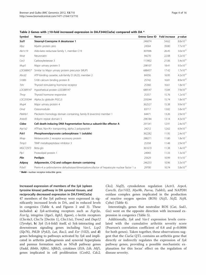
Table 2 Genes with ≥10-fold increased expression in DA.F344(Cia5a) compared with DA *
Symbol Name Entrez Gene ID Fold increase p-value
Scd1 Stearoyl-Coenzyme A desaturase 1 246074 54.62 8.8x10⁻⁵
Mpz Myelin protein zero 24564 39.80 7.7x10⁻⁵
Akr1c19 Aldo-keto reductase family 1, member C19 307096 26.45 3.0x10⁻⁶
Nnat Neuronatin 94270 22.08 5.2x10⁻⁷
Ces3 Carboxylesterase 3 113902 21.06 5.4x10⁻⁶
Mup5 Major urinary protein 5 298107 18.41 9.5x10⁻⁵
LOC688457 Similar to Major urinary protein precursor (MUP) 688457 17.42 1.7x10⁻⁴
Abcd2 ATP-binding cassette, sub-family D (ALD), member 2 84356 16.95 4.2x10⁻⁶
S100b S100 calcium binding protein B 25742 16.81 8.9x10⁻⁸
Tshr Thyroid stimulating hormone receptor 25360 16.61 1.8x10⁻⁷
LOC689147 Hypothetical protein LOC689147 689147 15.84 7.9x10⁻⁶
Thrsp Thyroid hormone responsive 25357 15.76 1.2x10⁻⁵
LOC259244 Alpha-2u globulin PGCL3 259244 15.74 1.8x10⁻⁴
Mup4 Major urinary protein 4 362527 15.38 9.9x10⁻⁵
Omd Osteomodulin 83717 13.82 5.8x10⁻⁶
Plekhb1 Pleckstrin homology domain containing, family B (evectins) member 1 64471 13.26 2.9x10⁻⁵
Ankrd5 Ankyrin repeat domain 5 296184 13.14 4.3x10⁻⁵
Cidea Cell death-inducing DNA fragmentation factor,a subunit-like effector A 291541 12.91 2.4x10⁻⁶
Atp1a2 ATPase, Na+/K+ transporting, alpha 2 polypeptide 24212 12.62 6.9x10⁻⁵
Pck1 Phosphoenolpyruvate carboxykinase 1 (soluble) 362282 11.95 2.4x10⁻⁵
Mrap Melanocortin 2 receptor accessory protein 288271 11.80 1.8x10⁻⁷
Timp3 TIMP metallopeptidase inhibitor 3 25358 11.48 2.9x10⁻⁹
MGC72973 Beta-glo 361619 11.38 1.4x10⁻⁶
Plp1 Proteolipid protein 1 24943 11.33 1.3x10⁻⁵
Plin Perilipin 25629 10.99 9.1x10⁻⁷
Adipoq Adiponectin, C1Q and collagen domain containing 246253 10.96 3.3x10⁻⁶
Pcbd1 Pterin-4 a-carbinolamine dehydratase/dimerizationcofactor of hepatocyte nuclear factor 1 a 29700 10.74 5.8x10⁻⁸
* Bold= nuclear receptor-inducible gene.
Brenner and Gulko BMC Genomics 2012, 13:710 Page 4 of 16http://www.biomedcentral.com/1471-2164/13/710
Increased expression of members of the Syk (spleentyrosine kinase) pathway in DA synovial tissues, andreciprocally decreased expression in DA.F344(Cia5a)47 members of the Syk pathway were expressed in sig-nificantly increased levels in DA, and in reduced levelsin congenics (Table 4, and Figures 2 and 3). Theseincluded: a) Syk-activating receptors such as Fcgr2a,Fcer1g, integrins (Itga5, Itgb2, Itgam), c-lectin receptors(Clec4a3, Clec7a [Dectin 1], Clec11a), Trem2 and Dap12(Tyrobp), b) Syk (5.4-fold) itself, c) Syk-interacting anddownstream signaling genes including Vav1, Lcp2(Slp76), Ptk2b (Pyk2), Lat, Rac2, and Ezr (Vil2), and d)genes belonging to pathways activated by Syk and impli-cated in arthritis pathogenesis and synovial hyperplasiaand pannus formation such as NFκB pathway genes(Fadd, Ikbkb, Nfkb1, Nfkb2), cytokines (Il1b, Ltb, Mif ),genes implicated in cell proliferation (Ccnb2, Cdc2,
Cks2, Nuf2), cytoskeleton regulation (Actr3, Arpc4,Coro1b, Ezr/Vil2, Myo9b, Parva, Tubb5), and NAPDHoxidase complex genes implicated in the productionof reactive oxygen species (ROS) (Ncf1, Ncf2, Ncf4,Cyba) (Table 4).Interestingly, genes that neutralize ROS (Cat, Sod1,
Gss) went on the opposite direction with increased ex-pression in congenics (Table 5).Additionally, Syk and Vav1 expression levels corre-
lated with the cumulative arthritis severity score(Pearson’s correlation coefficient of 0.8 and p=0.0006for both genes). Taken together, these observations sug-gest that the Cia5a QTL contains an arthritis gene thatdirectly or indirectly regulates the expression of Sykpathway genes, providing a possible mechanistic ex-planation for this locus’ effect on the regulation ofdisease severity.

Table 3 Mediators of inflammation and articular damage up-regulated in DA synovium and down-regulation inDA.F344(Cia5a)
Gene Symbol Gene Name Entrez Gene ID Fold DA/Cia5a p-value*
Cytokines and chemokines
Il1b interleukin 1 beta 24494 5.17 0.002
Il18 interleukin 18 29197 3.91 0.0002
Ltb lymphotoxin beta 361795 3.77 0.0002
Mif macrophage migration inhibitory factor 81683 2.37 0.0002
Aif1 allograft inflammatory factor 1 29427 2.48 0.0001
Ccl2 chemokine (C-C motif) ligand 2 24770 3.95 0.01
Ccl7 chemokine (C-C motif) ligand 7 287561 7.90 0.002
Cxcl1 chemokine (C-X-C motif) ligand 1 81503 10.88 0.0004
Cxcl13 chemokine (C-X-C motif) ligand 13 498335 7.53 0.000004
Proteases
Mmp3 matrix metallopeptidase 3 171045 23.94 0.0001
Mmp9 matrix metallopeptidase 9 81687 32.79 0.000000001
Mmp14 matrix metallopeptidase 14 81707 4.46 0.000000004
Mmp19 matrix metallopeptidase 19 304608 5.63 0.0000003
Ctsc cathepsin C 25423 2.64 0.00007
Ctsd cathepsin D 171293 1.90 0.000002
Ctse cathepsin E 25424 2.03 0.0002
Ctsk cathepsin K 29175 3.20 0.0000003
Ctss cathepsin S 50654 1.68 0.002
Extra-cellular matix
Cthrc1 collagen triple helix repeat containing 1 282836 17.93 0.000000003
Col12a1 collagen, type XII, alpha 1 25683 16.25 0.00000008
Emilin1 elastin microfibril interfacer 1 298845 13.55 0.000002
Adhesion molecules
Itga5 integrin alpha 5 (fibronectin receptor alpha) 315346 3.47 0.00000001
Itgam integrin alpha M 25021 3.58 0.0001
Itgav integrin alpha V 296456 1.61 0.0004
Itgb2 integrin beta 2 309684 3.44 0.0001
Itgb7 integrin, beta 7 25713 3.86 0.002
Cd44 Cd44 molecule 25406 1.83 0.005
Cdh11 cadherin 11 84407 1.87 0.003
Toll-like receptors and regulators of their activity
Cd14 CD14 60350 2.13 0.001
Irak4 interleukin-1 receptor-associated kinase 4 300177 1.65 0.0004
Lpb lipopolysaccharide binding protein 29469 4.36 0.0003
Myd88 myeloid differentiation primary response gene 88 301059 1.59 0.002
Pycard PYD and CARD domain containing 282817 1.95 0.0004
Tlr2 toll-like receptor 2 310553 4.36 0.000
Tlr6 toll-like receptor 6 305353 1.76 0.001
Brenner and Gulko BMC Genomics 2012, 13:710 Page 5 of 16http://www.biomedcentral.com/1471-2164/13/710

Table 3 Mediators of inflammation and articular damage up-regulated in DA synovium and down-regulation inDA.F344(Cia5a) (Continued)
Prostaglandin and leukotriene synthesis
Ptgs2 prostaglandin-endoperoxide synthase 2 29527 9.73 0.0006
Pla2g4a phospholipase A2, group IVA (cytosolic, calcium-dependent) 24653 2.04 0.0004
*t-test.
Brenner and Gulko BMC Genomics 2012, 13:710 Page 6 of 16http://www.biomedcentral.com/1471-2164/13/710
DA.F344(Cia5a) congenics have reduced synovialexpression of innate immune response-activating genes,including members of the inflammasomeExpression levels of genes implicated in innate immuneresponses were significantly increased in DA, and decreasedin DA.F344(Cia5a). In addition to the Syk pathway, andmediators of ROS synthesis, and regulators of cytokinetranscription such as members of the NFκB pathways dis-cussed above, DA.F344(Cia5a) congenics also had reducedexpression of AP-1 genes (Fos and JunB), Il1b and othermembers of the inflammasome (Card11, Nalp3 [bothdetected only in DA], and Pycard). Pattern recognitionreceptors such as Zbp1 and Lgp2 (both detected only inDA), and components of the toll-like receptor (TLR) path-way (Cd14, Ikbke, Irak4, Lbp, MyD88, Tlr2, Tlr6, Ticam1;Table 3) were expressed in increased levels in DA anddecreased in congenics. Interestingly, and in line withthese observations, the expression levels of negative
Figure 2 qPCR validation of the microarray results. Genes expressed inincreased levels in DA.F344(Cia5a) congenics (three genes, grey bars) wereThe same RNAs used in the microarray experiments were used for qPCR. Δ
regulators of TLR signaling such as Ptpn11 and Pparg wasconversely increased in congenics, suggesting that thearthritis gene located within the Cia5a QTL might medi-ate the balance between activating and inhibitory signalsimplicated in TLR signaling.Genes implicated in the synthesis of prostaglandins
and leukotrienes (Pla2g4a, Ptgs2/Cox2, Ptges) were alsoincreased in DA (Table 3), while genes that counteracteicosanoid-mediated inflammation (Ptgis, Cyp2j3) wereincreased in congenics (Table 5).
Increased expression of anti-inflammatory genes,including nuclear receptors (NRs), in synovial tissues fromDA.F344(Cia5a) CongenicsSeveral genes with known anti-inflammatory and cytokine-suppressing properties were expressed in increased levels inDA.F344(Cia5a) synovial tissues, and reduced in DA. These
increased levels in DA (six genes, black bars) and genes expressed inselected for qPCR confirmation. Fold-differences were log-transformed.Ct was used for statistical analyses; all genes had p≤0.04, t-test..

Table 4 Members of the Syk kinase pathway up-regulated in DA synovium compared with down-regulation inDA.F344(Cia5a)
Gene Symbol Gene Name Entrez GeneID
Fold DA/Cia5a
p-value*
Activating receptors
Fcer1g Fc fragment of IgE, high affinity I, receptor for; gamma polypeptide 25441 2.50 0.0004
Fcgr2a Fc fragment of IgG, low affinity IIa, receptor (CD32) 116591 3.08 0.005
Tcrg T cell receptor gamma locus 24821 3.32 0.0001
Trem2 triggering receptor expressed on myeloid cells 2 301227 2.69 0.0006
Tyrobp Tyro protein tyrosine kinase binding protein 361537 4.80 0.0001
Integrins
Itgam integrin alpha M 25021 3.58 0.0001
Itgav integrin alpha V 296456 1.61 0.0004
Itga5 integrin alpha 5 (fibronectin receptor alpha) 315346 3.47 0.00000001
Itgb2 integrin beta 2 309684 3.44 0.0001
Itgb7 integrin, beta 7 25713 3.86 0.002
c-type lectin receptors
Clec4a1 C-type lectin domain family 4, member a1, Dcir4 362430 2.38 0.001
Clec4a3 C-type lectin domain family 4, member a3, Dcir3 362431 3.00 0.0004
Clec7a C-type lectin domain family 7, member a, Dectin 1 502902 8.48 0.001
Clec11a C-type lectin domain family 11, member a, Scgf 29313 4.24 0.00000008
Syk and Syk-binding and intermediate partners
Ezr** ezrin 54319 2.42 0.0001
Lat linker for activation of T cells 81511 3.83 0.000001
Lcp2 lymphocyte cytosolic protein 2 155918 2.02 0.007
Ptk2b PTK2B protein tyrosine kinase 2 beta 50646 2.54 0.0000002
RhoG ras homolog gene family, member G (rho G) 308875 1.54 0.0008
RhoH ras homolog gene family, member H 305341 2.25 0.001
Syk spleen tyrosine kinase 25155 5.43 0.00004
Vav1 vav 1 guanine nucleotide exchange factor 25156 4.60 0.0001
NFκB genes andpathway
Ikbkb inhibitor of kappa light polypeptide gene enhancer in B-cells, kinase beta 84351 1.60 0.0006
Ikbke inhibitor of kappa light polypeptide gene enhancer in B-cells, kinaseepsilon
363984 1.64 0.0001
Nfkb1 nuclear factor of kappa light polypeptide gene enhancer in B-cells 1 81736 1.88 0.002
Nfkb2 nuclear factor of kappa light polypeptide gene enhancer in B-cells 2, p49/p100
309452 1.98 0.00003
Cytokine and chemokine transcription§
Ccl2 chemokine (C-C motif) ligand 2 24770 3.95 0.01
Ccl7 chemokine (C-C motif) ligand 7 287561 7.90 0.002
Il1b interleukin 1 beta 24494 5.17 0.002
Ltb lymphotoxin beta (TNF superfamily, member 3) 361795 3.77 0.0002
Brenner and Gulko BMC Genomics 2012, 13:710 Page 7 of 16http://www.biomedcentral.com/1471-2164/13/710
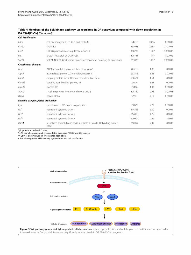
Table 4 Members of the Syk kinase pathway up-regulated in DA synovium compared with down-regulation inDA.F344(Cia5a) (Continued)
Cell Proliferation
Cdc2 cell division cycle 2, G1 to S and G2 to M 54237 24.16 0.00002
Ccnb2 cyclin B2 363088 22.95 0.000005
Cks2 CDC28 protein kinase regulatory subunit 2 498709 11.62 0.000006
Prc1 protein regulator of cytokinesis 1 308761 13.08 0.00002
Spc24 SPC24, NDC80 kinetochore complex component, homolog (S. cerevisiae) 363028 14.72 0.000002
Cytoskeletal changes
Actr3 ARP3 actin-related protein 3 homolog (yeast) 81732 1.88 0.0001
Arpc4 actin related protein 2/3 complex, subunit 4 297518 1.61 0.00005
Capzb capping protein (actin filament) muscle Z-line, beta 298584 1.64 0.0003
Coro1b coronin, actin-binding protein, 1B 29474 1.68 0.0001
Myo9b myosin IXb 25486 1.93 0.00003
Tiam2 T-cell lymphoma invasion and metastasis 2 308142 2.61 0.00003
Parva parvin, alpha 57341 2.19 0.00005
Reactive oxygen species production
Cyba cytochrome b-245, alpha polypeptide 79129 2.72 0.00001
Ncf1 neutrophil cytosolic factor 1 114553 6.00 0.0001
Ncf2 neutrophil cytosolic factor 2 364018 4.75 0.0003
Ncf4 neutrophil cytosolic factor 4 500904 2.46 0.004
Rac2¶ ras-related C3 botulinum toxin substrate 2 (small GTP binding proteinRac2)
366957 2.32 0.0007
Syk gene is underlined. * t-test;§=All four chemokine and cytokine listed genes are NFkB-inducible targets.** Ezrin is also involved in cytoskeleton regulation.¶ Rac also regulates NFkB activity, cytoskeleton and cell proliferation.
Figure 3 Syk pathway genes and Syk-regulated cellular processes. Genes, gene families and cellular processes with members expressed inincreased levels in DA synovial tissues, and significantly reduced levels in DA.F344(Cia5a) congenics.
Brenner and Gulko BMC Genomics 2012, 13:710 Page 8 of 16http://www.biomedcentral.com/1471-2164/13/710

Table 5 Anti-inflammatory genes and nuclear receptors up-regulated in DA.F344(Cia5a) congenics
Gene Symbol Gene Name Entrez Gene ID Fold difference DA/Cia5a p-value*
Anti-inflammatory and regulators of immune responses
Scd1 stearoyl-Coenzyme A desaturase 1 246074 54.62 0.0001
Timp3 TIMP metallopeptidase inhibitor 3 25358 11.48 0.000000003
Adipoq adiponectin, C1Q and collagen domaincontaining
246253 10.96 0.000003
Ptpn11 protein tyrosine phosphatase, non-receptortype 11
25622 4.18 0.00002
Cyp2j3 cytochrome P450, family 2, subfamily j,polypeptide 3
313375 3.38 0.002
Ptgis prostaglandin I2 (prostacyclin) synthase 25527 2.63 0.0003
Cat catalase 24248 1.91 0.002
Gss glutathione synthetase 25458 1.56 0.001
Sod1 superoxide dismutase 1, soluble 24786 1.53 0.00002
Nuclear receptors and an interacting protein
Rxrg retinoid X receptor gamma, Nr2b3 83574 5.39 0.0001
Pparg peroxisome proliferator-activated receptorgamma, Nr1c3
25664 5.32 0.00005
Rev-erba Nr1d1 252917 3.41 0.004
Arp1 Nr2f2 113984 2.45 0.0002
Nrip1 nuclear receptor interacting protein 1 304157 2.35 0.00004
Thrb thyroid hormone receptor beta, Nr1a2 24831 2.07 0.002
Thra thyroid hormone receptor alpha, Nr1a1 81812 1.97 0.001
Ncor1 nuclear receptor co-repressor 1 54299 1.89 0.003
Rora RAR-related orphan receptor alpha, Nr1f1 300807 1.88 0.0001
Lxra Liver X receptor alpha, Nr1h3 58852 1.52 0.0006
* t-test.
Brenner and Gulko BMC Genomics 2012, 13:710 Page 9 of 16http://www.biomedcentral.com/1471-2164/13/710
included the NRs Lxra, Pparg, Rev-erba, Rora, Thra, andThrb (Table 5).Scd1 was the gene with the most significantly
increased expression in DA.F344(Cia5a) congenics witha 55-fold difference compared with DA (Tables 2 and 5).Scd1 has been shown to reduce cytokine levels and to haveanti-inflammatory activity [19,20]. We have previouslyreported that Scd1 is expressed in significantly reducedlevels in synovial tissues from rats with severe arthritis,and increased in the synovial tissues of yet anotherarthritis-protected congenic strain [14].Adipoq and Timp3, which is an inhibitor of the TNFα
converting enzyme (TACE), were two additional anti-inflammatory genes expressed in significantly increasedlevels (>10-fold) in DA.F344(Cia5a) (Table 5).Scd1 and some of the other genes up-regulated in DA.
F344(Cia5a) synovial tissues such as Adipoq, Cidea,Cd36, Fabp4, Gpd1, Lpl, Lpin1, Mgst1, Plin, Pck1, Slc2a4and Srebf1, are known to be inducible by NRs (Table 2and Table 5). These observations suggest that NRs werenot only expressed in increased levels but also had
increased activity in synovial tissues from DA.F344(Cia5a) compared with DA.
Genes located within the Cia5a interval have significantlydifferent expression levels75 of the 7,925 genes expressed by all samples werelocated within the Cia5a interval. 21 of these 75 hadincreased expression in DA synovial tissues, and 11 wereincreased in the congenics. 14 of these 32 differentiallyexpressed genes had ≥2-fold-difference. Sphk1 andSectm1b were the genes contained within the Cia5ainterval with the most significantly increased expressionin DA (7.58 and 7.61-fold, respectively), while Itgb4 andDIgr1 and were those with the most significantlyincreased expression in DA.F344(Cia5a) congenics (2.77and 2.85-fold, respectively) (Table 6). Additionally, fourgenes located within the Cia5a interval were expressedonly or predominantly in DA synovium, while two othergenes were expressed predominantly in DA.F344(Cia5a)(Table 6). It is conceivable that these differences in ex-pression levels of genes located within the Cia5a interval

Table 6 Differentially expressed candidate genes located within the Cia5a interval on rat chromosome 10*
Gene Symbol Gene name Entrez GeneID
Difference p-value§
Increased in DA Fold DA/Cia5a
Igsf7 immunoglobulin superfamily, member 7 287813 4.42 0.00008
Lgals3bp lectin, galactoside-binding, soluble, 3 binding protein 245955 3.85 0.000001
RGD1309310 similar to mKIAA0195 protein 303677 3.89 0.00001
Sectm1b secreted and transmembrane 1B 287884 7.61 0.00001
Slc16a3 solute carrier family 16, member 3 80878 5.36 0.000005
Sphk1 sphingosine kinase 1 170897 7.58 0.0000003
Increased in DA.F344(Cia5a) Fold Cia5a/DA
Itgb4 integrin beta 4 25724 2.85 0.0004
RGD1311422 similar to CG8841-PA 287822 2.57 0.002
RGD1561778 similar to dendritic cell-derived immunoglobulin(Ig)-likereceptor 1, DIgR1
303666 2.77 0.002
Slc25a10 solute carrier family 25, member 10 170943 2.15 0.004
Expressed only or predominantly in DA Frequency DA:Cia5a
Cd300lf CD300 molecule-like family, member f 287818 6:0 0.0003
Fdxr ferredoxin reductase 79122 6:1 0.0047
Cd7 CD7 molecule 303747 6:0 0.0003
Sectm1a secreted and transmembrane 1A 287885 6:1 0.0047
Expressed only or predominantly in DA.F344(Cia5a)
Aanat arylalkylamine N-acetyltransferase 25120 0:7 0.0047
Hrnbp3 hexaribonucleotide binding protein 3 287847 1:6 0.1♯
*List contains the most signficantly differentially expressed genes; § t-test was used to compare means for fold-difference calculations and Fisher's Exact test tocompare frequencies. #not statistically significant.
Brenner and Gulko BMC Genomics 2012, 13:710 Page 10 of 16http://www.biomedcentral.com/1471-2164/13/710
could be explained at least in part by a polymorphism inthe 5’ untranslated region (UTR) that affects a transcrip-tion factor binding site in cis, thus affecting transcriptionefficiency, or a 3’ UTR polymorphism affecting mRNAstability.
Gene targets of microRNAs (miRNA) contained within theCia5a interval were not differentially expressedThe Cia5a interval contains six predicted miRNAs. Weconsidered the possibility that polymorphisms in one ofthose six miRNAs could account for the Cia5a effect ongene expression and arthritis severity. In that case, suchpolymorphism would affect the miRNA activity on thetranscription of its target genes. Therefore, we look forthe differential expression of targets of all six predictedmiRNAs located within the Cia5a interval. A list of tar-get and non-target genes was generated for each of thesix miRNAs, but no significant over-representation oftargets was detected (Figure 4), suggesting that poly-morphisms affecting the expression or function of themiRNAs contained within the Cia5a interval are lesslikely to explain the differences in gene expression iden-tified in this study.
Analyses of cell type specific genes suggests synovialtissue cellularity differences between DA andDA.F344(Cia5a) Congenics13 genes known to be specifically expressed by the celltypes of interest to this study were used to compare DAand DA.F344(Cia5a) synovial tissues (Additional file 3:Table S2). The expression levels of those genes suggestedincreased numbers of FLS (consistent with synovialhyperplasia), macrophages, dendritic cells (DC), neutro-phils and T cells in the synovial tissues of DA, comparedwith congenics (Additional file 3: Table S2). No genespecific for B cells, NK cells or Tregs were among thosedifferentially expressed between the two strains, suggest-ing that the number of these cells in the synovial tissuesof these two strains was not significantly different.
DiscussionDisease severity and articular damage are associated withincreased risk for disability, joint deformities andreduced life expectancy in patients with RA [21-23]. Yet,little is known about the genes implicated in the regula-tion of disease severity and articular damage genes inRA, and these genes could be the most relevant targets
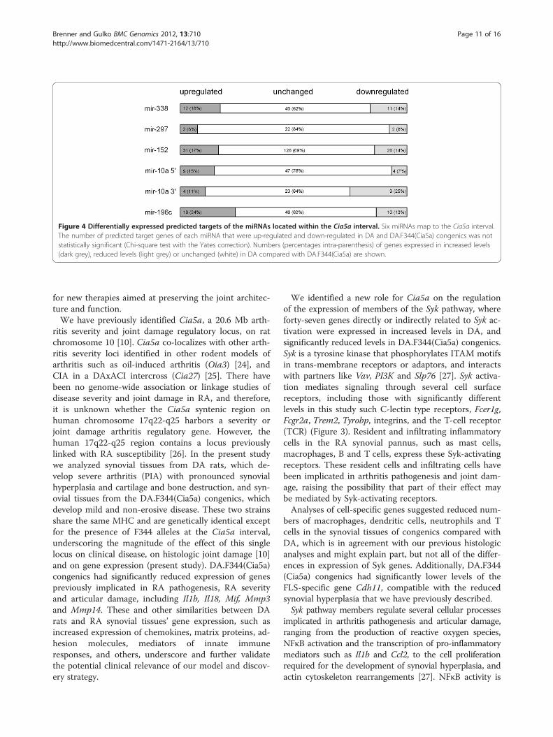
Figure 4 Differentially expressed predicted targets of the miRNAs located within the Cia5a interval. Six miRNAs map to the Cia5a interval.The number of predicted target genes of each miRNA that were up-regulated and down-regulated in DA and DA.F344(Cia5a) congenics was notstatistically significant (Chi-square test with the Yates correction). Numbers (percentages intra-parenthesis) of genes expressed in increased levels(dark grey), reduced levels (light grey) or unchanged (white) in DA compared with DA.F344(Cia5a) are shown.
Brenner and Gulko BMC Genomics 2012, 13:710 Page 11 of 16http://www.biomedcentral.com/1471-2164/13/710
for new therapies aimed at preserving the joint architec-ture and function.We have previously identified Cia5a, a 20.6 Mb arth-
ritis severity and joint damage regulatory locus, on ratchromosome 10 [10]. Cia5a co-localizes with other arth-ritis severity loci identified in other rodent models ofarthritis such as oil-induced arthritis (Oia3) [24], andCIA in a DAxACI intercross (Cia27) [25]. There havebeen no genome-wide association or linkage studies ofdisease severity and joint damage in RA, and therefore,it is unknown whether the Cia5a syntenic region onhuman chromosome 17q22-q25 harbors a severity orjoint damage arthritis regulatory gene. However, thehuman 17q22-q25 region contains a locus previouslylinked with RA susceptibility [26]. In the present studywe analyzed synovial tissues from DA rats, which de-velop severe arthritis (PIA) with pronounced synovialhyperplasia and cartilage and bone destruction, and syn-ovial tissues from the DA.F344(Cia5a) congenics, whichdevelop mild and non-erosive disease. These two strainsshare the same MHC and are genetically identical exceptfor the presence of F344 alleles at the Cia5a interval,underscoring the magnitude of the effect of this singlelocus on clinical disease, on histologic joint damage [10]and on gene expression (present study). DA.F344(Cia5a)congenics had significantly reduced expression of genespreviously implicated in RA pathogenesis, RA severityand articular damage, including Il1b, Il18, Mif, Mmp3and Mmp14. These and other similarities between DArats and RA synovial tissues’ gene expression, such asincreased expression of chemokines, matrix proteins, ad-hesion molecules, mediators of innate immuneresponses, and others, underscore and further validatethe potential clinical relevance of our model and discov-ery strategy.
We identified a new role for Cia5a on the regulationof the expression of members of the Syk pathway, whereforty-seven genes directly or indirectly related to Syk ac-tivation were expressed in increased levels in DA, andsignificantly reduced levels in DA.F344(Cia5a) congenics.Syk is a tyrosine kinase that phosphorylates ITAM motifsin trans-membrane receptors or adaptors, and interactswith partners like Vav, PI3K and Slp76 [27]. Syk activa-tion mediates signaling through several cell surfacereceptors, including those with significantly differentlevels in this study such C-lectin type receptors, Fcer1g,Fcgr2a, Trem2, Tyrobp, integrins, and the T-cell receptor(TCR) (Figure 3). Resident and infiltrating inflammatorycells in the RA synovial pannus, such as mast cells,macrophages, B and T cells, express these Syk-activatingreceptors. These resident cells and infiltrating cells havebeen implicated in arthritis pathogenesis and joint dam-age, raising the possibility that part of their effect maybe mediated by Syk-activating receptors.Analyses of cell-specific genes suggested reduced num-
bers of macrophages, dendritic cells, neutrophils and Tcells in the synovial tissues of congenics compared withDA, which is in agreement with our previous histologicanalyses and might explain part, but not all of the differ-ences in expression of Syk genes. Additionally, DA.F344(Cia5a) congenics had significantly lower levels of theFLS-specific gene Cdh11, compatible with the reducedsynovial hyperplasia that we have previously described.Syk pathway members regulate several cellular processes
implicated in arthritis pathogenesis and articular damage,ranging from the production of reactive oxygen species,NFκB activation and the transcription of pro-inflammatorymediators such as Il1b and Ccl2, to the cell proliferationrequired for the development of synovial hyperplasia, andactin cytoskeleton rearrangements [27]. NFκB activity is

Brenner and Gulko BMC Genomics 2012, 13:710 Page 12 of 16http://www.biomedcentral.com/1471-2164/13/710
regulated by Syk and by several other pathways includingTLRs and cytokine receptors [28]. The NFκB pathway hasa central role in the production of pro-inflammatory cyto-kines such as IL-1β, IL-6 and TNFα, in the developmentof synovial hyperplasia and in disease severity [29-31].Actin cytoskeleton rearrangements are also regulated bythe Syk pathway [27], and are required for the migrationof inflammatory cells into the synovial tissue, and for syn-ovial cells and synovial tissue invasion and destruction ofcartilage [13,32]. Therefore, our observations suggest thata gene located within the Cia5a interval is a new regulatorof the expression of Syk pathway genes implicated in keyprocesses in arthritis pathogenesis.The precise mechanisms whereby Cia5a regulates the
expression of Syk genes remain unclear, and might reflectdifferences in tissue cellularity, multiple cell-activatingprocesses, or a polymorphism in transcription factorlocated within the Cia5a interval that affects transcription.Studies by our group of synovial tissues obtained fromfour different congenic strains yielded different results ingene expression (Brenner et al., manuscript in prepar-ation) [12,14,33], suggesting that the Syk-regulatory effectof Cia5a is a specific observation, and not simply relatedcellularity differences or inflammation.Syk has been recently implicated in arthritis pathogen-
esis and joint damage, and Syk-deficient mice are pro-tected from autoantibody-induced erosive arthritis [34],and treatment with a SYK inhibitor significantly reduceddisease severity and joint erosions and damage incollagen-induced arthritis [35]. Both the total and phos-phorylated forms of SYK are expressed in increasedlevels in RA synovial tissues compared with osteoarth-ritis, and SYK inhibition reduced the expression of IL-6and MMP-3 [17]. More importantly, the use of a SYKinhibitor significantly reduced disease activity in patientswith RA [36], with 67%, 43% and 28% of patients achiev-ing ACR20, ACR50 and ACR70, respectively, in a 3-month double-blind and placebo-controlled study [37].Therefore, it is conceivable that the Syk pathway genesdifferentially expressed in this study could help identifypatients more likely to benefit from therapy with SYKinhibitors. Additionally, Syk is critical to TNFα-inducedresponses [38], raising the possibility that the Syk path-way 47-gene signature could be used to predictincreased TNFα activity prior to choosing a biologictherapeutic agent. Additionally, the increased expressionof Syk pathway genes could identify patients at increasedrisk to develop erosive disease and could become a prog-nostic tool. Lastly, the Cia5a gene itself has the potentialto become a new target for therapies aimed at reducingarticular damage via inhibition of Syk pathway genes, in-cluding processes downstream from Syk such as NFκB.While several genes with pro-inflammatory, proteo-
lytic, innate immunity and inflamasome-related activity
were expressed in reduced levels in DA.F344(Cia5a) con-genics, groups of genes with known anti-inflammatoryproperties were expressed in increased levels in con-genics. These genes included Timp3, Ptpn11, antagonistsof reactive oxygen species (Cat, Gss, Sod1) and nuclearreceptors. Nuclear receptors such as Lxra, Pparg andRora have been shown to interfere with NFκB and AP-1activation [39-41], and to have anti-inflammatory andarthritis-suppressive properties [42-45]. Rxrg was an-other nuclear receptor expressed in significantlyincreased levels in DA.F344(Cia5a) congenics. WhileRxrg itself has not been studied in the context of arth-ritis, it dimerizes with Lxra, Pparg, and with Vdr, and isrequired for their anti-inflammatory activity. Addition-ally, several nuclear receptor-inducible genes, includingthe inflammation-suppressor Scd1 [20] were expressedin increased levels in the synovial tissues of the con-genics. These observations suggest that not only nuclearreceptor levels were increased, but also their activity.We have recently identified a similar nuclear receptorexpression signature in another arthritis-protective con-genic strain, DA.ACI(Cia25) [14], suggesting that this ef-fect is not specific to the Cia5a locus, but more broadlycorrelates with preservation of both a normal synovialenvironment and articular architecture.The gene with the most significantly increased expres-
sion in DA compared with congenics was Tnn (TenascinN). While little is known about this secreted extra-cellular matrix glycoprotein, it has been implicated incancer-associated angiogenesis [46], and in integrin-dependent cancer motility [47]. Another member of thetenascin family, Tenascin C (Tnc), was recently shown tobe an endogenous activator of TLR4, an inducer of IL-6and TNFα, and was required for joint damage in arth-ritic mice [48], suggesting that Tnn could have a func-tion similar to Tnc in arthritis.Lastly, we considered the possibility that a polymorph-
ism in the 5’ UTR or 3’UTR region of the gene account-ing for Cia5a could interfere with its transcription and/or mRNA stability, respectively, leading to increased orreduced gene-specific mRNA levels. We looked for dif-ferentially expressed genes and genes preferentiallyexpressed by only one of the strains and located withinCia5a as a clue to the above possibility. Thirty-eightgenes met these criteria, and particularly the most sig-nificant sixteen genes are interesting candidates that willbe studied in detail (Table 6).
ConclusionIn conclusion, in the present study we identified a patternof gene expression regulated by Cia5a, which includedseveral inflammatory mediators and 47 members of theSyk pathway. Levels of several mediators of arthritis
Brenner and Gulko BMC Genomics 2012, 13:710 Page 13 of 16http://www.biomedcentral.com/1471-2164/13/710
pathogenesis, synovial hyperplasia and articular damagewere also reduced in DA.F344(Cia5a) congenics, under-scoring the importance of the gene accounting for thislocus. Increased expression of nuclear receptors correlatedwith joint preservation, and a new potential mediator ofinflammation, Tnn, was identified for the first time in syn-ovial tissues. Our observations suggest that the geneaccounting for Cia5a has the potential to become an im-portant new target for therapies aimed at preserving jointarchitecture free of damage, and reducing inflammation.
MethodsRatsDA/BklArbNsi (DA) rats were originally purchased fromBantin & Kingman (Freemont, CA), maintained at theArthritis and Rheumatism Branch, National Instituteof Arthritis and Musculoskeletal and Skin Disease, Na-tional Institutes of Health, and then transferred to theFeinstein Institute for Medical Research (FIMR; formerlyNorth Shore-LIJ Research Institute, Nsi). DA.F344(Cia5a) congenic rats were generated as previouslydescribed [9]. Briefly, a 20.6Mb interval from chromo-some 10 from the arthritis-resistant F344 strain wasintrogressed into arthritis-susceptible DA rats throughgenotype-guided breeding (Figure 1A). This strategyselected for F344 alleles at the Cia5a interval while ex-cluding donor genome contamination at other lociknown to regulate arthritis [10,49]. Experiments weredone with 8–12 week-old male rats homozygous at thecongenic interval. All experiments were conductedunder an Institutional Animal Care and Use Committee(IACUC)-approved protocol.
PIA and tissue collectionMale DA (n=6) and DA.F344(Cia5a) (n=8) congenic ratswere anesthetized and injected intradermally with 150 μlof pristane (MP Bio, Solon, OH) divided into two injec-tion sites at the base of the tail (day 0) [50]. Arthritis se-verity was assessed with a previously described 80-pointscoring system [51]. Ankle synovial tissues were col-lected 21 days post-induction of arthritis.
RNA extractionTotal RNA was extracted from synovial tissues using theRNeasy Mini Kit (Qiagen, Valencia, CA) accordingto the manufacturer's instructions and including aDNase treatment step. RNA was quantified and assessedfor purity using the NanoDrop spectrophotometer(Rockland, DE). RNA integrity was verified with theBioAnalyzer 2100 (Agilent, Palo Alto, CA).
MicroarrayAll reagents and procedures were previously optimizedfor use with the Illumina Whole-Genome Expression
platform [12]. Briefly, total RNA (200 ng) was amplifiedand biotinylated using the TotalPrep labeling kit(Ambion, Austin, TX). Each individual sample washybridized to one individual array in the RatRef-12 Ex-pression BeadChip (Illumina, San Diego, CA), whichcontains 22,522 probes covering 21,922 rat genesselected primarily from the NCBI RefSeq database (Re-lease 16). Hybridization was done in Illumina IntelliHybchambers, followed by washing and staining with Cy3-streptavidin. The BeadChip was scanned on a high-resolution Illumina BeadArray reader using a two-channel 0.8 μm resolution confocal laser scanner.
cDNA synthesis and quantitative real-time PCR (qPCR)expression analysisDifferences in the expression of selected genes from themicroarray analyses were validated with qPCR. TheqPCR conditions have been described elsewhere [12].Briefly, total RNA (200 ng) from each sample wasused for cDNA synthesis using Superscript III (Invitro-gen). Primers and qPCR probes were designed to tar-get the same exons as the corresponding IlluminaRatRef-12 Expression BeadChip probes (Additional file4: Table S1). We used Universal ProbeLibrary (Roche,Indianapolis, IN) and Taqman (ABI, Applied Biosys-tems, Foster City, CA) probes labeled with FAM at the5' end and TAMRA at 3' end. Reactions were preparedin duplicates with Eurogentec qPCR MasterMix(San Diego, CA), and run on an ABI Prism 7700 ther-mocycler using SDS software version 1.9.1 (ABI). Ct(threshold cycle) values were adjusted for Gapdh ineach sample (ΔCt). Expression levels (ΔCt) were com-pared using the t-test and a p-value ≤0.01 was consid-ered significant. Fold-differences were calculated withthe 2-ΔΔCt method [52].
MicroRNAs (miRNA)We considered the possibility that polymorphisms in amiRNA located within the Cia5a interval could accountfor the Cia5a effect on gene expression and arthritis se-verity. In that case, such polymorphism would affect themiRNA activity on the transcription of its target genes.Therefore, we looked for miRNAs mapping to the Cia5ainterval using the miRBase [53]. Target genes for themiRNAs contained within the Cia5a interval were pre-dicted with TargetScan [54]. Enrichment for differentiallyexpressed predicted targets of miRNAs located withinCia5a was calculated using the Chi-square test with theYates correction.
Cellular subset gene expression signaturesDifferences in tissue resident and infiltrating cell popula-tions can affect the interpretation of gene expressionanalyses. We looked for cell-specific genes using the

Brenner and Gulko BMC Genomics 2012, 13:710 Page 14 of 16http://www.biomedcentral.com/1471-2164/13/710
GNF Mouse GeneAtlas V3 (Affymetrix MOE430, GEOcode GSE10246), a database containing gene expressioninformation for 96 resting and stimulated mouse celltypes and tissues [55,56], as well as the BioGPS website(www.biogps.org, Scripps Research Institute). The GNFMouse GeneAtlas V3 did not include fibroblast-likesynoviocytes (FLS) or regulatory T cells (Tregs). There-fore, additional non-redundant cell signature genes wereobtained from the literature to represent FLS and Tregs[57-65]. We generated a list of genes specific for B cells,T cells, Treg cells, NK cells, FLS, dendritic cells, mastcells, macrophages and neutrophils. We next looked forthose cell-specific genes within the list of genes differen-tially expressed between DA and DA.F344(Cia5a) con-genics, as well as in the list of genes preferentiallyexpressed, or only expressed in one strain and not in theother in order to gain insight into differences in cellpopulations.
Microarray analysis and statisticsMicroarray fluorescence intensities were extracted usingBeadStudio 2.0 (Illumina). Fluorescent intensities werebackground-subtracted and then normalized using thecubic spline algorithm. Normalized data were log2-trans-formed prior to all analyses. Probes consistentlyexpressed in all arrays were included in the analyses.Genes with ≥1.5-fold difference in intensity between DAand DA.F344(Cia5a) and a t-test p-value ≤ 0.01 wereconsidered differentially expressed and selected for path-way detection analyses using IPA 5.5.1 (Ingenuity Sys-tems, Redwood City, CA), as well as public onlinedatabases (Ensembl, Genecards, Oncomine, BioGPS, RatGenome Database) and literature search (Pubmed).Strain-specific (genes only in one of the strains), or
preferential strain (genes expressed in a higher percent-age of rats of one strain, and in lower percentage of ratsof the other strain) gene expression was determined withthe Fisher’s exact test.Enrichment for biological functions and disease
groups was determined with the IPA software andcalculated using the Fisher’s exact test with theBenjamini-Hochberg correction and a cutoff p-valueof ≤0.05. Enrichment for differentially expressedgenes within specific cell subsets, or genes locatedwithin the Cia5a interval was calculated using theFisher’s exact test. Non-normally distributed arthritisseverity scores were compared using the Mann–Whitney test.Funded by a Postdoctoral Fellowship Award from the
New Jersey Chapter of the Arthritis Foundation to Dr.M. Brenner, and by the National Institutes of Healthgrants R01-AR46213, R01-AR052439 (NIAMS) and R01-AI54348 (NIAID) to Dr. P. Gulko.
Additional files
Additional file 1: Table S3. Functional categories related toangiogenesis and extra-cellular matrix turnover that were significantlydown-regulated in DA.F344(Cia5a) synovium.
Additional file 2: Table S4. Functional categories related topro-inflammatory signals, chemotaxis, and activation of myeloid cells thatwere significantly down-regulated in DA.F344(Cia5a) synovium*.
Additional file 3: Table S2. Detection frequency and expression valuesof cell subset specific genes in DA and DA.F344(Cia5a) synovial tissues.
Additional file 4: Table S1. Primers and probes used for qPCR and theexons they targeted.
Competing interestsThe authors have no competing financial interests to declare. The resultspresented in this manuscript are the basis for a recently submitted patentapplication.
Authors’ contributionsMB carried out the work with rats, including induction of arthritis, tissuedissection and all the steps in the microarray experiments, including asignificant role in the analyses, interpretation of the results and manuscriptwriting. PSG conceived and designed the study and did the statistical andpathway analyses analysis, as well as the manuscript writing. Both authorsread and approved the final manuscript.
Received: 20 August 2012 Accepted: 7 December 2012Published: 19 December 2012
References1. Kremer JM, Westhovens R, Leon M, Di Giorgio E, Alten R, Steinfeld S, Russell
A, Dougados M, Emery P, Nuamah IF, et al: Treatment of rheumatoidarthritis by selective inhibition of T-cell activation with fusion proteinCTLA4Ig. N Engl J Med 2003, 349(20):1907–1915.
2. Weinblatt ME, Kremer JM, Bankhurst AD, Bulpitt KJ, Fleischmann RM, Fox RI,Jackson CG, Lange M, Burge DJ: A trial of etanercept, a recombinanttumor necrosis factor receptor:Fc fusion protein, in patients withrheumatoid arthritis receiving methotrexate. N Engl J Med 1999,340(4):253–259.
3. Nishimoto N, Yoshizaki K, Miyasaka N, Yamamoto K, Kawai S, Takeuchi T,Hashimoto J, Azuma J, Kishimoto T: Treatment of rheumatoid arthritiswith humanized anti-interleukin-6 receptor antibody: a multicenter,double-blind, placebo-controlled trial. Arthritis Rheum 2004,50(6):1761–1769.
4. Wolfe F, Rasker JJ, Boers M, Wells GA, Michaud K: Minimal disease activity,remission, and the long-term outcomes of rheumatoid arthritis.Arthritis Rheum 2007, 57(6):935–942.
5. Marinou I, Maxwell JR, Wilson AG: Genetic influences modulating theradiological severity of rheumatoid arthritis. Ann Rheum Dis 2010,69(3):476–482.
6. Gulko PS, Kawahito Y, Remmers EF, Reese VR, Wang J, Dracheva SV, Ge L,Longman RE, Shepard JS, Cannon GW, et al: Identification of a newnon-major histocompatibility complex genetic locus on chromosome 2that controls disease severity in collagen- induced arthritis in rats.Arthrititis Rheum 1998, 41(12):2122–2131.
7. Kawahito Y, Cannon G, Gulko P, Remmers E, Longman R, Reese V, Wang J,Griffiths M, Wilder R: Localization of quantitative trait loci regulatingadjuvant induced arthritis in rats: evidence for genetic factors commonto multiple autoimmune diseases. J Immunol 1998, 161(8):4411–4419.
8. Remmers EF, Joe B, Griffiths MM, Dobbins DE, Dracheva SV, Hashiramoto A,Furuya T, Salstrom JL, Wang J, Gulko PS, et al: Modulation of multipleexperimental arthritis models by collagen-induced arthritis quantitativetrait loci isolated in congenic rat lines: different effects of non-majorhistocompatibility complex quantitative trait loci in males and females.Arthritis Rheum 2002, 46(8):2225–2234.
9. Joe B, Remmers EF, Dobbins DE, Salstrom JL, Furuya T, Dracheva S, Gulko PS,Cannon GW, Griffiths MM, Wilder RL: Genetic dissection ofcollagen-induced arthritis in chromosome 10 quantitative trait locus

Brenner and Gulko BMC Genomics 2012, 13:710 Page 15 of 16http://www.biomedcentral.com/1471-2164/13/710
speed congenic rats: evidence for more than one regulatory locus andsex influences. Immunogenetics 2000, 51(11):930–944.
10. Brenner M, Meng HC, Yarlett NC, Joe B, Griffiths MM, Remmers EF, WilderRL, Gulko PS: The Non-MHC quantitative trait locus Cia5 contains threemajor arthritis genes that differentially regulate disease severity, pannusformation, and joint damage in collagen- and pristane-induced arthritis.J Immunol 2005, 174(12):7894–7903.
11. Laragione T, Brenner M, Mello A, Symons M, Gulko PS: The arthritis severitylocus Cia5d is a novel genetic regulator of the invasive properties ofsynovial fibroblasts. Arthritis Rheum 2008, 58(8):2296–2306.
12. Laragione T, Brenner M, Li W, Gulko PS: Cia5d Regulates a new fibroblast-like synoviocyte invasion-associated gene expression signature. ArthritisRes Ther 2008, 10(4):R92.
13. Laragione T, Gulko PS: MTOR regulates the invasive properties of synovialfibroblasts in rheumatoid arthritis. Mol Med 2010, 16(9–10):352–358.
14. Brenner M, Linge CP, Li W, Gulko PS: Increased synovial expression ofnuclear receptors correlates with protection in pristane-induced arthritis:a possible novel genetically regulated homeostatic mechanism.Arthritis Rheum 2011, 63(10):2918–2929.
15. Rozzo SJ, Allard JD, Choubey D, Vyse TJ, Izui S, Peltz G, Kotzin BL: Evidencefor an interferon-inducible gene, Ifi202, in the susceptibility to systemiclupus. Immunity 2001, 15(3):435–443.
16. Karp CL, Grupe A, Schadt E, Ewart SL, Keane-Moore M, Cuomo PJ, Kohl J,Wahl L, Kuperman D, Germer S, et al: Identification of complement factor5 as a susceptibility locus for experimental allergic asthma. Nat Immunol2000, 1(3):221–226.
17. Cha HS, Boyle DL, Inoue T, Schoot R, Tak PP, Pine P, Firestein GS: A novelspleen tyrosine kinase inhibitor blocks c-Jun N-terminal kinase-mediatedgene expression in synoviocytes. J Pharmacol Exp Ther 2006,317(2):571–578.
18. Letellier E, Kumar S, Sancho-Martinez I, Krauth S, Funke-Kaiser A, LaudenklosS, Konecki K, Klussmann S, Corsini NS, Kleber S, et al: CD95-Ligand onperipheral myeloid cells activates Syk kinase to trigger their recruitmentto the inflammatory site. Immunity 2010, 32(2):240–252.
19. Chen C, Shah YM, Morimura K, Krausz KW, Miyazaki M, Richardson TA,Morgan ET, Ntambi JM, Idle JR, Gonzalez FJ: Metabolomics reveals thathepatic stearoyl-CoA desaturase 1 downregulation exacerbatesinflammation and acute colitis. Cell metabolism 2008, 7(2):135–147.
20. MacDonald ML, van Eck M, Hildebrand RB, Wong BW, Bissada N, Ruddle P,Kontush A, Hussein H, Pouladi MA, Chapman MJ, et al: Despiteantiatherogenic metabolic characteristics, SCD1-deficient mice haveincreased inflammation and atherosclerosis. Arterioscler Thromb Vasc Biol2009, 29(3):341–347.
21. van Zeben D, Breedveld FC: Prognostic factors in rheumatoid arthritis.J Rheumatol Suppl 1996, 44:31–33.
22. Gossec L, Dougados M, Goupille P, Cantagrel A, Sibilia J, Meyer O, Sany J,Daures JP, Combe B: Prognostic factors for remission in early rheumatoidarthritis: a multiparameter prospective study. Ann Rheum Dis 2004,63(6):675–680.
23. Wolfe F, Mitchell DM, Sibley JT, Fries JF, Bloch DA, Williams CA, Spitz PW,Haga M, Kleinheksel SM, Cathey MA: The mortality of rheumatoid arthritis.Arthritis Rheum 1994, 37(4):481–494.
24. Holm BC, Wei Xu H, Jacobsson L, Larsson A, Luthman H, Lorentzen JC: Ratsmade congenic for Oia3 on chromosome 10 become susceptible tosqualene-induced arthritis. Hum Mol Genet 2001, 10(6):565–572.
25. Brenner M, Laragione T, Yarlett NC, Li W, Mello A, Gulko P: Cia27 Is a novelnon-MHC arthritis severity locus on rat chromosome 10 syntenic to therheumatoid arthritis 17q22-q25 locus. Genes Immun 2006, 7(5):335–341.
26. Barton A, Eyre S, Myerscough A, Brintnell B, Ward D, Ollier WE, Lorentzen JC,Klareskog L, Silman A, John S, et al: High resolution linkage andassociation mapping identifies a novel rheumatoid arthritis susceptibilitylocus homologous to one linked to two rat models of inflammatoryarthritis. Hum Mol Genet 2001, 10(18):1901–1906.
27. Mocsai A, Ruland J, Tybulewicz VL: The SYK tyrosine kinase: a crucialplayer in diverse biological functions. Nat Rev Immunol 2010,10(6):387–402.
28. Oeckinghaus A, Hayden MS, Ghosh S: Crosstalk in NF-kappaB signalingpathways. Nat Immunol 2011, 12(8):695–708.
29. Li X, Makarov SS: An essential role of NF-kappaB in the "tumor-like"phenotype of arthritic synoviocytes. Proc Natl Acad Sci U S A 2006,103(46):17432–17437.
30. Jimi E, Aoki K, Saito H, D'Acquisto F, May MJ, Nakamura I, Sudo T, Kojima T,Okamoto F, Fukushima H, et al: Selective inhibition of NF-kappa B blocksosteoclastogenesis and prevents inflammatory bone destruction in vivo.Nat Med 2004, 10(6):617–624.
31. Miagkov AV, Kovalenko DV, Brown CE, Didsbury JR, Cogswell JP, StimpsonSA, Baldwin AS, Makarov SS: NF-kappaB activation provides the potentiallink between inflammation and hyperplasia in the arthritic joint.Proc Natl Acad Sci U S A 1998, 95(23):13859–13864.
32. Chan A, Akhtar M, Brenner M, Zheng Y, Gulko PS, Symons M: The GTPaseRac regulates the proliferation and invasion of fibroblast-likesynoviocytes from rheumatoid arthritis patients. Mol Med 2007,13(5–6):297–304.
33. Jenkins E, Brenner M, Laragione T, Gulko PS: Synovial expression ofTh17-related and cancer-associated genes is regulated by the arthritisseverity locus Cia10. Genes Immun 2012, 13(3):221–231.
34. Jakus Z, Simon E, Balazs B, Mocsai A: Genetic deficiency of Syk protectsmice from autoantibody-induced arthritis. Arthritis Rheum 2010,62(7):1899–1910.
35. Pine PR, Chang B, Schoettler N, Banquerigo ML, Wang S, Lau A, Zhao F,Grossbard EB, Payan DG, Brahn E: Inflammation and bone erosion aresuppressed in models of rheumatoid arthritis following treatment with anovel Syk inhibitor. Clin Immunol 2007, 124(3):244–257.
36. Weinblatt ME, Kavanaugh A, Burgos-Vargas R, Dikranian AH,Medrano-Ramirez G, Morales-Torres JL, Murphy FT, Musser TK, Straniero N,Vicente-Gonzales AV, et al: Treatment of rheumatoid arthritis with a Sykkinase inhibitor: a twelve-week, randomized, placebo-controlled trial.Arthritis Rheum 2008, 58(11):3309–3318.
37. Weinblatt ME, Kavanaugh A, Genovese MC, Musser TK, Grossbard EB,Magilavy DB: An oral spleen tyrosine kinase (Syk) inhibitor forrheumatoid arthritis. N Engl J Med 2010, 363(14):1303–1312.
38. Takada Y, Aggarwal BB: TNF activates Syk protein tyrosine kinase leadingto TNF-induced MAPK activation, NF-kappaB activation, and apoptosis.J Immunol 2004, 173(2):1066–1077.
39. Ogawa S, Lozach J, Benner C, Pascual G, Tangirala RK, Westin S, Hoffmann A,Subramaniam S, David M, Rosenfeld MG, et al: Molecular determinants ofcrosstalk between nuclear receptors and toll-like receptors. Cell 2005,122(5):707–721.
40. Ogawa D, Stone JF, Takata Y, Blaschke F, Chu VH, Towler DA, Law RE, HsuehWA, Bruemmer D: Liver x receptor agonists inhibit cytokine-inducedosteopontin expression in macrophages through interference withactivator protein-1 signaling pathways. Circ Res 2005, 96(7):e59–e67.
41. Konstantinopoulos PA, Vandoros GP, Sotiropoulou-Bonikou G, Kominea A,Papavassiliou AG: NF-kappaB/PPAR gamma and/or AP-1/PPAR gamma'on/off' switches and induction of CBP in colon adenocarcinomas:correlation with COX-2 expression. Int J Colorectal Dis 2007, 22(1):57–68.
42. Chintalacharuvu SR, Sandusky GE, Burris TP, Burmer GC, Nagpal S: Liver Xreceptor is a therapeutic target in collagen-induced arthritis.Arthritis Rheum 2007, 56(4):1365–1367.
43. Park MC, Kwon YJ, Chung SJ, Park YB, Lee SK: Liver X receptor agonistprevents the evolution of collagen-induced arthritis in mice.Rheumatology (Oxford) 2010, 49(5):882–890.
44. Cuzzocrea S, Mazzon E, Dugo L, Patel NS, Serraino I, Di Paola R, Genovese T,Britti D, De Maio M, Caputi AP, et al: Reduction in the evolution of murinetype II collagen-induced arthritis by treatment with rosiglitazone, aligand of the peroxisome proliferator-activated receptor gamma. ArthritisRheum 2003, 48(12):3544–3556.
45. Wiesenberg I, Chiesi M, Missbach M, Spanka C, Pignat W, Carlberg C:Specific activation of the nuclear receptors PPARgamma and RORA bythe antidiabetic thiazolidinedione BRL 49653 and the antiarthriticthiazolidinedione derivative CGP 52608. Mol Pharmacol 1998,53(6):1131–1138.
46. Martina E, Degen M, Ruegg C, Merlo A, Lino MM, Chiquet-Ehrismann R,Brellier F: Tenascin-W is a specific marker of glioma-associated bloodvessels and stimulates angiogenesis in vitro. FASEB J 2009, 24(3):778–787.
47. Scherberich A, Tucker RP, Degen M, Brown-Luedi M, Andres AC,Chiquet-Ehrismann R: Tenascin-W is found in malignant mammarytumors, promotes alpha8 integrin-dependent motility and requiresp38MAPK activity for BMP-2 and TNF-alpha induced expression in vitro.Oncogene 2005, 24(9):1525–1532.
48. Midwood K, Sacre S, Piccinini AM, Inglis J, Trebaul A, Chan E, Drexler S,Sofat N, Kashiwagi M, Orend G, et al: Tenascin-C is an endogenous

Brenner and Gulko BMC Genomics 2012, 13:710 Page 16 of 16http://www.biomedcentral.com/1471-2164/13/710
activator of toll-like receptor 4 that is essential for maintaininginflammation in arthritic joint disease. Nat Med 2009, 15(7):774–780.
49. Remmers EF, Longman RE, Du Y, O'Hare A, Cannon GW, Griffiths MM,Wilder RL: A genome scan localizes five non-MHC loci controllingcollagen-induced arthritis in rats. Nat Genet 1996, 14(1):82–85.
50. Vingsbo C, Sahlstrand P, Brun JG, Jonsson R, Saxne T, Holmdahl R:Pristane-induced arthritis in rats: a new model for rheumatoid arthritiswith a chronic disease course influenced by both majorhistocompatibility complex and non-major histocompatibility complexgenes. Am J Pathol 1996, 149(5):1675–1683.
51. Brenner M, Meng H, Yarlett N, Griffiths M, Remmers E, Wilder R, Gulko P:The non-MHC quantitative trait locus Cia10 contains a major arthritisgene and regulates disease severity, pannus formation and jointdamage. Arthritis Rheum 2005, 52(1):322–332.
52. Livak KJ, Schmittgen TD: Analysis of relative gene expression data usingreal-time quantitative PCR and the 2(−delta delta C(T)) method.Methods 2001, 25(4):402–408.
53. Griffiths-Jones S, Grocock RJ, van Dongen S, Bateman A, Enright AJ:MiRBase: microRNA sequences, targets and gene nomenclature.Nucleic Acids Res 2006, 34(Database issue)):D140–D144.
54. Lewis BP, Burge CB, Bartel DP: Conserved seed pairing, often flanked byadenosines, indicates that thousands of human genes are microRNAtargets. Cell 2005, 120(1):15–20.
55. Lattin JE, Schroder K, Su AI, Walker JR, Zhang J, Wiltshire T, Saijo K, Glass CK,Hume DA, Kellie S, et al: Expression analysis of G protein-coupledreceptors in mouse macrophages. Immunome Res 2008, 4(1):5.
56. Wu C, Orozco C, Boyer J, Leglise M, Goodale J, Batalov S, Hodge CL, Haase J,Janes J, Huss JW 3rd, et al: BioGPS: an extensible and customizable portalfor querying and organizing gene annotation resources. Genome Biol2009, 10(11):R130.
57. Feuerer M, Hill JA, Mathis D, Benoist C: Foxp3+ Regulatory T cells:differentiation, specification, subphenotypes. Nat Immunol 2009,10(7):689–695.
58. Hill JA, Feuerer M, Tash K, Haxhinasto S, Perez J, Melamed R, Mathis D,Benoist C: Foxp3 Transcription-factor-dependent and -independentregulation of the regulatory T cell transcriptional signature.Immunity 2007, 27(5):786–800.
59. Galligan CL, Baig E, Bykerk V, Keystone EC, Fish EN: Distinctive geneexpression signatures in rheumatoid arthritis synovial tissue fibroblastcells: correlates with disease activity. Genes Immun 2007, 8(6):480–491.
60. Palmer C, Diehn M, Alizadeh AA, Brown PO: Cell-type specific geneexpression profiles of leukocytes in human peripheral blood.BMC Genomics 2006, 7:115.
61. Fontenot JD, Rasmussen JP, Gavin MA, Rudensky AY: A function forinterleukin 2 in Foxp3-expressing regulatory T cells. Nat Immunol 2005,6(11):1142–1151.
62. Fontenot JD, Rasmussen JP, Williams LM, Dooley JL, Farr AG, Rudensky AY:Regulatory T cell lineage specification by the forkhead transcriptionfactor foxp3. Immunity 2005, 22(3):329–341.
63. Abbas AR, Baldwin D, Ma Y, Ouyang W, Gurney A, Martin F, Fong S,van Lookeren Campagne M, Godowski P, Williams PM, et al: Immuneresponse in silico (IRIS): immune-specific genes identified from acompendium of microarray expression data. Genes Immun 2005,6(4):319–331.
64. Hashimoto S, Nagai S, Sese J, Suzuki T, Obata A, Sato T, Toyoda N, Dong HY,Kurachi M, Nagahata T, et al: Gene expression profile in humanleukocytes. Blood 2003, 101(9):3509–3513.
65. Obata-Onai A, Hashimoto S, Onai N, Kurachi M, Nagai S, Shizuno K,Nagahata T, Matsushima K: Comprehensive gene expression analysis ofhuman NK cells and CD8(+) T lymphocytes. Int Immunol 2002,14(10):1085–1098.
doi:10.1186/1471-2164-13-710Cite this article as: Brenner and Gulko: The arthritis severity locus Cia5aregulates the expression of inflammatory mediators including Sykpathway genes and proteases in pristane-induced arthritis.BMC Genomics 2012 13:710.
Submit your next manuscript to BioMed Centraland take full advantage of:
• Convenient online submission
• Thorough peer review
• No space constraints or color figure charges
• Immediate publication on acceptance
• Inclusion in PubMed, CAS, Scopus and Google Scholar
• Research which is freely available for redistribution
Submit your manuscript at www.biomedcentral.com/submit