research article open access prognostic effect … scans was performed at 6-month intervals for 5...
TRANSCRIPT

Lee et al. Journal of Cardiothoracic Surgery (2015) 10:44 DOI 10.1186/s13019-015-0248-3
RESEARCH ARTICLE Open Access
Prognostic effect of matrix metalloproteinase-9 inpatients with resected Non small cell lung cancerChang Young Lee1†, Hyo Sup Shim2†, Seokkee Lee3, Jin Gu Lee1, Dae Joon Kim1 and Kyung Young Chung1*
Abstract
Background: The role of tumor matrix metalloproteinase-9 (MMP-9) expression in non-small cell lung cancer(NSCLC) remains controversial. In this study, we investigated the prognostic value of tumor MMP-9 expression andother clinicopathologic factors in patients with completely resected NSCLC.
Methods: This retrospective study included patients who underwent complete resection of pathological stageI–IIIA NSCLC at Severance Hospital, Seoul, Korea, between 1998 and 2009. Tumor samples of 417 patients werestained by immunohistochemistry, and the expression of MMP-9 in tumor cells was evaluated, using the medianimmunohistochemical score of 10 (range, 0-300) as the cut-off.
Results: Tumor MMP-9 expression was observed in 161 (38.6%) of 417 patients. Log-rank analysis showed asignificant association of tumor MMP-9 expression with shortened disease-free survival (p = 0.01) but not withoverall survival (p = 0.109). Multivariate analysis demonstrated that tumor MMP-9 expression was not an independentprognostic factor of recurrence (p = 0.142) or survival (p = 0.807). However, among patients with adenocarcinoma,tumor MMP-9 expression was associated with relapse (p = 0.003) and poor survival (p = 0.033). Furthermore, tumorMMP-9 expression was an independent prognostic indicator of relapse in patients with adenocarcinoma (p = 0.035).
Conclusions: Among patients with NSCLC, tumor MMP-9 expression was associated with poor outcomes in those withadenocarcinoma, but not in those with squamous cell carcinoma. In addition, MMP-9 expression was identified as anindependent predictor of relapse of completely resected lung adenocarcinoma.
Keywords: Matrix metalloproteinase-9 (MMP-9), Immunohistochemistry, Non-small cell lung cancer (NSCLC), Lungadenocarcinoma, Prognostic factor
BackgroundLung cancer is the leading cause of cancer death in theUnited States, and the 5-year survival for non-small celllung cancer (NSCLC) of all stages is only approximately15% [1,2]. Surgical resection is typically performed forearly-stage NSCLC, however even among patients whosetumors are successfully resected, the 5-year survival rateis only 50–60%, and for certain patients, recurrence oc-curs within a few years following resection. In addition,NSCLC patients with the same stage may show differentpatterns of disease progression [3-5]. Therefore, it is im-portant to identify molecular prognostic markers for
* Correspondence: [email protected]†Equal contributors1Department of Thoracic and Cardiovascular Surgery, Yonsei UniversityCollege of Medicine, 250 Seongsanno Seodaemun-Gu, CPO box 8044, Seoul,South KoreaFull list of author information is available at the end of the article
© 2015 Lee et al.; licensee BioMed Central. ThCommons Attribution License (http://creativecreproduction in any medium, provided the orDedication waiver (http://creativecommons.orunless otherwise stated.
NSCLC that may guide the use of adjuvant therapy aftersurgical resection.Numerous studies have demonstrated that the expres-
sion of matrix metalloproteinases (MMPs) is associatedwith lung cancer prognosis [6-8]. MMPs are associatedwith degradation of the extracellular matrix and arethought to play important roles in tumor invasion andmetastasis [9]. Among the many MMPs, MMP-9 (gelati-nase-B), a 92-kDa gelatinase that can catalyze type IVcollagen in the basal membrane, is considered a key en-zyme. MMP-9 has been reported to facilitate tumorgrowth, invasion, and angiogenesis [10,11].Recently, many studies have used immunohistochemical
(IHC) analysis to investigate MMP-9 expression inresected tumors. These have demonstrated a correlationbetween MMP-9 expression and prognosis [12-15]. How-ever, the clinical efficacy of tumor MMP-9 expression as a
is is an Open Access article distributed under the terms of the Creativeommons.org/licenses/by/4.0), which permits unrestricted use, distribution, andiginal work is properly credited. The Creative Commons Public Domaing/publicdomain/zero/1.0/) applies to the data made available in this article,

Lee et al. Journal of Cardiothoracic Surgery (2015) 10:44 Page 2 of 8
prognostic marker in patients with operable NSCLC re-mains controversial [16,17]. Moreover, there is disagree-ment on how best to define positive IHC staining forMMP-9.This study was designed to investigate the expression
of tumor MMP-9 in operable NSCLC and to analyze therelationship between tumor MMP-9 expression andprognosis. Furthermore, this study assessed the impactof tumor MMP-9 expression on the prognosis and out-come of patients with operable NSCLC.
MethodsPatientsThis was a retrospective study of 473 patients with stageI–IIIA NSCLC who underwent radical resection of pri-mary lung cancer at Severance Hospital between 1998and 2009. This study was approved by the InstitutionalReview Board of the Yonsei University College ofMedicine. The IRB waived the requirement of individualpatient consent because the analysis was retrospective innature. Patients were excluded according to the followingcriteria: (1) radiotherapy or chemotherapy prior to surgery,(2) tumor tissue not available, (3) pathological stage IIIBor stage IV disease, (4) complete resection not achieved(not R0), and (5) postoperative survival <60 days.In total, 417 patients met the selection criteria and
were included in the analysis. Preoperative evaluationsincluded routine chest radiography, bronchoscopy,
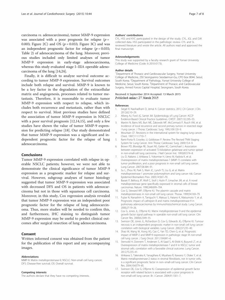
Figure 1 Immunohistochemical analyses of NSCLC representing differexpression; (B) tumor MMP-9 expression; (C) negative control.
computed tomography of the chest, abdominal sonog-raphy, and a bone scan or 18 F-fluorodeoxyglucosepositron emission tomography (18FDG-PET). Post-operatively, follow-up was achieved through regularclinic visits until the patient’s death. Patients wereexamined by chest computed tomography at 3-monthintervals for 2 years and at 6-month intervals thereafter.Furthermore, abdominal sonography or 18FDG-PET orbone scans was performed at 6-month intervals for 5 yearsand at 1-year intervals thereafter, and since 2007 18FDG-PET has been used instead of abdominal sonography.Paraffin-embedded tumor specimens were used to cre-
ate tissue microarray blocks with 2-mm diameter coresfor IHC staining. Two tissue cores were obtained fromeach patient. Pathologic staging was classified accordingto the 7th edition of the Union for International CancerControl tumor-node-metastasis classification of lungcancer.
Immunohistochemical stainingFormalin-fixed and paraffin-embedded tissues were sec-tioned at a thickness of 4 μm and stained using an auto-mated immunostainer (Discovery XT; Ventana MedicalSystems, Tucson, AZ, USA). The slides were dried at 60°Cfor 1 hour and deparaffinized using EZ Prep (VentanaMedical Systems) at 75°C for 8 minutes. Cell conditioningwas performed using CC1 solution (Ventana Medical
ent expression levels for tumor MMP-9. (A) No tumor MMP-9

Table 1 Patient characteristics
Tumor MMP-9 P
No ExpressionN (%)
ExpressionN (%)
Age 0.820
Below median 137 (53.5) 88 (54.7)
Above median 119 (46.5) 73 (45.3)
Sex 0.099
Male 179 (69.9) 100 (62.1)
Female 77 (30.1) 61 (37.9)
Histology 0.001
Squamous cell 130 (50.8) 45 (28.0)
Adenocarcinoma 103 (40.2) 99 (61.5)
Large cell 11 (4.3) 9 (5.5)
Others 12 (4.7) 8 (5.0)
Stage 0.001
I 158 (61.7) 77 (47.8)
II 70 (27.4) 51 (31.7)
IIIA 28 (10.9) 33 (20.5)
T stage 0.068
T1 75 (29.3) 33 (20.5)
T2 147 (57.4) 105 (65.2)
T3 32 (12.5) 18 (11.2)
T4 2 (0.8) 5 (3.1)
N stage 0.002
N0 204 (79.7) 108 (67.1)
N1 35 (13.7) 30 (18.6)
N2 17 (6.6) 23 (14.3)
Lymphovascular invasion 0.549
No 224 (87.5) 144 (89.4)
Yes 32 (12.5) 17 (10.6)
Postoperative Treatment 0.000
Chemotherapy 73 (28.5) 71 (44.1)
Radiation therapy 7 (2.7) 2 (1.2)
Combination therapy 19 (7.4) 20 (12.4)
No treatment 157 (61.3) 68 (42.2)
MMP-9: matrix metalloproteinase-9; Combination therapy:Chemotherapy + Radiation therapy.
Lee et al. Journal of Cardiothoracic Surgery (2015) 10:44 Page 3 of 8
Systems) at 100°C for 48 minutes. MMP-9 antibody(rabbit polyclonal antibody, 1:50 dilution; DiagnosticBiosystems, Pleasanton, CA, USA) was applied to theslides and incubated at 37°C for 32 minutes. Signalswere detected using a DAB Map Detection Kit (VentanaMedical Systems). Counterstaining was performed withhematoxylin (Ventana Medical Systems) for 4 minutes atroom temperature. We performed immunohistochemistrywithout the primary antibody as negative control.
Evaluation of tumor MMP-9 expressionIHC staining of tumor sections was reviewed and scoredindependently by 2 observers who were blinded to theclinical data. The staining intensity was classified asabsent (score of 0), weak (score of 1), moderate (score of2),or strong (score of 3). The extent of staining, definedas the percentage of positively stained cancer cells, wasevaluated using a continuous scale (range, 0-100 %). Thefinal IHC score for tumor MMP-9 expression wasobtained by multiplying the staining intensity and theextent of staining. For statistical analysis, expressionlevels were classified according to the median IHCscore (IHC score = 10) as no tumor MMP-9 expression(IHC score < 10) or tumor MMP-9 expression (IHCscore ≥ 10) (Figure 1).
Statistical analysisDifferences in MMP-9 expression and clinicopathologicvariables were analyzed using the χ2 test. Age was di-chotomized at the median value. Disease-free survival(DFS) was defined as the time from surgery to lung can-cer recurrence, and overall survival (OS) was defined asthe time between surgery and death from any cause. Re-lapse was defined as diagnosis of distant metastasis orlocal recurrence. Postoperative survival was analyzedusing the Kaplan-Meier method and compared using thelog-rank test. Multivariate analysis was performed usingthe Cox proportional hazards regression model with aforward selection procedure to study the effects of dif-ferent variables on recurrence and survival. A p valueless than 0.05 was considered statistically significant. Allstatistical manipulations were performed using the SPSSsoftware program (SPSS Inc., Chicago, IL, USA).
ResultsPatient populationA total of 417 patients were included in the study. Thestudy included 279 men and 138 women, with a medianage of 61 years (range, 30-81 years). The median follow-up period was 56.9 months (range, 3-168 months), andno patients were lost to follow-up. Relapse was observedin 114 patients, and in total, 150 patients died duringthe observation period. The patients’ characteristics areshown in Table 1.
Evaluation of MMP-9 expressionOf the 417 patients, 256 (61.4%) did not show tumorMMP-9 expression and 161 (38.6%) did show tumorMMP-9 expression. There was no significant differencein age, sex, and lymphovascular invasion (LVI) betweenthese 2 groups. The group with tumor MMP-9 expres-sion had a higher proportion of patients with adenocar-cinoma histology (40.2% vs. 61.5%; p = 0.001) and moreadvanced stage (p = 0.001). Adjuvant therapy was admin-istered more often in tumor MMP-9 expression group

Figure 2 Kaplan-Meier survival curves of the relationship between tumor MMP-9 expression and disease-free survival according totumor histology. (A) Overall tumor histology; (B) Squamous cell carcinoma; (C) Adenocarcinoma.
Lee et al. Journal of Cardiothoracic Surgery (2015) 10:44 Page 4 of 8
(38.7% vs. 57.8%; p = 0.000), but there was no differencein the frequency of adjuvant therapy for patients withstage II or IIIA (72.4% vs. 71.4%; p = 0.879). Occurrenceof relapse (21.9% vs. 36.0%; p = 0.002) and death (34.8%vs. 37.9%; p = 0.518) was higher among patients withtumor MMP-9 expression.
Analysis of DFS and OSUnivariate analysis revealed a relationship between tumorMMP-9 expression and DFS in patients with operableNSCLC. Patients with tumor MMP-9 expression had ashorter DFS than those without tumor MMP-9 expression(p = 0.01; Figure 2A). However, there was no significantcorrelation between tumor MMP-9 expression and OS(p = 0.109; Figure 3A).Cox regression analysis was performed to evaluate
the correlation between tumor MMP-9 expression andclinical outcomes. Tumor MMP-9 expression, age, LVI,tumor histology, and tumor stage were tested as inde-pendent possible prognostic variables. The results
Figure 3 Kaplan-Meier survival curves of the relationship between tuhistology. (A) Overall tumor histology; (B) Squamous cell carcinoma; (C) A
demonstrated that tumor MMP-9 expression was not asignificant independent prognostic predictor for DFS(p = 0.142), whereas LVI, stage, and tumor histologywere significant independent prognostic variables(Table 2). Similarly, MMP-9 expression was not an in-dependent predictor for OS (p = 0.807); LVI and tumorstage were the only significant prognostic indicators forOS (Table 3).
Analysis of DFS and OS by tumor histologyClinicopathologic findings according to tumor histologyare shown in Table 4. In patients with squamous cellcarcinoma, tumor MMP-9 expression was not signifi-cantly associated with DFS (p = 0.639: Figure 2B) or OS(p = 0.510; Figure 3B). The results of the Cox regressionanalysis showed that tumor MMP-9 expression was notan independent prognostic indicator for DFS (Table 2)or OS (Table 3).However, among patients with adenocarcinoma, there
was a significant negative correlation between tumor
mor MMP-9 expression and overall survival according to tumordenocarcinoma.

Table 2 Multivariate analysis of disease-free survival
Prognostic factor β P Relativerisk
95%confidenceinterval
Overall histology
Tumor MMP-9 expression 0.286 0.142 1.332 0.908–1.952
Lymphovascular invasion 0.586 0.028 1.797 1.065–3.032
Pathologic stage (I vs. II) 1.264 0.000 3.538 2.259–5.541
Pathologic stage (I vs. IIIA) 1.404 0.000 4.070 2.450–6.760
Tumor histology(squamous cell carcinoma vs.adeno)
0.685 0.002 1.985 1.299–3.033
Squamous cell carcinoma
Tumor MMP-9 expression −0.411 0.309 0.663 0.301–1.463
Adenocarcinoma
Tumor MMP-9 expression 0.580 0.035 1.787 1.041–3.067
MMP-9: matrix metalloproteinase-9; adeno: adenocarcinoma.
Lee et al. Journal of Cardiothoracic Surgery (2015) 10:44 Page 5 of 8
MMP-9 expression and both DFS (p = 0.003; Figure 2C)and OS (p = 0.033; Figure 3C). Cox regression analysiswas used to define clinical markers with independentpredictive value with respect to DFS and OS. TumorMMP-9 positivity was an independent prognostic factorfor DFS (p = 0.035; Table 2); however, OS was not associ-ated with MMP-9 expression (p = 0.259; Table 3).
DiscussionMMPs are a group of zinc-dependent endopeptidasesthat have been implicated in the degradation of extracel-lular matrix. The role of MMPs in tumor growth, metas-tasis, and angiogenesis has been widely investigated [18].MMPs are divided into 4 subclasses according to theirsubstrate specificity: collagenases, gelatinases, stromelysins,and elastases [19]. Among these, MMP-9 (gelatinase-B), acrucial factor in angiogenesis, plays a critical role in theprogression of a variety of tumor types.
Table 3 Multivariate analysis of overall survival
Prognostic factor β P Relativerisk
95%confidenceinterval
Overall histology
Tumor MMP-9 expression 0.043 0.807 1.044 0.741–1.469
Lymphovascular invasion 0.661 0.005 1.937 1.219–3.078
Pathologic stage (I vs. II) 0.904 0.000 2.469 1.686–3.616
Pathologic stage (I vs. IIIA) 1.123 0.000 3.073 1.938–4.875
Squamous cell carcinoma
Tumor MMP-9 expression −0.419 0.196 0.658 0.349–1.241
Adenocarcinoma
Tumor MMP-9 expression 0.295 0.259 1.343 0.805–2.242
MMP-9: matrix metalloproteinase-9.
Expression levels of MMP-9 in serum and tissue aresignificantly higher in patients with pancreatic ductaladenocarcinoma than in those with pancreatitis [20],and tumor MMP-9 expression is significantly elevated inpatients with breast cancer [21]. MMP-9 has been studiedas a prognostic factor for colorectal cancer in T3–T4node-negative patients; enhanced tumor MMP-9 expres-sion was found to be an independent marker of poorprognosis [22]. However, in ovarian cancer, tumorMMP-9 expression is associated with longer survival,whereas stromal MMP-9 expression is associated withshorter survival [23].Many recent reports of tumor MMP-9 expression in
patients with operable NSCLC have suggested thattumor MMP-9 expression is a predictor of poor progno-sis [12,14,15]. However, the prognostic impact of IHCdetection of tumor MMP-9 expression in operableNSCLC is controversial [16-19]. We therefore performedthis study to assess the prognostic impact of tumorMMP-9 expression, as determined by IHC staining, inpatients with operable NSCLC.There are certain limitations to studies of tumor
MMP-9 expression that may limit the interpretation ofthe results. First, there were differences among previousstudies in the definition of tumor MMP-9 positivity andthe appropriate cut-off value. Most of these studies ap-plied a scoring system that was based on the extent andintensity of staining for tumor MMP-9 expression andshowed that overexpression of tumor MMP-9 was asso-ciated with a poor prognosis [12,14,15]. Another studyused the median value of staining for tumor MMP-9 todefine tumor MMP-9 positivity [24]. However, there isno common cut-off value for defining positive tumorMMP-9 expression in NSCLC. In this study, an IHCscore was used to determine tumor expression levels ofMMP-9, and the median IHC score was used as the cut-off value [25]. An IHC staining score of 10 (range, 0-300) was used to divide patients into 2 groups, accordingto the presence or absence of tumor MMP-9 expression.In other words, an IHC score of less than 10 was definedas an absence of tumor MMP-9 expression. The import-ant point of our study was that tumor MMP-9 positivitywas not determined by a scoring system but by the pres-ence or absence of tumor MMP-9 expression. Despiteefforts to standardize this process, a widely acceptedscoring system has not yet been established. Thus, ourdichotomous distinction for tumor MMP-9 expressionwill reduce the impact of subjective judgment when de-termining tumor MMP-9 positivity. In our study, 38.6%of patients had tumor MMP-9 expression. However, if acut-off value of 20% was applied, tumor MMP-9 positivitywould be reduced to 27.3%. Our result of 38.6% seemsreasonable because it is consistent with the percentage ofpatients showing tumor MMP-9 positivity in previous

Table 4 Patient characteristics by tumor histology
Tumor MMP-9
Squamous cell carcinoma Adenocarcinoma
No Expression Expression P No Expression Expression P
N (%) N (%) N (%) N (%)
Age 0.464 0.160
Below median 64 (49.2) 25 (55.6) 57 (55.3) 45 (45.5)
Above median 66 (50.8) 20 (44.4) 46 (44.7) 54 (54.5)
Sex 0.021 0.166
Male 121 (93.1) 36 (80.8) 41 (39.8) 49 (49.5)
Female 9 (6.9) 9 (20.2) 62 (60.2) 50 (50.5)
Stage 0.021 0.006
I 65 (50.0) 15 (33.4) 77 (74.7) 55 (55.6)
II 49 (37.7) 19 (42.2) 15 (14.6) 24 (24.2)
IIIA 16 (12.3) 11 (24.4) 11 (10.7) 20 (20.2)
T stage 0.042 0.019
T1 29 (22.3) 4 (8.9) 42 (40.8) 24 (24.2)
T2 75 (57.7) 29 (64.5) 55 (53.3) 67 (67.7)
T3 25 (19.2) 10 (22.2) 5 (4.9) 5 (5.1)
T4 1 (0.8) 2 (4.4) 1 (1.0) 3 (3.0)
N stage 0.183 0.008
N0 93 (71.6) 28 (62.2) 90 (87.4) 68 (68.7)
N1 31 (23.8) 13 (28.9) 3 (2.9) 14 (14.1)
N2 6 (4.6) 4 (8.9) 10 (9.7) 17 (17.2)
Lymphovascular invasion 0.832 0.740
No 111 (85.4) 39 (86.7) 94 (91.3) 89 (89.9)
Yes 19 (14.6) 6 (13.3) 9 (8.7) 10 (10.1)
Postoperative Treatment 0.299 0.001
Chemotherapy 41 (31.5) 18 (40.0) 24 (22.3) 42 (42.4)
Radiation 7 (5.4) 1 (2.2) 0 1 (1.0)
Combination 10 (7.7) 6 (13.3) 7 (6.8) 13 (13.1)
No treatment 72 (55.4) 20 (44.4) 72 (69.9) 43 (43.4)
MMP-9: matrix metalloproteinase-9; Combination: Chemotherapy + Radiation therapy.
Lee et al. Journal of Cardiothoracic Surgery (2015) 10:44 Page 6 of 8
studies (range, 29.4-68.9 %) [12,14]. The patients of tumorMMP-9 expression had more advanced stage (p = 0.001;Table 1), therefore, they received more postoperative treat-ment (p = 0.000; Table 1). However, there was no differ-ence in adjuvant therapy for patients with stage II or IIIA(72.4% vs. 71.4%; p = 0.879) and there was no statisticaldifference in adjuvant therapy for histological type.Second, it is important to interpret data according to
tumor histology. In previous studies, all types of tumorhistology were included in the analysis of tumor MMP-9expression, and the value of the prognostic factors iden-tified in these studies is controversial [16-19]. Some re-ports have indicated that tumor MMP-9 expression is aprognostic factor for adenocarcinoma of the lung[11,24], and we analyzed that a higher proportion of the
adenocarcinoma patients had positive tumor MMP-9 ex-pression than squamous carcinoma in this study (28.0%vs. 61.5%; p = 0.001). Thus, histological distinction (squa-mous cell carcinoma vs. adenocarcinoma) seems to benecessary in the analysis of tumor MMP-9 expression.In the present study, the overall cohort included patientswith all histological types, including squamous cell car-cinoma, adenocarcinoma, and large cell carcinoma.When considering the total population, tumor MMP-9expression was associated with an increased risk of re-lapse (p = 0.01; Figure 2A) but was not predictive of OS(p = 0.109; Figure 3A). Furthermore, when including alltumor types, tumor MMP-9 expression was not an inde-pendent prognostic factor for the additional outcomestested. After stratifying by tumor histology (squamous cell
Lee et al. Journal of Cardiothoracic Surgery (2015) 10:44 Page 7 of 8
carcinoma vs. adenocarcinoma), tumor MMP-9 expressionwas associated with a poor prognosis for relapse (p =0.003; Figure 2C) and OS (p = 0.033; Figure 3C) and wasan independent prognostic factor for relapse (p = 0.035;Table 2) of adenocarcinoma of the lung. Moreover, previ-ous studies included only limited analyses of tumorMMP-9 expression in early-stage adenocarcinoma,whereas this study evaluated stage I–IIIA operable adeno-carcinoma of the lung [14,24].Finally, it is difficult to analyze survival outcome ac-
cording to tumor MMP-9 expression. Survival outcomesinclude both relapse and survival. MMP-9 is known tobe a key factor in the degradation of the extracellularmatrix and angiogenesis, processes related to tumor me-tastasis. Therefore, it is reasonable to evaluate tumorMMP-9 expression with respect to relapse, which in-cludes both recurrence and metastasis, rather than withrespect to survival. Most previous studies have definedthe association of tumor MMP-9 expression in NSCLCwith a poor survival prognosis [12,14,15], and only a fewstudies have shown the value of tumor MMP-9 expres-sion for predicting relapse [18]. Our study demonstratedthat tumor MMP-9 expression was a significant and in-dependent prognostic factor for the relapse of lungadenocarcinoma.
ConclusionsTumor MMP-9 expression correlated with relapse in op-erable NSCLC patients; however, we were not able todemonstrate the clinical significance of tumor MMP-9expression as a prognostic marker for relapse and sur-vival. However, subgroup analyses of tumor histologysuggested that tumor MMP-9 expression was associatedwith decreased DFS and OS in patients with adenocar-cinoma but not in those with squamous cell carcinoma.Moreover, in this study, Cox regression analysis revealedthat tumor MMP-9 expression was an independent poorprognostic factor for the relapse of lung adenocarcin-oma. Thus, more studies will be needed to confirm this,and furthermore, IHC staining to distinguish tumorMMP-9 expression may be useful to predict clinical out-comes after surgical resection of lung adenocarcinoma.
ConsentWritten informed consent was obtained from the patientfor the publication of this report and any accompanyingimages.
AbbreviationsMMP-9: Matrix metalloproteinase-9; NSCLC: Non-small cell lung cancer;DFS: Disease-free survival; OS: Overall survival.
Competing interestsThe authors declare that they have no competing interests.
Authors’ contributionsCYL, HSS and KYC participated in the design of the study. CYL, JGL and DJKcollected data. HSS participated in the pathologic review. CYL and SLreviewed literature and wrote the article. All authors read and approved thefinal manuscript.
AcknowledgementsThis study was supported by a faculty research grant of Yonsei UniversityCollege of Medicine (Code: 6-2010-0173).
Author details1Department of Thoracic and Cardiovascular Surgery, Yonsei UniversityCollege of Medicine, 250 Seongsanno Seodaemun-Gu, CPO box 8044, Seoul,South Korea. 2Department of Pathology, Yonsei University College ofMedicine, Seoul, South Korea. 3Department of Thoracic and CardiovascularSurgery, Armed Forces Capital Hospital, Seongnam, South Korea.
Received: 6 September 2014 Accepted: 13 March 2015
References1. Siegel R, Naishadham D, Jemal A. Cancer statistics, 2012. CA Cancer J Clin.
2012;62:10–29.2. Alberg AJ, Ford JG, Samet JM. Epidemiology of Lung Cancer: ACCP
Evidence-Based Clinical Practice Guidelines. CHEST. 2007;132:29S–55.3. Martini N, Bains MS, Burt ME, Zakowski MF, McCormack P, Rusch VW, et al.
Incidence of local recurrence and second primary tumors in resected stageI lung cancer. J Thorac Cardiovasc Surg. 1995;109:120–9.
4. Mountain CF. Revisions in the international system for staging lung cancer.Chest. 1997;111:1710–7.
5. Rami-Porta R, Crowley JJ, Goldstraw P. Review The Revised TNM StagingSystem for Lung Cancer. Ann Thorac Cardiovasc Surg. 2009;15:4–9.
6. Brown PD, Bloxidge RE, Stuart NS, Gatter KC, Carmichael J. Associationbetween expression of activated 72-kilodalton gelatinase and tumor spreadin non-small-cell lung carcinoma. J Natl Cancer Inst. 1993;85:574–8.
7. Liu D, Nakano J, Ishikawa S, Yokomise H, Ueno M, Kadota K, et al.Overexpression of matrix metalloproteinase-7 (MMP-7) correlates withtumor proliferation, and a poor prognosis in non-small cell lung cancer.Lung Cancer. 2007;58:384–91.
8. Su L, Zhou W, Park S, Wain JC, Lynch TJ, Liu G, et al. Matrixmetalloproteinase-1 promoter polymorphism and lung cancer risk. CancerEpidemiol Biomarkers Prev. 2005;14:567–70.
9. Basset P, Bellocq JP, Wolf C, Stoll I, Hutin P, Limacher JM, et al. A novelmetalloproteinase gene specifically expressed in stromal cells of breastcarcinomas. Nature. 1990;348:699–704.
10. Cox G, Steward WP, O’Byrne KJ. The plasmin cascade and matrixmetalloproteinases in non-small cell lung cancer. Thorax. 1999;54:169–79.
11. Fujise N, Nanashim A, Taniguchi Y, Matsuo S, Hatano K, Matsumoto Y, et al.Prognostic impact of cathepsin B and matrix metalloproteinase-9 inpulmonary adenocarcinomas by immunohistochemical study. Lung Cancer.2000;27:19–26.
12. Cox G, Jones JL, O’Byrne KJ. Matrix metalloproteinase 9 and the epidermalgrowth factor signal pathway in operable non-small cell lung cancer. ClinCancer Res. 2000;6:2349–55.
13. Swinson DE, Jones JL, Richardson D, Cox G, Edwards JG, O’Byrne KJ. Tumournecrosis is an independent prognostic marker in non-small cell lung cancer:correlation with biological variables. Lung Cancer. 2002;37:235–40.
14. Shao W, Wang W, Xiong XG, Cao C, Yan TD, Chen G, et al. Prognosticimpact of MMP‐2 and MMP‐9 expression in pathologic stage IA non‐smallcell lung cancer. J Surg Oncol. 2011;104:841–6.
15. Stenvold H, Donnem T, Andersen S, Al-Saad S, Al-Shibli K, Busund LT, et al.Overexpression of matrix metalloproteinase-7 and-9 in NSCLC tumor andstromal cells: correlation with a favorable clinical outcome. Lung Cancer.2012;75:235–41.
16. Ishikawa S, Takenaka K, Yanagihara K, Miyahara R, Kawano Y, Otake Y, et al.Matrix metalloproteinase-2 status in stromal fibroblasts, not in tumor cells,is a significant prognostic factor in non–small-cell lung cancer. Clin CancerRes. 2004;10:6579–85.
17. Swinson DE, Cox G, O’Byrne KJ. Coexpression of epidermal growth factorreceptor with related factors is associated with a poor prognosis innon-small-cell lung cancer. Br J Cancer. 2004;91:1301–7.

Lee et al. Journal of Cardiothoracic Surgery (2015) 10:44 Page 8 of 8
18. Liotta LA, Tryggvason K, Garbisa S, Hart I, Foltz CM, Shafie S. Metastaticpotential correlates with enzymatic degradation of basement membranecollagen. Nature. 1980;284:67–8.
19. Nelson AR, Fingleton B, Rothenberg ML, Matrisian LM. Matrixmetalloproteinases: biologic activity and clinical implications. J Clin Oncol.2000;18:1135–49.
20. Tian M, Cui YZ, Song GH, Zong MJ, Zhou XY, Chen Y, et al. Proteomicanalysis identifies MMP-9, DJ-1 and A1BG as overexpressed proteins inpancreatic juice from pancreatic ductal adenocarcinoma patients. BMCCancer. 2008;8:241.
21. Wu ZS, Wu Q, Yang JH, Wang HQ, Ding XD, Yang F, et al. Prognosticsignificance of MMP‐9 and TIMP‐1 serum and tissue expression in breastcancer. Int J Cancer. 2008;122:2050–6.
22. Cho YB, Lee WY, Song SY, Shin HJ, Yun SH, Chun HK. Matrixmetalloproteinase-9 activity is associated with poor prognosis in T3-T4node-negative colorectal cancer. Hum Pathol. 2007;38:1603–10.
23. Sillanpää S, Anttila M, Voutilainen K, Ropponen K, Turpeenniemi-Hujanen T,Puistola U, et al. Prognostic significance of matrix metalloproteinase-9(MMP-9) in epithelial ovarian cancer. Gynecol Oncol. 2007;104:296–303.
24. Pinto CA, Carvalho PE, Antonângelo L, Garippo A, Da Silva AG, Soares F,et al. Morphometric evaluation of tumor matrix metalloproteinase 9 predictssurvival after surgical resection of adenocarcinoma of the lung. Clin CancerRes. 2003;9:3098–104.
25. Pirker R, Pereira JR, von Pawel J, Krzakowski M, Ramlau R, Park K, et al. EGFRexpression as a predictor of survival for first-line chemotherapy plus cetuximabin patients with advanced non-small-cell lung cancer: analysis of data from thephase 3 FLEX study. Lancet Oncol. 2012;13:33–42.
Submit your next manuscript to BioMed Centraland take full advantage of:
• Convenient online submission
• Thorough peer review
• No space constraints or color figure charges
• Immediate publication on acceptance
• Inclusion in PubMed, CAS, Scopus and Google Scholar
• Research which is freely available for redistribution
Submit your manuscript at www.biomedcentral.com/submit