research article open access molecular analyses of fusarium
TRANSCRIPT
RESEARCH ARTICLE Open Access
Molecular analyses of Fusarium isolates recoveredfrom a cluster of invasive mold infections in aBrazilian hospitalChristina M Scheel1*†, Steven F Hurst1†, Gloria Barreiros2†, Tiyomi Akiti2†, Marcio Nucci2† and S Arunmozhi Balajee3†
Abstract
Background: Invasive fusariosis (IF) is a rare but often fatal fungal infection in immunosuppressed patients. In 2007,cases of IF above the expected epidemiologic baseline were detected in the hematology ward of a hospital in Riode Janeiro, Brazil. Possible sources of infection were investigated by performing environmental sampling andpatient isolate collection, followed by molecular typing. Isolates from dermatology patients with superficial fusariosiswere included in the study for comparison to molecular types found in the community.
Methods: Environmental sampling focused on water-related sources in and around the hematology ward. Initially,we characterized 166 clinical and environmental isolates using the Fusarium translation elongation factor 1α (EF-1α)genetic locus. Isolates included 68 collected from water-related sources in the hospital environment, 55 from 18hematology patients, and 43 from the skin/nails of 40 outpatients seen at the hospital dermatology clinic.Multi-locus sequence typing was performed on Fusarium solani species complex (FSSC) species 1 and 2 isolates toinvestigate their relatedness further.
Results: Most of the hematology samples were FSSC species 2, with species type FSSC 2-d the most commonlyisolated from these patients. Most of the outpatient dermatology samples were also FSSC 2, with type 2-d againpredominating. In contrast, environmental isolates from water sources were mostly Fusarium oxysporum speciescomplex (FOSC) and those from air samples mostly Fusarium incarnatum-equiseti species complex (FIESC). A third ofthe environmental samples were FSSC, with species types FSSC 1-a and FSSC 1-b predominating.
Conclusions: Fusarium isolate species types from hematology patient infections were highly similar to thoserecovered from dermatology patients in the community. Four species types (FSSC 1-a, 1-b, 2-d and 2-f) were sharedbetween hematology patients and the environment. Limitations in environmental sampling do not allow fornosocomial sources of infection to be ruled out. Future studies will focus on environmental factors that may haveinfluenced the prevalence of FSSC fusariosis in this hematology ward.
Keywords: Fusarium, Invasive fusariosis, Molecular epidemiology, Multi-locus sequence typing, Invasive fungalinfection, Bone marrow transplant, Hospital-associated infection, Community acquired disease, Brazil
* Correspondence: [email protected]†Equal contributors1Mycotic Diseases Branch, Centers for Disease Control and Prevention,Atlanta, GA, USAFull list of author information is available at the end of the article
© 2013 Scheel et al.; licensee BioMed Central Ltd. This is an Open Access article distributed under the terms of the CreativeCommons Attribution License (http://creativecommons.org/licenses/by/2.0), which permits unrestricted use, distribution, andreproduction in any medium, provided the original work is properly cited.
Scheel et al. BMC Infectious Diseases 2013, 13:49http://www.biomedcentral.com/1471-2334/13/49

BackgroundOver the last two decades, invasive mold infections havebecome increasingly common among severely ill hospitalpatients worldwide [1,2]. Fusariosis may manifest assuperficial or skin, nail and eye infections in healthy per-sons, whereas severely immunocompromised patientscan develop invasive disease [2-5]. Invasive fusariosis(IF) is a devastating fungal infection with high mortalityrates in patients with hematological malignancies and inhematopoietic stem cell transplant recipients [3].
Since 2005, an increase in cases of fusariosis wasobserved in a hospital in Rio de Janeiro, Brazil, bothamong immunosuppressed patients in the bone marrowtransplant unit (BMTU) and also among persons withsuperficial infections who were treated at the local derma-tology clinic. In 2007, the number of cases of fusariosis inthe BMTU above the expected epidemiologic baseline wasrecorded. Periodic environmental sampling in the hospital,focused on water and water-related sources within theBMTU, commenced in 2005 and was continued until2009, in an attempt to identify possible environmentalreservoirs of infection. Beginning in 2006, Fusarium iso-lates from hospital patients, hospital environmental sam-ples, and dermatology clinic patients were archived forfurther molecular analysis. Here we report the results ofcomparative sequence typing performed on these archivedisolates to assess the ecology of the hospital environment,determine the molecular genetic relatedness betweenpatient and environmental samples, and to estimate thedegree to which environmental Fusarium could be a reser-voir for infection within the hospital.
MethodsSampling and isolatesThis study was conducted in the University Hospital, FederalUniversity of Rio de Janeiro, Brazil. The University Hospitalis a 13-floor tertiary care teaching hospital with ~480beds. The BMTU is an isolated 18-bed ward located onthe eighth floor of the building. Rooms in the BMTU areunder positive pressure air flow, and 8 beds have high-efficiency particulate air (HEPA) filters. Sporadic cases ofIF had been diagnosed in the BMTU at a rate of 1.47 casesper 1000 admissions between 2000 and 2006. In 2007,there was a sharp increase in the rate of fusariosis, reach-ing 16.78 cases per 1000 BMTU admissions. During thisperiod, there was a concurrent increase of superficialfusariosis in outpatients seen at the dermatology clinic onthe hospital’s second floor.
Fifty-five Fusarium isolates were recovered from 18 he-matology patients (38 isolates from sterile site samples and17 from non-sterile sites) in the BMTU as a part of standardmedical care between December 2005 and September 2009.Forty-three Fusarium isolates were collected from skin andnail cultures of superficial lesions (onychomycosis and
intertrigus) of 40 dermatology outpatients between June2007 and October 2009 as part of standard medical care.After collection, all patient isolates were plated on Sabourauddextrose agar (SDA), Mycosel, brain/heart infusion (BHI),and yeast extract agars and incubated at 25°C.
Hospital environmental sampling was performed peri-odically in the BMTU between June 2005 and Aug 2006as part of an environmental surveillance program focusedon air, water, and water-related surfaces. In September2006 - October 2007, environmental sampling was per-formed in rooms of infected patients, with an emphasison bathroom surfaces (shower heads, drains, and tilegrout and sink surfaces and drains). Common areas of theBMTU were also swabbed as potential point sources offungal contamination. Thereafter, routine environmentalsurveillance of the BMTU continued through July 2009.Environmental sampling of the outpatient dermatologyclinic was not performed.
Surfaces were sampled using Culturette swabs (BectonDickinson, Franklin Lakes, NJ, USA), inoculated onto SDAplates containing 50 mg/L chloramphenicol (SDA+C), andincubated at 25°C. Water (100 ml) was collected in sterileglass bottles and was filtered through 0.45 μm membranes(Millipore, Billerica, MA) which were placed on SAB platesand incubated at 25°C. Air was sampled from patientrooms in November 2006, August 2007, and July 2009using a six-stage Anderson bioaerosol sampler (AndersonInstruments, Atlanta GA) with air impacting on an SDAplate for 30 min, for a total volume of 849 L per sample.Control air samples were also collected from other areas ofthe hospital, including the intensive care unit (2005, 2007and 2009), pediatrics unit (2007) and the nephrology dialy-sis unit (2007).Fusarium were isolated from 68 samples, ranging in
time of collection from June 2005 to October 2009.Provisional identification of hematology patient Fusariumisolates were obtained in order to decide if more dramaticmeasures should be implemented in the BMTU. Thisidentification was based on the micromorphology that istypical of this genus [6].
Molecular identification and subtypingAll available Fusarium isolates collected from patients andthe environment were sent to the Centers for DiseasesControl and Prevention, Atlanta, GA, USA for molecularidentification. At the CDC, isolates were sub-cultured onSDA+C with 50 mg/ml gentamycin agar, and incubated at30°C for approximately 72 hours. Genomic DNA wasextracted using the Qiagen DNeasy tissue kit (Qiagen,Valencia, CA) with slight modifications to the manufac-turer’s instructions. Briefly, small sections of sub-culturedFusarium (1 cm2) were cut from mycelial mats and placedin 5 ml polypropylene tubes containing 900 μl QiagenATL buffer and 60 U proteinase K. The mycelia in each
Scheel et al. BMC Infectious Diseases 2013, 13:49 Page 2 of 12http://www.biomedcentral.com/1471-2334/13/49
tube were homogenized using the Omni TH Mixer (OmniIntl., Kennesaw, GA) at slow speed for 30 s and then highspeed for 30 s, using a clean probe between each isolate.Homogenates were capped and incubated at 55°C for 1 hwith frequent vortexing and then cooled to RT. RNase A(Sigma-Aldrich Corp., St. Louis, MO) was then added to afinal concentration of 1 mg/ml and incubated for 5 min atRT, followed by the addition of 900 μl Qiagen buffer ALand vortexing. Homogenates were incubated at 70°C for10 min, then transferred to 1.7 ml microcentrifuge tubesand centrifuged at 15,000 X g for 10 min. Clear superna-tants (1 ml each) were transferred to clean microcentri-fuge tubes and 500 μl ethanol (Sigma-Aldrich Corp.) wasadded. The suspensions were vortexed and transferred toQiagen DNeasy columns, and manufacturer’s instructionswere followed throughout the remainder of the procedure.
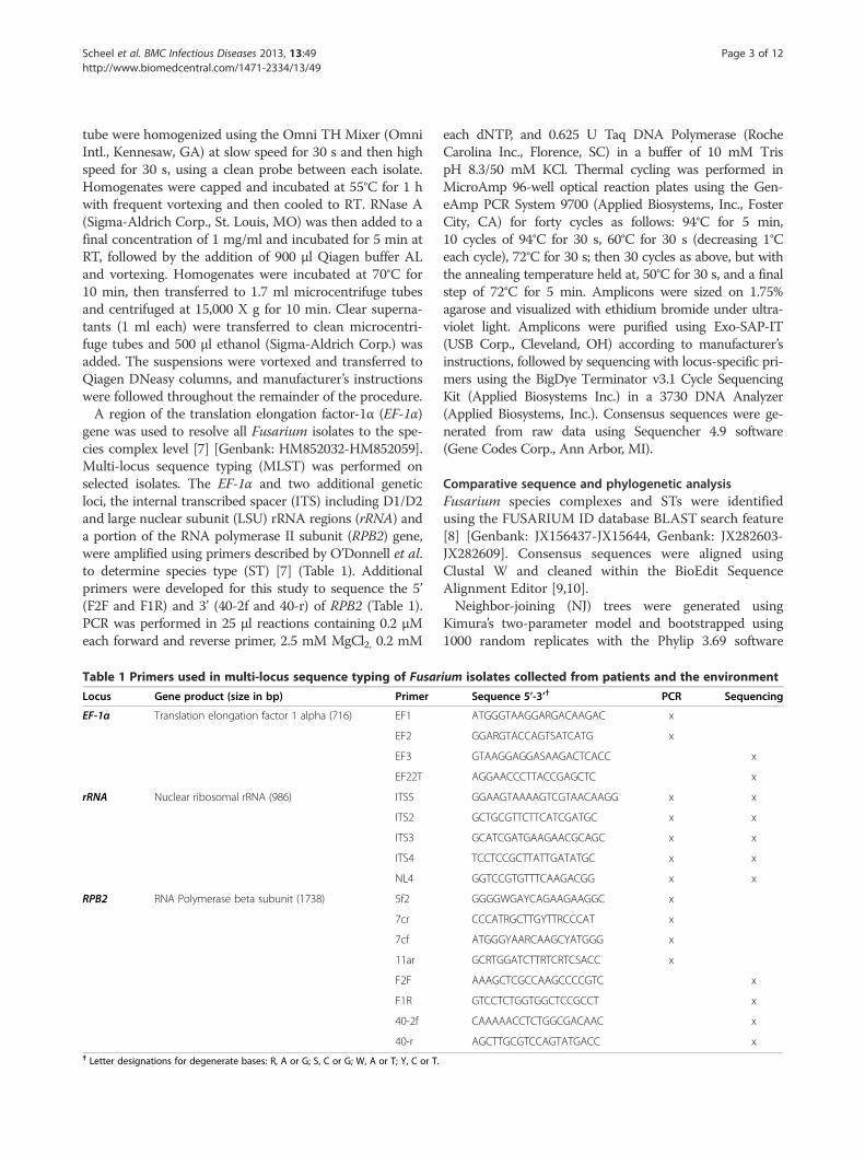
A region of the translation elongation factor-1α (EF-1α)gene was used to resolve all Fusarium isolates to the spe-cies complex level [7] [Genbank: HM852032-HM852059].Multi-locus sequence typing (MLST) was performed onselected isolates. The EF-1α and two additional geneticloci, the internal transcribed spacer (ITS) including D1/D2and large nuclear subunit (LSU) rRNA regions (rRNA) anda portion of the RNA polymerase II subunit (RPB2) gene,were amplified using primers described by O’Donnell et al.to determine species type (ST) [7] (Table 1). Additionalprimers were developed for this study to sequence the 5’(F2F and F1R) and 3’ (40-2f and 40-r) of RPB2 (Table 1).PCR was performed in 25 μl reactions containing 0.2 μMeach forward and reverse primer, 2.5 mM MgCl2, 0.2 mM
each dNTP, and 0.625 U Taq DNA Polymerase (RocheCarolina Inc., Florence, SC) in a buffer of 10 mM TrispH 8.3/50 mM KCl. Thermal cycling was performed inMicroAmp 96-well optical reaction plates using the Gen-eAmp PCR System 9700 (Applied Biosystems, Inc., FosterCity, CA) for forty cycles as follows: 94°C for 5 min,10 cycles of 94°C for 30 s, 60°C for 30 s (decreasing 1°Ceach cycle), 72°C for 30 s; then 30 cycles as above, but withthe annealing temperature held at, 50°C for 30 s, and a finalstep of 72°C for 5 min. Amplicons were sized on 1.75%agarose and visualized with ethidium bromide under ultra-violet light. Amplicons were purified using Exo-SAP-IT(USB Corp., Cleveland, OH) according to manufacturer’sinstructions, followed by sequencing with locus-specific pri-mers using the BigDye Terminator v3.1 Cycle SequencingKit (Applied Biosystems Inc.) in a 3730 DNA Analyzer(Applied Biosystems, Inc.). Consensus sequences were ge-nerated from raw data using Sequencher 4.9 software(Gene Codes Corp., Ann Arbor, MI).
Comparative sequence and phylogenetic analysisFusarium species complexes and STs were identifiedusing the FUSARIUM ID database BLAST search feature[8] [Genbank: JX156437-JX15644, Genbank: JX282603-JX282609]. Consensus sequences were aligned usingClustal W and cleaned within the BioEdit SequenceAlignment Editor [9,10].
Neighbor-joining (NJ) trees were generated usingKimura’s two-parameter model and bootstrapped using1000 random replicates with the Phylip 3.69 software
Table 1 Primers used in multi-locus sequence typing of Fusarium isolates collected from patients and the environment
Locus Gene product (size in bp) Primer Sequence 5’-3’† PCR Sequencing
EF-1α Translation elongation factor 1 alpha (716) EF1 ATGGGTAAGGARGACAAGAC x
EF2 GGARGTACCAGTSATCATG x
EF3 GTAAGGAGGASAAGACTCACC x
EF22T AGGAACCCTTACCGAGCTC x
rRNA Nuclear ribosomal rRNA (986) ITS5 GGAAGTAAAAGTCGTAACAAGG x x
ITS2 GCTGCGTTCTTCATCGATGC x x
ITS3 GCATCGATGAAGAACGCAGC x x
ITS4 TCCTCCGCTTATTGATATGC x x
NL4 GGTCCGTGTTTCAAGACGG x x
RPB2 RNA Polymerase beta subunit (1738) 5f2 GGGGWGAYCAGAAGAAGGC x
7cr CCCATRGCTTGYTTRCCCAT x
7cf ATGGGYAARCAAGCYATGGG x
11ar GCRTGGATCTTRTCRTCSACC x
F2F AAAGCTCGCCAAGCCCCGTC x
F1R GTCCTCTGGTGGCTCCGCCT x
40-2f CAAAAACCTCTGGCGACAAC x
40-r AGCTTGCGTCCAGTATGACC x† Letter designations for degenerate bases: R, A or G; S, C or G; W, A or T; Y, C or T.
Scheel et al. BMC Infectious Diseases 2013, 13:49 Page 3 of 12http://www.biomedcentral.com/1471-2334/13/49

package [11]. Resultant trees were then analyzed to gener-ate a consensus NJ tree that was annotated using FigTreev.1.3.1 software [12]. Maximum Likelihood (MJ) and Max-imum Parsimony (MP) algorithms were used to generatealternate trees for comparison to the NJ tree to ensure itsvalidity.
ResultsMolecular typing of EF-1αA total of 166 Fusarium isolates were available for ana-lysis. These included 98 clinical isolates (55 from 18hematological patients, 43 from 40 dermatology patients)and 68 isolates from environmental samples, including 14
air samples, 44 swab samples, and 10 water samples.Comparative EF-1α sequence analysis using the FUSAR-IUM ID database BLAST feature allowed for identificationof most study isolates to the species level, with sequenceidentities ranging from 94.1% to 100% (mean = 99.9,median = 100%) when compared with database isolates.Frequencies of different species complexes/species iso-lated from patients and the environment are shown inFigure 1. Isolates from BMTU patients and dermatologypatients showed a similar distribution. The majority of iso-lates (68%, 113/166) were identified as Fusarium solanispecies complex (FSSC). Among clinical isolates, FSSCspecies 2 (FSSC 2) predominated, and comprised 69%(38/55) of those collected from hematology patients, and
FSSC 269%
FSSC 115%
FSSC other7%
FOSC5%
GFSC4%
A
Hematology Patient Isolates (n = 55)
FSSC 274%
FSSC 17%
FSSC other12 %
FOSC7%
B
Dermatology Patient Isolates (n = 43)
FSSC 130%
FSSC 26%
FOSC50%
FIESC16%
C
Environmental Isolates (n = 68)Figure 1 Molecular typing of Fusarium isolates (n = 168) collected from patients and the environment. Molecular typing to speciescomplexes and species was accomplished by amplification and genetic sequencing of the translation elongation factor 1α (EF-1α) gene. Identities ofstudy isolates were resolved by comparison of EF-1α molecular sequences to sequences of known Fusarium species archived in a in a curateddatabase, FUSARIUM ID [8]. FSSC: Fusarium solani species complex; FOSC: Fusarium oxysporum species complex; FIESC: Fusarium incarnatum-equisetispecies complex; GFSC: Gibberella fujikuroi species complex.
Scheel et al. BMC Infectious Diseases 2013, 13:49 Page 4 of 12http://www.biomedcentral.com/1471-2334/13/49

Table 2 Speciation of clinical and environmental Fusarium isolates collected in a Brazilain hospital
Species ID (EF-1α Identity match%) Isolate number Collection date Sample type Isolate source
FSSC† (99.3) B8586 12/10/2005 Clinical HP01, blood
FSSC† (100) B8659 10/16/2007 Clinical DP33, skin scraping
FSSC† (99.1) B8684 7/13/2009 Clinical DP58, nail scraping
FSSC† (99.6) B8685 7/31/2009 Clinical DP60, skin scraping
FSSC 1-a B8648 7/16/2007 Clinical DP26, nail scraping
FSSC 1-a B8593 7/19/2006 Clinical HP03, lesion swab
FSSC 1-a B8710 7/31/2007 Environmental BR06, shower swab
FSSC 1-a B8711 7/31/2007 Environmental BR06, shower swab
FSSC 1-a B8712 7/31/2007 Environmental BR06, wall swab
FSSC 1-a B8714 7/31/2007 Environmental BR06, wall swab
FSSC 1-a B8715 7/31/2007 Environmental BR06, wall swab
FSSC 1-a B8716 7/31/2007 Environmental BR06, wall swab
FSSC 1-a B8720 8/14/2007 Environmental BR07, shower swab
FSSC 1-a B8721 8/14/2007 Environmental BR07, shower swab
FSSC 1-a B8690 9/21/2006 Environmental BR10, wall swab
FSSC 1-a B8694 10/5/2006 Environmental BR11, floor swab
FSSC 1-a B8741 10/25/2007 Environmental BR12, shower swab
FSSC 1-a B8696 11/1/2006 Environmental BR03, air
FSSC 1-b B8589 4/17/2006 Clinical HP02, blood
FSSC 1-b B8590 4/17/2006 Clinical HP02, blood
FSSC 1-b B8591 4/20/2006 Clinical HP02, skin biopsy
FSSC 1-b B8700 11/1/2006 Environmental BR03, floor swab
FSSC 1-b B8717 7/31/2007 Environmental BR06, wall swab
FSSC 1-b B8728 8/31/2007 Environmental BR03, sink swab
FSSC 1-b B8744 10/26/2007 Environmental BR01, water
FSSC 1-b B8745 10/26/2007 Environmental BR01, water
FSSC 1-b B8662 10/31/2007 Clinical DP36, nail scraping
FSSC 1-b B8681 5/20/2009 Clinical DP55, skin scraping
FSSC 1-b B8757 7/6/2009 Environmental BR03, shower swab
FSSC 1-b B8760 7/6/2009 Environmental BR03, air
FSSC 1-b B8637 7/28/2009 Clinical HP17, biopsy
FSSC 1-b B8638 9/4/2009 Clinical HP18, blood
FSSC 1-b B8639 9/4/2009 Clinical HP18, blood
FSSC 1-b B8640 9/4/2009 Clinical HP18, blood
FSSC 2-d B8592 7/12/2006 Clinical HP03, blood
FSSC 2-d B8697 11/1/2006 Environmental BR03, air
FSSC 2-d B8601 4/10/2007 Clinical HP07, skin biopsy
FSSC 2-d B8602 4/10/2007 Clinical HP07, vesicle
FSSC 2-d B8643 6/14/2007 Clinical DP21, nail scraping
FSSC 2-d B8603 6/18/2007 Clinical HP08, blood
FSSC 2-d B8604 6/18/2007 Clinical HP08, blood
FSSC 2-d B8605 6/18/2007 Clinical HP08, blood
FSSC 2-d B8606 6/18/2007 Clinical HP08, skin biopsy
FSSC 2-d B8607 6/19/2007 Clinical HP08, skin swab
Scheel et al. BMC Infectious Diseases 2013, 13:49 Page 5 of 12http://www.biomedcentral.com/1471-2334/13/49
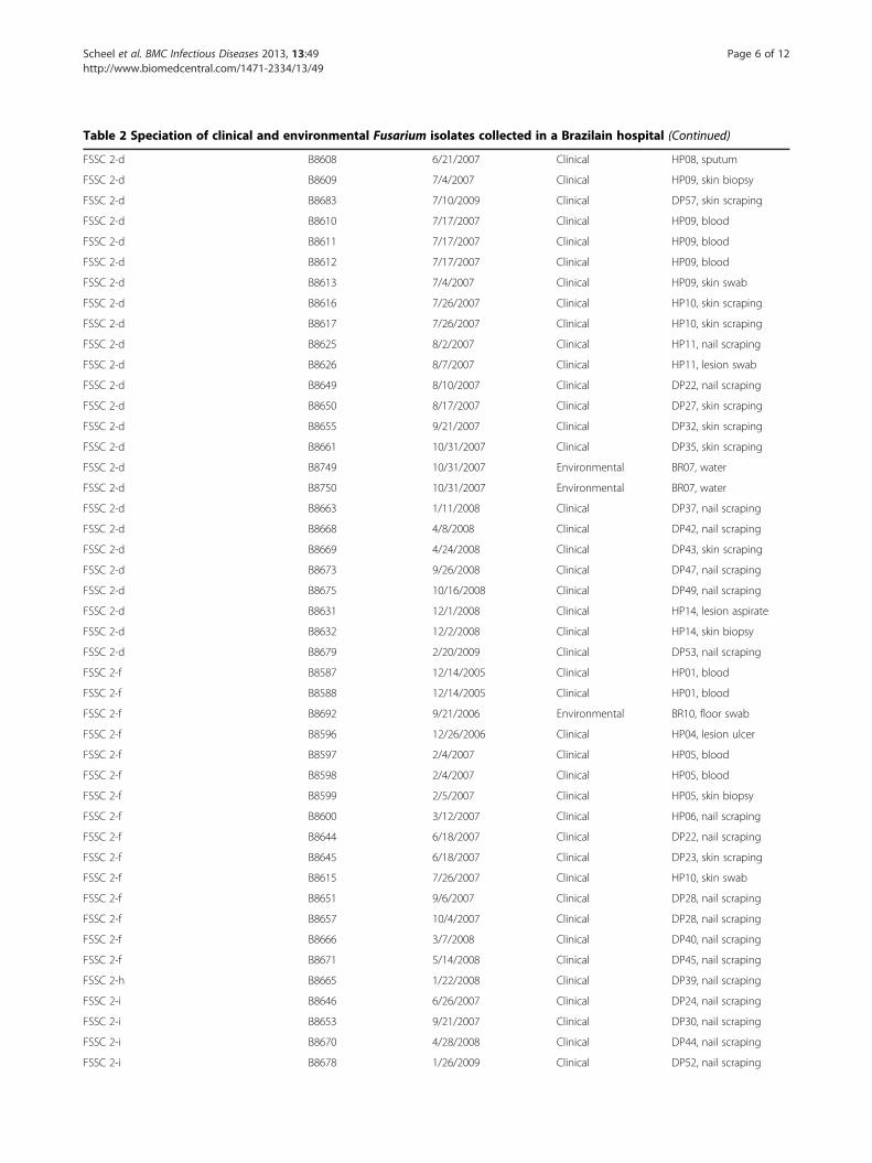
Table 2 Speciation of clinical and environmental Fusarium isolates collected in a Brazilain hospital (Continued)
FSSC 2-d B8608 6/21/2007 Clinical HP08, sputum
FSSC 2-d B8609 7/4/2007 Clinical HP09, skin biopsy
FSSC 2-d B8683 7/10/2009 Clinical DP57, skin scraping
FSSC 2-d B8610 7/17/2007 Clinical HP09, blood
FSSC 2-d B8611 7/17/2007 Clinical HP09, blood
FSSC 2-d B8612 7/17/2007 Clinical HP09, blood
FSSC 2-d B8613 7/4/2007 Clinical HP09, skin swab
FSSC 2-d B8616 7/26/2007 Clinical HP10, skin scraping
FSSC 2-d B8617 7/26/2007 Clinical HP10, skin scraping
FSSC 2-d B8625 8/2/2007 Clinical HP11, nail scraping
FSSC 2-d B8626 8/7/2007 Clinical HP11, lesion swab
FSSC 2-d B8649 8/10/2007 Clinical DP22, nail scraping
FSSC 2-d B8650 8/17/2007 Clinical DP27, skin scraping
FSSC 2-d B8655 9/21/2007 Clinical DP32, skin scraping
FSSC 2-d B8661 10/31/2007 Clinical DP35, skin scraping
FSSC 2-d B8749 10/31/2007 Environmental BR07, water
FSSC 2-d B8750 10/31/2007 Environmental BR07, water
FSSC 2-d B8663 1/11/2008 Clinical DP37, nail scraping
FSSC 2-d B8668 4/8/2008 Clinical DP42, nail scraping
FSSC 2-d B8669 4/24/2008 Clinical DP43, skin scraping
FSSC 2-d B8673 9/26/2008 Clinical DP47, nail scraping
FSSC 2-d B8675 10/16/2008 Clinical DP49, nail scraping
FSSC 2-d B8631 12/1/2008 Clinical HP14, lesion aspirate
FSSC 2-d B8632 12/2/2008 Clinical HP14, skin biopsy
FSSC 2-d B8679 2/20/2009 Clinical DP53, nail scraping
FSSC 2-f B8587 12/14/2005 Clinical HP01, blood
FSSC 2-f B8588 12/14/2005 Clinical HP01, blood
FSSC 2-f B8692 9/21/2006 Environmental BR10, floor swab
FSSC 2-f B8596 12/26/2006 Clinical HP04, lesion ulcer
FSSC 2-f B8597 2/4/2007 Clinical HP05, blood
FSSC 2-f B8598 2/4/2007 Clinical HP05, blood
FSSC 2-f B8599 2/5/2007 Clinical HP05, skin biopsy
FSSC 2-f B8600 3/12/2007 Clinical HP06, nail scraping
FSSC 2-f B8644 6/18/2007 Clinical DP22, nail scraping
FSSC 2-f B8645 6/18/2007 Clinical DP23, skin scraping
FSSC 2-f B8615 7/26/2007 Clinical HP10, skin swab
FSSC 2-f B8651 9/6/2007 Clinical DP28, nail scraping
FSSC 2-f B8657 10/4/2007 Clinical DP28, nail scraping
FSSC 2-f B8666 3/7/2008 Clinical DP40, nail scraping
FSSC 2-f B8671 5/14/2008 Clinical DP45, nail scraping
FSSC 2-h B8665 1/22/2008 Clinical DP39, nail scraping
FSSC 2-i B8646 6/26/2007 Clinical DP24, nail scraping
FSSC 2-i B8653 9/21/2007 Clinical DP30, nail scraping
FSSC 2-i B8670 4/28/2008 Clinical DP44, nail scraping
FSSC 2-i B8678 1/26/2009 Clinical DP52, nail scraping
Scheel et al. BMC Infectious Diseases 2013, 13:49 Page 6 of 12http://www.biomedcentral.com/1471-2334/13/49

Table 2 Speciation of clinical and environmental Fusarium isolates collected in a Brazilain hospital (Continued)
FSSC 2-i B8688 10/6/2009 Clinical DP63, nail scraping
FSSC 2-i B8687 10/8/2009 Clinical DP62, skin scraping
FSSC 2-k B8642 6/5/2007 Clinical DP20, skin scraping
FSSC 2-k B8647 7/2/2007 Clinical DP25, nail scraping
FSSC 2-k B8614 7/26/2007 Clinical HP10, skin swab
FSSC 2-k B8618 9/5/2007 Clinical HP10, sinovial fluid
FSSC 2-k B8619 9/5/2007 Clinical HP10, sinovial fluid
FSSC 2-k B8620 9/10/2007 Clinical HP10, sinovial fluid
FSSC 2-k B8621 9/10/2007 Clinical HP10, sinovial fluid
FSSC 2-k B8654 9/21/2007 Clinical DP31, skin scraping
FSSC 2-k B8656 9/24/2007 Clinical DP23, nail scraping
FSSC 2-k B8622 10/8/2007 Clinical HP10, sinovial fluid
FSSC 2-k B8623 10/9/2007 Clinical HP10, sinovial fluid
FSSC 2-k B8624 10/11/2007 Clinical HP10, sinovial fluid
FSSC 2-t B8594 7/19/2006 Clinical HP03, lesion swab
FSSC 2-t B8595 7/19/2006 Clinical HP03, lesion swab
FSSC 2-t B8658 10/16/2007 Clinical DP33, skin scraping
FSSC 2-u B8652 9/17/2007 Clinical DP29, nail scraping
FSSC 2-u B8674 10/13/2008 Clinical DP48, nail scraping
FSSC 3+4 (100) B8677 12/19/2008 Clinical DP51, skin scraping
FSSC 3+4 (100) B8636 1/29/2009 Clinical HP16, corneal ulcer swab
FSSC 5 (99.5) B8676 10/17/2008 Clinical DP50, nail scraping
FSSC 7 (99.7) B8634 1/5/2008 Clinical HP15, blood
FSSC 7 (99.7) B8635 1/5/2008 Clinical HP15, blood
FOSC† (100) B8698 11/1/2006 Environmental BR03, shower swab
FOSC† (100) B8660 10/26/2007 Clinical DP34, skin scraping
FOSC† (99.7) B8667 3/27/2008 Clinical DP41, nail scraping
FOSC† (100) B8672 8/18/2008 Clinical DP46, nail scraping
FOSC† (99.9) B8633 12/18/2008 Clinical HP15, skin scraping
FOSC 33 (100) B8699 11/1/2006 Environmental BR03, wall swab
FOSC 33 (100) B8701 11/1/2006 Environmental BR03, sink swab
FOSC 33 (100) B8702 11/1/2006 Environmental BR03, sink swab
FOSC 33 (100) B8691 9/21/2006 Environmental BR10, sink swab
FOSC 33 (100) B8695 10/5/2006 Environmental BR11, sink swab
FOSC 33 (100) B8713 7/31/2007 Environmental BR06, wall swab
FOSC 33 (100) B8719 8/14/2007 Environmental BR04, sink swab
FOSC 33 (100) B8722 8/15/2007 Environmental Nephrology, wall swab
FOSC 33 (100) B8723 8/27/2007 Environmental BR01, sink swab
FOSC 33 (100) B8724 8/27/2007 Environmental BR01, faucet swab
FOSC 33 (100) B8726 8/27/2007 Environmental BR02, sink swab
FOSC 33 (100) B8727 8/27/2007 Environmental BR02, water
FOSC 33 (100) B8729 8/31/2007 Environmental BR05, sink swab
FOSC 33 (100) B8730 8/31/2007 Environmental BR05, sink swab
FOSC 33 (100) B8731 10/16/2007 Environmental BR08, sink swab
FOSC 33 (100) B8732 10/16/2007 Environmental BR08, sink swab
Scheel et al. BMC Infectious Diseases 2013, 13:49 Page 7 of 12http://www.biomedcentral.com/1471-2334/13/49
74% (32/43) of those collected from dermatology patients(Figure 1). Fusarium oxysporum species complex (FOSC)isolates were found in much greater numbers in the hos-pital environment (50%, 34/68) than in those collectedfrom hematology (5%, 3/55) and dermatology (7%, 3/43)patients. Gibberella fujikuroi (GFSC) were isolated exclu-sively from hematology patients and Fusarium incarna-tum-equiseti (FIESC) exclusively from environmentalsources. FSSC 1 and FSSC 2 clinical and environmentalisolates shared between 99.7 -100% EF-1α identity with
FUSARIUM ID isolates, and were chosen for MLST dueto their predominance in patients.
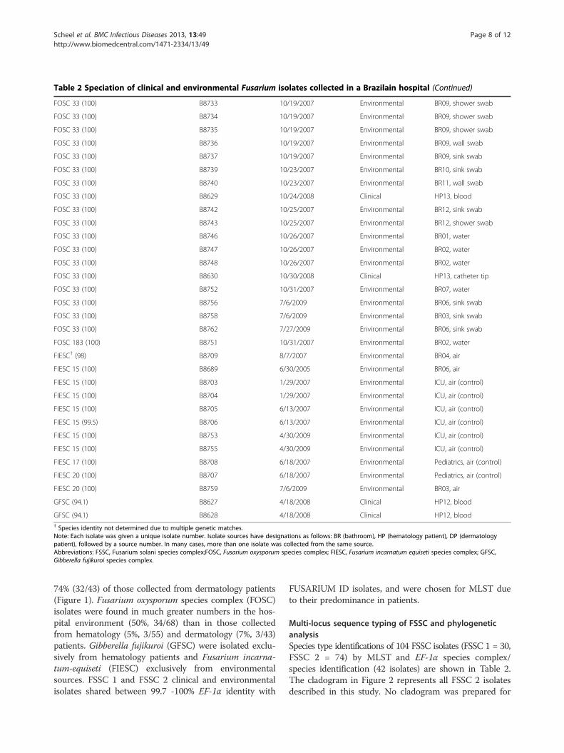
Multi-locus sequence typing of FSSC and phylogeneticanalysisSpecies type identifications of 104 FSSC isolates (FSSC 1 = 30,FSSC 2 = 74) by MLST and EF-1α species complex/species identification (42 isolates) are shown in Table 2.The cladogram in Figure 2 represents all FSSC 2 isolatesdescribed in this study. No cladogram was prepared for
Table 2 Speciation of clinical and environmental Fusarium isolates collected in a Brazilain hospital (Continued)
FOSC 33 (100) B8733 10/19/2007 Environmental BR09, shower swab
FOSC 33 (100) B8734 10/19/2007 Environmental BR09, shower swab
FOSC 33 (100) B8735 10/19/2007 Environmental BR09, shower swab
FOSC 33 (100) B8736 10/19/2007 Environmental BR09, wall swab
FOSC 33 (100) B8737 10/19/2007 Environmental BR09, sink swab
FOSC 33 (100) B8739 10/23/2007 Environmental BR10, sink swab
FOSC 33 (100) B8740 10/23/2007 Environmental BR11, wall swab
FOSC 33 (100) B8629 10/24/2008 Clinical HP13, blood
FOSC 33 (100) B8742 10/25/2007 Environmental BR12, sink swab
FOSC 33 (100) B8743 10/25/2007 Environmental BR12, shower swab
FOSC 33 (100) B8746 10/26/2007 Environmental BR01, water
FOSC 33 (100) B8747 10/26/2007 Environmental BR02, water
FOSC 33 (100) B8748 10/26/2007 Environmental BR02, water
FOSC 33 (100) B8630 10/30/2008 Clinical HP13, catheter tip
FOSC 33 (100) B8752 10/31/2007 Environmental BR07, water
FOSC 33 (100) B8756 7/6/2009 Environmental BR06, sink swab
FOSC 33 (100) B8758 7/6/2009 Environmental BR03, sink swab
FOSC 33 (100) B8762 7/27/2009 Environmental BR06, sink swab
FOSC 183 (100) B8751 10/31/2007 Environmental BR02, water
FIESC† (98) B8709 8/7/2007 Environmental BR04, air
FIESC 15 (100) B8689 6/30/2005 Environmental BR06, air
FIESC 15 (100) B8703 1/29/2007 Environmental ICU, air (control)
FIESC 15 (100) B8704 1/29/2007 Environmental ICU, air (control)
FIESC 15 (100) B8705 6/13/2007 Environmental ICU, air (control)
FIESC 15 (99.5) B8706 6/13/2007 Environmental ICU, air (control)
FIESC 15 (100) B8753 4/30/2009 Environmental ICU, air (control)
FIESC 15 (100) B8755 4/30/2009 Environmental ICU, air (control)
FIESC 17 (100) B8708 6/18/2007 Environmental Pediatrics, air (control)
FIESC 20 (100) B8707 6/18/2007 Environmental Pediatrics, air (control)
FIESC 20 (100) B8759 7/6/2009 Environmental BR03, air
GFSC (94.1) B8627 4/18/2008 Clinical HP12, blood
GFSC (94.1) B8628 4/18/2008 Clinical HP12, blood† Species identity not determined due to multiple genetic matches.Note: Each isolate was given a unique isolate number. Isolate sources have designations as follows: BR (bathroom), HP (hematology patient), DP (dermatologypatient), followed by a source number. In many cases, more than one isolate was collected from the same source.Abbreviations: FSSC, Fusarium solani species complex;FOSC, Fusarium oxysporum species complex; FIESC, Fusarium incarnatum equiseti species complex; GFSC,Gibberella fujikuroi species complex.
Scheel et al. BMC Infectious Diseases 2013, 13:49 Page 8 of 12http://www.biomedcentral.com/1471-2334/13/49
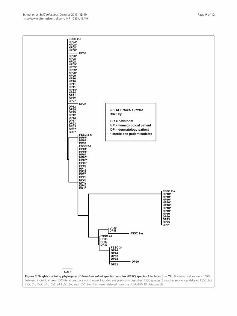
Figure 2 Neighbor-joining phylogeny of Fusarium solani species complex (FSSC) species 2 isolates (n = 74). Bootstrap values were 100%between individual taxa (1000 iterations, data not shown). Included are previously described FSSC species 2 voucher sequences (labeled FSSC 2-d,FSSC 2-f, FSSC 2-h, FSSC 2-I, FSSC 2-k, and FSSC 2-u) that were retrieved from the FUSARIUM ID database [8].
Scheel et al. BMC Infectious Diseases 2013, 13:49 Page 9 of 12http://www.biomedcentral.com/1471-2334/13/49

FSSC 1-a and 1-b isolates, since these STs differ by onlyone base pair.
FSSC 2-d and FSSC 2-f were the most common STs iso-lated from patients (Figure 2). FSSC 2-d was collectedfrom seven patients in the BTMU (20 isolates) and 12dermatology outpatients, while FSSC 2-f was isolated fromfive BTMU patients (8 isolates) and five dermatology out-patients (6 isolates). FSSC 2 isolates were rarely found inthe environment (n = 4); two FSSC 2-d isolates were col-lected from the same water source (BR07) on the sameday, one FSSC 2-d was isolated from an air sample(BR03), and FSSC 2-f was collected from a floor swab ofBR10. None of the environmental FSSC 2 isolates werecollected within the same month as those collected frompatients in the BMTU (Table 2).
FSSC 2-k (1 patient, 8 isolates) and FSSC 2-t (1 pa-tient, 2 isolates) were the other STs identified by MLSTin the BMTU, but none were collected in the environment(Figure 2). Other Fusarium isolated in hematology patientswere provisionally identified by EF-1α sequence compari-son and each was found in one BMTU patient. These wereFSSC 3+4 (HP16, corneal swab), FSSC 7 (HP15, blood),FOSC 33 (HP13, blood and catheter tip), GFSC (HP12,blood), and unspeciated FSSC (HP01, blood) and FOSC(HP15, skin) (Table 2).
Among environmental isolates, FOSC 33 comprised al-most half of all samples collected, and was isolated 32times between September 2006 and July 2009, with themajority (24) sampled between August and October 2007.This species was found in water-related surface swabs ofall 12 BMTU bathrooms, a nephrology wall swab (control),and water of two bathrooms (BR02, BR07). FOSC 33 wasalso isolated from blood and a catheter tip of a patient(HP13) in October 2008, but none were collected from theenvironment that month (Table 2).
Forty-three bathroom swabs were performed betweenSeptember 2006 and July 2009, and 6 different Fusariumspecies/STs were recovered; FOSC (unknown species) andFOSC 33, FSSC 1-a, 1-b, 2-d, and 2-f. Water samples posi-tive for Fusarium (n = 10) were collected in four bath-rooms (BR01, 02, 03, 07) and yielded four STs; FOSC 183,FOSC 33, FSSC 1-b and FSSC 2-d. Only BR01 and BR07were positive for FSSC (1-b, and 2-d, respectively), andwater in these rooms also yielded FOSC STs. FIESC wascollected from air in three bathrooms (BR03, BR04, BR06)and from 8 control samples. Three other STs were foundin air, FSSC 1-a, 1-b, and 2-d, and all were collected fromBR03.
DiscussionGiven the severe morbidity and mortality from Fusariuminfections, it is important to understand the sources ofthese organisms and determine the extent to which fusaria
may be transmitted in the hospital. One such study byAnaissie in 2001 implicated hospital water systems(drains, faucet aerators, shower heads) as potential reser-voirs for Fusarium, and suggested that these fungi can betransmitted through the water system due to their persist-ence in plumbing systems [13]. In the United States, Shortet al. recently confirmed these findings by performing anextensive MLST study of fusaria recovered from plumbingdrains in the United States [14]. They found that the 6most common sequence types in drains (FSSC 1-a, 1-b,2-d, 2-k, FOSC 33 and Fusarium dimerum) were also thesix most commonly associated with human infection. Ourstudy bolsters the findings of Short et al. with regard tocommon Fusarium STs isolated from water-relatedsources, since we also found that FOSC 33 was the mostcommon ST isolated in drains, faucets, and tiles ofshowers and sinks in the hospital. Unlike Short et al., wefound FSSC 1-a and FSSC 1-b to be more commonlyfound in these sources, as opposed to FSSC 2-d. Our studydiffers in two ways, however, since environmental sam-pling was conducted in Brazil, and only exterior plumbingsurfaces were sampled.
Several studies have shown that MLST of three loci(EF-1α, rRNA and RPB2) have the highest discriminatorypower in delineating FSSC using comparative sequenceanalyses [7,15,16]. We used this three-locus method andfound that subtypes of most BTMU inpatient clinical iso-lates belonged to FSSC species 2 (69%, 38/55), and wereprimarily 2-d (recovered from 36% of patients) and 2-f(recovered from 18% of patients). In contrast, FSSC isolatescomprised 36% of the environmental samples and wereshown to be predominantly species type 1-a (18%) and 1-b(13%), while only four environmental samples were FSSC 2.The remaining environmental samples were predominantlyFOSC from swabs and water (50% of environmentalsamples) and FIESC from air (79% of air samples). We didnot find any FIESC clinical isolates, although we did findtwo FOSC clinical cases. Our data suggest that most of theclinical cases of fusariosis in this immunosuppressed popu-lation could not be traced to a specific water-related envi-ronmental source in the hospital ward. However, becausewe found a small number of species type-level matchesbetween clinical and environmental isolates, we believe thatin this institution, environmental water-related sources andeven air must be considered as possible reservoirs for pa-tient infection.
In this study, we were unable to find any temporal associ-ation between BMTU patient fusarial infections and con-taminated water-related sources in the BMTU environment.We explored the epidemiology of community acquisition bystudying a population of dermatology outpatients diagnosedwith skin or nail fusariosis who were seen at the hospitalduring the same period. The spectrum of Fusarium STsrecovered from these patients largely mirrored those
Scheel et al. BMC Infectious Diseases 2013, 13:49 Page 10 of 12http://www.biomedcentral.com/1471-2334/13/49

recovered from BMTU inpatients (Figure 1). A study byRaad et al. used RAPD subtyping to demonstrate that Fu-sarium DNA collected from 10 infected patients did notmatch DNA from 15 environmental isolates collected fromshower head water, sink water, and drains in their institution[17]. Our study was far more extensive and yielded similarresults.
Both Anassie and Short [13,14] have implicated watersystems as reservoirs of fusariosis, but in our study wefound only FSSC 1 to be common in both hematologypatients and the environment. It is possible that Fusariumpatient subtypes could have been missed in the hospitalenvironment because of limitations in the environmentalinvestigation. Environmental sampling was sporadic andwas limited in scope, so potential sources of contaminationmay have been missed. Few samples were collected fromair in the BMTU, and surfaces of other fomites in the pa-tient rooms were not sampled; rather focus was placed onthe patient bathrooms, pursuing the hypothesis that water-related sources were Fusarium reservoirs. Additionally, themethods used for environmental sample collection maynot have been optimal for Fusarium recovery.
The diversity of Fusarium STs among dermatology pa-tients with superficial infections was greater than that ofBMTU patients, and the distribution of STs was highlysimilar between these patient populations. We found that asingle species, FSSC 2-d, predominated in both inpatienthematology and outpatient dermatology samples. Certainly,our combined patient data suggest that FSSC species re-main a cause of superficial colonization, and under appro-priate circumstances of immunosuppression, could causedisease in an IHC patient population. The increase infusariosis in this and other hospitals (personal communica-tion, Marcio Nucci) suggests that there may be a significantincrease in circulating Fusarium conidia in this geogra-phical region. Our findings raise important questions forfuture research, including a search for the environmentalreservoirs of Fusarium spp., an explanation for this surgeof environmental Fusarium, and the potential implicationsof these findings in infection control measures.
ConclusionsWe isolated similar Fusarium species in this hospital andoutpatient setting. The limited environmental investigationcoupled with molecular analysis did not show a definitivelink to clinical isolates causing infection. Future studies areneeded in order to identify reservoirs of Fusarium in thecommunity, as well as preventive measures for patients athigh risk for IF.
AbbreviationsBMTU: Bone marrow transplant unit; CDC: Centers for Disease Control andPrevention; EF-1α: Elongation factor 1-alpha; IF: Invasive Fusariosis;FIESC: Fusarium incarnatum-equiseti species complex; FOSC: Fusariumoxysporum species complex; FSSC: Fusarium solani species complex;
GFSC: Gibberella fujikuroi species complex; ITS: Internal transcribed spacerregion; ML: Maximum likelihood; MLST: Multi-locus sequence typing;MP: Maximum parsimony; NJ: Neighbor joining; RPB2: RNA polymerase IIsubunit; rRNA: rRNA DNA region; RT: Room temperature; SDA: Sabourauddextrose agar; SDA + C: Sabouraud dextrose agar with chloramphenicol;ST: Species type.
Competing interestsThe authors declare that they have no competing interest.
Authors’ contributionsSAB, MN and CMS conceived the study design and prepared article draft.CMS and SFH performed PCR and molecular analysis. GB and TA performedenvironmental sampling, fungal isolation and cultures. MN provided patientcare and management and patient isolate collection. All authors contributedto, reviewed, and approved the final draft of this article.
Authors’ noteThe findings and conclusions in this report are those of the authors and do notnecessarily represent the views of the Centers for Disease Control and Prevention.
AcknowledgementsWe thank Joyce Peterson of the Mycotic Diseases Branch Reference Team forsubculture, morphological identification and archiving of Fusarium isolates,and the Division of Foodborne, Waterborne, and Environmental DiseasesGenomics Unit for performing sequencing of Fusarium isolate loci.
Author details1Mycotic Diseases Branch, Centers for Disease Control and Prevention,Atlanta, GA, USA. 2University Hospital, Universidade Federal do Rio deJaneiro, Rio de Janeiro, Brazil. 3Center for Global Health, Centers for DiseaseControl and Prevention, Atlanta, GA, USA.
Received: 17 May 2012 Accepted: 23 January 2013Published: 30 January 2013
References1. Munoz P, Guinea J, Bouza E: Treatment options in emerging mold
infections. Curr Infect Dis Rep 2008, 10:473–479.2. Nucci M: Emerging moulds: Fusarium, Scedosporium and Zygomycetes in
transplant recipients. Curr Opin Infect Dis 2003, 16:607–612.3. Nucci M, Anaissie E: Fusarium infections in immunocompromised
patients. Clin Microbiol Rev 2007, 20:695–704.4. Lionakis MS, Kontoyiannis DP: Fusarium infections in critically ill patients.
Semin Respir Crit Care Med 2004, 25:159–169.5. Richardson M, Lass-Florl C: Changing epidemiology of systemic fungal
infections. Clin Microbiol Infect 2008, 14(Suppl 4):5–24.6. De Hoog GS, Guarro J, Gené J, Figueras MJ: Hyphomycetes. Genus: Fusarium,
Atlas of Clinical Fungi. 2nd Ed. Utrecht, The Netherlands: Centraalbureauvoor Schimmelcultures. Rues, Spain: Universitat Rovira I Virgili; 2000:681–705.
7. O’Donnell K, Sutton DA, Fothergill A, McCarthy D, Rinaldi MG, Brandt ME,Zhang N, Geiser DM: Molecular phylogenetic diversity, multilocushaplotype nomenclature, and in vitro antifungal resistance within theFusarium solani species complex. J Clin Microbiol 2008, 46:2477–2490.
8. O’Donnell K, Sutton DA, Rinaldi MG, Sarver BA, Balajee SA, Schroers HJ,Summerbell RC, Robert VA, Crous PW, Zhang N, Aoki T, Jung K, Park J, Lee YH,Kang S, Park B, Geiser DM: Internet-accessible DNA sequence database foridentifying fusaria from human and animal infections. J Clin Microbiol 2010,48:3708–3718. Available from: [http://isolate.fusariumdb.org/].
9. Thompson JD, Higgins DG, Gibson TJ: CLUSTAL W: improving thesensitivity of progressive multiple sequence alignment throughsequence weighting, position-specific gap penalties and weight matrixchoice. Nucleic Acids Res 1994, 22:4673–4680.
10. Hall TA: BioEdit: A user-friendly biological sequence alignment editor andanalysis program for Windows 95/98/NT. Nucl Acids Symp Ser 1999, 41:95–98.
11. Felsenstein J: PHYLIP (Phylogeny Inference Package) version 3.69. Distributed bythe author. Seattle: Department of Genetics, University of Washington; 1993.Available from: [http://evolution.genetics.washington.edu/phylip.html].
12. Rambaut A: Figtree: Tree figure drawing tool, version 1.3.1. Distributed by theauthor. Edinburgh, Scotland, United Kingdom: Institute of Evolutionary
Scheel et al. BMC Infectious Diseases 2013, 13:49 Page 11 of 12http://www.biomedcentral.com/1471-2334/13/49

Biology, University of Edinburgh; 2006–2009. Available from: [http://tree.bio.ed.ac.uk/software/figtree/].
13. Anaissie EJ, Kuchar RT, Rex JH, Francesconi A, Kasai M, Müller FM, Lozano-Chiu M, Summerbell RC, Dignani MC, Chanock SJ, Walsh TJ: Fusariosisassociated with pathogenic Fusarium species colonization of a hospitalwater system: a new paradigm for the epidemiology of opportunisticmold infections. Clin Infect Dis 2001, 33:1871–1878.
14. Short PG, O’Donnell K, Zhang N, Juba JH, Geiser DM: Widespreadoccurrence of diverse human pathogenic types of the fungus Fusariumdetected in plumbing drains. J Clin Microbiol 2011, 49:4264–4272.
15. Migheli Q, Balmas V, Harak H, Sanna S, Scherm B, Aoki T, O’Donnell K:Molecular phylogenetic diversity of dermatologic and other humanpathogenic fusarial isolates from hospitals in northern and central Italy.J Clin Microbiol 2010, 48:1076–1084.
16. Balajee SA, Borman AM, Brandt ME, Cano J, Cuenca-Estrella M, Dannaoui E,Guarro J, Haase G, Kibbler CC, Meyer W, O’Donnell K, Petti CA, Rodriguez-Tudela JL, Sutton D, Velegraki A, Wickes BL: Sequence-based identificationof Aspergillus, Fusarium, and Mucorales species in the clinical mycologylaboratory: where are we and where should we go from here? J ClinMicrobiol 2009, 47:877–884.
17. Raad I, Tarrand J, Hanna H, Albitar M, Janssen E, Boktour M, Bodey G,Mardani M, Hachern R, Kontoyiannis D, Whimbey E, Rolston K:Epidemiology, molecular mycology, and environmental sources ofinfection in patients with cancer. Infect Control Hosp Epidemiol 2002,23:532–537.
doi:10.1186/1471-2334-13-49Cite this article as: Scheel et al.: Molecular analyses of Fusarium isolatesrecovered from a cluster of invasive mold infections in a Brazilianhospital. BMC Infectious Diseases 2013 13:49.
Submit your next manuscript to BioMed Centraland take full advantage of:
• Convenient online submission
• Thorough peer review
• No space constraints or color figure charges
• Immediate publication on acceptance
• Inclusion in PubMed, CAS, Scopus and Google Scholar
• Research which is freely available for redistribution
Submit your manuscript at www.biomedcentral.com/submit
Scheel et al. BMC Infectious Diseases 2013, 13:49 Page 12 of 12http://www.biomedcentral.com/1471-2334/13/49