research article open access identification of a stem-like
TRANSCRIPT

RESEARCH ARTICLE Open Access
Identification of a stem-like cell population byexposing metastatic breast cancer cell lines torepetitive cycles of hypoxia and reoxygenationElizabeth Louie1, Sara Nik1, Juei-suei Chen1, Marlies Schmidt1, Bo Song2, Christine Pacson1, Xiu Fang Chen1,Seonhye Park1, Jingfang Ju2, Emily I Chen1*
Abstract
Introduction: The irregular vasculature of solid tumors creates hypoxic regions, which are characterized by cyclicperiods of hypoxia and reoxygenation. Accumulated evidence suggests that chronic and repetitive exposure tohypoxia and reoxygenation seem to provide an advantage to tumor growth. Although the development ofhypoxia tolerance in tumors predicts poor prognosis, mechanisms contributing to hypoxia tolerance remain to beelucidated. Recent studies have described a subpopulation of cancer stem cells (CSC) within tumors, which havestem-like properties such as self-renewal and the ability to differentiate into multiple cell types. The cancer stemcell theory suggests CSCs persist in tumors as a distinct population and cause relapse and metastasis by giving riseto new tumors. Since hypoxia is considered to be one of the critical niche factors to promote invasive growth oftumors, we hypothesize that repetitive cycles of hypoxia/reoxygenation also play a role in the enrichment of breastCSCs.
Methods: Two metastatic human breast cancer cell lines (MDA-MB 231 and BCM2) were used to optimize theconditions of hypoxia and reoxygenation cycles. The percentage of CSCs in the cycling hypoxia selectedsubpopulation was analyzed based on the CD44, CD24, ESA, and E-cadherin expression by three-color flowcytometry. Colony formation assays were used to assess the ability of this subpopulation to self-renew. Limitingdilution assays were performed to evaluate the tumor-initiating and metastatic ability of this subpopulation.Induction of EMT was examined by the expression of EMT-associated markers and EMT-associated microRNAs.
Results: Using an optimized hypoxia and reoxygenation regimen, we identified a novel cycling hypoxia-selectedsubpopulation from human breast cancer cell lines and demonstrated that a stem-like breast cancer cellsubpopulation could be expanded through repetitive hypoxia/reoxygenation cycles without genetic manipulation.We also found that cells derived from this novel subpopulation form colonies readily, are highly tumorigenic inimmune-deficient mice, and exhibit both stem-like and EMT phenotypes.
Conclusions: These results provide the validity to the newly developed hypoxia/reoxygenation culture system forexamining the regulation of CSCs in breast cancer cell lines by niche factors in the tumor microenvironment anddeveloping differential targeting strategies to eradicate breast CSCs.
IntroductionRecent studies have described a subpopulation of cancercells within tumors termed ‘cancer stem cells’ (CSCs),which have stem-like properties such as self-renewal and
the ability to differentiate into multiple cancer cell types[1-7]. The CSC theory suggests that such CSCs persist intumors as a distinct population and cause relapse andmetastasis by giving rise to new tumors [8-10]. AlthoughCSCs make up only a small fraction of a tumor, they pos-sess the unique capability to regenerate a tumor whereasmost tumor cells lack this regenerative capability [11,12].By means of a non-obese diabetic/severe combined
* Correspondence: [email protected] of Pharmacological Sciences, Stony Brook University, BST-125,Stony Brook, NY 11794, USAFull list of author information is available at the end of the article
Louie et al. Breast Cancer Research 2010, 12:R94http://breast-cancer-research.com/content/12/6/R94
© 2010 Louie et al.; licensee BioMed Central Ltd. This is an open access article distributed under the terms of the Creative CommonsAttribution License (http://creativecommons.org/licenses/by/2.0), which permits unrestricted use, distribution, and reproduction inany medium, provided the original work is properly cited.

immunodeficiency disease (NOD/SCID) xenotransplantassay in combination with specific cell surface markers(CD44+CD24-/low), CSCs were enriched from metastaticand primary breast tumors and were shown to have theability to reestablish tumor heterogeneity after transplan-tation [1]. Since then, additional CSC markers have beenproposed and studied to isolate putative tumor stem cellpopulations. However, as demonstrated by a recentreport from Stuelten and colleagues [13], the complexityof CSC markers continues to pose challenges for identify-ing and isolating the putative tumor stem cell popula-tions by the cell-sorting approach. In addition toinitiating tumors, CSCs are thought to be capable of initi-ating metastasis. The link between CSCs and metastasishas been suggested by several studies. First, breast CSCswere shown to invade through Matrigel, a basementmembrane matrix used routinely as an indicator of meta-static potential of cancer cells [14]. Second, a recentstudy demonstrated that there is a link between epithe-lial-mesenchymal transition (EMT) and breast CSCs [15].Furthermore, the prevalence of CD44+CD24- cells inbreast cancer patients indicates a link between highnumbers of stem-like cancer cells and metastasis [16].However, only a few studies have directly tested themetastatic capability of putative CSCs in vivo. Collectiveevidence from a few studies that directly tested thein vivo metastasis using sorted CSCs suggests thatthe CSC phenotype alone may exhibit invasive propertyin vitro but is inadequate to determine or predict in vivometastasis. For example, in pancreatic cancer, CSCs(CD133+ cells) were not able to metastasize wheninjected orthotopically at low numbers [17]. In mammarycarcinomas, CD44+CD24low cells were invasive in vitrobut the phenotype was not sufficient for metastasis whencells were injected intracardiacally in vivo [14]. Therefore,we set out to investigate alternative mechanisms thatcould enrich for breast CSCs with tumor-initiating andmetastatic capabilities.To identify factors that distinguish the malignant
subpopulation within breast tumors, we began to exploreenvironmental influences known to associate withaggressively metastatic breast tumors. It has been postu-lated that hypoxia contributes directly to the develop-ment of more aggressive cancers by exerting selectivepressure on the tumor cell population to favor cells thatcan survive decreased O2 and nutrients [18-20]. Duringtumor development, rapid expansion of cancer cells cre-ates a hypoxic microenvironment that is followed by per-iods of reoxygenation to promote tumor progression.These two aspects of tumor progression (hypoxia andreoxygenation) cooperate to provide growth advantagesessential for the progressive development of aggressivetumors [21]. Although extensive efforts have beendevoted to understanding the effect of hypoxia on tumor
progression, two areas of tumor biology remain unclear.What is the effect of fluctuating oxygen tension ontumor progression? How does hypoxia drive an irreversi-ble phenotype without genetic manipulation? It is knownthat both hypoxia and consecutive hypoxia/reoxygena-tion can exert a variety of effects on tumor cell biology,including activation of pro-survival signal transductionpathways, aberrant genetic and epigenetic alterations,and increased tumor angiogenesis. In some studies,hypoxia/reoxygenation was shown to drive expression ofproteins associated with poor prognosis [22]. Further-more, susceptibility of genomic instability can vary afteracute or chronic exposure to hypoxia followed by reoxy-genation [23]. Therefore, we hypothesize that hypoxia/reoxygenation cycles may provide the driving force toselect for a highly metastatic breast CSC subpopulation.To study the effect of hypoxia/reoxygenation cycles
on breast cancer, we exposed two metastatic humanbreast cancer cell lines (MDA-MB 231 and BCM2) tocycles of chronic hypoxia and nutrient deprivation.After one cycle of hypoxia and reoxygenation, weobserved a small cell population that survived hypoxiaas spherical clusters under hypoxic conditions and thatresumed proliferation after reoxygenation. We then iso-lated and exposed this novel subpopulation to additionalcycles of hypoxia/reoxygenation and established a dis-tinct subpopulation of cells from these two breast can-cer cell lines. As expected, this novel subpopulationshowed increasing viability under hypoxia and the abil-ity to proliferate as either an adherent monolayer orsubstrate-independent tumor spheres. Interestingly, anincreased fraction of the cell population was found toexpress CD44+/CD24-/ESA+ cell surface markers. How-ever, this novel subpopulation was distinguished bybeing highly tumorigenic and metastatic and showedupregulation of EMT markers. These findings stronglysuggest that we have succeeded in isolating a uniquemetastatic CSC population by exposing breast cancercells to repetitive cycles of hypoxia and reoxygenation.
Materials and methodsCell lines and tissue cultureMDA-MB 231 was purchased from American Type Cul-ture Collection (Manassas, VA, USA); BCM2 cell linewas obtained from Brunhilde Felding-Habermann(Scripps Research Institute, La Jolla, CA, USA). MDA-MB 231 and BCM2 cell lines were cultured in minimumessential medium (MEM) (Invitrogen Corporation,Carlsbad, CA, USA) supplemented with 10% fetal bovineserum (HyClone, Logan, UT, USA; Thermo FisherScientific Inc., Waltham, MA, USA), 2 mM L-glutamine,1 mM sodium pyruvate, 1 mM non-essential aminoacids, and 1% vitamin (HyClone). All cell lines weregrown at 37°C and in 5% carbon dioxide.
Louie et al. Breast Cancer Research 2010, 12:R94http://breast-cancer-research.com/content/12/6/R94
Page 2 of 14

Flow cytometryNon-confluent cultures were trypsinized into single-cellsuspension, counted, washed with phosphate-bufferedsaline (PBS), and fixed with 10% paraformaldehyde. Cellswere stained with antibodies specific for human cell sur-face markers: CD326/ESA-FITC, CD24-PE, and CD44-PE-Cy7 (BD Pharmingen, San Jose, CA, USA). E-cadherinantibody was purchased from Cell Signaling Technology,Inc. (Danvers, MA, USA). A total of 2 × 10⁵ cells wereincubated with antibodies for 30 minutes on ice.Unbound antibody was washed, and cells were analyzedon a Guava EasyCyte Plus Flow Cytometer (MilliporeCorporation, Billerica, MA, USA). The Student t test wasused to calculate the significance of increased CD44+/CD24-/ESA+ and E-cad-/CD44+/CD24- populations inthe cycling hypoxia-selected subpopulations.
Viability and cell proliferation assayCells were collected from either the media (floating subpo-pulation) or the adherent subpopulation after eachhypoxic cycle and dissociated into single-cell suspensionby trypsin/EDTA (trypsin/ethylenediaminetetraaceticacid). Guava ViaCount Reagent (Millipore Corporation)was used to determine the viability and cell numbers aftereach hypoxic cycle. For cell proliferation assay, 5 × 10⁴
cells were seeded in triplicate wells per cell line per timepoint in 12-well tissue culture plates. Cells were detached(adherent culture) or dissociated (spherical culture) bytrypsin/EDTA and resuspended in PBS for cell countingusing the ViaCount assay. In accordance with the manu-facturer’s instructions, an appropriate volume of theViaCount Reagent was mixed with 50 μL of cell suspen-sion in PBS. After 5 minutes of incubation at room tem-perature in the dark, ViaCount/cell mixture was loadedinto a 96-well microtiter plate and read by the Guava FlowCytometer. Both live and dead cells were detected and dis-played in dot plots. The Guava ViaCount software carriedout calculation of viability and cell numbers for each sam-ple automatically.
Colony-forming assaysFor the colony formation assay, cells were trypsinized togenerate single-cell suspensions and counted by a hemo-cytometer. Single-cell suspensions (500 cells per well)were plated on 96-well plates with an ultralow attach-ment surface. Three wells were seeded for each cell line,and triplicate experiments were performed per cell line(n = 3). The EVOS microscope (Advanced MicroscopyGroup, Bothell, WA, USA) was used to count tumorspheres and take images of each well on days 1, 3, 5, and9. Immediately after the cells were seeded, each well waschecked under the microscope to verify the sparseness ofeach spherical culture, and only wells containing single-cell suspension with no cell cluster were chosen for
tumor-sphere counting after 9 days. Colonies of at least60 μm in diameter (determined by using an eyepiece gra-ticule with crossed scales) were counted on day 9 afterplating. Colony-forming efficiency (CFE) was calculatedby dividing the number of colonies (> 60 μm) formed bythe original number of single cells seeded and isexpressed as a percentage.
Animals and surgeryAll animal procedures were performed in accordance withan approved protocol by the Stony Brook University Insti-tutional Animal Care and Use Committee. NOD/SCIDmice were purchased from the Jackson Laboratory (BarHarbor, ME, USA). Four- to six-week-old female micewere used for tumor injections. Human breast cancer cellswere suspended in sterile Hank’s buffered salt solution(HBSS) and injected into the third thoracic mammarygland. Tumor formation was assessed by palpation at leastonce a week. A caliper was used to measure the lengthand width of tumors at least once a week. A standard for-mula for calculating tumor volume (in cubic millimeters)was used: length × (width)2/2. Duplicate experiments wereperformed for each cell number.
Quantification of lung metastasisFive to six weeks after the resection of primary tumorsfrom tumor-bearing NOD/SCID mice, lungs were har-vested and fixed in the Bouin’s fixative solution overnight.Nodules on the surface of lungs were visualized under thedissecting microscope, and a USB digital camera (LeicaMicrosystems, Bannockburn, IL, USA) from the micro-scope was used to take gross images of each lung. Toquantify the tumor burden, each image was first annotatedwith the National Institutes of Health ImageJ software toidentify measurable nodules on the surface of the lung.The length and width of each nodule were measured withpixels and then converted to millimeters to calculate thetumor volume. A standard formula for calculating tumorvolume (cubic millimeters) was used: length × (width)2/2.Metastatic tumor burden of each animal was calculated bycombining tumor volumes from four lobes of lungs. Aver-age tumor burden and standard deviations were derivedfrom at least four animals per group.
Western blot analysisSubcellular fractionation of cell lysates was preparedusing the ProteoExtract Subcellular Proteome ExtractionKit (Calbiochem-EMD Biosciences, San Diego, CA,USA), and protein concentrations were determined byEZQ Protein Quantification Kit (Invitrogen Corporation).Proteins from nuclear or cytoplasmic fractions (40 μg)were resolved by 4% to 20% SDS-PAGE and transferredonto nitrocellulose membranes for immunoblotting.Immunoblotting assays were carried out by standard
Louie et al. Breast Cancer Research 2010, 12:R94http://breast-cancer-research.com/content/12/6/R94
Page 3 of 14

procedures using Snail (Cell Signaling Technology, Inc.)and vimentin antibodies (Novus Biologicals, Littleton,CO, USA). Anti-b-actin antibody (Sigma-Aldrich, St.Louis, MO, USA) was used to confirm equal proteinloading of cytoplasmic fractions. Anti-histone H3 anti-body (Millipore Corporation) was used to confirm equalprotein loading of nuclear fractions. Secondary antibodiesconjugated with either AF680 (Invitrogen Corporation)or CW800 (LI-COR Biosciences, Lincoln, NE, USA) wereused to visualize protein bands using the Odyssey Infra-red Imager (LI-COR Biosciences).
Analysis of the mRNA and microRNA expression byquantitative reverse transcription-polymerase chainreaction assayTotal RNA was isolated from each cell line using an RNAExtraction Kit (Qiagen Inc., Valencia, CA, USA) in accor-dance with the manufacturer’s instructions. One point twomicrograms of total RNA from each cell line was reverse-transcribed using random primers and the High-CapacitycDNA Synthesis Kit (Applied Biosystems, Foster City, CA,USA). The resulting cDNAs were mixed with the SYBRPCR [polymerase chain reaction] master mix (Applied Bio-systems) and run on the StepOnePlus Applied BiosystemsReal-time PCR machine. One cycle of denaturing step(10 minutes at 95°C) was applied, followed by 35 cycles ofamplification (15 seconds at 95°C and 1 minute at 58°C),with fluorescence measured during the extension. Primersused to amplify the human Snail gene have the followingsequences: 5’-CCTCCCTGTCAGATGAGGAC-3’ (for-ward) and 5’-CCAGGCTGAGGTATTCCTG-3’ (reverse).Primers used to amplify the human Slug gene have the fol-lowing sequences: 5’-GGGGAGAAGCCTTTTTCTTG-3’(forward) and 5’-TCCTCATGTTTGTGCAGGAG-3’(reverse). Primers used to amplify the human Twist genehave the following sequences: 5’-GGAGTCCGCAGTCT-TACGAG-3’ (forward) and 5’-TCTGGAGGACCTGGTA-GAGG-3’ (reverse). The relative quantification (RQ) valuereflects the fold changes of mRNA expression in each cellline compared with the parental cell lines. RQ was calcu-lated using the comparative CT (ΔΔCT) method and Ste-pOne software version 2.0.1 (Applied Biosystems) andnormalized by the expression of the housekeeping gene,GAPDH (glyceraldehyde-3-phosphate dehydrogenase).Three independent experiments were performed to deriveaverage RQ and standard deviations (n = 3).For microRNA (miRNA) analysis, total RNAs, includ-
ing miRNAs, were isolated from the cell lines using TRI-zol reagent (Invitrogen Corporation) in accordance withthe manufacturer’s instructions. Ten nanograms of totalRNA from each cell line was converted to cDNA usingthe High-Capacity cDNA synthesis kit (Applied Biosys-tems). The miRNA sequence-specific reverse transcrip-tion-PCR (RT-PCR) primers for miR-200c, miR-205, and
an endogenous control RNU6B were purchased fromAmbion (Austin, TX, USA). Real-time quantitative RT-PCR (qRT-PCR) analysis was performed using the 7500Real-Time PCR System from Applied Biosystems. TheTaqMan master mix was added to the cDNA and primermix to amplify and quantify target miRNAs (No AmpEr-ase UNG; Applied Biosystems). The following PCR cyclewas used for miRNA amplifications: 95°C for 10 minutesand 40 cycles of 95°C for 15 seconds and 60°C for 60 sec-onds. The RQ value reflects the fold changes of miRNAexpression in each cell line compared with the parentalcell lines. RQ was calculated using the comparative CT
(ΔΔCT) method and StepOne software version 2.0.1(Applied Biosystems) and normalized by the expressionof the internal control RNU6B. Three independentexperiments were performed to derive average RQ andstandard deviations (n = 3).
ResultsEstablishing optimized conditions for hypoxia andreoxygenation selectionTo study the effect of repetitive hypoxia/reoxygenationon breast cancer cells, we optimized conditions ofhypoxia and reoxygenation with regard to the numberof cycles, exposure time, cell numbers, and other para-meters. We used two metastatic breast cancer cell lines,MDA-MB 231 and BCM2 [24], to ensure that ourcurrent protocol was not cell line-specific. To establishefficient and reproducible hypoxia/reoxygenation condi-tions, we used the ProOxC system (BioSpherix, Lacona,NY, USA), which allows precise oxygen control over anextended time period via an oxygen sensor and auto-matic feedback mechanisms to maintain a consistentlylow oxygen environment. The optimized procedureincluded exposing these breast cancer cell lines (1 × 10⁷
cells in a T125 tissue culture flask, approximately 60%confluency) to hypoxia (1% O2) and nutrient deprivationfor 7 days, mimicking the avascular microenvironment,and reoxygenating surviving cells for 1 to 3 weeksdepending on the viability of surviving cells (Figure 1).The same growth media used to maintain monolayercultures (see Materials and methods) were used toprepare the cells and maintain the adherent and non-adherent cells in culture. After each hypoxic cycle, 10%of the adherent culture became non-adherent (floatingcell population). Using this protocol, we consistentlyobserved novel surviving populations that were non-adherent and that demonstrated a propensity to formspherical clusters under hypoxic conditions. However,unlike the adherent hypoxia-resistant cells, only 10% ofthe total non-adherent cells were viable after the firstround of hypoxic cycle (F1). After reoxygenating andgrowing F1 cells as spherical cultures, we exposed thesame amount of F1 cells to a second hypoxic cycle and
Louie et al. Breast Cancer Research 2010, 12:R94http://breast-cancer-research.com/content/12/6/R94
Page 4 of 14

found an increase in viability (30%) (F2). After threerounds of hypoxia and oxygenation, we found that theviability of the non-adherent cells increased to 70% (F3).Additional rounds of hypoxia did not increase the per-centage of survival significantly. In addition to findingincreased hypoxia viability, we found distinct character-istics in this novel subpopulation. For comparison pur-poses, we exposed the adherent hypoxia-resistant cellsto two additional rounds of hypoxia/reoxygenationselection and generated the hypoxia-exposed adherentcell population (A3).
The newly isolated cycling hypoxia-selected breast cancersubpopulation has increased tumor-initiating capabilitySince hypoxia has been implicated in the promoting ofaggressive tumors, we first examined the tumor-initiat-ing ability of the parental and newly isolated cyclinghypoxia-selected subpopulation in immune-deficientmice. We orthotopically injected either parent MDA-MB 231 and BCM2 or the cycling hypoxia-selected sub-populations (MDA-MB 231 F3 and BCM2 F3) intoNOD/SCID mice as a limiting dilution assay from 5 ×10⁵ to 5 × 10² cells per mammary gland. Although therewas no noticeable difference in the frequency of tumorformation between these two cell populations (MDA-MB 231 and MDA-MB 231 F3) when 5 × 10⁵ cells wereinjected, a dramatic difference in tumor initiation andgrowth was observed when 10-fold, 100-fold, and 1,000-fold fewer cells were injected in NOD/SCID mice(Figure 2a). With as few as 500 cells, MDA-MB 231 F3and BCM2 F3 cells could form tumors in NOD/SCIDmice, whereas both parent cell lines were not able to doso at the same dilution (Table 1 and Figure 2b,c).Furthermore, tumors progressed rapidly in mice injectedwith lower numbers of MDA-MB 231 F3 cells andBCM2 F3 cells (5 × 10⁴ cells) than equivalent numbers
of MDA-MB 231 cells (Figure 2a-c), indicating thatMDA-MB 231 F3 cells formed more aggressive tumors.We also injected 500 hypoxia-exposed adherent cells(MDA-MB 231 A3 and BCM2 A3) in the mammary fatpad of female NOD/SCID mice. None of the miceinjected with 500 MDA-MB 231 A3 or BCM2 A3 cellsdeveloped mammary tumors by 90 days (MDA-MB 231A3) or 98 days (BCM2 A3) (Table 1). We concludethat the newly isolated cycling hypoxia-selected non-adherent subpopulation is highly tumorigenic comparedwith the hypoxia-exposed adherent and parental cell lines.
The newly isolated cycling hypoxia-selected breast cancersubpopulation comprises putative breast cancer stem cellpopulation (CD44+/CD24-/ESA+)Evidence of stem-like cancer cells has been establishedin various types of cancer, including breast cancer[1,3,4,7,8,12,25]. This small subpopulation has beenshown to be highly tumorigenic and exhibits some stemcell characteristics such as self-renewal and the ability toform tumor spheres [12,26]. Since the newly isolatedcycling hypoxia-selected subpopulation is highly tumori-genic, we hypothesized that this subpopulation isenriched with stem-like breast cancer cells. Using thethree-color flow cytometry analysis, we found that thecycling hypoxia-selected subpopulation displayed anincreased proportion of cells with the surface markerexpression of CD44+/CD24-/ESA+, which has beenreported as putative breast CSCs. The percentage of theCD44+/CD24-/ESA+ cell population was significantlyincreased in the cycling hypoxia-selected subpopulationin comparison with the parental breast cancer cell lines(3-fold increase in MDA-MB 231 F3 cells, Figure 3a,c;13-fold increase in BCM2 F3, Figure 3b,c; Figure S1 inAdditional file 1). Since it has been reported thathypoxia exposure alone can increase the stem-like cell
Figure 1 An illustration of the hypoxia and reoxygenation regimen. Conditions for hypoxia/reoxygenation cycles were optimized using twohuman metastatic breast cancer cell lines (MDA-MB 231 and BCM2). The viability of non-adherent cells is listed after each hypoxic cycle. FBS,fetal bovine serum.
Louie et al. Breast Cancer Research 2010, 12:R94http://breast-cancer-research.com/content/12/6/R94
Page 5 of 14

population, we also examined the percentage of CD44+/CD24-/ESA+ and CD44+/CD24+/ESA+ cell populationsin the hypoxia-exposed adherent cells (MDA-MB 231A3 and BCM2 A3) and found only a slight increase in
the percentage of CD44+/CD24-/ESA+ cell population inthe hypoxia-exposed adherent cells compared with theparental breast cancer cell lines (1.8-fold increase inMDA-MB 231 A3 cells, Figure 3c; 3.5-fold increase in
Figure 2 Growth curves of primary tumors generated from the parental cell lines and cycling hypoxia-selected subpopulations. (a)Growth curves of primary tumors in non-obese diabetic/severe combined immunodeficiency disease (NOD/SCID) mice injected with 5 × 10⁵ or5 × 104 MDA-MB 231 and MDA-MB 231 F3 cells. Tumors were first detected by palpation. Standard errors are derived from duplicateexperiments (n = 6 per cell number for each experiment). (b) Growth curves of tumors in NOD/SCID mice injected with 5 × 10³ or 5 × 10²
MDA-MB 231 and MDA-MB 231 F3 cells. Standard errors were derived from duplicate experiments (n = 5 per cell number for each experiment).(c) Growth curves of tumors in NOD/SCID mice injected with 5 × 10⁴ or 5 × 10² BCM2 and BCM2 F3 cells. Standard errors were derived fromduplicate experiments (n = 5 per cell number for each experiment).
Louie et al. Breast Cancer Research 2010, 12:R94http://breast-cancer-research.com/content/12/6/R94
Page 6 of 14

Table 1 Limiting dilution tumor formation of parental, hypoxia-exposed adherent, and cycling hypoxia-selected breastcancer cells in vivo
Cell type Days Number injected and tumors formed
5 × 10⁵ 5 × 10⁴ 5 × 10³ 5 × 10²
MDA-MB 231 (total cell population) 24-90 5/6 2/6 0/5 0/5
MDA-MB 231 A3 (hypoxia-exposed adherent cells) 90 - - - 0/5
MDA-MB 231 F3 (cycling hypoxia-selected subpopulation) 10-90 6/6 6/6 5/5 4/5
BCM2 (total cell population) 35-98 - 5/5 - 0/5
BCM2 A3 (hypoxia-exposed adherent cells) 98 - - - 0/5
BCM2 F3 (cycling hypoxia-selected subpopulation) 28-98 - 5/5 - 4/5
Figure 3 Cell surface expression of ESA, CD44, and CD24. Three-color flow cytometry analysis was performed to detect the CD44+/CD24-/ESA+ cell population. (a) Top panel: Unstained, isotype control, and ESA-stained cells are shown in the histogram. ESA staining of MDA-MB 231(total cell population) and MDA-MB 231 F3 (cycling hypoxia-selected subpopulation) cells. M1 marker gates ESA+ cells, and M2 marker gates theESA- cells. Bottom panel: CD44 and CD24 expression of ESA+ cells in each cell line. The percentages of the CD44+/CD24- and CD44+/CD24+ cellswithin the ESA+ cell population are indicated in the Quad plot. (b) The same flow cytometry analysis for BCM2 (total cell population) and BCM2F3 (cycling hypoxia-selected subpopulation) cells. (c) Quantitative comparison of CD44+/CD24-/ESA+ cell population and CD44+/CD24+/ESA+ cellpopulation in cycling hypoxia-selected subpopulations (MDA-MB 231 F3 and BCM2 F3) and their parental breast cancer cell lines. At least threereplications were performed to derive the average percentage of each cell population in each cell line and standard deviations. Asterisks indicatestatistical significance by two-tail t test (n = 3, P < 0.05). ESA, epithelial-specific antigen.
Louie et al. Breast Cancer Research 2010, 12:R94http://breast-cancer-research.com/content/12/6/R94
Page 7 of 14

BCM2 A3, Figure 3c). Thus, our data suggest that cyclicexposures of hypoxia and reoxygenation are crucial forthe enrichment of stem-like breast cancer cells in thisnovel non-adherent subpopulation.It has been shown that tumor-initiating cells or CSCs
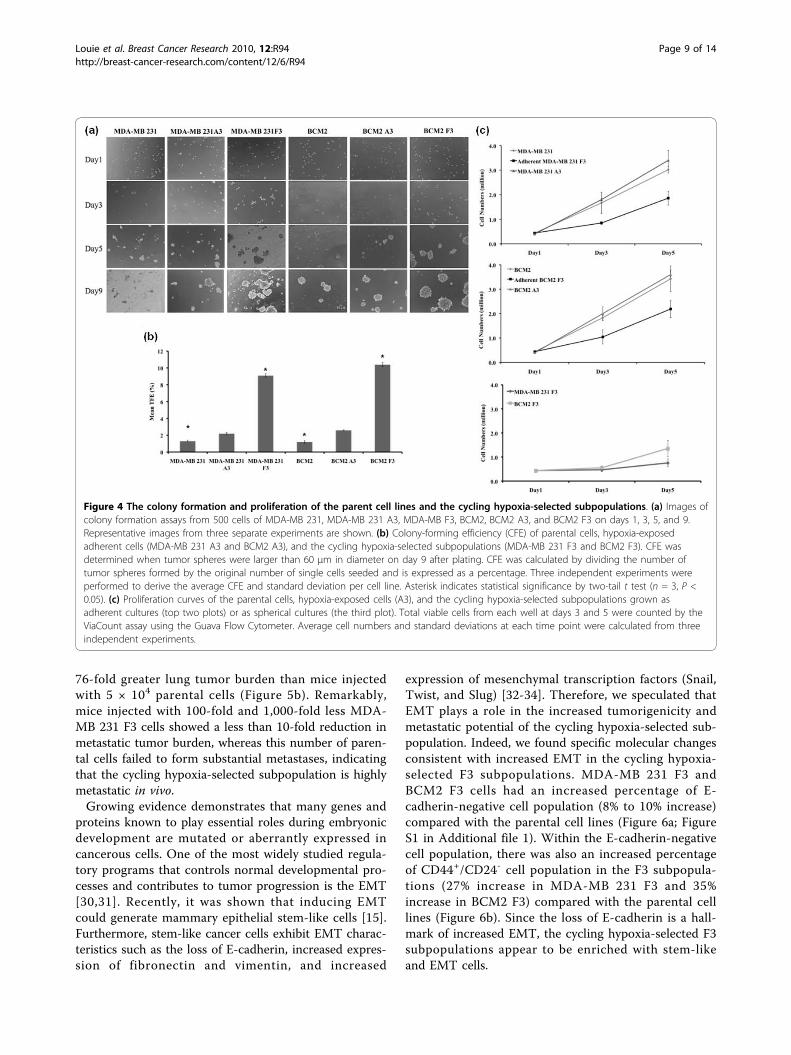
resemble stem cells in their ability to grow as sphereswhen cultured under conditions in which they cannotattach to a solid substratum [6,27]. To test whether theincreased tumor-initiating capability observed in thecycling hypoxia-selected subpopulations correlates withtheir ability to grow as tumor spheres in culture, cellsfrom parental cell populations (MDA-MB 231 andBCM2), hypoxia-exposed adherent cells (MDA-MB 231A3 and BCM2 A3), and cycling hypoxia-selected subpo-pulations (MDA-MB 231 F3 and BCM2 F3) were platedat low density (500 cells per 0.32 cm2) onto ultralowattachment plates in MEM. Images of cells from eachwell were taken at days 1, 3, 5, and 9 to visualize theformation of colonies. After day 5, we noticed that theappearance of compact and asymmetric solid tumorspheres developed in all wells containing MDA-MB 231F3 and BCM2 F3 cells whereas only small clusters ofcells were found in wells containing MDA-MB 231 andBCM2 cells (Figure 4a). By day 9, we observed thatsome large tumor spheres began to form in the parentalcell lines. The time line of tumor-sphere formation wascomparable between hypoxia-exposed adherent cells(MDA-MB 231 A3 and BCM2 A3) and their parentalcell lines. To determine the CFE of each cell population,tumor spheres of at least 60 μm were counted on day 9after plating. CFE was calculated by dividing the numberof tumor spheres formed by the original number of sin-gle cells seeded and was expressed as a percentage.Three independent experiments were performed toderive the average CFE and standard deviation per cellline. Although there is around a 2-fold increase in CFEin hypoxia-exposed cells (MDA-MB 231 A3 and BCM2A3) compared with the parental cell lines, a largerincrease (7- to 8-fold) of CFE was found in the cyclinghypoxia-selected subpopulations compared with the par-ental cell lines (Figure 4b). Therefore, according to ourresults, the increased tumor-initiating capability of thecycling hypoxia-selected subpopulations is in accordancewith the increased ability to grow as spherical coloniesin culture.In addition to showing increased ability to grow as
spherical colonies, the stem-like cancer cells wereshown to proliferate slower than the bulk cancer cells[28,29]. This attribute of stem-like cancer cells has sig-nificant clinical implications such as chemoresistanceand radioresistance. To determine whether the cyclinghypoxia-selected subpopulations cycle slower than theparental and hypoxia-exposed adherent cells, equalnumbers of cells from each cell population were plated
on day 1, and an increase in cell numbers over 5 dayswas used to assess the rate of proliferation. To comparethe rate of proliferation under the same condition as theparental and hypoxia-exposed adherent cells, we platedthe cycling hypoxia-selected subpopulation as adherentcells (adherent MDA-MB 231 F3 and adherent BCM2F3) and measured the increase in cell numbers togetherwith the other adherent cell populations. The prolifera-tion of the cycling hypoxia-selected subpopulation asspherical culture was performed separately. While theparental and hypoxia-exposed adherent cells displaysimilar rates of proliferation, the cycling hypoxia-selected subpopulations grow slower as adherent cellsand even slower as suspension cultures (Figure 4c).Together, results from established assays for CSC activ-ity show that the cycling hypoxia-selected subpopulationhas an increased ability of tumor initiation and tumor-sphere formation. More importantly, these findings sug-gested that our hypoxia/reoxygenation scheme resultedin an enrichment of bona fide breast CSCs withoutselecting for user-defined markers or targeted geneticmanipulation.
The newly isolated cycling hypoxia-selected breast cancersubpopulation is highly metastatic and exhibitsincreasing epithelial-mesenchymal transition phenotypeAside from their role in tumor initiation, stem-like can-cer cells have been hypothesized to contribute directlyto cancer metastasis. However, the collective evidencefrom a few studies examining the metastatic potential ofCSCs in vivo suggests that CSC phenotype alone is notenough to determine metastasis. Particularly, a study bySheridan and colleagues [14] demonstrates that CD44+/CD24- stem-like phenotype is not sufficient for hom-ing and proliferation at sites of metastasis, despite a dis-play of increased invasive property in vitro.Using the human cancer cell xenograft model, we
were able to quantify lung metastases 4 weeks after thesurgical resection of primary tumors. Images of lungswere taken by a digital camera built into the microscopeand were used for quantifying metastases. The NationalInstitutes of Health ImageJ software was used to mea-sure the diameters (width and length) of each visiblemetastasis on the lung, and total tumor volumes werecalculated from each animal (for a description, seeMaterials and methods). Overall, lungs harvested frommice with primary tumors derived from the cyclinghypoxia-selected MDA-MB 231 F3 cells showed signifi-cantly more metastases than lungs harvested from micewith parental cell primary tumors (Figure 5a). Miceinjected with 5 × 10⁵ MDA-MB 231 F3 cells showed a43-fold greater lung tumor burden, by volume, thanmice injected with 5 × 10⁵ parental cells (Figure 5b).Mice injected with 5 × 10⁴ MDA-MB 231 F3 cells had
Louie et al. Breast Cancer Research 2010, 12:R94http://breast-cancer-research.com/content/12/6/R94
Page 8 of 14
76-fold greater lung tumor burden than mice injectedwith 5 × 10⁴ parental cells (Figure 5b). Remarkably,mice injected with 100-fold and 1,000-fold less MDA-MB 231 F3 cells showed a less than 10-fold reduction inmetastatic tumor burden, whereas this number of paren-tal cells failed to form substantial metastases, indicatingthat the cycling hypoxia-selected subpopulation is highlymetastatic in vivo.Growing evidence demonstrates that many genes and
proteins known to play essential roles during embryonicdevelopment are mutated or aberrantly expressed incancerous cells. One of the most widely studied regula-tory programs that controls normal developmental pro-cesses and contributes to tumor progression is the EMT[30,31]. Recently, it was shown that inducing EMTcould generate mammary epithelial stem-like cells [15].Furthermore, stem-like cancer cells exhibit EMT charac-teristics such as the loss of E-cadherin, increased expres-sion of fibronectin and vimentin, and increased
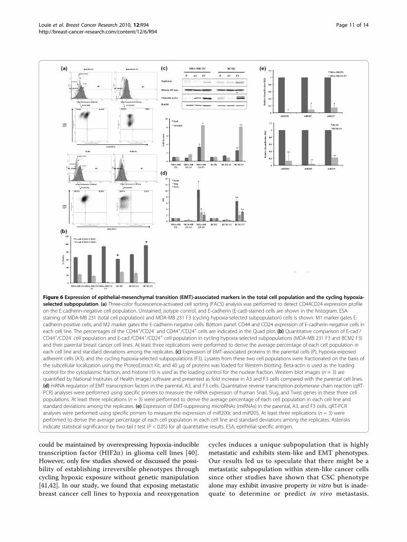
expression of mesenchymal transcription factors (Snail,Twist, and Slug) [32-34]. Therefore, we speculated thatEMT plays a role in the increased tumorigenicity andmetastatic potential of the cycling hypoxia-selected sub-population. Indeed, we found specific molecular changesconsistent with increased EMT in the cycling hypoxia-selected F3 subpopulations. MDA-MB 231 F3 andBCM2 F3 cells had an increased percentage of E-cadherin-negative cell population (8% to 10% increase)compared with the parental cell lines (Figure 6a; FigureS1 in Additional file 1). Within the E-cadherin-negativecell population, there was also an increased percentageof CD44+/CD24- cell population in the F3 subpopula-tions (27% increase in MDA-MB 231 F3 and 35%increase in BCM2 F3) compared with the parental celllines (Figure 6b). Since the loss of E-cadherin is a hall-mark of increased EMT, the cycling hypoxia-selected F3subpopulations appear to be enriched with stem-likeand EMT cells.
Figure 4 The colony formation and proliferation of the parent cell lines and the cycling hypoxia-selected subpopulations. (a) Images ofcolony formation assays from 500 cells of MDA-MB 231, MDA-MB 231 A3, MDA-MB F3, BCM2, BCM2 A3, and BCM2 F3 on days 1, 3, 5, and 9.Representative images from three separate experiments are shown. (b) Colony-forming efficiency (CFE) of parental cells, hypoxia-exposedadherent cells (MDA-MB 231 A3 and BCM2 A3), and the cycling hypoxia-selected subpopulations (MDA-MB 231 F3 and BCM2 F3). CFE wasdetermined when tumor spheres were larger than 60 μm in diameter on day 9 after plating. CFE was calculated by dividing the number oftumor spheres formed by the original number of single cells seeded and is expressed as a percentage. Three independent experiments wereperformed to derive the average CFE and standard deviation per cell line. Asterisk indicates statistical significance by two-tail t test (n = 3, P <0.05). (c) Proliferation curves of the parental cells, hypoxia-exposed cells (A3), and the cycling hypoxia-selected subpopulations grown asadherent cultures (top two plots) or as spherical cultures (the third plot). Total viable cells from each well at days 3 and 5 were counted by theViaCount assay using the Guava Flow Cytometer. Average cell numbers and standard deviations at each time point were calculated from threeindependent experiments.
Louie et al. Breast Cancer Research 2010, 12:R94http://breast-cancer-research.com/content/12/6/R94
Page 9 of 14

Several transcription factors have been implicated inthe transcriptional repression of E-cadherin. One of thefirst discovered and most important transcriptionalrepressors of E-cadherin is Snail [35]. Expression ofSnail represses expression of E-cadherin and inducesEMT in different cell types, including breast cancer cells
[36,37]. Therefore, we speculated that molecularmachinery found to promote EMT might also be upre-gulated in the cycling hypoxia-selected subpopulation.Consistent with a decrease in E-cadherin expression,MDA-MB 231 F3 and BCM2 F3 cells had increasednuclear expression of Snail and cytoplasmic expressionof a mesenchymal marker vimentin (Figure 6c). Quanti-tative analysis of Snail and vimentin protein expressionby Western blotting revealed a 3- to 4-fold increase innuclear Snail expression and an 8- to 9-fold increase invimentin expression in the MDA-MB 231 F3 and BCM2F3 cells compared with the parental and hypoxia-exposed A3 subpopulations (Figure 6c). Furthermore,the cycling hypoxia-selected F3 subpopulations hadincreased mRNA levels of three mesenchymal transcrip-tion factors - Snail (7- to 10-fold), Slug (2- to 3-fold),and Twist (2- to 4-fold) - as measured by qRT-PCR(Figure 6d).In addition to EMT-regulatory factors, regulatory mole-
cules such as miRNAs can play a role in promoting stem-like and EMT phenotypes in cancer cells. Using a qRT-PCR-based method, we found dramatic decreases ofepithelial miRNAs (miR200c and miR205) in the F3 sub-populations compared with the parental breast cancer celllines (Figure 6e). It has been reported that human breastCSCs and normal human mammary stem/progenitor cellsshowed decreased expression of miR200c and othermiR200 members and that restoring miR200c in breastCSCs inhibits their ability to expand clonally and formtumors in vivo [38]. Hence, downregulation of miR200cand miR205 in the cycling hypoxia-selected subpopulationcorroborates with the stem-like phenotype exhibited inthis subpopulation. In addition to miRNA200c andmiR205, the cycling hypoxia-selected subpopulationshowed decreased miR215 expression (Figure 6e). miR215suppresses EMT by suppressing the mesenchymal tran-scription factor ZEB2 and increasing the E-cadherin level[39]. Therefore, downregulation of miR-215 could pro-mote EMT phenotype in downregulation of E-cadherinand promote EMT phenotype in the cycling hypoxia-selected subpopulation. Together, our results reinforce theidea that EMT and stem cell factors work cooperatively inhighly tumorigenic cancer subpopulations and raise inter-esting questions about their contribution to the increasedmetastatic capability observed in the cycling hypoxia-selected subpopulation.
DiscussionSome studies have shown that external influences, suchas hypoxia, can drive a ‘reversible’ phenotype that canenhance stem-like properties of cells to ensure survivalof the tumor. Also, hypoxia-inducible factors have beenshown to play a role in CSC self-renewal and tumorgrowth. It was reported, for example, that CSC state
Figure 5 Lung metastases from the tumor-initiating assays.(a) Representative images of lungs from tumor-bearing non-obesediabetic/severe combined immunodeficiency disease (NOD/SCID)mice are shown (n = 6). A higher magnification of lung metastasesderived from orthotopic injection of 5 × 10⁵ MDA-MB 231 or MDA-MB 231 F3 cells is shown in the lower right corner. (b) Quantitativeanalysis of the metastatic tumor burden in the lung. Images oflungs were taken using a digital microscope camera. Images wereimported into the National Institutes of Health ImageJ software forquantitative analysis. The sum of metastatic tumor volume was firstcalculated from four lobes of lungs per animal (see Materials andmethods), and the lung metastasis per group is presented as tumorvolume (cubic millimeters). The number of animals used per groupis listed in Table 1.
Louie et al. Breast Cancer Research 2010, 12:R94http://breast-cancer-research.com/content/12/6/R94
Page 10 of 14
could be maintained by overexpressing hypoxia-inducibletranscription factor (HIF2a) in glioma cell lines [40].However, only few studies showed or discussed the possi-bility of establishing irreversible phenotypes throughcycling hypoxic exposure without genetic manipulation[41,42]. In our study, we found that exposing metastaticbreast cancer cell lines to hypoxia and reoxygenation
cycles induces a unique subpopulation that is highlymetastatic and exhibits stem-like and EMT phenotypes.Our results led us to speculate that there might be ametastatic subpopulation within stem-like cancer cellssince other studies have shown that CSC phenotypealone may exhibit invasive property in vitro but is inade-quate to determine or predict in vivo metastasis.
Figure 6 Expression of epithelial-mesenchymal transition (EMT)-associated markers in the total cell population and the cycling hypoxia-selected subpopulation. (a) Three-color fluorescence-activated cell sorting (FACS) analysis was performed to detect CD44CD24 expression profileon the E-cadherin-negative cell population. Unstained, isotype control, and E-cadherin (E-cad)-stained cells are shown in the histogram. ESAstaining of MDA-MB 231 (total cell population) and MDA-MB 231 F3 (cycling hypoxia-selected subpopulation) cells is shown. M1 marker gates E-cadherin-positive cells, and M2 marker gates the E-cadherin-negative cells. Bottom panel: CD44 and CD24 expression of E-cadherin-negative cells ineach cell line. The percentages of the CD44+/CD24- and CD44+/CD24+ cells are indicated in the Quad plot. (b) Quantitative comparison of E-cad-/CD44+/CD24- cell population and E-cad-/CD44+/CD24+ cell population in cycling hypoxia-selected subpopulations (MDA-MB 231 F3 and BCM2 F3)and their parental breast cancer cell lines. At least three replications were performed to derive the average percentage of each cell population ineach cell line and standard deviations among the replicates. (c) Expression of EMT-associated proteins in the parental cells (P), hypoxia-exposedadherent cells (A3), and the cycling hypoxia-selected subpopulations (F3). Lysates from these two cell populations were fractionated on the basis ofthe subcellular localization using the ProteoExtract Kit, and 40 μg of proteins was loaded for Western blotting. Beta-actin is used as the loadingcontrol for the cytoplasmic fraction, and histone H3 is used as the loading control for the nuclear fraction. Western blot images (n = 3) arequantified by National Institutes of Health ImageJ software and presented as fold increase in A3 and F3 cells compared with the parental cell lines.(d) mRNA regulation of EMT transcription factors in the parental, A3, and F3 cells. Quantitative reverse transcription-polymerase chain reaction (qRT-PCR) analyses were performed using specific primers to measure the mRNA expression of human Snail, Slug, and Twist genes in these three cellpopulations. At least three replications (n = 3) were performed to derive the average percentage of each cell population in each cell line andstandard deviations among the replicates. (e) Expression of EMT-suppressing microRNAs (miRNAs) in the parental, A3, and F3 cells. qRT-PCRanalyses were performed using specific primers to measure the expression of miR200c and miR205. At least three replications (n = 3) wereperformed to derive the average percentage of each cell population in each cell line and standard deviations among the replicates. Asterisksindicate statistical significance by two-tail t test (P < 0.05) for all quantitative results. ESA, epithelial-specific antigen.
Louie et al. Breast Cancer Research 2010, 12:R94http://breast-cancer-research.com/content/12/6/R94
Page 11 of 14

Furthermore, we found that the stem-like and EMT phe-notypes observed in the cycling hypoxia-selected subpo-pulation are not reversible, because we obtained the samemolecular profile from this subpopulation by culturingthem as spherical cultures for several months in normaloxygen content and culturing them as adherent cells (Fig-ure S2 in Additional file 2).Nevertheless, we believe hypoxia is only a partial driv-
ing force for the metastatic CSC enrichment. Our studyshowed that reoxygenation might also play a role inselecting stem-like breast cancer cells. The occurrenceof reoxygenation following hypoxic exposures of variousdegrees is inherent in the dynamic nature of the tumorvasculature [43,44]. Fluctuating oxygen tensions intumors could lead to reoxygenation-induced DNAdamage and potentially increased genomic instability[45]. A hypoxia-dependent decrease in DNA repaircould lead to the accumulation of unrepaired lesions intumors and contribute to tumor progression [46]. Arecent study by Pires and colleagues [23] showed thatcells that are exposed acutely to hypoxia are able torestart replication regardless of the presence of activecheckpoint response and reoxygenation-induced DNAdamage. However, chronic exposure of cells to hypoxiainduces disassembly of the replisome, preventing repli-cation restart after reoxygenation [23]. In our study, wefound that the emergence of a small non-adherent sub-population (approximately 1%) survived after the firsthypoxia/reoxygenation cycle and speculate that cyclicexposures of hypoxia and reoxygenation may select forthe stem-like subpopulation with the ability to overcomereplication arrest whereas the majority of non-adherentcells cannot. Also, as shown in our study, the survivingcycling hypoxia-selected subpopulation acquires addi-tional molecular advantages after exposure to severalcycles of hypoxia/reoxygenation.Phillips and colleagues [47] reported that the non-adher-
ent population of monolayer cultures of breast cancer cellshas the ability to initiate mammosphere formation afterirradiation. Other groups have also reported that exposingcancer cells to environmental factors such as serum depri-vation or hypoxia alone can increase the stem-like pheno-type or the number of stem-like cancer cells [40,48,49].Collectively, our results support the idea that a stem-likesubpopulation in the tumor could expand selectively inresponse to changes in the microenvironment. However, itis unclear whether the same stem-like subpopulation or adifferent one is generated by various conditions. Tocompare the non-adherent population described byPhillips and colleagues [47] and our F3 non-adherent sub-population, we studied the expression level of Snail, anEMT transcription factor, in these two cell populations.Our results showed that the expression of Snail is robustlyincreased in cycling hypoxia-selected F3 cells compared
with the non-nutrient-deprived floating cell populationand the parental cell lines (Figure S2 in Additional file 2).Although our data indicate that these two cell populationsare not the same, it is possible that this small viable float-ing cell population expands and gives rise to the F3 cellpopulation after exposing cancer cells to cycling hypoxiaand reoxygenation. We are currently exploring this possi-bility and hope to elucidate the mechanism and relevanceof the cycling hypoxia-selected cell population in breastcancer progression.
ConclusionsAlthough many studies have suggested the potential ofCSCs as the seeds for distal metastasis, few studies havedirectly tested the metastatic capability of putative CSCsin vivo. Collective evidence from a few studies thatdirectly tested the in vivo metastasis using sorted CSCssuggests that the CSC phenotype alone may exhibit inva-sive property in vitro but is not sufficient to determine orpredict in vivo metastasis. Here, we show that a non-adherent, stem-like, and metastatic CSC-enriched subpo-pulation could be isolated by exposing human metastaticbreast cancer cell lines to cycles of chronic hypoxia fol-lowed by reoxygenation. Since very few studies havedemonstrated the formation of macro-metastasis fromlow numbers of sorted CSCs and currently proposedCSC markers might not be sufficient to identify all stemcell populations [13], we believe that our study presents apromising approach to isolate stem-like and metastaticbreast CSCs as opposed to the cell-sorting strategy basedon putative stem cell surface markers. Also, it will be ofgreat interest to investigate the possibility that repetitivecycles of hypoxia/reoxygenation lead to the selectiveexpansion of a pre-existing metastatic CSC subpopula-tion. Our results demonstrated the possibility of isolatinghighly metastatic breast CSCs using the hypoxia/reoxy-genation regimen we established. With the recent successof identifying selective inhibitors targeting CSCs, webelieve that the newly isolated cycling hypoxia-selectedsubpopulation may present a new opportunity for chemi-cal screening and discovery of compounds with selectivetoxicity for metastatic breast CSCs.
Additional material
Additional file 1: Figure S1. Gating parameters of ESA+/CD24-/CD44+,ESA+/CD24+/CD44+, E-cad+/CD24-/CD44+, and E-cad+/CD24+/CD44+ cellsusing the Guava EasyCyte Flow Cytometer.
Additional file 2: Figure S2. mRNA regulation of Snail in the parental,non-nutrient deprived (Flo), cycling hypoxia-selected cells grown insuspension culture (F3), and cycling hypoxia-selected cells grown inmonolayer culture (F3 AD). Quantitative qRT-PCR analyses wereperformed using specific primers to measure the mRNA expression ofthe human Snail gene. Minimum three replications (n = 3) wereperformed to derive the average percentage of each cell population ineach cell line and standard deviations among the replicates.
Louie et al. Breast Cancer Research 2010, 12:R94http://breast-cancer-research.com/content/12/6/R94
Page 12 of 14

AbbreviationsCFE: colony-forming efficiency; CSC: cancer stem cell; EDTA:ethylenediaminetetraacetic acid; EMT: epithelial-mesenchymal transition; ESA:epithelial-specific antigen; MEM: minimum essential medium; MIRNA:microRNA; NOD/SCID: non-obese diabetic/severe combinedimmunodeficiency disease; PBS: phosphate-buffered saline; RQ: relativequantification; RT-PCR: reverse transcription-polymerase chain reaction; QRT-PCR: quantitative reverse transcription-polymerase chain reaction.
AcknowledgementsThe authors would like to thank Howard Crawford for valuable comments.This work was supported by grants to EC from the Susan KomenFoundation and the Mary Anita Conroy Memorial Breast Cancer Fund fromthe Manhasset Women’s Coalition Against Breast Cancer. BS and JJ aresupported by the Stony Brook University Translational Research LaboratoryStart-up fund and MH075020.
Author details1Department of Pharmacological Sciences, Stony Brook University, BST-125,Stony Brook, NY 11794, USA. 2Department of Pathology, Stony BrookUniversity Medical Center, Stony Brook, NY 11794, USA.
Authors’ contributionsEL helped to carry out the optimization of hypoxia/reoxygenation protocol,carried out the flow cytometry experiments and animal experiments, andprepared the manuscript. SC and MS helped to carry out the optimization ofhypoxia/reoxygenation protocol. SN designed and performed the Westernblot analysis and helped to design and perform tumor-sphere formationassays. CP helped to design and perform tumor-sphere formation assays. SPperformed the real-time polymerase chain reaction analysis. XC performedthe quantification of lung metastases. BS and JJ helped to perform andprovide results for the miRNA analysis. EC helped to perform animalexperiments and prepare the manuscript. All authors read and approved thefinal manuscript.
Competing interestsThe authors declare that they have no competing interests.
Received: 20 April 2010 Revised: 12 August 2010Accepted: 10 November 2010 Published: 10 November 2010
References1. Al-Hajj M, Wicha MS, Benito-Hernandez A, Morrison SJ, Clarke MF:
Prospective identification of tumorigenic breast cancer cells. Proc NatlAcad Sci USA 2003, 100:3983-3988.
2. Horst D, Scheel SK, Liebmann S, Neumann J, Maatz S, Kirchner T, Jung A:The cancer stem cell marker CD133 has high prognostic impact butunknown functional relevance for the metastasis of human coloncancer. J Pathol 2009, 219:427-434.
3. Ghosh N, Matsui W: Cancer stem cells in multiple myeloma. Cancer Lett2009, 277:1-7.
4. Bednar F, Simeone DM: Pancreatic cancer stem cells and relevance tocancer treatments. J Cell Biochem 2009, 107:40-45.
5. Wright MH, Calcagno AM, Salcido CD, Carlson MD, Ambudkar SV,Varticovski L: Brca1 breast tumors contain distinct CD44+/CD24- andCD133+ cells with cancer stem cell characteristics. Breast Cancer Res 2008,10:R10.
6. Singh SK, Clarke ID, Terasaki M, Bonn VE, Hawkins C, Squire J, Dirks PB:Identification of a cancer stem cell in human brain tumors. Cancer Res2003, 63:5821-5828.
7. Lang SH, Frame FM, Collins AT: Prostate cancer stem cells. J Pathol 2009,217:299-306.
8. O’Brien CA, Kreso A, Dick JE: Cancer stem cells in solid tumors: anoverview. Semin Radiat Oncol 2009, 19:71-77.
9. Marotta LL, Polyak K: Cancer stem cells: a model in the making. Curr OpinGenet Dev 2009, 19:44-50.
10. Waterworth A: Introducing the concept of breast cancer stem cells. BreastCancer Res 2004, 6:53-54.
11. Reya T, Morrison SJ, Clarke MF, Weissman IL: Stem cells, cancer, andcancer stem cells. Nature 2001, 414:105-111.
12. Al-Hajj M, Clarke MF: Self-renewal and solid tumor stem cells. Oncogene2004, 23:7274-7282.
13. Stuelten CH, Mertins SD, Busch JI, Gowens M, Scudiero DA, Burkett MW,Hite KM, Alley M, Hollingshead M, Shoemaker RH, Niederhuber JE: Complexdisplay of putative tumor stem cell markers in the NCI60 tumor cell linepanel. Stem Cells 2010, 28:649-660.
14. Sheridan C, Kishimoto H, Fuchs RK, Mehrotra S, Bhat-Nakshatri P, Turner CH,Goulet R Jr, Badve S, Nakshatri H: CD44+/CD24- breast cancer cells exhibitenhanced invasive properties: an early step necessary for metastasis.Breast Cancer Res 2006, 8:R59.
15. Mani SA, Guo W, Liao MJ, Eaton EN, Ayyanan A, Zhou AY, Brooks M,Reinhard F, Zhang CC, Shipitsin M, Campbell LL, Polyak K, Brisken C, Yang J,Weinberg RA: The epithelial-mesenchymal transition generates cells withproperties of stem cells. Cell 2008, 133:704-715.
16. Abraham BK, Fritz P, McClellan M, Hauptvogel P, Athelogou M, Brauch H:Prevalence of CD44+/CD24-/low cells in breast cancer may not beassociated with clinical outcome but may favor distant metastasis. ClinCancer Res 2005, 11:1154-1159.
17. Hermann PC, Huber SL, Herrler T, Aicher A, Ellwart JW, Guba M, Bruns CJ,Heeschen C: Distinct populations of cancer stem cells determine tumorgrowth and metastatic activity in human pancreatic cancer. Cell Stem Cell2007, 1:313-323.
18. Graeber TG, Osmanian C, Jacks T, Housman DE, Koch CJ, Lowe SW,Giaccia AJ: Hypoxia-mediated selection of cells with diminishedapoptotic potential in solid tumours. Nature 1996, 379:88-91.
19. Brown NS, Bicknell R: Hypoxia and oxidative stress in breast cancer.Oxidative stress: its effects on the growth, metastatic potential andresponse to therapy of breast cancer. Breast Cancer Res 2001, 3:323-327.
20. Wouters BG, van den Beucken T, Magagnin MG, Lambin P, Koumenis C:Targeting hypoxia tolerance in cancer. Drug Resist Updat 2004, 7:25-40.
21. Huang LE, Bindra RS, Glazer PM, Harris AL: Hypoxia-induced geneticinstability-a calculated mechanism underlying tumor progression. J MolMed 2007, 85:139-148.
22. Postovit LM, Abbott DE, Payne SL, Wheaton WW, Margaryan NV, Sullivan R,Jansen MK, Csiszar K, Hendrix MJ, Kirschmann DA: Hypoxia/reoxygenation:a dynamic regulator of lysyl oxidase-facilitated breast cancer migration.J Cell Biochem 2008, 103:1369-1378.
23. Pires IM, Bencokova Z, Milani M, Folkes LK, Li JL, Stratford MR, Harris AL,Hammond EM: Effects of acute versus chronic hypoxia on DNA damageresponses and genomic instability. Cancer Res 70:925-935.
24. Chen EI, Hewel J, Krueger JS, Tiraby C, Weber MR, Kralli A, Becker K,Yates JR, Felding-Habermann B: Adaptation of energy metabolism inbreast cancer brain metastases. Cancer Res 2007, 67:1472-1486.
25. Cheshier SH, Kalani MY, Lim M, Ailles L, Huhn SL, Weissman IL: Aneurosurgeon’s guide to stem cells, cancer stem cells, and brain tumorstem cells. Neurosurgery 2009, 65:237-249, discussion 249-250; quiz N236.
26. Dontu G, Al-Hajj M, Abdallah WM, Clarke MF, Wicha MS: Stem cells innormal breast development and breast cancer. Cell Prolif 2003, 36(Suppl1):59-72.
27. Ponti D, Costa A, Zaffaroni N, Pratesi G, Petrangolini G, Coradini D, Pilotti S,Pierotti MA, Daidone MG: Isolation and in vitro propagation oftumorigenic breast cancer cells with stem/progenitor cell properties.Cancer Res 2005, 65:5506-5511.
28. Fillmore CM, Kuperwasser C: Human breast cancer cell lines contain stem-like cells that self-renew, give rise to phenotypically diverse progenyand survive chemotherapy. Breast Cancer Res 2008, 10:R25.
29. Gupta PB, Onder TT, Jiang G, Tao K, Kuperwasser C, Weinberg RA,Lander ES: Identification of selective inhibitors of cancer stem cells byhigh-throughput screening. Cell 2009, 138:645-659.
30. Kalluri R: EMT: when epithelial cells decide to become mesenchymal-likecells. J Clin Invest 2009, 119:1417-1419.
31. Yilmaz M, Christofori G: EMT, the cytoskeleton, and cancer cell invasion.Cancer Metastasis Rev 2009, 28:15-33.
32. Kang Y, Massague J: Epithelial-mesenchymal transitions: twist indevelopment and metastasis. Cell 2004, 118:277-279.
33. Javle MM, Gibbs JF, Iwata KK, Pak Y, Rutledge P, Yu J, Black JD, Tan D,Khoury T: Epithelial-mesenchymal transition (EMT) and activatedextracellular signal-regulated kinase (p-Erk) in surgically resectedpancreatic cancer. Ann Surg Oncol 2007, 14:3527-3533.
34. Cano A, Perez-Moreno MA, Rodrigo I, Locascio A, Blanco MJ, del Barrio MG,Portillo F, Nieto MA: The transcription factor snail controls epithelial-
Louie et al. Breast Cancer Research 2010, 12:R94http://breast-cancer-research.com/content/12/6/R94
Page 13 of 14

mesenchymal transitions by repressing E-cadherin expression. Nat CellBiol 2000, 2:76-83.
35. Nieto MA: The snail superfamily of zinc-finger transcription factors. NatRev Mol Cell Biol 2002, 3:155-166.
36. Bolos V, Peinado H, Perez-Moreno MA, Fraga MF, Esteller M, Cano A: Thetranscription factor Slug represses E-cadherin expression and inducesepithelial to mesenchymal transitions: a comparison with Snail and E47repressors. J Cell Sci 2003, 116:499-511.
37. Elloul S, Elstrand MB, Nesland JM, Trope CG, Kvalheim G, Goldberg I,Reich R, Davidson B: Snail, Slug, and Smad-interacting protein 1 as novelparameters of disease aggressiveness in metastatic ovarian and breastcarcinoma. Cancer 2005, 103:1631-1643.
38. Shimono Y, Zabala M, Cho RW, Lobo N, Dalerba P, Qian D, Diehn M, Liu H,Panula SP, Chiao E, Dirbas FM, Somlo G, Pera RA, Lao K, Clarke MF:Downregulation of miRNA-200c links breast cancer stem cells withnormal stem cells. Cell 2009, 138:592-603.
39. Wang B, Herman-Edelstein M, Koh P, Burns W, Jandeleit-Dahm K, Watson A,Saleem M, Goodall GJ, Twigg SM, Cooper ME, Kantharidis P: E-cadherinexpression is regulated by miR-192/215 by a mechanism that isindependent of the profibrotic effects of TGF{beta}. Diabetes 2010,59:1794-1802.
40. Heddleston JM, Li Z, McLendon RE, Hjelmeland AB, Rich JN: The hypoxicmicroenvironment maintains glioblastoma stem cells and promotesreprogramming towards a cancer stem cell phenotype. Cell Cycle 2009,8:3274-3284.
41. van den Beucken T, Koritzinsky M, Wouters BG: Translational control ofgene expression during hypoxia. Cancer Biol Ther 2006, 5:749-755.
42. Koritzinsky M, Magagnin MG, van den Beucken T, Seigneuric R, Savelkouls K,Dostie J, Pyronnet S, Kaufman RJ, Weppler SA, Voncken JW, Lambin P,Koumenis C, Sonenberg N, Wouters BG: Gene expression during acuteand prolonged hypoxia is regulated by distinct mechanisms oftranslational control. Embo J 2006, 25:1114-1125.
43. Brown JM, Wilson WR: Exploiting tumour hypoxia in cancer treatment.Nat Rev Cancer 2004, 4:437-447.
44. Cardenas-Navia LI, Mace D, Richardson RA, Wilson DF, Shan S,Dewhirst MW: The pervasive presence of fluctuating oxygenation intumors. Cancer Res 2008, 68:5812-5819.
45. Aguilera A, Gomez-Gonzalez B: Genome instability: a mechanistic view ofits causes and consequences. Nat Rev Genet 2008, 9:204-217.
46. Bindra RS, Crosby ME, Glazer PM: Regulation of DNA repair in hypoxiccancer cells. Cancer Metastasis Rev 2007, 26:249-260.
47. Phillips TM, McBride WH, Pajonk F: The response of CD24(-/low)/CD44+breast cancer-initiating cells to radiation. J Natl Cancer Inst 2006,98:1777-1785.
48. Tavaluc RT, Hart LS, Dicker DT, El-Deiry WS: Effects of low confluency,serum starvation and hypoxia on the side population of cancer celllines. Cell cycle 2007, 6:2554-2562.
49. Blazek ER, Foutch JL, Maki G: Daoy medulloblastoma cells that expressCD133 are radioresistant relative to CD133- cells, and the CD133+ sectoris enlarged by hypoxia. Int J Radiat Oncol Biol Phys 2007, 67:1-5.
doi:10.1186/bcr2773Cite this article as: Louie et al.: Identification of a stem-like cellpopulation by exposing metastatic breast cancer cell lines to repetitivecycles of hypoxia and reoxygenation. Breast Cancer Research 2010 12:R94.
Submit your next manuscript to BioMed Centraland take full advantage of:
• Convenient online submission
• Thorough peer review
• No space constraints or color figure charges
• Immediate publication on acceptance
• Inclusion in PubMed, CAS, Scopus and Google Scholar
• Research which is freely available for redistribution
Submit your manuscript at www.biomedcentral.com/submit
Louie et al. Breast Cancer Research 2010, 12:R94http://breast-cancer-research.com/content/12/6/R94
Page 14 of 14