research article open access factors influencing the
TRANSCRIPT
Kurome et al. BMC Biotechnology 2013, 13:43http://www.biomedcentral.com/1472-6750/13/43
RESEARCH ARTICLE Open Access
Factors influencing the efficiency of generatinggenetically engineered pigs by nuclear transfer:multi-factorial analysis of a large data setMayuko Kurome1†, Ludwig Geistlinger2†, Barbara Kessler1, Valeri Zakhartchenko1, Nikolai Klymiuk1,Annegret Wuensch1, Anne Richter1, Andrea Baehr1, Katrin Kraehe1, Katinka Burkhardt1, Krzysztof Flisikowski3,Tatiana Flisikowska3, Claudia Merkl3, Martina Landmann3, Marina Durkovic3, Alexander Tschukes3, Simone Kraner3,Dirk Schindelhauer3, Tobias Petri2, Alexander Kind3, Hiroshi Nagashima4, Angelika Schnieke3, Ralf Zimmer2
and Eckhard Wolf1*
Abstract
Background: Somatic cell nuclear transfer (SCNT) using genetically engineered donor cells is currently the mostwidely used strategy to generate tailored pig models for biomedical research. Although this approach facilitates asimilar spectrum of genetic modifications as in rodent models, the outcome in terms of live cloned piglets is quitevariable. In this study, we aimed at a comprehensive analysis of environmental and experimental factors that aresubstantially influencing the efficiency of generating genetically engineered pigs. Based on a considerably largedata set from 274 SCNT experiments (in total 18,649 reconstructed embryos transferred into 193 recipients),performed over a period of three years, we assessed the relative contribution of season, type of geneticmodification, donor cell source, number of cloning rounds, and pre-selection of cloned embryos for earlydevelopment to the cloning efficiency.
Results: 109 (56%) recipients became pregnant and 85 (78%) of them gave birth to offspring. Out of 318 clonedpiglets, 243 (76%) were alive, but only 97 (40%) were clinically healthy and showed normal development. Theproportion of stillborn piglets was 24% (75/318), and another 31% (100/318) of the cloned piglets died soon afterbirth. The overall cloning efficiency, defined as the number of offspring born per SCNT embryos transferred,including only recipients that delivered, was 3.95%. SCNT experiments performed during winter using fetalfibroblasts or kidney cells after additive gene transfer resulted in the highest number of live and healthy offspring,while two or more rounds of cloning and nuclear transfer experiments performed during summer decreased thenumber of healthy offspring.
Conclusion: Although the effects of individual factors may be different between various laboratories, our resultsand analysis strategy will help to identify and optimize the factors, which are most critical to cloning success inprograms aiming at the generation of genetically engineered pig models.
Keywords: Transgenic pig, Knockout pig, Somatic cell nuclear transfer, Multi-factorial analysis
* Correspondence: [email protected]†Equal contributors1Molecular Animal Breeding and Biotechnology, and Laboratory forFunctional Genome Analysis (LAFUGA), Gene Center, LMU Munich, Munich,GermanyFull list of author information is available at the end of the article
© 2013 Kurome et al.; licensee BioMed Central Ltd. This is an Open Access article distributed under the terms of the CreativeCommons Attribution License (http://creativecommons.org/licenses/by/2.0), which permits unrestricted use, distribution, andreproduction in any medium, provided the original work is properly cited.
Kurome et al. BMC Biotechnology 2013, 13:43 Page 2 of 13http://www.biomedcentral.com/1472-6750/13/43
BackgroundSomatic cell nuclear transfer (SCNT) has become widelyused for the generation of genetically engineered largeanimals, especially since germ line competent pluripo-tent stem cells – the key to sophisticated reverse gene-tics in rodents – are not available in these species [1-4].Genetic modification of pigs by SCNT facilitated genetargeting [5-7], inducible transgene expression [8], andthe first successful examples of zinc finger nuclease me-diated targeted gene modifications [9,10] to generatetailored large animal models and donor animals forxenotransplantation.During the last decade, transgenic pigs have gained
importance in the field of biomedical research becauseof major anatomical and physiological similarities withhumans [11] as well as the need for non-rodent basedstudies to investigate disease mechanisms, the efficacyand safety of new therapies, and to identify biomarkersfor companion diagnostics. Genetically tailored pig mo-dels have already been developed to investigate cysticfibrosis [12], diabetes mellitus [13-16], and neurodegen-erative diseases [17] (reviewed in [18]). Multiple lines ofgenetically modified pigs have also been generated forxenotransplantation (reviewed in [19]), most notablyα1,3-galactosyl transferase knockout pigs lacking α1,3-Gal, the major xeno-antigen [5]. SCNT has facilitatedthe generation of donor pigs carrying multi-transgenecombinations designed to overcome immune rejectionand to ensure functional compatibility between xeno-graft and recipient, e.g. regulation of blood coagulation.Although the first successful SCNT experiments using
cultured porcine cells were performed more than a de-cade ago [20-22], the efficiency of cloning (live offspringper reconstructed embryos transferred to recipients) isstill low, usually ranging from 1 to 5%, and cloned ani-mals may suffer from various developmental defects.Genetic modification of nuclear donor cells necessarily
involves a series of procedures, such as transfection ortransduction, drug selection and extended growth in cul-ture, which could possibly affect their ability to supportnormal development.To date, several studies have reported key factors in
the production of cloned pigs and suggested a numberof approaches to improve efficiency. However, the ma-jority of these studies have addressed only single factors,e.g. SCNT procedure [23-26], oocyte and embryo culturesystems [27,28], donor cell type [29,30], and the methodof genetic modification [31,32]. Combined assessment ofmultiple factors and comparative analysis of their rela-tive contribution to cloning efficiency have not yet beenperformed to our knowledge.Here, we investigate the impact of five factors on the
crucial stages of a cloning experiment and ultimately theimpact on cloning efficiency. We used a large data set
comprising three years of porcine SCNT experiments,during which more than 300 cloned pigs were generatedusing different genetically modified cell cultures. Thedata contains simultaneous variations in season, type ofgenetic modification (additive gene transfer vs. gene tar-geting), donor cell source (mesenchymal stem cells,postnatal fibroblasts, fetal fibroblasts, and kidney cells),number of cloning rounds, and pre-selection of clonedembryos for early development. We assessed the impactpattern of the variable factors on pregnancy and deliveryrates as well as the numbers of born, live and healthyoffspring. Cloning efficiency was calculated as the num-ber of cloned piglets born relative to the number ofSCNT embryos transferred to recipients that gave birth.
ResultsGeneral informationA total of 18,649 SCNT embryos were transferred into193 recipients. The average number of embryos trans-ferred per recipient was 97 (range: 43–216). 109 recipi-ents (56%) became pregnant and 85 (78%) of those gavebirth to offspring. The pregnancy rate was significantlyincreased when more than 100 NT embryos were trans-ferred to a recipient. Experiments in which over 135NT embryos were transferred resulted in the maximumoverall pregnancy rate of 79.3% (Additional file 1). Re-cipients that became pregnant displayed no tendency fordelivering live offspring in dependence on the number ofembryos transferred (Additional file 2). Of the 318cloned piglets born, 243 (76%) were alive, but only 97(40%) were clinically healthy, defined as the absenceof any visible anatomical or physiological disturbance,and showed normal development. The proportion ofstillborn piglets was 24% (75/318), and another 31%(100/318) of the cloned piglets died soon after birth. Themajor reason for early neonatal death within 2 weekswas underweight (<1000 g) and/or weakness of unknowncauses, which was observed in several transgenic litters.In addition, we observed malformations such as over-sized tongue (30 cases, 9.4%), cleft palate (2 cases, 0.6%)or atresia ani (1 case, 0.3%), abnormalities of the legs(6 cases, 1.9%), patent urachus (1 case, 0.3%) and umbili-cal hernia (6 cases, 1.9%). In 3 cases (0.9%), piglets sho-wed contracted tendons in the forelegs, which improvedwith increasing body weight and did not affect survival.39 piglets (12%) were lost for other reasons (killed bythe mother or died from infection). The health status ofthe remaining 7 cloned piglets could not be estimatedas they have been used for experiments immediatelyafter birth. The overall cloning efficiency, defined asthe number of offspring born per SCNT embryos trans-ferred, including only recipients that delivered, was3.95%. A detailed description of the data set is shownin Table 1.
Table 1 Data summary
Total no. of transferred SCNT embryos 18,649
Average no. of transferred embryos per recipient 97
Range of transferred embryos per recipient 43-216
No. of different cell sources used for SCNT 41
Type of genetic modification
Additive gene transfer (no. of constructs) 142
Homologous recombination (no. of target genes) 63
Total no. of recipient pigs 193
Pregnant recipients 109 (56%)
Delivering recipients 85 (78%)
Total no. of cloned offspring 318
Live cloned pigs 243 (76%)
Healthy cloned pigs 97 (40%)1 Mesenchymal stem cells, postnatal fibroblasts, fetal fibroblasts, andkidney cells.2 See Table 8 for details.3 See Table 9 for details.
Table 2 Stratification and data distribution of theinvestigated experimental factors
Factor No. of embryotransfers (%)
Season1
- Spring 39 (20.2)
- Summer 59 (30.6)
- Autumn 58 (30.0)
- Winter 37 (19.2)
Type of genetic modification2
- de novo - AGT 57 (29.5)
- HR 48 (24.9)
- replication of transgenic pig 88 (45.6)
Donor cell source3
- MSC 36 (18.7)
- PF 24 (12.4)
- FF 51 (26.4)
- KC 82 (42.5)
Cloning rounds
- 1 time 110 (57.0)
- 2 times 62 (32.1)
- 3 times 21 (10.9)
Selection of SCNT embryos for early development4
- no selection 45 (23.3)
- selection after 1 day 13 (6.7)
- selection after 2 days 15 (7.8)
- mixed selection 120 (62.2)
Additional file 3 and Additional file 4 show in more detail the distribution inseason and embryo selection of specific SCNT configurations with respect togenetic modification, donor cell source and number of cloning rounds.1 Embryo transfer date.2 AGT: additive gene transfer, HR: homologous recombination.3 Mesenchymal stem cells (MSC), postnatal fibroblasts (PF), fetal fibroblasts(FF), and kidney cells (KC).4 No selection: all SCNT embryos transferred, selection for 1 day: 1-cell stageSCNT embryos transferred, selection for 2 days: 2-cell to 4-cell stage SCNTembryos transferred, mixed selection: mixed SCNT embryos transferred(no selection/1 day and 1 day/2 days).
Kurome et al. BMC Biotechnology 2013, 13:43 Page 3 of 13http://www.biomedcentral.com/1472-6750/13/43
Impact of individual factorsWe assessed the influence on the cloning outcome offive factors: the season the embryo transfer (ET) wasperformed in, the type of genetic modification, the donorcell source, the number of cloning rounds, and selectionof SCNT embryos for development before transfer tothe recipient. The stratification and distribution of eachvaried factor is summarized in Table 2 (more details can befound in Methods, Additional file 3 and Additional file 4).
SeasonThe seasonal influence on the assessed parameters ispresented in Table 3. Spring was used as the referencecategory and statistically significant differences of resultsobtained in other seasons are indicated relative to the re-ference category. The oocyte maturation rate was highestin spring (77.1%), slightly lower in autumn (75.8%) andsummer (74.4%), and significantly decreased in winter(71.3%; p < 0.05). Similarly, significantly fewer pregnancieswere established in winter (1:2 chance) than in spring(2:1 chance). In contrast, the proportion of offspringper SCNT embryos transferred (cloning efficiency)was highest when ET was performed in winter (5.3%),as compared to spring (3.5%; p < 0.05). Similarly, theaverage number of live cloned offspring from ET per-formed during winter (4.3) was significantly higher thanduring spring (2.6; p < 0.05). The lowest number ofhealthy cloned piglets was observed if the ET was done insummer (0.8 vs. 2.2 when ET was performed in winter).
Type of genetic modificationGenetic modifications were categorized into three clas-ses: additive gene transfer, homologous recombination,and replication of already existing transgenic pigs. The
effects of these classes of genetic modification on outcomeare summarized in Table 4. Homologous recombinationwas used as the reference category. No significant diffe-rence was apparent between these three classes of modifi-cation with regard to cloning efficiency, pregnancy anddelivery rate. However, the numbers of live and healthycloned offspring per litter, respectively, were significantlyhigher (p < 0.05) in the additive gene transfer group thanin the homologous recombination group (3.5 vs. 2.3 and1.5 vs. 0.6, respectively).
Nuclear donor cell sourceFour different cell sources – mesenchymal stem cells,fetal fibroblasts, postnatal fibroblasts, and kidney cells –
Table 3 Seasonal variation pattern of the cloning outcome
Season Temperature (°C)1 Oocytematuration (%)
Chance forpregnancy
Chancefor delivery
Cloningefficiency (%)
No. of livecloned piglets
No. of healthycloned piglets
Spring 9.6 77.1 2 4.2 3.5 2.6 1.4
Summer 18.1 74.4 1.2 3.6 3.8 3.0 0.8*
Autumn 9.2 75.8 1.8 3.1 4.0 2.6 1.4
Winter 0.1 71.3* 0.6* 3.7 5.3* 4.3* 2.2
Spring was used as the reference category. For details on the statistical analysis and definition of the cloning benchmarks listed, please see Methods.* Statistically significant differences (p < 0.05).1 Average temperature in Munich during the experiments.
Kurome et al. BMC Biotechnology 2013, 13:43 Page 4 of 13http://www.biomedcentral.com/1472-6750/13/43
were used and their effect on cloning success was deter-mined (Table 5). Mesenchymal stem cells were used asthe reference category. The fusion rate of mesenchymalstem cells (93%) was significantly (p < 0.05) higher, whilethat of postnatal fibroblasts (80%) was lower than thoseof other donor cells. The pregnancy rate was highestwith fetal fibroblasts, and lowest with postnatal fibro-blasts used as donor cells, but the differences betweendonor cell sources were not statistically significant. Incontrast, the delivery rate was higher with mesenchymalstem cells than with fetal fibroblasts and kidney cells.The cloning efficiency was not affected by the sourceof donor cells. The proportion of live and healthycloned offspring in the fetal fibroblast and kidney cellgroups was higher than in the mesenchymal stem cellreference group.
Number of cloning roundsIn this data set, up to three rounds of nuclear transferwere performed. One cloning round was used as thereference category (Table 6). Although no statisticallysignificant difference was apparent in pregnancy anddelivery rates, cloning efficiency decreased significantly(p < 0.05) with cloning round (4.4%, 3.5% and 2.9% forone, two and three cloning rounds, respectively). Thenumber of live and healthy offspring after two roundswas significantly (p < 0.05) lower than after the first clo-ning round (2.2 vs. 3.2 and 0.5 vs. 1.7, respectively). Thiseffect was not seen after three rounds of SCNT.
Table 4 Variation of the cloning outcome depending on the t
Genetic modification1 Chance forpregnancy
Chance fordelivery
HR 1 4
AGT 1.8 6.2
Replication of transgenic pigs 1 2.4
Homologous recombination (HR) was used as the reference category. For details onsee Methods.* Statistically significant differences (p < 0.05).1 HR: homologous recombination, AGT: additive gene transfer, Replication of transg
Selection of cloned embryos for initiation of developmentThe effect of selection of SCNT embryos on the cloningoutcome is shown in Table 7. As reference category, weused the cases where no selection was performed. Preg-nancy and delivery rates were not significantly affectedby in vitro culture of cloned embryos and selection forearly development. However, transfer of in vitro cul-tured SCNT embryos, which had developed to 2-cellto 4-cell stage, resulted in the highest proportion ofoffspring per embryos transferred (6.8% vs. 4.5% in thegroup where no selection was performed; p < 0.05).The numbers of live and healthy offspring were not af-fected by the pre-selection of cloned embryos for earlydevelopment.
Statistically significant effects on different phases ofdevelopmentFrom in vitro oocyte maturation to cloned offspringAs shown in Figure 1, the maturation of oocytes was sig-nificantly impaired in winter (reduced by almost 6 per-centage points as compared to spring). We found highfusion rates to be associated with the use of mesenchy-mal stem cells (up to 13 percentage points better thanother cell sources). Cloning efficiency and, thus, thechance for full term development was improved when2-cell to 4-cell embryos, selected after 2 days in vitroculture, were transferred to the recipient. In contrast,the cloning efficiency was negatively affected by repeatedSCNT (two rounds of cloning).
ype of genetic modification
Cloningefficiency (%)
No. of livecloned piglets
No. of healthycloned piglets
3.8 2.3 0.6
4.2 3.5* 1.5*
3.9 2.7 1.2
the statistical analysis and definition of the cloning benchmarks listed, please
enic pigs: replication of already existing transgenic pig lines.
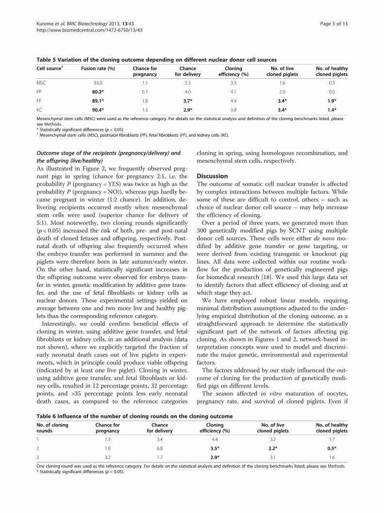
Table 5 Variation of the cloning outcome depending on different nuclear donor cell sources
Cell source1 Fusion rate (%) Chance forpregnancy
Chancefor delivery
Cloningefficiency (%)
No. of livecloned piglets
No. of healthycloned piglets
MSC 93.0 1.1 5.3 3.5 1.6 0.3
PF 80.2* 0.7 4.0 4.1 2.0 0.5
FF 89.1* 1.8 3.7* 4.4 3.4* 1.9*
KC 90.4* 1.3 2.9* 3.8 3.4* 1.4*
Mesenchymal stem cells (MSC) were used as the reference category. For details on the statistical analysis and definition of the cloning benchmarks listed, pleasesee Methods.* Statistically significant differences (p < 0.05).1 Mesenchymal stem cells (MSC), postnatal fibroblasts (PF), fetal fibroblasts (FF), and kidney cells (KC).
Kurome et al. BMC Biotechnology 2013, 13:43 Page 5 of 13http://www.biomedcentral.com/1472-6750/13/43
Outcome stage of the recipients (pregnancy/delivery) andthe offspring (live/healthy)As illustrated in Figure 2, we frequently observed preg-nant pigs in spring (chance for pregnancy 2:1, i.e. theprobability P (pregnancy = YES) was twice as high as theprobability P (pregnancy = NO)), whereas pigs hardly be-came pregnant in winter (1:2 chance). In addition, de-livering recipients occurred mostly when mesenchymalstem cells were used (superior chance for delivery of5:1). Most noteworthy, two cloning rounds significantly(p < 0.05) increased the risk of both, pre- and post-nataldeath of cloned fetuses and offspring, respectively. Post-natal death of offspring also frequently occurred whenthe embryo transfer was performed in summer and thepiglets were therefore born in late autumn/early winter.On the other hand, statistically significant increases inthe offspring outcome were observed for embryo trans-fer in winter, genetic modification by additive gene trans-fer, and the use of fetal fibroblasts or kidney cells asnuclear donors. These experimental settings yielded onaverage between one and two more live and healthy pig-lets than the corresponding reference category.Interestingly, we could confirm beneficial effects of
cloning in winter, using additive gene transfer, and fetalfibroblasts or kidney cells, in an additional analysis (datanot shown), where we explicitly targeted the fraction ofearly neonatal death cases out of live piglets in experi-ments, which in principle could produce viable offspring(indicated by at least one live piglet). Cloning in winter,using additive gene transfer, and fetal fibroblasts or kid-ney cells, resulted in 12 percentage points, 32 percentagepoints, and >35 percentage points less early neonataldeath cases, as compared to the reference categories
Table 6 Influence of the number of cloning rounds on the clo
No. of cloningrounds
Chance forpregnancy
Chancefor delivery
1 1.3 3.4
2 1.0 6.8
3 3.2 1.7
One cloning round was used as the reference category. For details on the statistica* Statistically significant differences (p < 0.05).
cloning in spring, using homologous recombination, andmesenchymal stem cells, respectively.
DiscussionThe outcome of somatic cell nuclear transfer is affectedby complex interactions between multiple factors. Whilesome of these are difficult to control, others – such aschoice of nuclear donor cell source – may help increasethe efficiency of cloning.Over a period of three years, we generated more than
300 genetically modified pigs by SCNT using multipledonor cell sources. These cells were either de novo mo-dified by additive gene transfer or gene targeting, orwere derived from existing transgenic or knockout piglines. All data were collected within our routine work-flow for the production of genetically engineered pigsfor biomedical research [18]. We used this large data setto identify factors that affect efficiency of cloning and atwhich stage they act.We have employed robust linear models, requiring
minimal distribution assumptions adjusted to the under-lying empirical distribution of the cloning outcome, as astraightforward approach to determine the statisticallysignificant part of the network of factors affecting pigcloning. As shown in Figures 1 and 2, network-based in-terpretation concepts were used to model and discrimi-nate the major genetic, environmental and experimentalfactors.The factors addressed by our study influenced the out-
come of cloning for the production of genetically modi-fied pigs on different levels.The season affected in vitro maturation of oocytes,
pregnancy rate, and survival of cloned piglets. Even if
ning outcome
Cloningefficiency (%)
No. of livecloned piglets
No. of healthycloned piglets
4.4 3.2 1.7
3.5* 2.2* 0.5*
2.9* 3.1 1.6
l analysis and definition of the cloning benchmarks listed, please see Methods.
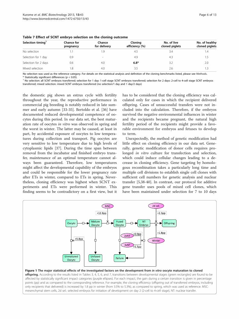
Table 7 Effect of SCNT embryo selection on the cloning outcome
Selection timing1 Chance forpregnancy
Chancefor delivery
Cloningefficiency (%)
No. of livecloned piglets
No. of healthycloned piglets
No selection 1.1 1.9 4.5 3.4 1.4
Selection for 1 day 0.9 - 4.9 4.3 1.5
Selection for 2 days 0.6 4.0 6.8* 3.2 2.0
Mixed selection 1.8 4.0 3.5 2.6 1.3
No selection was used as the reference category. For details on the statistical analysis and definition of the cloning benchmarks listed, please see Methods.* Statistically significant differences (p < 0.05).1 No selection: all SCNT embryos transferred; selection for 1 day: 1-cell stage SCNT embryos transferred; selection for 2 days: 2-cell to 4-cell stage SCNT embryostransferred; mixed selection: mixed SCNT embryos transferred (no selection/1 day and 1 day/2 days)
Kurome et al. BMC Biotechnology 2013, 13:43 Page 6 of 13http://www.biomedcentral.com/1472-6750/13/43
the domestic pig shows an estrus cycle with fertilitythroughout the year, the reproductive performance incommercial pig breeding is notably reduced in late sum-mer and early autumn [33-35]. Bertoldo et al. [36] havedocumented reduced developmental competence of oo-cytes during this period. In our data set, the best matur-ation rate of oocytes in vitro was observed in spring andthe worst in winter. The latter may be caused, at least inpart, by accidental exposure of oocytes to low tempera-tures during collection and transport. Pig oocytes arevery sensitive to low temperature due to high levels ofcytoplasmic lipids [37]. During the time span betweenremoval from the incubator and finished embryo trans-fer, maintenance of an optimal temperature cannot al-ways been guaranteed. Therefore, low temperaturesmight affect the developmental capability of the embryosand could be responsible for the lower pregnancy rateafter ETs in winter, compared to ETs in spring. Never-theless, cloning efficiency was highest when SCNT ex-periments and ETs were performed in winter. Thisfinding seems to be contradictory on a first view, but it
Figure 1 The major statistical effects of the investigated factors on thoffspring. According to the results listed in Tables 3, 4, 5, 6, and 7, transitioaffected by statistically significant impact categories (purple ellipses). For eapoints (pp) and as compared to the corresponding reference. For exampleonly recipients that delivered) is increased by 1.8 pp in winter (from 3.5% tmesenchymal stem cells, 2d sel.: selected embryos for initiation of develop
has to be considered that the cloning efficiency was cal-culated only for cases in which the recipient deliveredoffspring. Cases of unsuccessful transfers were not in-cluded into the calculation. Therefore, if the embryossurvived the negative environmental influences in winterand the recipients became pregnant, the natural highfertility period of the recipients might provide a favo-rable environment for embryos and fetuses to developto term.Unexpectedly, the method of genetic modification had
little effect on cloning efficiency in our data set. Gene-rally, genetic modification of donor cells requires pro-longed in vitro culture for transfection and selection,which could induce cellular changes leading to a de-crease in cloning efficiency. Gene targeting by homolo-gous recombination takes a particularly long time andmultiple cell divisions to establish single cell clones withsufficient cell numbers for genetic analysis and nucleartransfer [5,38-40]. In contrast, our protocol for additivegene transfer uses pools of mixed cell clones, whichhave been maintained under selection for 7 to 10 days
e development from in vitro oocyte maturation to clonedns between developmental stages (green rectangles) are found to bech impact, the gain during a certain transition is given in percentage, the cloning efficiency (offspring out of transferred embryos, includingo 5.3%), as compared to spring, which was used as reference. MSC:ment on day 2 (2-cell to 4-cell stage), NT: nuclear transfer.

Figure 2 The major statistical effects of the investigated factors on the outcome stage of the recipients (pregnancy/delivery) and theoffspring (live/healthy). For each impact, the gain during a certain transition is given in the respective outcome unit. For example, there are onaverage 1.7 more alive piglets for cloning in winter, as compared to the reference (cloning in spring). MSC: mesenchymal stem cells, AGT:additive gene transfer, KC: kidney cells, FF: fetal fibroblasts, NT: nuclear transfer, END: early neonatal death.
Kurome et al. BMC Biotechnology 2013, 13:43 Page 7 of 13http://www.biomedcentral.com/1472-6750/13/43
[8,18,41]. It might therefore be expected that extendedin vitro culture of donor cells required for homologousrecombination would negatively influence the cloning ef-ficiency, compared to cells modified by additive genetransfer. However, our data showed no statistically sig-nificant difference in cloning efficiency between additivegene transfer, homologous recombination or replicationof already existing transgenic pigs. It can be hypothe-sized that the conditions for transfection and selectiondid not adversely affect the developmental potential ofdonor cells, since we kept the passage numbers forSCNT donor cells as low as possible - less than 8 pas-sages for additive gene transfer and less than 10 passagesfor gene targeting. Additionally, all wild-type primarycell lines used in this study were karyotyped and showed68% to 90% normal karyotypes.Interestingly, our analysis indicated that the number of
live and healthy offspring was decreased when nucleardonor cells had undergone homologous recombination.However, this may – at least in part – be explained bythe fact that 65% of nuclear transfers, designed to gener-ate gene-targeted pigs, were carried out using only 4particular mesenchymal stem cell lines, which later onturned out to be consistently poor in producing livecloned offspring.Another important aspect to be considered in the con-
text of genetic modification is the potential for lethal ortoxic effects of modifications per se. For the experimentsinvolving additive gene transfer this is unlikely, since livecloned piglets expressing the transgenes were obtainedwith all constructs. Nevertheless, we cannot rule out thatcloned fetuses or offspring died due to a detrimentalrandom integration of the construct. Of the gene tar-geting experiments, only mutation of the X-linked
dystrophin (DMD) gene in male clones may cause asevere phenotype. In fact, DMD mutant male pigletsshowed severe muscular dystrophy already at birth, anda proportion died shortly later [42]. For all other targetgenes, the heterozygous knockout had either no specificphenotype or a phenotype that develops later in life.A critical factor for the establishment of genetically
engineered pig lines by SCNT is the viability of thecloned founder animals up to sexual maturity. In ourdata set, more than half of cloned pigs were stillborn(23.6%) or died soon after birth (31.4%). Associated pa-thological changes, such as underweight (average weightof the cloned piglets born under 1000 g: 686.4 +/−181.0 g; range: 375 – 973 g), which is one of the majorcauses of early neonatal death, or cleft palate, contractedtendons, or enlarged tongues, have also been observedby other groups [43-47]. We have the impression thatthe percentage of underweight piglets (among normalweight littermates) is higher in cloned litters. However,we cannot prove this observation by statistical data, asthe birth weights of naturally bred piglets are not rou-tinely recorded in our facility. The average birth weightof healthy cloned pigs was higher than that of pigletsthat died in the neonatal period, or that of stillbornpiglets (1409.2 +/− 343.1 g, 974.8 +/− 394.1 g, and1065.5 +/− 479.0 g, respectively). These abnormalitiescould not be associated with any particular parameter,like donor cell source or genetic modification, andmight be a general side effect in pig cloning. Previousstudies reported that phenotypically abnormal clonedanimals could produce normal offspring [48,49], sug-gesting that phenotypic abnormalities of the cloneswere more likely due to epigenetic rather than to ge-netic alterations.
Kurome et al. BMC Biotechnology 2013, 13:43 Page 8 of 13http://www.biomedcentral.com/1472-6750/13/43
In our data set, cloned piglets with enlarged tongueswere mainly observed in offspring cloned from bonemarrow derived mesenchymal stem cells, originatingfrom 4 different animals (25 of 30 cases). However, thisdoes not seem to be a general feature of mesenchymalstem cells, since in more recent cloning experimentswith adipose tissue derived mesenchymal stem cells ahigh proportion of viable offspring without malforma-tions was obtained (T. Flisikowska and A. Schnieke,unpublished data). Some groups reported that mesen-chymal stem cells are superior to fibroblasts for SCNTin pigs [50-53], although this has not been generally ob-served [40,54,55]. Our results did not show any diffe-rences in the cloning efficiency among the different cellsources tested, although there was a tendency for ahigher pregnancy rate when mesenchymal stem cellswere used. The observation that the numbers of live andhealthy cloned piglets were significantly lower in themesenchymal stem cells group than other donor cellsources may be due to the fact that mesenchymal stemcells were only used for gene targeting. Thus, it cannotbe distinguished at this stage, whether the low outcomeof live and healthy piglets can be attributed to the cellsource or type of genetic modification. In addition,different cell lines derived from the same cell sourceshowed a considerable degree of variation in cloning ef-ficiency (Additional file 5).Re-cloning by using cells from a cloned animal for NT
is a reasonable approach for the reproduction of specifictransgenic animals, for example if animals of a definedgenotype are required for an experiment or if the pheno-type hinders natural breeding. However, the majority ofstudies on re-cloning have demonstrated that additionalrounds of cloning lead to a decrease in cloning efficiency[49,56-58]. Our data also showed that repeated cloningrounds significantly decreased cloning efficiency (R1:4.4%, R2: 3.5% and R3: 2.9%), and the number of livecloned offspring in the second round was in average onepiglet less as compared to the initial cloning round. Itshould be mentioned that the lowest cloning efficiencyfor R3 may also be related to the high number of em-bryos transferred in these experiments. Xing et al. [59]recently demonstrated that reduced developmental po-tential of pig embryos generated by multiple rounds ofcloning was associated with altered gene expression pat-terns, and a previous report stated that the reduction ofcloning efficiency with additional rounds of cloning maybe caused by accumulation of epigenetic errors [60].The last factor addressed by our study was in vitro cul-
ture of cloned embryos and selection for normal deve-lopment before transfer to recipients. This is possiblesince the in vitro culture systems for pig embryos havebeen markedly improved within the last decade [27,28].Indeed, culture of embryos for two days and selection of
2-cell to 4-cell stage embryos for ET resulted in thehighest proportion of offspring per SCNT embryostransferred. This suggests that SCNT embryos, which un-dergo normal cleavage in vitro within the expected timeframe, have a greater chance of full term developmentin vivo.
ConclusionWe have investigated the influence of important experi-mental and environmental factors on the cloning out-come in a considerably large data set comprising over270 porcine nuclear transfer experiments. Besides as-sessment of the cloning efficiency, we determined therespective steps of the cloning process from oocyte tooffspring that are most critically influenced. We ob-served varying effects of individual factors, depending onthe combination with other chosen factors and the pa-rameters tested. Most importantly, more live and healthyoffspring were obtained when fetal fibroblasts or kidneycells were modified by additive gene transfer and the re-sulting SCNT embryos were transferred in the winterperiod. Although our results cannot be simply extrapo-lated to other cloning labs, the approach used in thisstudy may help to identify and optimize the specific fac-tors most critical to cloning success in programs aimingto generate genetically engineered pigs.
MethodsEthics statementAll animal procedures in this study were performed ac-cording to the German Animal Welfare Act and to aprotocol approved by the Regierung von Oberbayern,under the reference numbers (55.2.1.54-2531-26-06;55.2.1.54-2531-77-07; 55.2.1.54-2531-78-07; 55.2.1.54-2531-136-07; 55.2.1.54-2531-54-08; 55.2.1.54-2531-86-10; 55.2.1.54-2532-68-11).
Generation of genetically modified pigsGenetically modified cells derived by transfection of pri-mary cells or established from already existing trans-genic pig lines were used as donors. The cells derivedfrom transfection were genetically modified by additivegene transfer (Table 8) or by homologous recombination(Table 9). The latter group included bacterial artificialchromosome (BAC) targeting [7] and the use of classicaltargeting vectors [5]. The cells re-established from al-ready existing transgenic pig lines were collected from18 different transgenic pigs. Individual information onall cell lines used for these analyses is shown in theAdditional file 6 and Additional file 7.The following cell sources were used: mesenchymal
stem cells, postnatal fibroblasts, fetal fibroblasts, and kid-ney cells. Mesenchymal stem cells, multi-potent tissuestem cells, as well as fibroblasts and kidney cells are
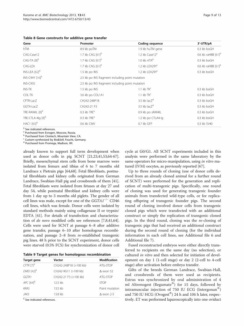
Table 8 Gene constructs for additive gene transfer
Gene Promoter Coding sequence 3’-UTR/pA
hTM 8.9 kb poTM 1.9 kb huTM gene 0.3 kb boGH
CAG-Case12 1.7 kb CAG [61]§ 1.2 kb Case12a 0.6 kb raHBB [61]§
CAG-TA [8]§ 1.7 kb CAG [61]§ 1.0 kb rtTAb 0.3 kb boGH
CAG-LEA 1.7 kb CAG [61]§ 1.2 kb LEA29Yc 0.6 kb raHBB [61]§
INS-LEA [62]§ 1.5 kb po INS 1.2 kb LEA29Yc 0.3 kb boGH
INS-C94Y [16]§ 2.5 kb po INS fragment including point mutation
INS-C93S 2.5 kb po INS fragment including point mutation
INS-TK 1.5 kb po INS 1.1 kb TKc 0.3 kb boGH
COL-TK 3.6 kb po COL1A1 1.1 kb TKc 0.3 kb boGH
CFTR-LacZ CH242-248P18 3.5 kb lacZd 0.3 kb boGH
GGTA-LacZ CH242-21 F3 3.5 kb lacZd 0.3 kb boGH
TRE-RANKL [8]§ 0.3 kb TREa 0.9 kb po sRANKL 0.3 kb boGH
TRE-CTLA-4Ig [8]§ 0.3 kb TREa 1.2 kb po CTLA4-Ig 0.3 kb boGH
HAC1 [63]§ 0.6 kb CMV 0.7 kb GFP 0.3 kb SV40§ See indicated references.a Purchased from Evrogen, Moscow, Russia.b Purchased from Clontech, Mountain View, CA.c Custom-synthesized by Bio&Sell, Feucht, Germany.d Purchased from Promega, Madison, WI.
Kurome et al. BMC Biotechnology 2013, 13:43 Page 9 of 13http://www.biomedcentral.com/1472-6750/13/43
already known to support full term development whenused as donor cells in pig SCNT [21,23,41,53,64-67].Briefly, mesenchymal stem cells from bone marrow wereisolated from femurs and tibias of 6 to 7 months oldLandrace x Pietrain pigs [64,68]. Fetal fibroblasts, postna-tal fibroblasts and kidney cells originated from GermanLandrace, Swabian-Hall pig and crossbreeds of them [41].Fetal fibroblasts were isolated from fetuses at day 27 andday 54, while postnatal fibroblast and kidney cells werefrom 1 day up to 3 months old piglets. The gender of allcell lines was male, except for one of the GGTA1−/− CD46cell lines, which was female. Donor cells were isolated bystandard methods mainly using collagenase II or trypsin/EDTA [41]. For details of transfection and characteriza-tion of de novo modified cells see references [7,8,41,64].Cells were used for SCNT at passage 6–8 after additivegene transfer, passage 6–10 after homologous recombi-nation, and passage 2–8 from re-established transgenicpig lines. 48 h prior to the SCNT experiment, donor cellswere starved (0.5% FCS) for synchronization of donor cell
Table 9 Target genes for homologous recombination
Target gene Vector Modification
CFTR [7]§ CH242-248P18 (>100 kb) ATG-STOP
DMD [42]§ CH242-9G11 (>100 kb) Δ exon 52
GGTA1 CH242-21 F3 (>100 kb) ATG-STOP
APC [64]§ 12.5 kb STOP
KRAS 13.5 kb Point mutation
JAK3 13.8 kb Δ exon 2-5§ See indicated references.
cycle at G0/G1. All SCNT experiments included in thisanalysis were performed in the same laboratory by thesame operators for micro-manipulation, using in vitro ma-tured (IVM) oocytes, as previously reported [67].Up to three rounds of cloning (use of donor cells de-
rived from an already cloned animal for a further roundof SCNT) were performed for the generation and repli-cation of multi-transgenic pigs. Specifically, one roundof cloning was used for generating transgenic founderanimals from transfected wild-type cells, or for replica-ting offspring of transgenic founder pigs. The secondround of cloning involved donor cells from transgeniccloned pigs which were transfected with an additionalconstruct or simply the replication of transgenic clonedpigs. In the third round, cloning was the re-cloning oftransgenic pigs that had received an additional constructduring the second round of cloning (for the individualinformation in each cell lines, see Additional file 6 andAdditional file 7).Fused reconstructed embryos were either directly trans-
ferred to recipients on the same day (no selection), orcultured in vitro and then selected for initiation of devel-opment on day 1 (1-cell stage) or day 2 (2-cell to 4-cellstage) after activation before embryo transfer.Gilts of the breeds German Landrace, Swabian-Hall,
and crossbreeds of them were used as recipients.Estrus was synchronized by oral administration of 4ml Altrenogest (RegumateW) for 15 days, followed byintramuscular injection of 750 IU ECG (IntergonanW)and 750 IU HCG (OvogestW) 24 h and 104 h later, respec-tively. ET was performed laparoscopically into one oviduct
d
Kurome et al. BMC Biotechnology 2013, 13:43 Page 10 of 13http://www.biomedcentral.com/1472-6750/13/43
[69]. Pregnancy was confirmed by ultrasonographic exam-ination on day 21, repeated every 2 – 3 weeks.
Data descriptionThe analysis is based on data from cloning experiments,performed in the period from April 2008 to February2011, at the Chair for Molecular Animal Breeding andBiotechnology in Munich, Germany. The location is situ-ated at an altitude of 444 m, and at latitude and longi-tude of 48°22’N and 11°49’E, respectively.Changes in the experimental setup, described in the
previous section, included variations of the season theET was performed in, the type of genetic modification,the donor cell source, the number of cloning rounds,and selection of SCNT embryos for development beforetransfer to the recipient. The stratification and distribu-tion of each varied factor is summarized in Table 2.
1. Season: Experiments were performed covering thewhole year range, i.e. an approximately balancedsample size in each season – spring (March-May),summer (June-August), autumn (September-November) and winter (December-February) – wasensured. However, 10% more experiments wereperformed in summer and autumn. The averagetemperature in each season was 9.6°C, 18°C, 9.2°C,0.1°C, respectively (http://www.dwd.de).
2. Type of genetic modification: Genetically modifiedcells were derived in roughly 30% of all experimentsby additive gene transfer, in 25% of the experimentsby homologous recombination, and in most cases(45%) established from transgenic pigs.
3. Donor cell source: Regarding the source of nucleardonor cells, most of the experiments wereperformed with kidney cells (43%), followed by fetalfibroblasts (26%), mesenchymal stem cells (19%),and postnatal fibroblasts (12%).
4. Number of cloning rounds: The vast majority of allcloning experiments were carried out with oneround of cloning (57%), one third (32%) with tworounds, and the remaining experiments (11%) withthree rounds of cloning.
5. Selection of SCNT embryos for initiation ofdevelopment: In 23% of all experiments, all SCNTembryos were transferred to recipients on thesame day on which the nuclear transfer wascarried out (no selection for development). Inother experiments, the SCNT embryos werecultured either 1 day (7%) or 2 days (8%) afteractivation and selected for initiation of normaldevelopment (1-cell stage on day 1, 2-cell to4-cell stage on day 2). In most of the cases (62%)mixed populations of SCNT embryos (no selection,1 day culture, 2 days culture) were transferred to the
recipients. Those were not included in the analysis ofthis specific factor.
Cloning benchmarksThe success of each cloning experiment was progres-sively assessed based on the outcome of distinct evalu-ation stages. After the cloned embryos were transferredto the recipient, we first determined whether it becamepregnant or not.For a sample stratum under investigation, the chance
for pregnancy is hence defined as the probability ratio
P pregnancy ¼ YESð Þ =P pregnancy ¼ NOð Þ ð1ÞThe probabilities result from the relative frequencies
of the corresponding event in the stratum.Analogously, the chance for delivery is defined as
P delivery ¼ YESð Þ = P delivery ¼ NOð Þ ð2ÞFor delivering recipients, we counted the number of
offspring born, the number of live offspring amongthem, and if there were any, the number of healthyoffspring.In addition, we calculated for the experiments re-
sulting in at least one delivered offspring the cloning effi-ciency as
delivered cloned offspring=SCNT embryos transferre
ð3ÞAs a benchmark for oocyte and donor cell quality, re-
spectively, we also took the oocyte maturation rate, cal-culated as
successfully matured oocytes=oocytes entering IVM;
ð4Þand the fusion rate, calculated as
successfully fused karyoplast� cytoplast complexes=
complexes submitted to electrofusion;
ð5Þinto account.
Statistical analysisGeneralized linear models [70] were computed for eachexperimental factor (season, genetic modification, cellsource, cloning rounds and SCNT embryo selection) inorder to estimate its impact on each cloning outcomestage (pregnancy and delivery rate as well as numbersof total, live, and healthy offspring) and the cloningefficiency.As all explaining variables, i.e. the experimental fac-
tors, are categorial, we designed the linear predictor ofthe regression models using indicator (dummy) variables
Kurome et al. BMC Biotechnology 2013, 13:43 Page 11 of 13http://www.biomedcentral.com/1472-6750/13/43
[71], yielding effects with respect to the correspondinglychosen reference category (spring for season, additivegene transfer for genetic modification, mesenchymal stemcells for cell source, one round for cloning rounds, andno culture for SCNT embryo selection). This design cor-responds to an ANOVA model [72], where the samplemean of each stratum of the experimental factor underinvestigation is tested for deviation from the samplemean of the reference category assuming the samplemeans to be t-distributed. Consequently, all p-valuesreported here are t-test [73] derived, and should, thus,be interpreted as a statistical significance measure forequality of means, i.e. the lower the p-value, the moresignificant is the difference in the means. The link func-tion of the regression models was selected according tothe goodness of fit between the empirical distribution ofthe response (outcome) variable and the correspondingcommon distribution. Briefly, logistic regression was car-ried out for the binary factors (pregnancy and delivery),Poisson regression for the counts of live and healthyoffspring, and Gaussian regression for the cloning effi-ciency (as well as for maturation and fusion rate).
Additional files
Additional file 1: Correlation of the number of embryos transferredwith pregnancy rate. The absolute number of embryo transfers(left y-axis) that resulted in pregnancy of the recipient depending on thenumber of embryos transferred (x-axis) is shown in black over thenumber of all observations in grey. The red curve indicates the overallpregnancy rate (right y-axis) when more than x embryos have beentransferred.
Additional file 2: Correlation of the number of embryos transferredwith the number of live piglets. The number of transferred embryos isshown on the x-axis and the number of live piglets on the y-axis. Novisible correlation can be detected (Pearson correlation 0.2).
Additional file 3: Seasonal distribution of specific SCNTconfigurations with respect to genetic modification, cell type andcloning round. For each season on the x-axis, the bar height denotesthe total number of embryo transfers performed (as indicated on they-axis). The three vertical slots in each of the bars correspond to thedistribution of the respective categories of genetic modification (gen.mod), cell type (cell.type), and cloning rounds (clon.rds). The categoriesare alphanumerically encoded as denoted at the top: geneticmodification = (1 = homologous recombination (HR), 2 = additive genetransfer (AGT), 3 = replication of transgenic pigs (replic. of tg pigs)), celltype = (1 = mesenchymal stem cells (MSC), 2 = postnatal fibroblasts (PF),3 = fetal fibroblasts (FF), and 4 = kidney cells (KC)), cloning rounds =(1 = 1 round, 2 = 2rounds, 3 = 3rounds).
Additional file 4: Distribution of selected embryos derived fromspecific SCNT configurations with respect to genetic modification,cell type and cloning round. For a particular selection timing on thex-axis, the bar height denotes the total number of embryo transfersperformed (as indicated on the y-axis). The three vertical slots in each ofthe bars correspond to the distribution of the respective categories ofgenetic modification (gen.mod), cell type (cell.type), and cloning rounds(clon.rds). The categories are alphanumerically encoded as denoted atthe top: genetic modification = (1 = homologous recombination (HR),2 = additive gene transfer (AGT), 3 = replication of transgenic pigs (replic.of tg pigs)), cell type = (1 = mesenchymal stem cells (MSC), 2 = postnatalfibroblasts (PF), 3 = fetal fibroblasts (FF), and 4 = kidney cells (KC)), cloning
rounds = (1 = 1 round, 2 = 2 rounds, 3 = 3 rounds). Data for mixedselection timing not shown.
Additional file 5: Degree of variation in cloning efficiency withincell types. The variation in cloning efficiency on the y-axis is shownfor the different cell lines within the four cell type categories (MSC:mesenchymal stem cells, FF: fetal fibroblasts, PF: postnatal fibroblasts, andKC: kidney cells). The numbers in brackets on the x-axis denote thenumber of embryo transfers (in total and for the corresponding fractionthat delivered offspring, respectively). Details on the cell lines used canbe found in Additional file 6 and Additional file 7.
Additional file 6: List of de novo modified cell lines by additivegene transfer or homologous recombination.
Additional file 7: List of transgenic cell lines from already existingtransgenic pig.
AbbreviationsBAC: Bacterial artificial chromosome; ET: Embryo transfer; IVM: In vitromaturation; SCNT: Somatic cell nuclear transfer.
Competing interestsThe authors declare that they have no competing interests.
Authors’ contributionsMK, LG, RZ and EW conceived and designed the study. MK, BK, VZ, NK, AW,AR, AB, KK, KB, KF, TF, CM, TL, MD, AT, SK, DS, HN, AS, EW were involved insomatic cell nuclear transfer experiments. LG, MK, TP, RZ, EW analyzed thedata and MK, LG, AK, RZ, EW drafted the manuscript. All authors read andapproved the final manuscript.
AcknowledgmentsWe are grateful to Tuna Guengoer, Eva-Maria Jemiller, Christian Erdle andSigfried Elsner for their excellent technical support. This work was financiallysupported by the German Research Council (FOR 535 ‘Xenotransplantation’,FOR 793 ‘Mechanisms of Fracture Healing in Osteoporosis’, Transregio-CRC127 ‘Biology of xenogeneic cell, tissue and organ transplantation – frombench to bedside’), by the Federal Ministry for Education and Research(Leading-Edge Cluster ‘m4 – Personalised Medicine and Targeted Therapies’),the Bavarian Research Council (FORZebRA, Az. 802–08), the Mildred ScheelStiftung für Krebsforschung, the Mukoviszidose Institut gemeinnützigeGesellschaft für Forschung und Therapieentwicklung mbH and by the DFGInternational Research Training Group (1563/1 RECESS). The funders had norole in study design, data collection and analysis, decision to publish, orpreparation of the manuscript.
Author details1Molecular Animal Breeding and Biotechnology, and Laboratory forFunctional Genome Analysis (LAFUGA), Gene Center, LMU Munich, Munich,Germany. 2Practical Informatics and Bioinformatics, Institute for Informatics,LMU Munich, Munich, Germany. 3Livestock Biotechnology, Center of Life andFood Sciences Weihenstephan, TU Munich, Freising, Germany. 4InternationalInstitute for Bio-Resource Research, Meiji University, Kawasaki, Japan.
Received: 22 November 2012 Accepted: 9 April 2013Published: 20 May 2013
References1. Hall V: Porcine embryonic stem cells: a possible source for cell
replacement therapy. Stem Cell Rev 2008, 4(4):275–282.2. Niemann H, Kues WA: Transgenic farm animals: an update. Reprod Fertil
Dev 2007, 19(6):762–770.3. Nowak-Imialek M, Kues W, Carnwath JW, Niemann H: Pluripotent stem
cells and reprogrammed cells in farm animals. Microsc Microanal 2011,17(4):474–497.
4. Prather RS, Hawley RJ, Carter DB, Lai L, Greenstein JL: Transgenic swine forbiomedicine and agriculture. Theriogenology 2003, 59(1):115–123.
5. Lai L, Kolber-Simonds D, Park KW, Cheong HT, Greenstein JL, Im GS,Samuel M, Bonk A, Rieke A, Day BN, et al: Production of alpha-1,3-galactosyltransferase knockout pigs by nuclear transfer cloning. Science2002, 295(5557):1089–1092.
Kurome et al. BMC Biotechnology 2013, 13:43 Page 12 of 13http://www.biomedcentral.com/1472-6750/13/43
6. Rogers CS, Hao Y, Rokhlina T, Samuel M, Stoltz DA, Li Y, Petroff E,Vermeer DW, Kabel AC, Yan Z, et al: Production of CFTR-null andCFTR-DeltaF508 heterozygous pigs by adeno-associated virus-mediatedgene targeting and somatic cell nuclear transfer. J Clin Invest 2008,118(4):1571–1577.
7. Klymiuk N, Mundhenk L, Kraehe K, Wuensch A, Plog S, Emrich D,Langenmayer MC, Stehr M, Holzinger A, Kroner C, et al: Sequentialtargeting of CFTR by BAC vectors generates a novel pig model of cysticfibrosis. J Mol Med 2012, 90(5):597–608.
8. Klymiuk N, Bocker W, Schonitzer V, Bahr A, Radic T, Frohlich T, Wunsch A,Kessler B, Kurome M, Schilling E, et al: First inducible transgene expressionin porcine large animal models. FASEB J 2012, 26:1088–1099.
9. Whyte JJ, Zhao J, Wells KD, Samuel MS, Whitworth KM, Walters EM, LaughlinMH, Prather RS: Gene targeting with zinc finger nucleases to producecloned eGFP knockout pigs. Mol Reprod Dev 2011, 78(1):2.
10. Hauschild J, Petersen B, Santiago Y, Queisser AL, Carnwath JW, Lucas-HahnA, Zhang L, Meng X, Gregory PD, Schwinzer R, et al: Efficient generation ofa biallelic knockout in pigs using zinc-finger nucleases. Proc Nat Acad SciUSA 2011, 108(29):12013–12017.
11. Swindle MM, Smith AC: Comparative anatomy and physiology of the pig.Scand J Lab Anim Sci 1998, 25:11–21.
12. Rogers CS, Stoltz DA, Meyerholz DK, Ostedgaard LS, Rokhlina T, Taft PJ,Rogan MP, Pezzulo AA, Karp PH, Itani OA, et al: Disruption of the CFTRgene produces a model of cystic fibrosis in newborn pigs. Science 2008,321(5897):1837–1841.
13. Renner S, Fehlings C, Herbach N, Hofmann A, von Waldthausen DC,Kessler B, Ulrichs K, Chodnevskaja I, Moskalenko V, Amselgruber W, et al:Glucose intolerance and reduced proliferation of pancreatic beta-cells intransgenic pigs with impaired glucose-dependent insulinotropicpolypeptide function. Diabetes 2010, 59(5):1228–1238.
14. Renner S, Römisch-Margl W, Prehn C, Krebs S, Adamski J, Göke B, Blum H,Suhre K, Roscher AA, Wolf E: Changing metabolic signatures of aminoacids and lipids during the pre-diabetic period in a pig model withimpaired incretin function and reduced β-cell mass. Diabetes 2012,61(8):2166–2175.
15. Umeyama K, Watanabe M, Saito H, Kurome M, Tohi S, Matsunari H, Miki K,Nagashima H: Dominant-negative mutant hepatocyte nuclear factor1alpha induces diabetes in transgenic-cloned pigs. Transgenic Res 2009,18(5):697–706.
16. Renner S, Braun-Reichhart C, Blutke A, Herbach N, Emrich D,Wuensch A, Kessler B, Kurome M, Baehr A, Klymiuk N, et al: Permanentneonatal diabetes in INSC94Y transgenic pigs. Diabetes 2013,62(5):1505–1511.
17. Kragh PM, Nielsen AL, Li J, Du Y, Lin L, Schmidt M, Bogh IB, Holm IE,Jakobsen JE, Johansen MG, et al: Hemizygous minipigs produced byrandom gene insertion and handmade cloning express the Alzheimer’sdisease-causing dominant mutation APPsw. Transgenic Res 2009,18(4):545–558.
18. Aigner B, Renner S, Kessler B, Klymiuk N, Kurome M, Wunsch A, Wolf E:Transgenic pigs as models for translational biomedical research. J MolMed (Berl) 2010, 88(7):653–664.
19. Klymiuk N, Aigner B, Brem G, Wolf E: Genetic modification of pigsas organ donors for xenotransplantation. Mol Reprod Dev 2010,77(3):209–221.
20. Polejaeva IA, Chen SH, Vaught TD, Page RL, Mullins J, Ball S, Dai Y, Boone J,Walker S, Ayares DL, et al: Cloned pigs produced by nuclear transfer fromadult somatic cells. Nature 2000, 407(6800):86–90.
21. Onishi A, Iwamoto M, Akita T, Mikawa S, Takeda K, Awata T, Hanada H, PerryAC: Pig cloning by microinjection of fetal fibroblast nuclei. Science 2000,289(5482):1188–1190.
22. Betthauser J, Forsberg E, Augenstein M, Childs L, Eilertsen K, Enos J,Forsythe T, Golueke P, Jurgella G, Koppang R, et al: Production of clonedpigs from in vitro systems. Nat Biotechnol 2000, 18(10):1055–1059.
23. Yin XJ, Tani T, Yonemura I, Kawakami M, Miyamoto K, Hasegawa R, Kato Y,Tsunoda Y: Production of cloned pigs from adult somatic cells bychemically assisted removal of maternal chromosomes. Biol Reprod 2002,67(2):442–446.
24. Kurome M, Fujimura T, Murakami H, Takahagi Y, Wako N, Ochiai T, MiyazakiK, Nagashima H: Comparison of electro-fusion and intracytoplasmicnuclear injection methods in pig cloning. Cloning Stem Cells 2003,5(4):367–378.
25. Whitworth KM, Li R, Spate LD, Wax DM, Rieke A, Whyte JJ, Manandhar G,Sutovsky M, Green JA, Sutovsky P, et al: Method of oocyte activationaffects cloning efficiency in pigs. Mol Reprod Dev 2009, 76(5):490–500.
26. Petersen B, Lucas-Hahn A, Oropeza M, Hornen N, Lemme E, Hassel P,Queisser AL, Niemann H: Development and validation of a highly efficientprotocol of porcine somatic cloning using preovulatory embryo transferin peripubertal gilts. Cloning Stem Cells 2008, 10(3):355–362.
27. Yoshioka K: Development and application of a chemically definedmedium for the in vitro production of porcine embryos. J Reprod Dev2011, 57(1):9–16.
28. Vajta G, Zhang Y, Machaty Z: Somatic cell nuclear transfer in pigs:recent achievements and future possibilities. Reprod Fertil Dev 2007,19(2):403–423.
29. Kuhholzer B, Hawley RJ, Lai L, Kolber-Simonds D, Prather RS: Clonal lines oftransgenic fibroblast cells derived from the same fetus result in differentdevelopment when used for nuclear transfer in pigs. Biol Reprod 2001,64(6):1695–1698.
30. Li Z, Shi J, Liu D, Zhou R, Zeng H, Zhou X, Mai R, Zeng S, Luo L, Yu W, et al:Effects of donor fibroblast cell type and transferred cloned embryonumber on the efficiency of pig cloning. Cell Reprogram 2013, 15(1):35–42.
31. Nakayama A, Sato M, Shinohara M, Matsubara S, Yokomine T, Akasaka E,Yoshida M, Takao S: Efficient transfection of primarily cultured porcineembryonic fibroblasts using the Amaxa Nucleofection system. CloningStem Cells 2007, 9(4):523–534.
32. Skrzyszowska M, Samiec M, Slomski R, Lipinski D, Maly E: Development ofporcine transgenic nuclear-transferred embryos derived from fibroblastcells transfected by the novel technique of nucleofection or standardlipofection. Theriogenology 2008, 70(2):248–259.
33. Love RJ: Seasonal infertility in pigs. Vet Rec 1981, 109(18):407–409.34. Peltoniemi OA, Love RJ, Heinonen M, Tuovinen V, Saloniemi H: Seasonal
and management effects on fertility of the sow: a descriptive study.Anim Reprod Sci 1999, 55(1):47–61.
35. Claus R, Weiler U: Influence of light and photoperiodicity on pigprolificacy. J Reprod Fertil Suppl 1985, 33:185–197.
36. Bertoldo M, Holyoake PK, Evans G, Grupen CG: Oocyte developmentalcompetence is reduced in sows during the seasonal infertility period.Reprod Fertil Dev 2010, 22(8):1222–1229.
37. Zhou GB, Li N: Cryopreservation of porcine oocytes: recent advances.Mol Hum Reprod 2009, 15(5):279–285.
38. Roh S, Shim H, Hwang WS, Yoon JT: In vitro development of greenfluorescent protein (GFP) transgenic bovine embryos after nucleartransfer using different cell cycles and passages of fetal fibroblasts.Reprod Fertil Dev 2000, 12(1–2):1–6.
39. Wongsrikeao P, Nagai T, Agung B, Taniguchi M, Kunishi M, Suto S, Otoi T:Improvement of transgenic cloning efficiencies by culturing recipientoocytes and donor cells with antioxidant vitamins in cattle. Mol ReprodDev 2007, 74(6):694–702.
40. Ahn KS, Won JY, Heo SY, Kang JH, Yang HS, Shim H: Transgenesis andnuclear transfer using porcine embryonic germ cells. Cloning Stem Cells2007, 9(4):461–468.
41. Richter A, Kurome M, Kessler B, Zakhartchenko V, Klymiuk N, Nagashima H,Wolf E, Wuensch A: Potential of primary kidney cells for somatic cellnuclear transfer mediated transgenesis in pig. BMC Biotechnol 2012, 12:84.
42. Klymiuk N, Thirion C, Burkhardt K, Wuensch A, Krause S, Richter A, Kessler B,Zakhartchenko V, Kurome M, Nagashima H, et al: Tailored Pig Model ofDuchenne Muscular Dystrophy [abstract]. Reprod Fertil Dev 2012, 24:#238.
43. Estrada J, Sommer J, Collins B, Mir B, Martin A, York A, Petters RM, PiedrahitaJA: Swine generated by somatic cell nuclear transfer have increasedincidence of intrauterine growth restriction (IUGR). Cloning Stem Cells2007, 9(2):229–236.
44. Carter DB, Lai L, Park KW, Samuel M, Lattimer JC, Jordan KR, Estes DM,Besch-Williford C, Prather RS: Phenotyping of transgenic cloned piglets.Cloning Stem Cells 2002, 4(2):131–145.
45. Park MR, Cho SK, Lee SY, Choi YJ, Park JY, Kwon DN, Son WJ, Paik SS, Kim T,Han YM, et al: A rare and often unrecognized cerebromeningitis andhemodynamic disorder: a major cause of sudden death in somatic cellcloned piglets. Proteomics 2005, 5(7):1928–1939.
46. Prather RS, Sutovsky P, Green JA: Nuclear remodeling and reprogrammingin transgenic pig production. Exp Biol Med 2004, 229(11):1120–1126.
47. Piedrahita JA, Mir B, Dindot S, Walker S: Somatic cell cloning: the ultimateform of nuclear reprogramming? J Am Soc Nephrol 2004, 15(5):1140–1144.
Kurome et al. BMC Biotechnology 2013, 13:43 Page 13 of 13http://www.biomedcentral.com/1472-6750/13/43
48. Tamashiro KL, Wakayama T, Akutsu H, Yamazaki Y, Lachey JL, Wortman MD,Seeley RJ, D’Alessio DA, Woods SC, Yanagimachi R, et al: Cloned mice havean obese phenotype not transmitted to their offspring. Nat Med 2002,8(3):262–267.
49. Cho SK, Kim JH, Park JY, Choi YJ, Bang JI, Hwang KC, Cho EJ, Sohn SH, UhmSJ, Koo DB, et al: Serial cloning of pigs by somatic cell nuclear transfer:restoration of phenotypic normality during serial cloning. Dev Dyn 2007,236(12):3369–3382.
50. Faast R, Harrison SJ, Beebe LF, McIlfatrick SM, Ashman RJ, Nottle MB:Use of adult mesenchymal stem cells isolated from bone marrowand blood for somatic cell nuclear transfer in pigs. Cloning Stem Cells2006, 8(3):166–173.
51. Jin HF, Kumar BM, Kim JG, Song HJ, Jeong YJ, Cho SK, Balasubramanian S,Choe SY, Rho GJ: Enhanced development of porcine embryos clonedfrom bone marrow mesenchymal stem cells. Int J Dev Biol 2007,51(1):85–90.
52. Kumar BM, Jin HF, Kim JG, Ock SA, Hong Y, Balasubramanian S, Choe SY,Rho GJ: Differential gene expression patterns in porcine nuclear transferembryos reconstructed with fetal fibroblasts and mesenchymal stemcells. Dev Dyn 2007, 236(2):435–446.
53. Lee SL, Kang EJ, Maeng GH, Kim MJ, Park JK, Kim TS, Hyun SH, Lee ES,Rho GJ: Developmental ability of miniature pig embryos cloned withmesenchymal stem cells. J Reprod Dev 2010, 56(2):256–262.
54. Zhu H, Craig JA, Dyce PW, Sunnen N, Li J: Embryos derived from porcineskin-derived stem cells exhibit enhanced preimplantation development.Biol Reprod 2004, 71(6):1890–1897.
55. Sung LY, Gao S, Shen H, Yu H, Song Y, Smith SL, Chang CC, Inoue K, Kuo L,Lian J, et al: Differentiated cells are more efficient than adult stemcells for cloning by somatic cell nuclear transfer. Nat Genet 2006,38(11):1323–1328.
56. Wakayama T, Shinkai Y, Tamashiro KL, Niida H, Blanchard DC, Blanchard RJ,Ogura A, Tanemura K, Tachibana M, Perry AC, et al: Cloning of mice to sixgenerations. Nature 2000, 407(6802):318–319.
57. Kubota C, Tian XC, Yang X: Serial bull cloning by somatic cell nucleartransfer. Nat Biotechnol 2004, 22(6):693–694.
58. Peura TT, Lane MW, Lewis IM, Trounson AO: Development of bovineembryo-derived clones after increasing rounds of nuclear recycling.Mol Reprod Dev 2001, 58(4):384–389.
59. Xing X, Magnani L, Lee K, Wang C, Cabot RA, Machaty Z: Gene expressionand development of early pig embryos produced by serial nucleartransfer. Mol Reprod Dev 2009, 76(6):555–563.
60. Robl JM, Wang Z, Kasinathan P, Kuroiwa Y: Transgenic animal productionand animal biotechnology. Theriogenology 2007, 67(1):127–133.
61. Matsuda T, Cepko CL: Electroporation and RNA interference in the rodentretina in vivo and in vitro. Proc Nat Acad Sci USA 2004, 101(1):16–22.
62. Klymiuk N, Buerck L, Bähr A, Offers M, Kessler B, Wuensch A, Kurome M,Thormann M, Lochner K, Nagashima H, et al: Xenografted islet-cell-clustersfrom INSLEA29Y transgenic pigs rescue diabetes and prevent immunerejection in humanized mice. Diabetes 2012, 61(6):1527–1532.
63. Rocchi L, Braz C, Cattani S, Ramalho A, Christan S, Edlinger M, Ascenzioni F,Laner A, Kraner S, Amaral M, et al: Escherichia coli-cloned CFTR locirelevant for human artificial chromosome therapy. Hum Gene Ther 2010,21(9):1077–1092.
64. Flisikowska T, Merkl C, Landmann T, Eser S, Rezaei N, Cui X, Kurome M,Zakhartchenko V, Kessler B, Wieland H, et al: A porcine model of familialadenomatous polyposis. Gastroenterology 2012, 143(5):1173–1175.
65. Park KW, Cheong HT, Lai L, Im GS, Kuhholzer B, Bonk A, Samuel M, Rieke A,Day BN, Murphy CN, et al: Production of nuclear transfer-derived swinethat express the enhanced green fluorescent protein. Anim Biotechnol2001, 12(2):173–181.
66. Park KW, Lai L, Cheong HT, Cabot R, Sun QY, Wu G, Rucker EB, Durtschi D,Bonk A, Samuel M, et al: Mosaic gene expression in nuclear transfer-derived embryos and the production of cloned transgenic pigs fromear-derived fibroblasts. Biol Reprod 2002, 66(4):1001–1005.
67. Kurome M, Ueda H, Tomii R, Naruse K, Nagashima H: Production oftransgenic-clone pigs by the combination of ICSI-mediated genetransfer with somatic cell nuclear transfer. Transgenic Res 2006,15(2):229–240.
68. Leuchs S, Saalfrank A, Merkl C, Flisikowska T, Edlinger M, Durkovic M, RezaeiN, Kurome M, Zakhartchenko V, Kessler B, et al: Pigs with an inducibleoncogenic mutation of p53. PLoS One 2012, 7(10):e43323.
69. Besenfelder U, Modl J, Muller M, Brem G: Endoscopic embryo collectionand embryo transfer into the oviduct and the uterus of pigs.Theriogenology 1997, 47(5):1051–1060.
70. Nelder J, Wedderburn R: Generalized linear models. J Roy Stat Soc A 1972,135:370–384.
71. Suits D: Use of dummy variables in regression equations. J Am Stat Assoc1957, 52:548–551.
72. Lentner M, Bishop T: Experimental design and analysis. Blacksburg: ValleyBook Co; 1993.
73. Welch B: The generalization of student’s problem when several differentpopulation variances are involved. Biometrika 1947, 34:28–35.
doi:10.1186/1472-6750-13-43Cite this article as: Kurome et al.: Factors influencing the efficiency ofgenerating genetically engineered pigs by nuclear transfer: multi-factorial analysis of a large data set. BMC Biotechnology 2013 13:43.
Submit your next manuscript to BioMed Centraland take full advantage of:
• Convenient online submission
• Thorough peer review
• No space constraints or color figure charges
• Immediate publication on acceptance
• Inclusion in PubMed, CAS, Scopus and Google Scholar
• Research which is freely available for redistribution
Submit your manuscript at www.biomedcentral.com/submit