research article on the role of transient depolarization
TRANSCRIPT

R E S E A RCH A RTI CLE
https://doi.org/10.1085/jgp.201711940 1287J. Gen. Physiol. 2018 Vol. 150 No. 9 1287–1298Rockefeller University Press
Photoreceptors in the compound eyes of most insect species express two functional types of depolarization-activated potassium currents: a transient A-type current (IA) and a sustained delayed rectifier current (IDR). The role of Shaker-dependent IA in Drosophila melanogaster photoreceptors was previously investigated by comparing intracellular recordings from Shaker and wild-type photoreceptors. Shaker channels were proposed to be involved in low-frequency signal amplification in dim light and reduction of the metabolic cost of information transfer. Here, I study the function of IA in photoreceptors of the cockroach Panchlora nivea using the patch-clamp method. Responses to Gaussian white-noise stimuli reveal that blockade of IA with 4-aminopyridine has no discernible effect on voltage responses or information processing. However, because open-channel blockers are often ineffective at low membrane potentials, no conclusion on the role of IA could be made on the basis of negative results of pharmacological tests. Using a relatively large set of control data, a physiological variability analysis was performed to discern the role of IA. Amplitudes of the IA window current and half-activation potentials correlate strongly with membrane corner frequencies, especially in dim light, indicating that IA facilitates transmission of higher frequencies. Consistent with voltage-dependent inactivation of IA, these correlations decrease with depolarization in brighter backgrounds. In contrast, correlations involving IDR are comparatively weak. Upon reexamining photoreceptor conductance in wild-type and Shaker strains of D. melanogaster, I find a biphasic voltage dependence near the resting potential in a minority of photoreceptors from both strains, indicating that Shaker channels are not crucial for early amplification of voltage signals in D. melanogaster photoreceptors. Leak current in Shaker photoreceptors at the level of the soma is not elevated. These results suggest a novel role for IA in facilitating transmission of high-frequency signals in microvillar photoreceptors.
On the role of transient depolarization-activated K+ current in microvillar photoreceptorsRoman V. Frolov
Rockefeller University Press
IntroductionPhotoreceptors in the majority of insect species express two functional types of voltage-activated potassium currents (Kv) that counter membrane depolarization and modulate graded voltage responses to light stimulation: a transient A-type (IA) and one or more sustained delayed rectifier (IDR) currents (Laughlin and Weckström, 1993; Weckström and Laughlin, 1995; Frolov et al., 2016). IA is a rapidly activating current that inactivates fully within less than a second at voltages corresponding to responses in moderately bright light. In Drosophila melanogaster, IA is mediated by Shaker channels (Hardie et al., 1991). Early patch-clamp recordings revealed that these two conductances had very different voltage dependencies, with the half-activation potential (HAP) of IA more than 20 mV more negative than that of IDR (Hevers and Hardie, 1995).
The role of IA in D. melanogaster photoreceptors was inves-tigated by comparing electrophysiological phenotypes of Shaker
null mutants (ShKS133) and wild-type flies in intracellular re-cording experiments (Juusola et al., 2003; Niven et al., 2003a,b). In addition to the deletion of IA, several changes were found in the mutant photoreceptors, including a large leak conductance, strongly reduced signal and noise variances with low voltage gain, high membrane corner frequencies, a uniformly low sig-nal-to-noise ratio, and low information rates.
It was proposed that IA is responsible for low-frequency amplification of voltage responses in dim light through the fol-lowing mechanism. At rest, IA conductance is partly activated, whereas IDR channels are closed and do not start opening until the membrane is substantially depolarized, usually by tens of millivolts. Because inactivation of IA at rest is insignificant, IA forms a persistent current. However, as the membrane is depo-larized by light-activated channels, inactivation of IA increases drastically. Because persistent IA is observed in a relatively
Correspondence to Roman V. Frolov: rvfrolov@ gmail .com.
© 2018 Frolov This article is distributed under the terms of an Attribution–Noncommercial–Share Alike–No Mirror Sites license for the first six months after the publication date (see http:// www .rupress .org/ terms/ ). After six months it is available under a Creative Commons License (Attribution–Noncommercial–Share Alike 4.0 International license, as described at https:// creativecommons .org/ licenses/ by -nc -sa/ 4 .0/ ).
Faculty of Science, Nano and Molecular Materials Research Unit, University of Oulu, Oulu, Finland.
Dow
nloaded from http://rupress.org/jgp/article-pdf/150/9/1287/1235959/jgp_201711940.pdf by guest on 15 February 2022

Frolov Transient K+ current in microvillar photoreceptors
Journal of General Physiologyhttps://doi.org/10.1085/jgp.201711940
1288
narrow voltage range between the resting potential and several millivolts above it, this constitutes a “window” current. As IA in-activates progressively with membrane depolarization, the total membrane conductance might decrease depending on the acti-vation threshold for Shab channels, and amplify low-frequency voltage signals (Niven et al., 2003b). It was also argued that as the metabolic cost of photoreceptor signaling in Shaker null mutants is higher than in wild-type flies, expression of Shaker channels reduces it accordingly (Niven et al., 2003a).
The role of IA was examined in several other species, in-cluding crane flies and locusts (Weckström and Laughlin, 1995; Laughlin, 1996). In locust photoreceptors, the total Kv current undergoes striking circadian changes from a sustained current during the day to a strongly inactivating, IA-like current during the night (Cuttle et al., 1995). This arrangement is thought to be instrumental in reducing the metabolic cost of photoreceptor functioning (Weckström and Laughlin, 1995). In the crane fly, photoreceptor properties evolved for optimal operation in the dark, with only one rapidly inactivating repolarizing Kv conduc-tance found. Its function was suggested to suppress high-fre-quency noise (Laughlin, 1996). In the cockroach Periplaneta americana, patch-clamp recordings found a relatively small IA, of unknown molecular basis, and a large IDR, which is mostly based on ether-a-go-go channels (Salmela et al., 2012; Immonen et al., 2017). Computer modeling indicated that voltage responses to contrast-modulated light in the absence of IA would be no dif-ferent from control and would not be altered even if IA was in-creased ten-fold (Salmela et al., 2012).
In the present work, I investigated the role of IA in photo-receptor functioning using whole-cell patch-clamp record-ings combined with acute pharmacological blockade of IA in photoreceptors of Panchlora nivea, a flying cockroach species characterized by a prominent IA and a small delayed rectifier (Frolov et al., 2017). Although, in contrast to D. melanogaster, voltage activation ranges for IA and IDR coincide in P. nivea, IA is characterized by a prominent window current between −60 and −40 mV (Frolov et al., 2017), which can be expected to modulate voltage responses in relatively dim light stimulation backgrounds. I show that the IA appears to be responsible for expansion of signaling bandwidth under low and intermediate illumination conditions.
In addition, because the conclusions of the D. melanogaster Shaker studies cited above were primarily based on data ob-tained from intracellular recordings, I reexamined the underly-ing assumptions of the early nonlinear amplification hypothesis (Juusola et al., 2003; Niven et al., 2003b) by comparing photo-receptor conductances in wild-type and ShKS133 mutant flies using prolonged patch-clamp recordings. I found that although the dependence of total membrane conductance on membrane potential in some photoreceptors was indeed consistent with the previous observations and could provide some signal am-plification, such photoreceptors were in the minority. Impor-tantly, similar observations were also made in ShKS133 mutants, implying that this phenomenon is not based solely on Shaker channels. In addition, I show that at the level of photoreceptor soma, leak conductance is not increased in ShKS133 in comparison to control flies.
Materials and methodsAnimalsCuban cockroaches (P. nivea) of both sexes were purchased from Virginia Cheeseman, Entomological Supplier. D. melanogaster wild-type Canton Special (CS), mutant ShKS133, and wild-type Drosophila virilis strains were maintained in the laboratory.
Patch-clamp recordingsAnimal preparation and whole-cell patch-clamp recordings from P. nivea photoreceptors were performed as described previously (Frolov et al., 2017). In brief, data were acquired using an Ax-opatch 1-D patch-clamp amplifier with pClamp software (Axon Instruments/Molecular Devices). Electrodes were made from bo-rosilicate glass (World Precision Instruments) and had resistance of 4–8 MΩ. Bath solution contained (in mM) 120 NaCl, 5 KCl, 4 MgCl2, 1.5 CaCl2, 10 N-Tris-(hydroxymethyl)-methyl-2-ami-no-ethanesulfoncic acid (TES), 25 proline, and 5 alanine, pH 7.15. Patch pipette solution contained (in mM) 120 K-glutamate plus 20 KCl, 10 TES, 2 MgCl2, 4 Mg-ATP, 0.4 Guanosine 5′-triphosphate sodium salt hydrate (Na-GTP), and 1 NAD, pH 7.15. All chemicals were purchased from Sigma-Aldrich. The liquid junction poten-tial (LJP) between bath and intracellular solution was −12 mV. Only green-sensitive photoreceptors were used in experiments. A light-emitting diode with a peak wavelength at 525 nm com-bined with a series of neutral density filters (0 to 8 log units of attenuation) was used for light stimulation.
D. melanogaster data were acquired by Dr. S. Krause in 2006 (University of Oulu, Oulu, Finland). Preparation and patch-clamp recordings were performed as described previously (Krause et al., 2008). In contrast to P. nivea experiments, recording elec-trodes had a resistance of 12–15 MΩ, allowing whole-cell record-ings with a series resistance of <20 MΩ. The bath solution was the same as above. Patch pipette solution contained (in mM) 140 KCl, 10 TES, 2 MgCl2, 4 Mg-ATP, 0.4 Na-GTP, and 1 NAD, pH 7.15. The LJP in Drosophila experiments was −4 mV. All voltage values cited in the text were corrected for the LJPs. The series resistance was compensated by 80%. Recordings were performed at room temperature (21–22°C).
Data analysisTo evaluate photoreceptor performance, a white-noise con-trast-modulated light stimulus was used (Frolov et al., 2017). The stimulus consisted of 30 repetitions of a 2-s Gaussian randomly modulated sequence, with a mean contrast of 0.36 and a corner frequency of 50 Hz, preceded by an adapting 1 s steady light. Re-sponses to the stimulus were analyzed using Matlab 7.5 (Math-Works). All 2-s sequence response repeats were averaged in the time domain yielding a 2-s signal trace, which was consequently subtracted from each individual 2-s response to get noise traces, spectra of which (N(f)) were also averaged. The signal gain T(f)| was calculated by dividing the cross-spectrum of photoreceptor input C(f) and output S(f) (photoreceptor signal) S(f)·C*(f) by the autospectrum of the input C(f)·C*(f) and taking the absolute value of the resulting frequency response function
T ( f ) = S ( f ) · C * ( f ) _ C ( f ) · C * ( f ) .
Dow
nloaded from http://rupress.org/jgp/article-pdf/150/9/1287/1235959/jgp_201711940.pdf by guest on 15 February 2022

Frolov Transient K+ current in microvillar photoreceptors
Journal of General Physiologyhttps://doi.org/10.1085/jgp.201711940
1289
The signal-to-noise ratio (SNR) was calculated as S(f)/N(f), and the information rate was obtained using the Shannon equation in the frequency range of 1–50 Hz as
IR = ∫ 1
50 lo g 2 (
| S(f) | _
| N(f) | + 1 ) df.
StatisticsDuring statistical analysis, the Shapiro-Wilk normality test was first applied to data samples to determine if parametric statistical tests can be used. All experimental samples in this study have passed the normality test and were analyzed with parametric statistical meth-ods as indicated. Data are presented as mean ± SD. Spearman’s rank order correlation coefficient (SRO CC, ρ) was used in analyses of cor-relations. Throughout the text, n stands for experimental group size.
ResultsIA inhibition by 4-aminopyridine (4-AP) in P. niveaAs described in the previous study, photoreceptors in P. nivea express two Kv currents: a strong transient IA and a relatively small IDR (Frolov et al., 2017). To determine the role of IA in pho-toreceptor functioning, I first attempted to abolish IA pharmaco-logically while leaving IDR intact. In D. melanogaster, IA can be selectively blocked by 1 mM 4-AP (Elkins and Ganetzky, 1988), with similar results also obtained in P. americana (Salmela et al., 2012). I tested if 4-AP could block IA in P. nivea. Although 1 mM 4-AP could strongly reduce IA, complete suppression was usu-ally observed at higher concentrations. However, concentrations above 2 mM often additionally reduced both IDR and light-in-duced current (LIC), which was undesirable. Therefore, 2 mM 4-AP was used, and all photoreceptors where this concentration reduced IA by less than 85%, or notably affected IDR or LIC, were discarded from the analysis. Fig. 1, A and B, demonstrates a typ-ical example of IA inhibition. The effect of 4-AP was reversible (Fig. 1 C). On average, 2 mM 4-AP suppressed IA by ∼90% in the photoreceptors used for analysis (Fig. 1 D).
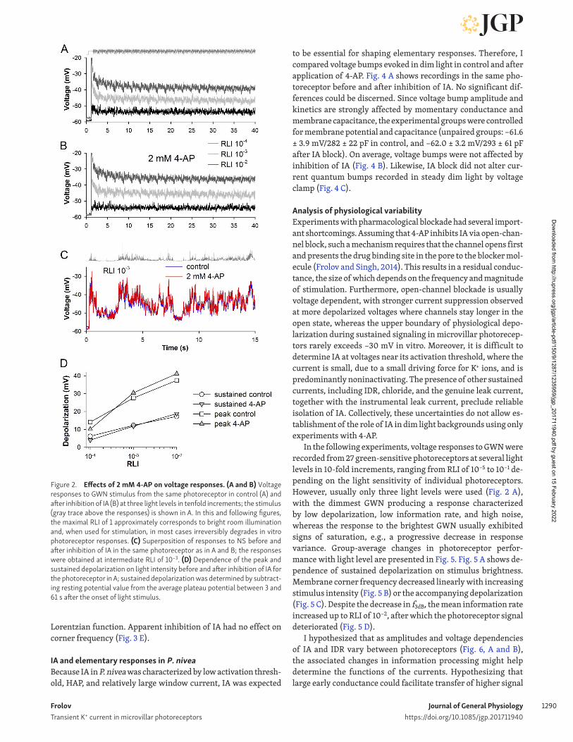
Effects of IA inhibition on voltage responses and information rates in P. niveaVoltage responses and information processing were evaluated using a 61-s stimulus consisting of an adapting 1-s prepulse followed by 30 repeats of a 2-s Gaussian noise sequence with a corner frequency of 50 Hz (Gaussian white-noise [GWN] stim-ulus) Also, a 60-s naturalistic contrast-modulated light stimu-lus (NS) was used. Fig. 2, A and B, shows voltage responses of the same photoreceptor to the GWN stimulus presented at three light levels in 10-fold increments before and after inhibition of IA. Fig. 2 C shows responses of the same photoreceptor to a mod-erately bright NS in control and after inhibition of IA. Applica-tion of 4-AP did not alter the resting potential (data not shown). There was no significant effect of application of 2 mM 4-AP on membrane depolarization during light responses (Fig. 2 D).
Information rates were calculated from SNR functions (see Methods). Typical signal and noise traces at a relative light in-tensity (RLI) of 10−2 before and after application of 2 mM 4-AP are shown in Fig. 3 A. Kv currents recorded in this photoreceptor are
shown in Fig. 1, A–C, and voltage responses in Fig. 2, A and B. It can be seen that both signal and noise traces before and after ap-plication of 4-AP are nearly identical. Consistently, only marginal differences could be seen between signal gain and SNR functions under two conditions (Fig. 3, B and C; data correspond to traces in Fig. 2, A and B). On average, information rates were not signifi-cantly different (Fig. 3 D). Corner frequencies were obtained by fitting the voltage dependencies of signal gain with a first-order
Figure 1. Inhibition of IA with 4-AP in P. Nivea. Delayed rectifier and tran-sient K+ currents were recorded in the same P. nivea photoreceptor in control (A), after inhibition of IA with 2 mM 4-AP (B), and after washout (C), from an HP of −92 mV using a protocol displayed in the inset in the middle row. IA was obtained by subtracting current traces evoked by the second part of the protocol from the traces evoked by the first part. Current traces are shown for the voltage range of −72 to −12 mV. (B) Voltage-current relations for IA in control and after application of 2 mM 4-AP; here and elsewhere, error bars represent mean ± SD.
Dow
nloaded from http://rupress.org/jgp/article-pdf/150/9/1287/1235959/jgp_201711940.pdf by guest on 15 February 2022
Frolov Transient K+ current in microvillar photoreceptors
Journal of General Physiologyhttps://doi.org/10.1085/jgp.201711940
1290
Lorentzian function. Apparent inhibition of IA had no effect on corner frequency (Fig. 3 E).
IA and elementary responses in P. niveaBecause IA in P. nivea was characterized by low activation thresh-old, HAP, and relatively large window current, IA was expected
to be essential for shaping elementary responses. Therefore, I compared voltage bumps evoked in dim light in control and after application of 4-AP. Fig. 4 A shows recordings in the same pho-toreceptor before and after inhibition of IA. No significant dif-ferences could be discerned. Since voltage bump amplitude and kinetics are strongly affected by momentary conductance and membrane capacitance, the experimental groups were controlled for membrane potential and capacitance (unpaired groups: −61.6 ± 3.9 mV/282 ± 22 pF in control, and −62.0 ± 3.2 mV/293 ± 61 pF after IA block). On average, voltage bumps were not affected by inhibition of IA (Fig. 4 B). Likewise, IA block did not alter cur-rent quantum bumps recorded in steady dim light by voltage clamp (Fig. 4 C).
Analysis of physiological variabilityExperiments with pharmacological blockade had several import-ant shortcomings. Assuming that 4-AP inhibits IA via open-chan-nel block, such a mechanism requires that the channel opens first and presents the drug binding site in the pore to the blocker mol-ecule (Frolov and Singh, 2014). This results in a residual conduc-tance, the size of which depends on the frequency and magnitude of stimulation. Furthermore, open-channel blockade is usually voltage dependent, with stronger current suppression observed at more depolarized voltages where channels stay longer in the open state, whereas the upper boundary of physiological depo-larization during sustained signaling in microvillar photorecep-tors rarely exceeds −30 mV in vitro. Moreover, it is difficult to determine IA at voltages near its activation threshold, where the current is small, due to a small driving force for K+ ions, and is predominantly noninactivating. The presence of other sustained currents, including IDR, chloride, and the genuine leak current, together with the instrumental leak current, preclude reliable isolation of IA. Collectively, these uncertainties do not allow es-tablishment of the role of IA in dim light backgrounds using only experiments with 4-AP.
In the following experiments, voltage responses to GWN were recorded from 27 green-sensitive photoreceptors at several light levels in 10-fold increments, ranging from RLI of 10−5 to 10−1 de-pending on the light sensitivity of individual photoreceptors. However, usually only three light levels were used (Fig. 2 A), with the dimmest GWN producing a response characterized by low depolarization, low information rate, and high noise, whereas the response to the brightest GWN usually exhibited signs of saturation, e.g., a progressive decrease in response variance. Group-average changes in photoreceptor perfor-mance with light level are presented in Fig. 5. Fig. 5 A shows de-pendence of sustained depolarization on stimulus brightness. Membrane corner frequency decreased linearly with increasing stimulus intensity (Fig. 5 B) or the accompanying depolarization (Fig. 5 C). Despite the decrease in f3dB, the mean information rate increased up to RLI of 10−2, after which the photoreceptor signal deteriorated (Fig. 5 D).
I hypothesized that as amplitudes and voltage dependencies of IA and IDR vary between photoreceptors (Fig. 6, A and B), the associated changes in information processing might help determine the functions of the currents. Hypothesizing that large early conductance could facilitate transfer of higher signal
Figure 2. Effects of 2 mM 4-AP on voltage responses. (A and B) Voltage responses to GWN stimulus from the same photoreceptor in control (A) and after inhibition of IA (B) at three light levels in tenfold increments; the stimulus (gray trace above the responses) is shown in A. In this and following figures, the maximal RLI of 1 approximately corresponds to bright room illumination and, when used for stimulation, in most cases irreversibly degrades in vitro photoreceptor responses. (C) Superposition of responses to NS before and after inhibition of IA in the same photoreceptor as in A and B; the responses were obtained at intermediate RLI of 10−3. (D) Dependence of the peak and sustained depolarization on light intensity before and after inhibition of IA for the photoreceptor in A; sustained depolarization was determined by subtract-ing resting potential value from the average plateau potential between 3 and 61 s after the onset of light stimulus.
Dow
nloaded from http://rupress.org/jgp/article-pdf/150/9/1287/1235959/jgp_201711940.pdf by guest on 15 February 2022

Frolov Transient K+ current in microvillar photoreceptors
Journal of General Physiologyhttps://doi.org/10.1085/jgp.201711940
1291
frequencies, peak IA and sustained IDR amplitudes at different membrane potentials were correlated to f3dB values obtained from responses to GWN at three backgrounds, RLI 10−4 to 10−2. Fig. 6 C shows dependencies of the correlations for IA on mem-brane potential. A clear maximum can be seen at −52 mV, which
corresponds to the maximal IA window current (see Fig. 7 E in Frolov et al., 2017). Two correlations shown in Fig. 6 D illustrate these findings. The highest SRO CC of 0.73 was observed in the dimmest 10−4 background, which is consistent with greater avail-ability of IA channels at this than at more depolarizing 10−3 and 10−2 backgrounds. IDR amplitudes also correlated positively with the corner frequencies, but the correlations were comparatively weak and increased with membrane depolarization, except at the 10−4 background, where the maximal correlation of 0.56 was found at −42 mV (Fig. 6 E).
A similar analysis was performed using HAPs for IA and IDR as proxies for the current amplitudes. A low HAP reflects an early activating current and vice versa. HAP values were obtained by fitting conductance-voltage relations with a Boltzmann’s charge-voltage equation (Fig. 7, A and B). On average, HAP was −32.8 ± 8.2 mV for IA and −35.3 ± 6.6 mV (n = 27) for IDR. Vari-ability in HAP was greater for IA than IDR (Fig. 7, A and B). There was a moderate positive statistically significant correlation be-tween HAP values for two currents (Fig. 7 C). Statistically signif-icant negative correlations were found between HAP for IA and f3dB at RLIs from 10−4 to 10−2 (Fig. 7, D and F). Interestingly, HAP for IDR correlated significantly with f3dB at RLIs of 10−4 but not in brighter backgrounds (Fig. 7, E and F). However, even in the relatively dim background of 10−4, the correlation was stronger for IA than for IDR (Fig. 7, D–F). The information rate correlated
Figure 3. Inhibition of IA by 4-AP does not affect signal processing in P. nivea in moderate and bright light. (A) 2-s signal and noise traces in control and after inhibition of IA were recorded in the same photoreceptor, with the stimulus shown in black; the signal was obtained by averaging 30 response repeats from the 60-s voltage response to GWN at the RLI of 10−2. (B and C) Voltage signal gain (B) and SNR (C) functions in control and in the presence of 2 mM 4-AP obtained from responses in Fig. 2, A and B; in both panels, triangles denote control and circles denote the presence of 4-AP (the legend to symbols is divided between the two panels). (D and E) Mean information rates (D) and the corresponding corner frequencies (E) at three light levels before and after application of 2 mM 4-AP; f3dB values were obtained from gain functions by fitting them with a first-order Lorentzian function; data from the same five photoreceptors. IR, information rate.
Figure 4. 4-AP does not affect elementary voltage responses. (A) Volt-age bump responses in steady dim light from the same photoreceptor before and after application of 2 mM 4-AP. (B) Mean voltage bumps were produced from unpaired recordings in control and after IA inhibition; group averages were obtained in the following way: first, mean bumps were obtained for each photoreceptor; second, a subsample of photoreceptors was selected so that their mean resting potentials were nearly the same for the two experimental conditions. (C) Mean current bumps were not altered in the presence of 2 mM 4-AP; bumps were evoked by 1-ms flashes of low-intensity light from a HP of −82 mV; mean bumps were determined for each photoreceptor, and then group averages were obtained.
Dow
nloaded from http://rupress.org/jgp/article-pdf/150/9/1287/1235959/jgp_201711940.pdf by guest on 15 February 2022

Frolov Transient K+ current in microvillar photoreceptors
Journal of General Physiologyhttps://doi.org/10.1085/jgp.201711940
1292
strongly with f3dB in the bright 10−2 background but not at dim-mer light levels (Fig. 7 G). These results suggest that IA facilitates transfer of higher-frequency signals.
What mechanism could cause the expansion of signaling bandwidth by IA? Graded voltage response is a superposition of transient bump-like voltage responses. It is possible that the fast-activating IA decreases the speed of membrane charging and relaxation by reducing the momentary membrane time constant, and through this facilitates resolution of individual responses. Fig. 8 shows examples of mean voltage bumps evoked by continu-ous dim light at resting potential in six photoreceptors character-ized by different membrane capacitance and HAP for IA values. Since the input resistance crucially depends on membrane po-tential, the recordings were arbitrarily combined into two groups with similar resting potentials as indicated. It can be seen that photoreceptors with more negative HAP values generate voltage bumps with faster onsets and smaller half widths than photore-ceptors with more positive HAP for IA.
IA in D. melanogaster: RevisitedElectrophysiological studies addressing the role of IA in photore-ceptors of D. melanogaster mainly relied on intracellular record-ings in wild-type and Shaker null mutant flies (ShKS133; Juusola et al., 2003; Niven et al., 2003b). Two main findings that can be assessed in independent voltage-clamp experiments are (1) the nonlinear dependence of sustained membrane conductance in the dark on voltage, and (2) the increased leak conductance in ShKS133 photoreceptors. The first effect was attributed to inacti-vation of Shaker channels and is crucial for the early nonlinear amplification hypothesis, whereas the second one is the founda-tion of the compensatory changes hypothesis.
Voltage-activated conductances were studied in patch-clamp recordings from photoreceptors in dissociated ommatidia from a wild-type CS and IA null mutant ShKS133 strains. Fig. 9 A shows averaged current traces evoked in wild-type photoreceptors from a holding potential (HP) of −64 mV by 100-ms voltage pulses between −74 and +36 mV in 10-mV increments. Fig. 9 B shows averaged current traces recorded under the same conditions in ShKS133 photoreceptors. Shaker IA can be obtained by subtracting currents in Fig. 9 B from the currents in Fig. 9 A. It can be seen that IA in D. melanogaster is much smaller than IA in P. nivea (Fig. 9 C). In D. melanogaster, values of HAP were −14.6 mV for IA and +11.9 ± 2.0 mV (n = 9) for IDR (current values at the end
Figure 5. General properties of responses to GWN. (A) Mean sustained membrane depolarization at different light levels during responses to GWN; numbers stand for the number of photoreceptors at each background in A–D; RP, resting potential (n = 27). (B and C) Changes in membrane corner fre-quency with light level (B) and the associated depolarization (C). (D) Changes in information rates.
Figure 6. Correlations (Correl.) between IA and IDR amplitudes and cor-ner frequency. (A and B) Conductance-voltage relations for IDR (A) and peak IA (B) were obtained from 27 photoreceptors. At each voltage in the range from −62 to +28 mV, the correlations between IA and IDR conductance, on the one hand, and corner frequencies at RLIs of 10−4, 10−3, and 10−2, on the other hand, were measured using SRO CC. (C) Voltage dependencies of correlation coefficients for IA/f3dB correlations; P < 0.03 for all correlations. (D) Examples of two correlations for IA at −52 mV at RLI of 10−4 and 10−2. (E) Voltage depen-dencies of correlation coefficients for IDR/f3dB correlations; at RLI of 10−4, P < 0.04 for voltages −62 through −22 mV; at RLIs of 10−3 and 10−2, P < 0.03 for −42 mV and all more positive voltages.
Dow
nloaded from http://rupress.org/jgp/article-pdf/150/9/1287/1235959/jgp_201711940.pdf by guest on 15 February 2022

Frolov Transient K+ current in microvillar photoreceptors
Journal of General Physiologyhttps://doi.org/10.1085/jgp.201711940
1293
of 100-ms pulses were used). Accordingly, the HAP for IDR in ShKS133 photoreceptors was +11.0 ± 6.7 mV (n = 8).
The early nonlinear amplification hypothesis postulates that Shaker channels in D. melanogaster begin activating at around resting potential. At such negative voltages, they do not undergo inactivation and therefore underlie what is effectively a sus-tained K+ conductance. Then, when the cell is depolarized by a few millivolts, inactivation of Shaker channels sets in, and the total membrane conductance decreases if the IDR activation threshold is not yet reached and Shab channels do not begin opening en masse. As a result, the amplitudes of small light-in-duced voltage responses at potentials slightly above rest become higher than similar responses evoked at the resting potential, where the effective IA conductance is higher. Two testable pre-dictions follow from this: (1) the decrease in conductance upon small depolarization should be observed in wild-type but not in ShKS133 photoreceptors, and (2) the decrease in conductance should be more prominent after prolonged depolarizations as IA inactivation is slow at such membrane potentials.
The results shown in Fig. 9, D and E, do not generally support the Shaker-based early amplification hypothesis. Fig. 9 D shows a prolonged 6-s Kv recording from a wild-type photoreceptor. Examining the current traces evoked by voltage pulses near the resting potential in the second half of the recording (colored traces inset) after subtracting the offset and scaled-up leak cur-rents (the current between −74 and −64 mV was defined as leak) revealed that in this cell, the total voltage-activated K+ conduc-tance indeed decreases between −64 and −54 mV (blue and green traces). However, such a pronounced decrease in conductance was observed only in one out of five photoreceptors (Fig. 9 E,
blue traces). Surprisingly, examination of prolonged recordings in ShKS133 photoreceptors revealed that one out of three photore-ceptors was also characterized by a decrease in conductance at −54 mV. Next, 100-ms recordings were examined (Fig. 9 F). It can be seen that a minority of photoreceptors from both wild-type and ShKS133 groups were characterized by decreasing conduc-tance between −54 and −44 mV. This was observed in photorecep-tors where the activation threshold for IDR was especially high (Fig. 9, E and F). On average, the current at the end of 100-ms voltage pulses between −44 and −24 mV was significantly larger in wild-type than Shaker photoreceptors (P < 0.007 at all three potentials; Fig. 9 G). However, the differences became statisti-cally insignificant at the end of 6-s responses (Fig. 9 H).
Intracellular recordings in ShKS133 photoreceptors were char-acterized by a relatively large leak conductance, which was in-terpreted as a compensatory development (Niven et al., 2003a). If this leak conductance is mediated by ion channels expressed in the soma, it would manifest in whole-cell patch-clamp record-ings as well. However, comparative analysis of leak current in control and ShKS133 flies showed that the total leak conductance, which consists of the instrumental and physiological leak con-ductances, was not significantly different between the experi-mental groups: at the end of 100-ms voltage pulses between –74 and −64 mV, the leak conductance equaled 1.05 ± 0.58 nS in con-trol (n = 9) versus 0.69 ± 0.37 nS in ShKS133 photoreceptors (n = 8).
DiscussionIn this work, I investigated the role of IA in signal processing in P. nivea photoreceptors and reexamined the situation in D. melano-
Figure 7. HAPs and information processing. (A and B) Normalized (Norm.) conductance-voltage relations for IDR (A) and peak IA (B); data from the same 27 photoreceptors as in Fig. 6, A and B. (C) Correlation (Correl.) between HAPs for IDR and IA; the correlation was statistically significant as indicated. (D and E) Correlations between HAPs for IA (D) or IDR (E) and corner frequency at RLI of 10−4. (F) SRO CC values for correlations between HAPs for IA or IDR and f3dB at three backgrounds; numbers in parentheses indicate the number of cells; n/s, not significant. (G) Correlation between f3dB and information rate at RLI of 10−2.
Dow
nloaded from http://rupress.org/jgp/article-pdf/150/9/1287/1235959/jgp_201711940.pdf by guest on 15 February 2022

Frolov Transient K+ current in microvillar photoreceptors
Journal of General Physiologyhttps://doi.org/10.1085/jgp.201711940
1294
gaster. I found some evidence for IA involvement in conditioning voltage responses in P. nivea, whereas the findings in D. melan-ogaster were inconsistent with the long-standing hypotheses on the role of IA (Juusola et al., 2003; Niven et al., 2003a,b).
The problem of IA is complex and needs to be separated into three distinct subproblems: (1) Why do photoreceptors need a rapidly activating Kv conductance, (2) what is the utility of a rapidly inactivating Kv conductance, and (3) what is the use for IA as it is—a rapidly activating and inactivating current?
The first question was addressed previously in several com-parative studies, and it appears that having a large, fast-activat-ing conductance is a prerequisite for fast vision (Weckström and Laughlin, 1995; Frolov et al., 2016). However, such current can play a dual role. The first one would be counteracting similarly fast high-frequency LIC transients and attenuating high-fre-quency signals and noise. On the other hand, by rapidly increas-ing membrane conductance and thus decreasing the momentary membrane time constant, it should facilitate transfer of high-fre-quency signals by preventing their excessive low-pass filtering. This effect would be more salient in the vicinity of resting poten-tial, where the driving force for K+ ions is relatively small and the opening of fast-activating Kv channels would not elicit a strong counteracting repolarizing current while providing a substan-tial conductance. Although in this situation, the conductance can
also be expected to reduce the gain of high-frequency signals, the efficiency of the signal transfer is eventually determined at the first synapse. The synapse, however, appears to be a dynamically adapting differentiating device, which emphasizes transmission of fast over slow components of the graded signal (Juusola et al., 1995; Baden et al., 2013), thus favoring the high-frequency facilitation function. Consistently, here I demonstrated that in P. nivea photoreceptors, the signal transfer bandwidth as mea-sured by membrane corner frequency is proportional to the amplitude of IA.
The utility of a rapidly inactivating Kv conductance was also studied previously in several species (Cuttle et al., 1995; Weckström and Laughlin, 1995; Laughlin, 1996). The current opinion is that it helps preventing unnecessary metabolic ex-penses and suppresses high-frequency noise.
The answer to the third question is probably related to the lifestyle and behavior of the species. P. nivea is a nocturnal spe-cies, and D. melanogaster is also mainly active during low light periods. If strong photoreceptor depolarizations occur infre-quently and briefly in these and similar species, then it might be sufficient for them to express a fast-activating and -inactivating Kv conductance rather than a fast-activating sustained conduc-tance such as found in diurnal blowflies (Weckström et al., 1991).
IA in P. niveaThe experiments in P. nivea consisted of two parts: acute phar-macological inhibition of IA with 4-AP and analysis of normal variability in a relatively large group of photoreceptors. While the tests with 4-AP allowed rapid removal of IA with immediate registration of associated effects, such results could be inter-preted with confidence only for fairly depolarized responses. Due to the uncertain extent of IA blockade in the lower half of the physiological voltage range, i.e., 10- to 15-mV depolarization from the resting potential, the absence of changes in photoreceptor signaling in relatively dim backgrounds cannot be interpreted as the absence of IA function. On the other hand, during stimula-tion with bright GWN, which elicits relatively highly depolarized sustained responses, most of IA can be expected to be removed by inactivation (see voltage dependence of IA inactivation in Fig. 7 E of Frolov et al., 2017). Use of 4-AP in such circumstances might add little to that removal. The variability analysis was based on the premise that if amplitudes of Kv currents differ from cell to cell in the physiological voltage range, then the accompanying variability in the higher photoreceptor functions might help to explain the roles of the currents. In this analysis, both direct conductance and indirect HAP values were used as measures of the currents.
There are several possible functions or electrophysiological situations where IA could improve photoreceptor signaling. First, by rapidly increasing membrane conductance and decreas-ing the membrane time constant, IA could accelerate the initial transient depolarizing responses of dark-adapted photorecep-tors stimulated by relatively bright light. However, neither the experiments with 4-AP described here nor the previous modeling study in P. americana (Salmela et al., 2012) support this function. There was no detectable effect of IA removal on the amplitude, width, or onset kinetics of the large initial depolarizing transient.
Figure 8. Voltage bumps in photoreceptors with different HAP for IA. Voltage bumps were evoked by dim continuous light from resting potential; mean voltage bumps for each photoreceptor are shown. Cm, membrane capac-itance; RP, resting membrane potential. (A) Mean voltage bumps in three pho-toreceptors with resting potentials between −58 and −60 mV. Here and in B, the original mean bumps are shown to the left, and normalized bumps to the right. (B) Mean bumps in three photoreceptors with resting potentials between −62 and −63 mV.
Dow
nloaded from http://rupress.org/jgp/article-pdf/150/9/1287/1235959/jgp_201711940.pdf by guest on 15 February 2022

Frolov Transient K+ current in microvillar photoreceptors
Journal of General Physiologyhttps://doi.org/10.1085/jgp.201711940
1295
This might be explained by the relatively slow onset and large half width of the transients evoked by flashes of bright light, as IA channels would be strongly inactivated by the time the peak of the transient is reached. However, it should be noted that the transients seem to be consistently slower and wider in patch-clamp than in intracellular recordings (data not shown), proba-bly because of the absence of additional conductances associated with either the axon or the instrumental leak due to membrane piercing (see below), so that a role for IA in modulating the tran-sient in vivo cannot be ruled out.
However, the most likely function for IA is the facilitation of transfer of high-frequency components of the stimulus by mod-ulating small-voltage transients that form the sustained light re-sponse (Fig. 8). Strong and statistically significant correlations found between the measures of IA and f3dB values (Figs. 6 and 7) are consistent with this hypothesis. Notably, the strength of the correlations between HAP for IA and f3dB decreased in brighter backgrounds consistently with the progressive inactivation of IA with increasing membrane depolarization (Fig. 7 F; see also Fig. 7 E in Frolov et al., 2017). Interestingly, correlations between the actual IA conductance values and f3dB were voltage depen-dent, with the strongest one observed precisely at the voltage linked to the maximal IA window current, −52 mV (Fig. 6 C). The correlations decreased steadily as the membrane was fur-ther depolarized, pointing to a growing disconnect between the
peak IA conductance at each membrane potential determined in voltage-clamp experiments and the actual persistent IA during sustained voltage responses to GWN.
The strongest correlations between the actual IDR conduc-tance values and f3dB, and also between HAC for IDR and f3dB, were found in the relatively dim light, at RLI of 10−4 (Figs. 6 E and 7 F). What could explain this finding? As the level of sustained depo-larization is determined by the balance between depolarizing LIC and repolarizing Kv conductances, in relatively bright light, the level of depolarization is set by the interplay between LIC and IDR since IA is mostly inactivated. Bright light activates a sufficiently large IDR conductance as not to restrict the signaling bandwidth (or to restrict it to a similar degree in cells with different HAP for IDR). In contrast, both IA and IDR are likely to be involved in repolarization in relatively dim light so that the correlation between HAP for IDR and f3dB at RLI of 10−4 could reflect the dif-ferences in IDR activation thresholds between photoreceptors.
Examination of individual voltage bumps in similar physio-logical variability analysis could possibly reveal the electrophys-iological mechanisms by which IA and IDR influence membrane corner frequency. However, a much larger experimental dataset would be needed to achieve this goal than currently available because there are many factors modulating elementary voltage responses, such as membrane capacitance, conductance and HAP values for IA and IDR, quantum bump amplitude and latency, etc.,
Figure 9. Voltage-activated K+ conductances in D. melanogaster photoreceptors. (A) Voltage-activated K+ current in wild-type D. melanogaster was recorded from an HP of −64 mV using 100-ms voltage pulses applied in 10-mV increments from −74 to +36 mV; each pulse was preceded by a 100-ms pulse to −104 mV to recover IA from inactivation; the traces are averages of recordings from six photoreceptors. (B) Averaged (n = 5) current traces from the Shaker mutant obtained as in A. (C) Comparison of IA in D. melanogaster and P. nivea; D. melanogaster IA was acquired by subtracting traces in B from the traces in A; P. nivea IA is the average of currents from nine photoreceptors obtained as in Fig. 1 A. (D) Prolonged 6-s recordings of Kv current in wild-type D. melanogaster photoreceptors; the current was recorded as in A with a 400-ms prepulse to −104 mV. Inset left shows the IA. Inset right shows four current traces elicited by voltage steps between −74 and −44 mV as indicated; the average current value at −74 mV was subtracted from all traces; the corresponding resulting average current values are shown at right. (E) Conductance-voltage relations for sustained IDR currents in wild-type and Shaker photoreceptors in the voltage range between −64 and −24 mV; current values were obtained by averaging currents between 4 and 6 s after the onset of voltage steps; leak conductance was determined between −74 and −64 mV and subtracted from all traces; black dotted line indicates zero conductance. (F) Conductance-voltage relations for IDR between 70 and 100 ms after the onset of voltage steps. (G) Average conductance-voltage relations for IDR from F. In wild-type photoreceptors, IDR was significantly higher than in Shaker photoreceptors at three potentials as indicated. (H) Average conductance-voltage relations for IDR from E.
Dow
nloaded from http://rupress.org/jgp/article-pdf/150/9/1287/1235959/jgp_201711940.pdf by guest on 15 February 2022

Frolov Transient K+ current in microvillar photoreceptors
Journal of General Physiologyhttps://doi.org/10.1085/jgp.201711940
1296
and their effects need to be explored while controlling membrane potential as strictly as possible.
IA in D. melanogasterDespite the differences in activation ranges of IA and IDR be-tween D. melanogaster and P. nivea, sustained voltage responses in bright light in both species are mostly counteracted by IDR, and it follows from the results in P. nivea that IA can hardly affect information transfer in bright light. How can this be reconciled with findings in D. melanogaster, where Shaker null mutant flies exhibited strongly decreased information capacity across the en-tire range of light intensities (Niven et al., 2003b)? Apparently, the reduction in information capacity, especially in bright light where IA should be completely inactivated, cannot be caused by the loss of IA conductance per se. In general, differences be-tween wild-type and mutant phenotypes can arise from multiple sources, including the effects of the mutation, the accompanying physiological compensatory changes, differences in genetic back-grounds, and interactions between the background and the gene of interest (Linder, 2006). Moreover, the functions of a gene are not always limited to the function of its namesake protein (see, e.g., Gomez-Ospina et al., 2013). In the D. melanogaster study, the changes in the mutant photoreceptors had all the hallmarks of a highly increased leak conductance as the underlying cause: reduced response variance and voltage gain, reduced impedance in the lower frequency region, and increased membrane corner frequency (Niven et al., 2003b).
In the following study, when the additional leak conductance found in Shaker null mutants was incorporated into a mathe-matical model, the resulting metabolic cost of visual informa-tion processed by photoreceptors increased twofold (Niven et al., 2003a). The increased leak was considered a necessary devel-opmental compensation for the absent IA, and it was concluded that expression of Shaker channels reduces the cost of informa-tion. However, increased leak current in Shaker null mutants is a strong confounding factor, altering the whole electrophysio-logical phenotype of the mutant photoreceptors. No valid con-clusion on the specific electrogenic function of Shaker channels,
such as their putative involvement in information processing or energy metabolism, is possible without separating the effects of IA knockout from those of the increased leak. Moreover, in-creased leak might not be the only compensatory development in the mutant, as changes in light-induced conductance were proposed in the subsequent study by the same authors (Niven et al., 2004). This problem needs to be addressed in future exper-imental studies.
It should be noted that a similar increase in leak current was found in the study of small conductance Ca2+-activated K+ chan-nels in D. melanogaster photoreceptors (Abou Tayoun et al., 2011), indicating that it is a nonspecific development. Its origin remains unclear, and here I provided evidence that the leak current in Shaker photoreceptors at the level of the soma does not exceed that in wild-type photoreceptors. This suggests that it might be caused by changes in the axon, i.e., in the presynaptic terminal.
Finally, IA was proposed to amplify relatively small voltage responses to light at potentials slightly above the resting poten-tial (Juusola et al., 2003; Niven et al., 2003b). While it cannot be contested that transition of Shaker-based IA from a persistent to inactivating conductance with progressive membrane depolariza-tion would increase membrane resistance if IA was the only volt-age-dependent conductance in the cell, in practice the situation is more complex because photoreceptors express several voltage-de-pendent channels. Due to natural variation in the amplitudes and voltage dependencies of such conductances, Shaker can be ex-pected to amplify voltage signals only under specific conditions, when a decrease in IA with depolarization is not offset by opening of other channels. Indeed, results presented here indicate that the decrease in conductance with increasing membrane potential is a valid phenomenon. However, it was observed only in a minor frac-tion of photoreceptors characterized by a high activation thresh-old for IDR. According to Fig. 9, E and F, the total conductance could decrease by as much as 0.5 nS; considering that the average leak conductance is ∼1 nS, such a 0.5-nS decrease can lead to a notice-able low-frequency amplification of voltage responses. Further-more, as some of the leak conductance is probably caused by the imperfect electrode-membrane seal interface, the actual amplifi-
Table 1. Mean HAP values for IA and IDR in insect photoreceptors with a prominent IA (see Frolov et al., 2016).
Species HAP, mV IDR at −40 mV, nS IDR at −40 mV, pS/pF Reference
IA IDR at 400 ms
Carausius morosus −12.4 ± 2.0 −23.2 ± 1.9 2.5 21 Frolov et al., 2012
Corixa punctata −25.5 ± 7.9 −25.7 ± 7.3 6.6 15 Frolov, 2015
D. melanogaster −14.6a +2.7 ± 9.4 1.1 20b Fig. 9
D. virilis −22.4 ± 5.9 −27.5 ± 2.5c 9.0c 106c Not published
Gerris lacustris −19.0 ± 6.0 −18.0 ± 10.0 3.3 57 Frolov and Weckström, 2014
Gryllus bimaculatus −20.5 ± 3.8 −22.1 ± 11.2d 5.2 46 Frolov et al., 2014
P. nivea −34.6 ± 7.4 −34.5 ± 7.1 5.3 20 Frolov et al., 2017
aFrom the current traces in Fig. 9 C.bAverage photoreceptor capacitance value of 55 pF was used (Frolov, 2016). D. virilis values were obtained from unpublished data.cAt the end of 100 ms pulses; the average capacitance value of 85 pF was used (Frolov, 2016).dFor regular green-sensitive photoreceptors in dark-adapted crickets (Frolov et al., 2014).
Dow
nloaded from http://rupress.org/jgp/article-pdf/150/9/1287/1235959/jgp_201711940.pdf by guest on 15 February 2022

Frolov Transient K+ current in microvillar photoreceptors
Journal of General Physiologyhttps://doi.org/10.1085/jgp.201711940
1297
cation might be even stronger. However, since the decreasing con-ductance was found in equal measure both in control and ShKS133 photoreceptors, it cannot be plausibly attributed to IA only. The question of amplification has been revisited by the same authors in the upcoming modeling study in D. melanogaster (Heras et al., 2018), who suggest that amplification has no real function, and the main function of the Shaker conductance is through activation to increase bandwidth at low light levels.
D. melanogaster as an outlierRecent comparative electrophysiological studies of microvillar photoreceptors imply that the very positive HAP for IDR found in D. melanogaster is an exception rather than a typical situation. Table 1 compares some properties of IA and IDR in seven species characterized by a prominent IA current, including a D. mela-nogaster relative, D. virilis. In all cases except D. melanogaster, HAPs for IDR were equal or more negative than the HAPs for IA. (However, it should be noted that the utility of the HAP concept is limited because it strongly depends on the inactivation state of the channel pool: for instance, in the photoreceptor shown in Fig. 9 D, the nominal value of the momentary HAP decreases from +14 mV at 100 ms after the onset of voltage steps to 0 mV between 4 and 6 s due to a disproportional inactivation of IDR at positive voltages.) As a consequence of such a positive HAP for IDR in D. melanogaster, IDR conductance in the lower part of the physiological voltage range (in Table 1, an arbitrary value of −40 mV was used) was smallest in the fruit fly. However, if instead of conductance per se, IDR conductance densities (conductance divided by capacitance) were estimated, then D. melanogaster cannot be considered an outsider. The IDR conductance density value has been linked to visual ecological and behavioral traits of the species (Frolov, 2016). Although the reason for such an un-usual arrangement of Kv conductances in photoreceptors of D. melanogaster is not clear, the difference between the HAPs is not fixed and subject to modulating influences capable of eliminat-ing the difference altogether as it was shown previously (Hevers and Hardie, 1995).
ConclusionsResults presented here suggest that IA might play the same roles in both D. melanogaster and P. nivea photoreceptors, i.e., facilitating transmission of high-frequency signals. In D. mela-nogaster, discovery of this function was probably obfuscated by the increased membrane corner frequencies in the Shaker null mutant clearly caused by high leak conductance (Niven et al., 2003b; Heras et al., 2018). On the one hand, such compensa-tory developments in mutants lacking important proteins limit utility of the mutational analysis. On the other hand, pharmaco-logical inhibition can be unreliable, as exemplified in this study. Combining these two approaches often helps resolving difficult cases, but it is not always feasible. The physiological variability analysis I offered here circumvents this problem by exploiting naturally occurring variabilities in electrophysiological proper-ties of photoreceptors in the same species or breed. While it has several shortcomings, including nonequivalence of correlation and causation, and a need to obtain a large number of data points, its advantages are appealing.
AcknowledgmentsThe author thanks Dr. Jeremy Niven for many insightful discus-sions and Prof. Andrew S. French for help with the manuscript.
The author declares no competing financial interests.Sharona E. Gordon served as editor.
Submitted: 6 November 2017Revised: 11 May 2018Accepted: 28 June 2018
ReferencesAbou Tayoun, A.N., X. Li, B. Chu, R.C. Hardie, M. Juusola, and P.J. Dolph. 2011.
The Drosophila SK channel (dSK) contributes to photoreceptor perfor-mance by mediating sensitivity control at the first visual network. J. Neurosci. 31:13897–13910. https:// doi .org/ 10 .1523/ JNE URO SCI .3134 -11 .2011
Baden, T., T. Euler, M. Weckström, and L. Lagnado. 2013. Spikes and ribbon synapses in early vision. Trends Neurosci. 36:480–488. https:// doi .org/ 10 .1016/ j .tins .2013 .04 .006
Cuttle, M.F., W. Hevers, S.B. Laughlin, and R.C. Hardie. 1995. Diurnal modu-lation of photoreceptor potassium conductance in the locust. J. Comp. Physiol. A Neuroethol. Sens. Neural Behav. Physiol. 176:307–316. https:// doi .org/ 10 .1007/ BF00219056
Elkins, T., and B. Ganetzky. 1988. The roles of potassium currents in Dro-sophila flight muscles. J. Neurosci. 8:428–434. https:// doi .org/ 10 .1523/ JNE URO SCI .08 -02 -00428 .1988
Frolov, R.V. 2015. Biophysical properties of photoreceptors in Corixa punctata facilitate diurnal life-style. Vision Res. 111(Pt A):75–81. https:// doi .org/ 10 .1016/ j .visres .2015 .03 .026
Frolov, R.V. 2016. Current advances in invertebrate vision: insights from patch-clamp studies of photoreceptors in apposition eyes. J. Neuro-physiol. 116:709–723. https:// doi .org/ 10 .1152/ jn .00288 .2016
Frolov, R.V., and S. Singh. 2014. Celecoxib and ion channels: a story of unex-pected discoveries. Eur. J. Pharmacol. 730:61–71. https:// doi .org/ 10 .1016/ j .ejphar .2014 .02 .032
Frolov, R., and M. Weckström. 2014. Developmental changes in biophysical properties of photoreceptors in the common water strider (Gerris la-custris): better performance at higher cost. J. Neurophysiol. 112:913–922. https:// doi .org/ 10 .1152/ jn .00239 .2014
Frolov, R., E.V. Immonen, M. Vähäsöyrinki, and M. Weckström. 2012. Postembry-onic developmental changes in photoreceptors of the stick insect Carau-sius morosus enhance the shift to an adult nocturnal life-style. J. Neurosci. 32:16821–16831. https:// doi .org/ 10 .1523/ JNE URO SCI .2612 -12 .2012
Frolov, R.V., E.V. Immonen, and M. Weckström. 2014. Performance of blue- and green-sensitive photoreceptors of the cricket Gryllus bimaculatus. J. Comp. Physiol. A Neuroethol. Sens. Neural Behav. Physiol. 200:209–219. https:// doi .org/ 10 .1007/ s00359 -013 -0879 -6
Frolov, R., E.V. Immonen, and M. Weckström. 2016. Visual ecology and potas-sium conductances of insect photoreceptors. J. Neurophysiol. 115:2147–2157. https:// doi .org/ 10 .1152/ jn .00795 .2015
Frolov, R.V., A. Matsushita, and K. Arikawa. 2017. Not flying blind: a compar-ative study of photoreceptor function in flying and non-flying cock-roaches. J. Exp. Biol. 220:2335–2344. https:// doi .org/ 10 .1242/ jeb .159103
Gomez-Ospina, N., G. Panagiotakos, T. Portmann, S.P. Pasca, D. Rabah, A. Budzillo, J.P. Kinet, and R.E. Dolmetsch. 2013. A promoter in the coding region of the calcium channel gene CAC NA1C generates the transcrip-tion factor CCAT. PLoS One. 8:e60526. https:// doi .org/ 10 .1371/ journal .pone .0060526
Hardie, R.C., D. Voss, O. Pongs, and S.B. Laughlin. 1991. Novel potassium chan-nels encoded by the Shaker locus in Drosophila photoreceptors. Neuron. 6:477–486. https:// doi .org/ 10 .1016/ 0896 -6273(91)90255 -X
Heras, F.J.H., M. Vähäsöyrinki, and J.E. Niven. 2018. Modulation of voltage-de-pendent K+ conductances in photoreceptors trades off investment in contrast gain for bandwidth. bioRxiv. https:// doi .org/ 10 .1101/ 344325 (Preprint posted June 12, 2018)
Hevers, W., and R.C. Hardie. 1995. Serotonin modulates the voltage depen-dence of delayed rectifier and Shaker potassium channels in Drosoph-ila photoreceptors. Neuron. 14:845–856. https:// doi .org/ 10 .1016/ 0896 -6273(95)90228 -7
Dow
nloaded from http://rupress.org/jgp/article-pdf/150/9/1287/1235959/jgp_201711940.pdf by guest on 15 February 2022

Frolov Transient K+ current in microvillar photoreceptors
Journal of General Physiologyhttps://doi.org/10.1085/jgp.201711940
1298
Immonen, E.V., A.S. French, P.H. Torkkeli, H. Liu, M. Vähäsöyrinki, and R.V. Frolov. 2017. EAG channels expressed in microvillar photoreceptors are unsuited to diurnal vision. J. Physiol. 595:5465–5479. https:// doi .org/ 10 .1113/ JP273612
Juusola, M., R.O. Uusitalo, and M. Weckström. 1995. Transfer of graded poten-tials at the photoreceptor-interneuron synapse. J. Gen. Physiol. 105:117–148. https:// doi .org/ 10 .1085/ jgp .105 .1 .117
Juusola, M., J.E. Niven, and A.S. French. 2003. Shaker K+ channels contrib-ute early nonlinear amplification to the light response in Drosophila photoreceptors. J. Neurophysiol. 90:2014–2021. https:// doi .org/ 10 .1152/ jn .00395 .2003
Krause, Y., S. Krause, J. Huang, C.H. Liu, R.C. Hardie, and M. Weckström. 2008. Light-dependent modulation of Shab channels via phosphoinosit-ide depletion in Drosophila photoreceptors. Neuron. 59:596–607. https:// doi .org/ 10 .1016/ j .neuron .2008 .07 .009
Laughlin, S.B. 1996. Matched filtering by a photoreceptor membrane. Vision Res. 36:1529–1541. https:// doi .org/ 10 .1016/ 0042 -6989(95)00242 -1
Laughlin, S.B., and M. Weckström. 1993. Fast and slow photoreceptors — a comparative study of the functional diversity of coding and conduc-tances in the Diptera. J. Comp. Physiol. A Neuroethol. Sens. Neural Behav. Physiol. 172:593–609. https:// doi .org/ 10 .1007/ BF00213682
Linder, C.C. 2006. Genetic variables that influence phenotype. ILAR J. 47:132–140. https:// doi .org/ 10 .1093/ ilar .47 .2 .132
Niven, J.E., M. Vähäsöyrinki, and M. Juusola. 2003a. Shaker K(+)-channels are predicted to reduce the metabolic cost of neural information in Dro-sophila photoreceptors. Proc. Biol. Sci. 270(Suppl 1):S58–S61. https:// doi .org/ 10 .1098/ rsbl .2003 .0010
Niven, J.E., M. Vähäsöyrinki, M. Kauranen, R.C. Hardie, M. Juusola, and M. Weckström. 2003b. The contribution of Shaker K+ channels to the in-formation capacity of Drosophila photoreceptors. Nature. 421:630–634. https:// doi .org/ 10 .1038/ nature01384
Niven, J.E., M. Vähäsöyrinki, M. Juusola, and A.S. French. 2004. Interactions between light-induced currents, voltage-gated currents, and input sig-nal properties in Drosophila photoreceptors. J. Neurophysiol. 91:2696–2706. https:// doi .org/ 10 .1152/ jn .01163 .2003
Salmela, I., E.V. Immonen, R. Frolov, S. Krause, Y. Krause, M. Vähäsöyrinki, and M. Weckström. 2012. Cellular elements for seeing in the dark: volt-age-dependent conductances in cockroach photoreceptors. BMC Neuro-sci. 13:93. https:// doi .org/ 10 .1186/ 1471 -2202 -13 -93
Weckström, M., and S.B. Laughlin. 1995. Visual ecology and voltage-gated ion channels in insect photoreceptors. Trends Neurosci. 18:17–21. https:// doi .org/ 10 .1016/ 0166 -2236(95)93945 -T
Weckström, M., R.C. Hardie, and S.B. Laughlin. 1991. Voltage-activated potas-sium channels in blowfly photoreceptors and their role in light adaptation. J. Physiol. 440:635–657. https:// doi .org/ 10 .1113/ jphysiol .1991 .sp018729
Dow
nloaded from http://rupress.org/jgp/article-pdf/150/9/1287/1235959/jgp_201711940.pdf by guest on 15 February 2022