research article - journal of cell...
TRANSCRIPT

IntroductionHemidesmosomes (HD) are specialized attachment structuresof the basement membrane zone (BMZ) which bind laminin-5(Rousselle et al., 1991) through α3β1 integrin (Carter et al.,1991) andα6β4 integrin (Sonnenberg et al., 1991). Duringwound healing, basal keratinocytes along the wound edgeundergo transition from static adherent structures to motile,regenerative sheets of cells (Martin, 1997). Growth factorssuch as epidermal growth factor (EGF), secreted into thewound site by macrophages and keratinocytes induce HDdisassembly, keratinocyte proliferation and migration(Barrandon and Green, 1987; Mainiero et al., 1996;Marikovsky et al., 1993; Martin, 1997). Keratinocytes at thewound front can migrate on dermal collagen using α2β1integrin and MMP-1 (Pilcher et al., 1997) or laminin-5 throughα3β1 integrin (Goldfinger et al., 1999).
α6β4 integrin has generally been viewed as a mediator ofattachment and HD formation at sites more distal from thewound edge (Kurpakus et al., 1991; Nguyen et al., 2000a) oreven as an inhibitor of keratinocyte motility (Hintermann et al.,2001). However, several lines of evidence suggest α6β4integrin may play a more direct and active role in keratinocytemigration. α6β4 integrin interacts with receptor tyrosine
kinases such as EGFR, ErbB-2 and Met (Falcioni et al., 1997;Hintermann et al., 2001; Trusolino et al., 2001). Stimulation ofkeratinocytes with EGF induces tyrosine phosphorylation ofthe cytoplasmic domain of β4 which is implicated in both HDdisassembly and epithelial motility (Mainiero et al., 1996).α6β4 may play an active role in chemotactic migration throughlysophosphatidic acid (LPA) by activation of a cAMP-specificphosphodiesterase and RhoA GTPase (O’Connor et al., 2000;O’Connor et al., 1998). and α6β4 can localize with filamentousactin and stabilize lamellipodial membrane protrusions(Rabinovitz and Mercurio, 1997; Rabinovitz et al., 1999).
Since growth factor stimulation is required to inducekeratinocyte migration during wound healing, we examined therole of laminin-5, α3β1 integrin and α6β4 integrin in thisprocess. We re-expressed wild-type and attachment-defectiveβ4 integrin in β4 null patient keratinocytes and studied theeffects of α6β4 integrin ligation on EGF-mediated keratinocytechemotaxis. We found that EGF-induced keratinocytemigration response depends upon the interaction betweenlaminin-5 and the β4 integrin ectodomain. When bound tolaminin-5, α6β4 integrin promoted EGF-dependent cellmigration through Rac1 activation. Without laminin-5 ligationthrough α6β4 integrin, EGF induced keratinocyte chemotaxis
3543
Growth factor-induced cell migration and proliferation areessential for epithelial wound repair. Cell migration duringwound repair also depends upon expression of laminin-5, aligand for α6β4 integrin. We investigated the role of α6β4integrin in laminin-5-dependent keratinocyte migration byre-expressing normal or attachment-defective β4 integrinin β4 integrin null keratinocytes. We found that expressionof β4 integrin in either a ligand bound or ligand unboundstate was necessary and sufficient for EGF-induced cellmigration. In a ligand bound state, β4 integrin supportedEGF-induced cell migration though sustained activation ofRac1. In the absence of α6β4 integrin ligation, Rac1activation became tempered and EGF chemotaxisproceeded through an alternate mechanism that dependedupon α3β1 integrin and was characterized by cellscattering. α3β1 integrin also relocalated from cell-cell
contacts to sites of basal clustering where it displayedincreased conformational activation. The aberrantdistribution and activation of α3β1 integrin in attachment-defective β4 cells could be reversed by the activation ofRac1. Conversely, in WT β4 cells the normal cell-celllocalization of α3β1 integrin became aberrant after theinhibition of Rac1. These studies indicate that theextracellular domain of β4 integrin, through its ability tobind ligand, functions to integrate the divergent effects ofgrowth factors on the cytoskeleton and adhesion receptorsso that coordinated keratinocyte migration can beachieved.
Key words: α6β4 Integrin, EGF, Laminin-5, Keratinocyte,Chemotaxis
Summary
α6β4 integrin regulates keratinocyte chemotaxisthrough differential GTPase activation andantagonism of α3β1 integrinAlan J. Russell 1,*, Edgar F. Fincher 1, Linda Millman 1, Robyn Smith 1, Veronica Vela 1, Elizabeth A. Waterman 1,Clara N. Dey 1, Shireen Guide 1, Valerie M. Weaver 2 and M. Peter Marinkovich 1,‡
1Program in Epithelial Biology, Stanford University School of Medicine, Stanford, CA 94305, USA2Pathology & Institute for Medicine and Engineering, University of Pennsylvania, Philadelphia, PA 19104, USA*Present address: Cytokinetics Inc, 280 East Grand Ave, South San Franscisco, CA 94080, USA‡Author for correspondence (e-mail: [email protected])
Accepted 12 May 2003Journal of Cell Science 116, 3543-3556 © 2003 The Company of Biologists Ltddoi:10.1242/jcs.00663
Research Article

3544
through α3β1 integrin. We show evidence that these twopathways are antagonistic and suggest a mechanism throughwhich α6β4 may coordinate these two signals to regulateintegrated epithelial movement during wound healing.
Materials and MethodsCell linesPrimary keratinocytes were obtained from an patient withepidermolysis bullosa with pyloric atresia (EB-PA) resulting from acompound heterozygote mutation in the β4 integrin gene(C738X/4791delCA) (Pulkkinen et al., 1998). Cells wereimmortalized with HPV18 E6 and E7 genes (Kaur et al., 1989).Additional studies were carried out on primary keratinocytes from anEB-PA patient deficient in β4 as a result of a premature terminationcodon (C658X). Neonatal human foreskin keratinocytes (NHK) andimmortalized patient cells were cultured in serum-free medium (SFM)(Gibco). Modified human 293 PHOENIX cells (a gift from Dr G.Nolan, Stanford University, Stanford, CA) were cultured in DMEMsupplemented with 10% fetal calf serum, 100 IU/ml penicillin and 100µg/ml streptomycin.
AntibodiesMouse mAb 3E1, ASC-8 and rat mAb GoH3 recognizing theextracellular domains of β4 and α6 respectively, and rabbit polyclonalantiserum to β4 wwere obtained from Chemicon (Temecula, CA).Mouse mAb ASC-8 is inhibitory to α6β4 attachment and was used inall inhibition assays at 10 µg/ml. The mouse mAb 121 raised againstHD1/plectin and the mouse mAb 233 raised against BP180 were agift from Dr K. Owaribe (Nagoya University, Nagoya, Japan). Therabbit laminin-5 antisera has been characterized (Marinkovich et al.,1992). Anti-laminin-5 mAb BM165 (Rousselle et al., 1991) waspurified through protein G affinity chromatography. BM165 preventsattachment to the α3 subunit of laminin-5 and was used at 10 µg/ml.Mouse anti-α3 integrin mAb P1B5 and mouse mAb HUTS-4 againstthe active conformation of β1 integrins were obtained from Chemicon.P1B5 is inhibitory to α3β1 integrin attachment and was used at 10µg/ml. Mouse mAb 349 and rat mAb 346-11A against human paxillinand integrin were obtained from Transduction Labs and Pharmingenrespectively (Lexington, NY). Rabbit sera 119 and P1 raised againstRhoA and Cdc42 respectively were obtained from Santa CruzBiotechnology (Santa Cruz, CA). Mouse mAb 23A8 against Rac1 wasobtained from Upstate Biotech (Lake Placid, NY). Mouse mAb 9E10to the myc tag was obtained from Oncogene Research Products(Boston, MA). Phosphotyrosine western blots were carried out withmouse mAb 4G10 (Upstate Biotech, Lake Placid, NY). Rabbitantibodies to p44/42 MAP kinase and phospho-p44/42 MAP kinasewere obtained from New England Biolabs Inc (Beverly, MA). FITC-and TRITC-conjugated phalloidin was purchased from SigmaChemical Co. (St Louis, MO). TRITC-conjugated goat anti-rabbit,Cy5-conjugated goat anti-rat and FITC-conjugated donkey anti-mouse secondary antibodies were purchased from JacksonImmunoResearch (Westgrove, PA). The sheep anti-mouse and donkeyanti-rabbit horseradish peroxidase-conjugated secondary antibodieswere obtained from Amersham (Arlington Heights, IL).
cDNA constructs and vectorsβ4pRK-5 was a generous gift from Dr F. G. Giancotti (Sloan KetteringCancer Institute, NY). Previous reports have shown that a β4 integrincDNA from this lab contained an in frame deletion of 7 amino acids(880-886) in the membrane proximal region (Dans et al., 2001).Therefore, before use we sequenced this region to ensure no deletionswere present.β4 cDNA was cloned as a 5.6 kb EcoRI fragment intothe EcoRI site of retroviral expression vector LZRS (Kinsella and
Nolan, 1996) containing the encephalomyocarditis virus (EMCV)-IRES and blasticidin-resistance sequences (Deng et al., 1998). Anattachment-deficient (AD) β4 construct was produced through cloningthe EcoRI β4 cDNA insert into the EcoRI site of pSK and performingmutagenesis using the GeneEditor in vitro site-directed mutagenesissystem (Promega, Madison WI). Primers used for the point mutationof β4 sequences were as follows: β4(AD) (D230A, P232A, E233A,incorporating a novel NaeI site) 5′ GGCAACCTGGCTGCTG-CTGCCGGCGGCTTCG 3′. Positive clones were sequenced andligated into the EcoRI site of LZRS-IRES-blasticidin. Dominantinhibitory Rho family GTPase constructs cloned into the GFP fusionvector EGFP-C1 (Clontech) were a generous gift from Dr EugeneButcher (Stanford Medical Center, CA). GFP tagged GTPase insertswere cloned into LZRS-IRES-blasticidin by PCR using the EcoRItailed primer GTPaseF, 5′ CCCCCCGAATTACAGATCCGCTA-GCGCTACCGGTC 3′ and GTPaseR 5′ CGGTACCGTCGACTG-CAGAATTC 3′. PCR products were digested with EcoRI and clonedinto LZRS and verified by sequencing. Myc tagged V12Rac1 was akind gift of Dr Alan Hall (University College London, UK) and wascloned as an EcoRI fragment into LZRS-IRES-blasticidin. TheGTPase pull-down construct pGEX-2T-RBD against GTP-RhoA wasa kind gift from Dr Martin A. Schwartz (Scripps Research Insitute,La Jolla, CA) while pGEX-2T-PAK against GTP-Rac1 and GTP-Cdc42 was a kind gift from Dr John Collard (Netherlands CancerInstitute, Amsterdam, The Netherlands).
Retroviral transductionAmphotropic retrovirus was produced in modified 293 cells aspreviously described (Kinsella and Nolan, 1996). 1×10⁵ keratinocyteswere seeded into 6-well tissue culture plates and incubated for 24hours 15 minutes prior to infection, 5 µg/ml polybrene (Sigma) wasadded to both viral supernatant and keratinocyte media. Media wasremoved and 4 ml retroviral supernatant added. Plates werecentrifuged at 300 g for 1 hour at 32°C using a Beckman GS-6Rcentrifuge. Cells were incubated at 37°C for 24 hours followed byreplacement with fresh SFM and selection with 5 µg/ml blasticidin(Calbiochem, La Jolla, CA).
Biochemical methodsPhosphorylation of α6β4 by EGF was assessed by immunoprecipitationof β4 from EGF-treated cell lysates. Briefly, keratinocytes in culturewere starved of growth factors by incubating in keratinocyte SFMwithout additives (SFM/WA) for 16 hours. Cells were then treated withrecombinant human EGF (100 ng/ml) before washing with ice cold PBSfollowed by the addition of lysis buffer (20 mM Tris pH 8.0, 137 mMNaCl, 1% NP-40, 10% glycerol, 1 mM PMSF, 10 µg/ml aprotinin, 1µg/ml leupeptin, 10 mM EDTA, 500 µM Na3VO4) for 20 minutes onice. Equalized lysates (1 mg) were added to 3 µg mAb 3E1 and 100 µlprotein A/G immobilized beads (Pierce, Rockford, IL) and incubatedfor 17 hours at 4°C. Beads were washed with lysis buffer twice thenonce with ice-cold water before being boiled for 10 minutes with 7 Murea sample buffer (125 mM Tris pH 6.95, 7 M urea, 1 mM EDTA, 2%SDS, 0.1% bromophenol blue, 10% β-ME). After SDS-PAGE ofsamples, the degree of phosphorylation was ascertained by western blotwith mAb 4G10. GTPase activation assays were carried out using amodified GST pull-down protocol (Ren et al., 1999). Briefly, cells weregrowth starved as above, treated with 2 ng/ml EGF, harvested atintervals, washed once with ice cold PBS and extracted with lysis buffer(50 mM Tris pH 7.2, 0.1% SDS, 1% Triton X-100, 0.5% deoxycholate,50 mM NaCl, 1% NP-40, 10% glycerol, 1 mM PMSF, 10 µg/mlaprotinin, 10 µg/ml leupeptin and 1 mM Na3VO4). Lysates wereimmediately incubated for 30 minutes with GST-PAK or GST-RBDbeads at 4°C washed three times with lysis buffer, once with ice-coldwater and eluted with 35 µl 7 M urea sample buffer with 20% β-mercaptoethanol before electrophoresis on a 12% SDS-PAGE gel.
Journal of Cell Science 116 (17)

3545α6β4 integrin in keratinocyte chemotaxis
Cell scattering and adhesion assaysCell scattering was ascertained by examining clonal growth after 4days. Briefly, cells were plated at low density (<5000 cells per 60 mmplate) and allowed to grow in each selected medium for 4 days. Forstudies with EGF-free medium, cells were plated in normal SFM for16 hours then the medium was changed to SFM/WA. Cell scatteringwas quantified by counting colonies of less than eight cells, definingunscattered colonies as having at least 90% of the cells in contact witheach other. Each count was performed with at least 50 colonies andrepeated three times. Cell adhesion assays were performed using acrystal violet assay attachment assay (Wayner et al., 1991), coating96-well plates with 10 µg/ml affinity purified laminin-5 andincubating cells for 60 minutes at 37°C. Laminin-5 secreted by cellswas visualized by matrix extraction. Briefly, cells were allowed toadhere to 6-well plates as described then were removed with 2 ml 20mM ammonium hydroxide for 5 minutes at room temperature. Plateswere rinsed three times with PBS then 200 µl matrix extraction bufferadded (8 M urea, 1% SDS, 10 mM Tris-HCl (pH 6.8), 5% β-mercaptoethanol) before removal by scraping. Western blotting wasperformed with 10 µg of each lysate.
Migration assaysMonolayer scratch assays were performed by plating 106 cells into 60mm tissue culture plates and incubating cells in SFM for 24 hours.Medium was changed to SFM/WA for 16 hours. Fresh mitomycin-C(Sigma) was added at 10 µg/ml and cells incubated 3 hours on ice.Cells were washed twice with SFM/WA and scratched with a 1 mmcell scraper. Plates were washed three times with SFM/WA andmarked areas photographed using a Zeiss Axiovert 25 microscope(50× magnification). Cells were incubated with or without 2 ng/mlEGF and photographed at defined time intervals. Migration wasquantified by calculating percentage change in the area betweenmigrating cell sheets using NIH image software and >3 repeats perdata point. Chemotaxis assays were performed using a modifiedBoyden chamber assay (Leavesley et al., 1992). Briefly, 6.5 mm, 8.0µm pore size transwell inserts (Costar, Corning, NY) were coated withextracellular matrix (ECM) diluted in 250 µl PBS for 3 hours at 37°C,rinsed twice with PBS and blocked with 5% BSA/PBS for 60 minutesat 37°C then placed in 750 µl medium in 24-well plates. 5×10⁴ growthfactor-starved keratinocytes were added to the upper chamber andincubated for 16 hours Chambers were washed twice with PBS, fixedwith 3% paraformaldehyde/PBS for 15 minutes and stained with 0.1%crystal violet for 15 minutes. Non-migrating cells were removed byswabbing and cells quantified by counting three fields of view (100×)on a Zeiss Axioscope. Experiments were performed in triplicate andrepeated at least twice.
Immunofluorescence microscopyFor HD components, cells were cultured in HAMF12:DMEM (1:3)containing 10% fetal calf serum, 0.4 µg/ml hydrocortisone and 10–6
M isoproterenol (both from Sigma Chemical Co., St Louis, MO).Cells were then fixed with 3% paraformaldehyde permeabilized with0.5% Triton X-100 in PBS at room tempeerature (RT) for 30 minutes.For focal adhesion (FA) components, cells were fixed with 3%formaldehyde/0.5% Triton X-100 buffer (20 mM Tris, pH 7.4, 50 mMNaCl, 1 mM EGTA, 5 mM EDTA, 50 mM sodium pyrophosphate,100 µM Na3VO4, 1 µg/ml aprotinin, 1 µg/ml leupeptin) for 30 minutesat RT. Cells were blocked with 1% BSA for 60 minutes before stainingwith appropriate primary and secondary antibodies. Actin was labeledwith FITC or TRITC-phalloidin diluted at 1 ng/ml. Labeled slideswere viewed using an Applied Precision deltavision deconvolutionsystem and a Bio-Rad confocal microscope.
Quantification of lamellipodial areaCells were seeded into 8 chamber slides in SFM. After 6 hours,medium was changed to SFM/WA and cells were incubated at 37°Cfor 16 hours Cells were treated with recombinant EGF (2 ng/ml) andfixed with 3.4% formaldehyde at RT for 15 minutes. Cells werestained with TRITC-phalloidin and visualized on a Leitz Aristaplanmicroscope, capturing images with a digital spot camera (NationalInstruments, Austin TX). Lamellipodial area was calculated asdescribed (Rabinovitz and Mercurio, 1997) using NIH imagesoftware. Each value is expressed as the mean of >50 cells.
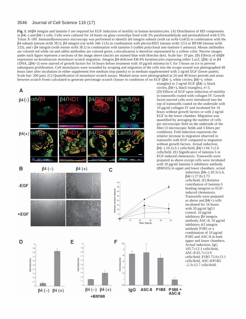
Resultsα6β4 integrin and laminin-5 are essential for EGFinduced migration in keratinocytesWe utilized α6β4 integrin null EB-PA keratinocytes to studyα6β4 integrin in keratinocyte migration. Control vector (LacZ)or full-length β4 integrin were retrovirally expressed in EB-PAcells to create β4(–) and β4(+) cells respectively. β4(–) cellsshowed normal laminin-5 secretion but no detectable β4integrin and the HD proteins BP180, BP230 and HD1/plectinwere diffusely localized (Fig. 1A top panels; BP230 notshown). In contrast, β4(+) cells basally accumulated β4integrin which co-localized with α6 integrin, HD1/plectin,BP180, BP230 and laminin-5 (Fig. 1A bottom panels; BP230not shown). Flow cytometry showed that β4(+) cells expressedcell surface β4 integrin at a level comparable to normalkeratinocytes (73.2±12.2% of control, n=3).
Without EGF, neither β4(–) nor β4(+) cells migrated in amonolayer scratch assay (Fig. 1B,C; <10.9±5.6% scratchclosure over 48 hours), however upon addition of EGF onlyβ4(+) cells migrated significantly into the wound scratch(β4(–) 10.9±5.6% closure vs. β4(+), 35.3±1.6% scratch closureover 48 hours, P<0.05). Similar observations were obtainedwhen migration was assayed using ECM-coated transwellchambers. EGF also induced the migration of β4(+) cells butnot β4(–) cells across transwells coated with collagen IV,collagen I, fibronectin or laminin-1 (Fig. 1D, collagen IVshown, overall fold induction: β4(+) 2.04±0.18 versus β4(–)1.08±0.03). These ECM substrates are not ligands for α6β4integrin (with the exception of laminin-1), therefore weinvestigated whether interactions between α6β4 integrin andautocrine laminin-5 were responsible for mediating cellmigration. In the presence of BM165, an antibody to laminin-5 that inhibits cell adhesion, EGF-induced migration of β4(+)cells was significantly reduced, suggesting ligation of β4integrin by laminin-5 was necessary for EGF-inducedkeratinocyte chemotaxis (Fig. 1E, collagen IV shown). Similarresults were also obtained with collagen I, fibronectin and,laminin-1 (data not shown).
To identify the cellular receptors for laminin-5 responsiblefor mediating EGF-induced chemotaxis in keratinocytes, thetranswell assays were repeated using β4(+) cells in thepresence of α6β4 integrin (mAb ASC-8) and α3β1 integrin(mAb P1B5) inhibitory antibodies or an IgG control (Fig. 1F).Collagen IV was selected as a migration substrate as it isneither a ligand for α6β4 nor for α3β1 integrin. These studiesshowed that although β4 integrin expression is necessary forattachment, either α6β4 or α3β1 integrin is sufficient for EGF-induced chemotaxis in keratinocytes. Thus, both α6β4 integrinand laminin-5 expression are essential for EGF-induced
3546 Journal of Cell Science 116 (17)
Fig. 1.α6β4 integrin and laminin-5 are required for EGF induction of motility in human keratinocytes. (A) Distribution of HD componentsin β4(–) and β4(+) cells. Cells were cultured for 24 hours on glass coverslips fixed with 3% paraformaldehyde and permeabilized with 0.5%Triton X-100. Immunofluorescence microscopy was performed to identify α6 integrin subunit (with rat mAb GoH3) in combination with theβ4 subunit (mouse mAb 3E1); β4 integrin (rat mAb 346-11A) in combination with plectin/HD1 (mouse mAb 121) or BP180 (mouse mAb233), and r β4 integrin (with mouse mAb 3E1) in combination with laminin-5 (rabbit polyclonal anti-laminin-5 antisera). Mouse antibodiesare colored red while rat and rabbit antibodies are colored green, colocalization is therefore represented by a yellow color. Narrow imagesunder each figure represent z-sections of the image above (nuclei are stained blue with Hoechst dye). Scale bar: 10 µm. (B) Effects of α6β4expression on keratinocyte monolayer scratch migration. Integrin β4-deficient EB-PA keratinocytes expressing either LacZ, [β4(–)] or β4cDNA, [β4(+)] were starved of growth factors for 16 hours before treatment with 10 µg/ml mitomycin C for 3 hours on ice to preventsubsequent proliferation. Cell monolayers were wounded by scraping and migration of the cells into the scrape wound was photographed 48hours later after incubation in either supplement free medium (top panels) or in medium supplemented with 2 ng/ml EGF (lower panels;Scale bar: 200 µm). (C) Quantification of monolayer scratch assays. Marked areas were photographed at 24 and 48 hours periods and areasbetween scratch fronts calculated to generate percentage scratch closure in conditions of no EGF (β4(–), white circles; β4(+), white
triangles) or 2 ng/ml EGF (β4(–), blackcircles; β4(+), black triangles), n=3.(D) Effects of EGF upon induction of motilityin transwells coated with collagen IV. Growthfactor-starved cells were introduced into thetop of transwells coated on the underside with10 µg/ml collagen IV and incubated for 16hours without growth factors or with 2 ng/mlEGF in the lower chamber. Migration wasquantified by averaging the number of cellsper microscopic field on the underside of thefilter (3 microscopic fields and 3 filters percondition). Fold induction represents therelative increase in migration observed intranswells with EGF compared to migrationwithout growth factors. Actual induction,β4(–) 10.3±3.1 cells/field, β4(+) 94.7±2.6cells/field. (E) Significance of laminin-5 inEGF-induced chemotaxis. Transwells wereprepared as above except cells were incubatedwith 10 µg/ml laminin-5 inhibitory antibody(BM165) in upper and lower chambers. actual
induction, β4(–) 20.3±5.4,β4(+) 27.8±3.73cells/field. (F) Relativecontribution of laminin-5binding integrins to EGF-induced chemotaxis.Transwells were preparedas above and β4(+) cellsincubated for 16 hourswith 20 µg/ml IgG1control, 10 µg/mlinhibitory β4 integrinantibody ASC-8, 10 µg/mlinhibitory α3 integrinantibody P1B5 or acombination of 10 µg/mlP1B5 and ASC-8 in bothupper and lower chambers.Actual induction, IgG105.7±12.1 cells/field,ASC-8 65.7±13.9cells/field, P1B5 72.0±13.1cells/field, ASC-8/P1B5–2.3±12.7 cells/field.

3547α6β4 integrin in keratinocyte chemotaxis
chemotaxis in keratinocytes but this process can be mediatedby laminin-5 induced ligation of either α6β4 or α3β1 integrin.
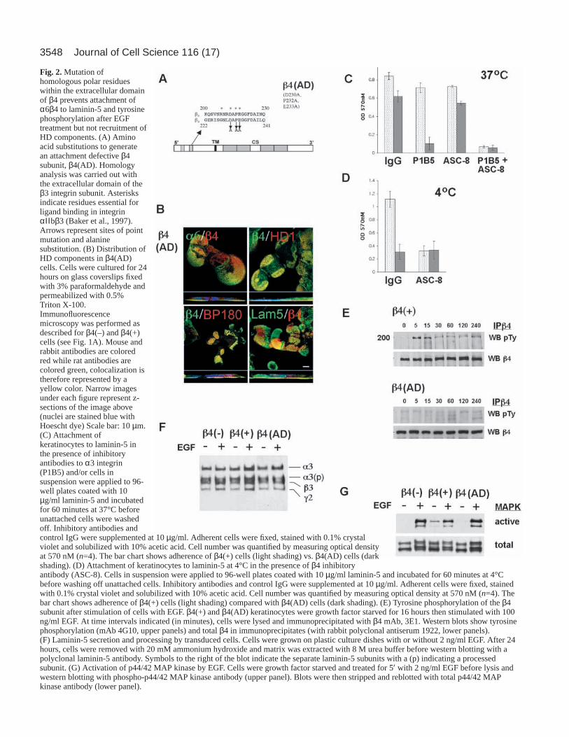
Mutation of β4 integrin extracellular domain permitsrecruitment of HD components but prevents laminin-5attachment and EGF induced β4 integrinphosphorylationTo further examine the contribution of α6β4 integrin ligationto EGF induced chemotaxis we designed and expressed anattachment-defective β4 integrin mutant in the EB-PA cells. Anextracellular ligand-binding mutant of β4 integrin wasdesigned according to published data that identified tworegions in the β3 integrin extracellular domain that wereessential for attachment in the platelet integrin αIIbβ3(asterisks, Fig. 2A) (Baker et al., 1997). Homology analysis ofthe exodomains of β3 and β4 integrins revealed a high level ofconservation between these two ligand attachment regions.Accordingly, a mutant β4 cDNA construct was engineered thatincorporated three substitutions within the second homologydomain at D230A, P232A and E233A and termed adhesiondefective β4, or β4(AD).
EB-PA cells expressing β4(AD) had strong basal expressionof the β4 integrin that co-localized in type I HD clusters withα6 integrin, plectin, BP180 and secreted laminin-5, in a patternthat was similar to that exhibited by the β4(+) cells (Fig. 2B).Recruitment of HD components by β4(AD) is in agreementwith previous reports that β4 integrin recruitment to HDs isdriven by cytoplasmic interactions with BP180 and plectin andnot by α6β4 attachment to laminin-5 (Homan et al., 1998;Nievers et al., 2000; Nievers et al., 1998).
β4(AD) and β4(+) cells were studied by attachment assaysusing affinity purified laminin-5 (Fig. 2C). Contributions ofα6β4 and α3β1 integrin to adhesion were analyzed usinginhibitory antibodies (ASC8; α6β4 integrin, and P1B5; α3β1integrin, respectively). Both cell types attached at comparablelevels to laminin-5, however inhibition of α3β1 integrincompletely prevented β4(AD) attachment while not affectingβ4(+) cells. Note that these assays measure substrateattachment and not the strength of substrate attachment, whichcould be enhanced in β4(+) cells. α6β4 integrin can uniquelymediate attachment to laminin-5 even at 4°C (Xia et al., 1996).While β4(+) cells could attach effectively at 4°C, β4 (AD) cellscould not (Fig. 4D). However, pre-incubating β4(+) cells withβ4 integrin inhibitory antibody (ASC-8) reduced the adhesionlevel of the β4 (+) cells at 4°C to the same level exhibited byβ4(AD) cells. These studies demonstrate that although β4(AD)cells fail to adhere through α6β4 integrins they retain normalfunction of α3β1 integrin. We conclude that the adhesiondefect of β4(AD) cells is a consequence of direction mutationof the extracellular domain of the β4 integrin subunit ratherthan of a non-specific effect upon α3β1 integrin expression orfunction.
α6β4 integrin becomes tyrosine phosphorylated followingstimulation with high concentrations of EGF (Mainiero et al.,1996). To further evaluate the ligand binding characteristics ofour β4 integrin mutant (AD) cells, we tested the ability of EGFto induce tyrosine phosphorylation of β4 integrin. AlthoughEGF induced phosphorylation of β4 integrin in β4(+) cells, nophosphorylation of β4 integrin was observed in β4(AD) cells(Fig. 2E, compare top panel to the lower panel). All three cell
types secreted and processed laminin-5 to a similar degreeregardless of EGF treatment (Fig. 2F). Finally, we examinedthe possibility that expression of the β4(AD) mutant inducednon-specific effects on EGFR expression or signaling. Flowcytometry of surface EGFR of β4(+) compared with β4(AD)cells revealed similar levels of expression (mean fluorescence162.1±10.6 vs. 199.4±11.6). Signaling by the EGFR was testedby examination of the effects of EGF on MAP kinase activation(Fig. 2G). Treatment of all cell types resulted in strongactivation of p44/42 MAP kinase. Separate studies alsoconfirmed that the kinetics of this activation was unchanged(data not shown). Taken together, these experiments show thatmutation of conserved extracellular residues within the β4subunit prevents attachment to laminin-5 and inhibits ligation-dependent tyrosine phosphorylation of β4 integrin. However,loss of attachment function does not prevent β4 integrin frommediating its other cellular functions including the recruitmentof HD components.
Ligation of α6β4 integrin is required for sustainedactivation of Rac1, lamellipodia formation and RhoA-independent chemotaxisMembers of the Rho GTPase family drive chemotaxis by EGFin many cell types (Nobes and Hall, 1995). Therefore, weexamined the effects of α6β4 expression and ligation uponEGF-dependent Rho GTPase activity (Fig. 3A,B). β4(–) cellsshowed transient EGF induced stimulation of Rac1 activity andmodest activation of Cdc42. Expression of wild-type α6β4integrin resulted in rapid and sustained activation of both Rac1and Cdc42 (for at least 2 hours). In contrast, expression ofβ4(AD) resulted in a truncated Rac1 activation profile similarto β4(–) cells. Interestingly, while β4(–) and β4(+) cellsexhibited similar EGF-dependent RhoA activity β4(AD) cellsshowed higher RhoA activation both before and after EGFtreatment (Fig. 3B). Thus we concluded that α6β4 expressionand ligation are essential for sustained EGF-dependent Rac1and Cdc42 activation. Interestingly, in the absence of α6β4integrin laminin-5 interactions, Rac1/Cdc42 activation istruncated whereas RhoA activity appears to be amplified.
Since Rac1 induces membrane ruffling and lamellipodiaextension, structures of known importance to cell migration(Lauffenburger and Horwitz, 1996; Mitchison and Cramer,1996), we next investigated the relevance of α6β4 integrin-dependent Rac1 activation to lamellipodia formation after EGFexposure. Cells were treated with EGF and fixed at timeintervals following EGF stimulation and lamellipodial area wasquantified (Fig. 3C). EGF induced membrane extension in allcells tested, and this effect peaked after 5 minutes. However,β4(–) and β4(AD) cells failed to sustain lamellipodia inductionbeyond 20 minutes. In contrast, β4(+) cells maintainedlamellipodia for at least 2 hours following EGF exposure.Therefore, sustained lamellipodia formation in β4(+) cellsmirrors the activation of Rac1 in that it requires both α6β4integrin expression and ligation to laminin-5. To more directlytest this observation, NHK were incubated with β4 inhibitoryantibody (ASC-8) and lamellipodial induction was measuredin response to EGF (Fig. 3D). Consistently, inhibition of α6β4integrin ligation markedly truncates sustained lamellipodiaformation similar to that observed in β4(–) and β4(AD) cells.
To further explore the significance of Rac1 activation in
3548 Journal of Cell Science 116 (17)
Fig. 2.Mutation ofhomologous polar residueswithin the extracellular domainof β4 prevents attachment ofα6β4 to laminin-5 and tyrosinephosphorylation after EGFtreatment but not recruitment ofHD components. (A) Aminoacid substitutions to generatean attachment defective β4subunit, β4(AD). Homologyanalysis was carried out withthe extracellular domain of theβ3 integrin subunit. Asterisksindicate residues essential forligand binding in integrinαΙΙ bβ3 (Baker et al., 1997).Arrows represent sites of pointmutation and alaninesubstitution. (B) Distribution ofHD components in β4(AD)cells. Cells were cultured for 24hours on glass coverslips fixedwith 3% paraformaldehyde andpermeabilized with 0.5%Triton X-100.Immunofluorescencemicroscopy was performed asdescribed for β4(–) and β4(+)cells (see Fig. 1A). Mouse andrabbit antibodies are coloredred while rat antibodies arecolored green, colocalization istherefore represented by ayellow color. Narrow imagesunder each figure represent z-sections of the image above(nuclei are stained blue withHoescht dye) Scale bar: 10 µm.(C) Attachment ofkeratinocytes to laminin-5 inthe presence of inhibitoryantibodies to α3 integrin(P1B5) and/or cells insuspension were applied to 96-well plates coated with 10µg/ml laminin-5 and incubatedfor 60 minutes at 37°C beforeunattached cells were washedoff. Inhibitory antibodies andcontrol IgG were supplemented at 10 µg/ml. Adherent cells were fixed, stained with 0.1% crystalviolet and solubilized with 10% acetic acid. Cell number was quantified by measuring optical densityat 570 nM (n=4). The bar chart shows adherence of β4(+) cells (light shading) vs. β4(AD) cells (darkshading). (D) Attachment of keratinocytes to laminin-5 at 4°C in the presence of β4 inhibitoryantibody (ASC-8). Cells in suspension were applied to 96-well plates coated with 10 µg/ml laminin-5 and incubated for 60 minutes at 4°Cbefore washing off unattached cells. Inhibitory antibodies and control IgG were supplemented at 10 µg/ml. Adherent cells were fixed, stainedwith 0.1% crystal violet and solubilized with 10% acetic acid. Cell number was quantified by measuring optical density at 570 nM (n=4). Thebar chart shows adherence of β4(+) cells (light shading) compared with β4(AD) cells (dark shading). (E) Tyrosine phosphorylation of the β4subunit after stimulation of cells with EGF. β4(+) and β4(AD) keratinocytes were growth factor starved for 16 hours then stimulated with 100ng/ml EGF. At time intervals indicated (in minutes), cells were lysed and immunoprecipitated with β4 mAb, 3E1. Western blots show tyrosinephosphorylation (mAb 4G10, upper panels) and total β4 in immunoprecipitates (with rabbit polyclonal antiserum 1922, lower panels).(F) Laminin-5 secretion and processing by transduced cells. Cells were grown on plastic culture dishes with or without 2 ng/ml EGF. After 24hours, cells were removed with 20 mM ammonium hydroxide and matrix was extracted with 8 M urea buffer before western blotting with apolyclonal laminin-5 antibody. Symbols to the right of the blot indicate the separate laminin-5 subunits with a (p) indicating a processedsubunit. (G) Activation of p44/42 MAP kinase by EGF. Cells were growth factor starved and treated for 5′ with 2 ng/ml EGF before lysis andwestern blotting with phospho-p44/42 MAP kinase antibody (upper panel). Blots were then stripped and reblotted with total p44/42 MAPkinase antibody (lower panel).
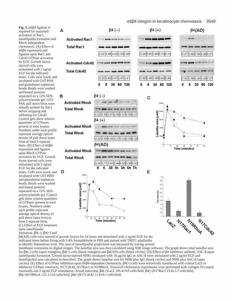
3549α6β4 integrin in keratinocyte chemotaxisFig. 3.α6β4 ligation isrequired for sustainedactivation of Rac1,lamellipodia formation andRhoA independentchemotaxis. (A) Effect ofα6β4 expression andligation upon Rac1 andCdc42 GTPase activationby EGF. Growth factor-starved cells werestimulated with 2 ng/mlEGF for the indicatedtimes. Cells were lysed, andincubated with GST-PAKand glutathione-sepharosebeads. Beads were washedand bound proteinsseparated on a 12% SDS-polyacrylamide gel. GST-PAK pull down blots wereinitially probed for Rac1before stripping andreblotting for Cdc42.Control gels show relativequantities of GTPasespresent in total lysates.Numbers under each profilerepresent average opticaldensity of pull down lanesfrom at least 2 separateblots. (B) Effect of α6β4expression and ligationupon RhoA GTPaseactivation by EGF. Growthfactor starved cells werestimulated with 2 ng/mlEGF for the indicatedtimes. Cells were lysed, andincubated with GST-RBDand glutathione-sepharosebeads. Beads were washedand bound proteinsseparated on a 12% SDS-polyacrylamide gel. Controlgels show relative quantitiesof GTPases present in totallysates. Numbers undereach profile representaverage optical density ofpull down lanes from atleast 2 separate blots.(C) Effect of EGF treatmentupon lamellipodiaformation. β4(–), β4(+) andβ4(AD) cells were starved of growth factors for 16 hours and stimulated with 2 ng/ml EGF for theindicated times before fixing with 3.4% formaldehyde in PBS and stained with TRITC-phalloidinto identify filamentous actin. The area of lamellipodial projections was measured by tracing aroundmembrane extensions on digital images. The lamellar area was then calculated using NIH image software. The graph shows total lamellar areafor β4(–) cells (open triangles), β4(+) cells (black triangles) and β4(AD) cells (black circles). (D) Effect of β4 inhibitory antibody ASC-8 uponlamellipodia formation. Growth factor-starved NHKs incubated with 10 µg/ml IgG or ASC-8 were stimulated with 2 ng/ml EGF andlamellipodial area calculated as described. The graph shows lamellar area for NHK plus IgG (black circles) and NHK plus ASC-8 (opencircles). (E) Effect of GTPase inhibition upon α6β4-dependent chemotaxis. β4(+) cells were retrovirally transduced with control LacZ orinhibitory GTPase constructs, N17Cdc42, N17Rac1 or N19RhoA. Transwell chemotaxis experiments were performed with collagen IV-coatedtranswells and 2 ng/ml EGF stimulation. Actual induction, β4(+)LacZ 106.4±9.0 cells/field, β4(+)N17Rac1 23.8±3.7 cells/field,β4(+)N19RhoA 121.1±3.0 cells/field, β4(+)N17Cdc42 11.8±9.1 cells/field.

3550
keratinocyte chemotaxis, we retrovirally expressed dominantinhibitory forms of GFP-tagged N17Rac1, N17Cdc42 orN19RhoA in β4(+) cells and tested for effects on chemotaxisusing the transwell assay (expression verified by western blotand immunofluorescence microscopy, data not shown).Transwell assays conducted with collagen IV revealedsignificant inhibition of EGF-induced chemotaxis in β4(+)cells after N17Rac1 or N17Cdc42 were expressed (Fig. 3D).However, expression of N19RhoA did not inhibit induction.These results suggest that expression and ligation of α6β4integrin is required for sustained stimulation of Rac1,lamellipodia formation and chemotaxis in a process thatappears to be independent of activated RhoA.
Attachment defective α6β4 integrin undergoeschemotaxis through an alternate pathway involvingRhoA and integrin α3β1We observed that expression and ligation of α6β4 integrin isrequired for sustained activation of Rac1 and lamellipodiaformation. However, we previously observed that blockingα6β4 ligation with the inhibitory antibody ASC-8 does notblock chemotaxis of β4(+) cells (Fig. 1F). We therefore usedβ4(AD) cells and asked whether the absence of α6β4 integrinligation results in chemotaxis through an alternative EGF-dependent mechanism. In both monolayer scratch (Fig. 4A, 24-hour time point shown, full closure in 48 hours; n=3) andtranswell migration assays (using collagen I, collagen IV,fibronectin or laminin-1; Fig. 4B, average fold induction6.16±1.51, collagen IV shown), EGF-induced chemotaxis wasenhanced in β4(AD) cells. To ascertain the contribution oflaminin-5 and integrin α3β1 to this process we repeated thetranswell assay in the presence of inhibitory antibodies.Treatment of β4(AD) cells with either laminin-5 or α3β1integrin antibodies inhibited the chemotactic response to EGF(Fig. 4B), indicating that laminin-5-α3β1 integrin interactionsare required for chemotaxis in β4(AD) cells.
The GTPase activation profile of β4(AD) cells showedelevated levels of RhoA activation before and after treatmentwith EGF. We expressed inhibitory Rac1, RhoA and Cdc42constructs in β4(AD) cells to determine whether changes inGTPase activation profiles reflected altered Rho family GTPaserequirements for β4(AD) chemotaxis. Chemotaxis wasuniformly inhibited by N17Rac1, N17Cdc42 and N19Rhoexpression (Fig. 4C) suggesting cell motility in response toEGF was now also dependent upon RhoA. Activation of RhoAin epithelial cells is often associated with cell scattering(Sander et al., 1999). In support of a role for RhoA in β4(AD)migration, β4(AD) cells appeared to migrate predominantly asindividual cells following stimulation with EGF as opposed tomigrating as an intact sheet of cells (Fig. 4A). The scatteringphenotype of β4(AD) cells was quantified by examiningcolony formation following growth in normal EGF-supplemented medium (Fig. 4D). Four days after plating (5000cells per 60 mm plate) β4(AD) cells showed extensive colonyscattering (65.3±1.6% of total colonies), while β4(–) and β4(+)cells predominantly formed epithelial colonies with intact cell-cell interactions (18.2±4.2% and 23.1±4.2% scatteredrespectively).
To verify that the scattering effects that we observed wereindeed due to loss of β4 integrin adhesion, colony scattering
experiments were carried out using β4(+) cells treated with theβ4 integrin inhibitory antibody (ASC-8) following 16 hours ofEGF treatment (Fig. 4E). β4 (+) cells incubated with the β4integrin adhesion blocking antibody (ASC-8) exhibitedincreased cell scattering (P<0.05; Fig. 4E, right), similar to thatexhibited by β4(AD) cells, although the scattering wasmaintained for a shorter duration, possibly due to antibodyinternalization and turnover.
These data illustrated that while ligation of α6β4 mediatesEGF induced chemotaxis through Rac1, in the absence of α6β4ligation (but not expression) chemotaxis appears to bemediated through an alternative pathway that depends uponα3β1 integrin and utilizes RhoA. If true then in the absence ofβ4 integrin ligand binding, loss of RhoA activity should inhibitEGF-induced keratinocyte migration. We tested this hypothesisby conducting transwell assays in the presence of β4 integrinligand blocking antibody (ASC-8) using β4(+) cells thatexpressed a dominant-negative RhoA (Fig. 4F). As anticipated,when α6β4 integrin ligation is prevented, EGF-inducedchemotaxis is decreased, suggesting an increased dependencyon RhoA for migration upon inhibition of α6β4 ligation.
Expression and ligation of α6β4 integrin change thedistribution and conformational activation of α3β1integrinOur studies thus far suggested that EGF-induced keratinocytechemotaxis through α3β1 integrin becomes altered followingexpression and ligation of α6β4 integrin. Additional studieswere therefore conducted to further examine the effects ofα6β4 integrin on α3β1 integrin function in our keratinocytemodel. Since reports have suggested that keratinocyteimmortalization with HPV18 E6 and E7 genes might alteradhesion and cytoskeletal organization (Nguyen et al., 2000a),we also conducted studies using retrovirally transducedprimary cells isolated from a second EB-PA patient. Becauseα3β1 integrin regulates the actin cytoskeleton, we firstanalyzed the effect of expression and ligation of α6β4 integrinon the distribution of actin and the FA component paxillinusing immunofluorescence. β4(–) cells had diffusely organizedactin and paxillin (Fig. 5A) with basally distributed α3β1integrin (Fig. 5D). In contrast, β4(+) cells displayed organized,cortical stress fibers and FAs (Fig. 5B) with basal laterallydistributed α3β1 integrin (Fig. 5E), comparable to normalkeratinocytes (Symington et al., 1993). Importantly, whencompared with β4(+) cells, β4(AD) cells had reduced α3β1integrin at sites of cell-cell contact and exhibited prominentstress fibers, increased focal adhesions (which is a phenotypethat is consistent with enhanced RhoA activity; Fig. 5C) andclustered basal α3β1 integrin (Fig. 5F). Furthermore, parallelcultures immunostained with the β1 integrin activation specificantibody, HUTS-4, suggested that these cells had a notableincrease in β1 integrin activity (Fig. 5I). In contrast, β4(–) cellshad more punctate basal β1 integrin activation (Fig. 5G) andβ4(+) cells had only limited peripheral staining e(Fig. 5H),similar to normal keratinocytes (Penas et al., 1998). Usingimmunofluorescence, we compared the distribution of β1integrin binding partners (α1, α2, α3, α4, α5) to determine thespecific contribution of different heterodimer partners to thisactivation (data not shown). Data showed that only α3 integrinrelocalized from sites of cell-cell to cell-matrix adhesions in
Journal of Cell Science 116 (17)

3551α6β4 integrin in keratinocyte chemotaxis
association with activated β1 integrin in β4(AD) cells. Thesedata suggested that the β4(AD) cell phenotype is potentiallylinked to enhanced α3β1 integrin activation at the basalmembrane of cells, which is consistent with focal adhesions.
α6β4 integrin controls relocalization and inactivation ofα3β1 integrin through Rac1 activation and suppressionof RhoAOur data indicate that chemotaxis of β4(AD) cells requires
Fig. 4.EGF stimulates enhancedchemotaxis and reduced epithelialintegrity in β4(AD) cells dependentupon an alternate pathway utilizingRhoA and integrin α3β1. (A) Effect ofβ4(AD) expression upon monolayerscratch migration. Cells were preparedas in Fig. 1A and incubated withoutgrowth factors (top panel) or with 2ng/ml EGF (lower panel) for 24 hoursat 37°C. Scale bar: 300 µm. (B) Effectof β4(AD) expression upon transwellchemotaxis. Transwell experimentswith β4(AD) keratinocytes wereperformed with collagen IV-coatedtranswells and 2 ng/ml EGF. Cells wereincubated for 16 hours with mediasupplemented in upper and lowerchambers with IgG1, inhibitory β4integrin antibody ASC-8, inhibitory α3integrin antibody P1B5 or inhibitorylaminin-5 antibody BM165. Actualinduction IgG 60.0±4.2 cells/field,ASC-8 60.0±4.4 cells/field, P1B510.0±1.2 cells/field, BM165 16.1±1.1cells/field. (C) Effect of GTPaseinhibition upon β4(AD)-dependentchemotaxis. β4(AD) cells wereretrovirally transduced with controlLacZ or inhibitory GTPase constructs,N17Cdc42, N17Rac1 or N19RhoA.Chemotaxis experiments wereperformed with collagen IV-coatedtranswells and 2 ng/ml EGF. Actualinduction, β4(AD)LacZ 136±23.1cells/field, β4(AD)N17Rac1 15.0±3.7cells/field, β4(AD)N19RhoA 21.0±3.6cells/field, β4(AD)N17Cdc42 8.4±2.7cells/field. (D) Effect of β4(AD)expression upon cell scattering. Cellswere incubated at low density for 4days in SFM before beingphotographed under phase contrastillumination. Scale bar: 40 µm.(E) Effect of EGF upon cell scattering.Cells were grown in growth factor freeSFM for 4 days and scattered coloniescounted using criteria described inmaterials and methods (light shadedcolumns). Cells were then incubatedfor 16 hours with 2 ng/ml EGF andcolony scatter counts repeated (darkshaded columns). For antibodytreatments, β4(+) cells were incubatedfor 3 days in normal SFM then 1 day in growth factor free SFM with 10 µg/ml mouse IgG or β4 integrin inhibitor ASC-8, before colony counts(light shaded columns). Colony scattering was measured after 6 hours with 2 ng/ml EGF (dark shaded columns). Each point represents the datafrom at least 50 colonies (n=3). (F) Effect of N19RhoA expression upon β4(+) transwell chemotaxis. Transwell experiments with β4(+)LacZand β4(+)N19RhoA keratinocytes were performed with fibronectin-coated transwells and 2 ng/ml EGF. Cells were incubated for 16 hours withmedia supplemented in upper and lower chambers with IgG1 or inhibitory β4 integrin antibody ASC-8. Actual induction, β4(+)LacZ IgG116.5±18.5 cells/field, β4(+)LacZ ASC-8 113.5±16.9 cells/field, β4(+)N19RhoA IgG 77.7±7.5 cells/field, β4(+)N19RhoA ASC-8 34±21.3cells/field.

3552
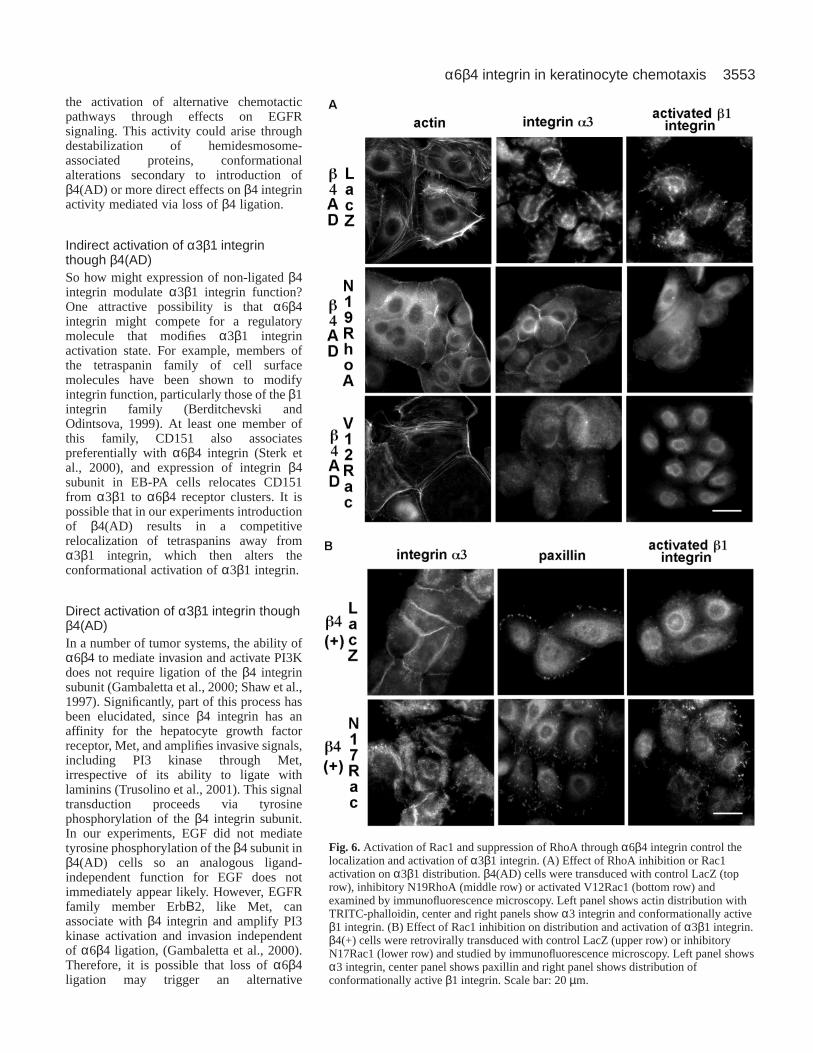
RhoA. RhoA is essential for the formation of actin stress fibersand focal adhesions and its functions can be antagonized byRac1 (Nimnual et al., 2003; Sander et al., 1999). We thereforeasked whether RhoA could play a role in the basal clusteringand activation of α3β1 integrin and if this could be antagonizedby Rac1 activation through α6β4 integrin. We expressedinhibitory N19RhoA or activated V12Rac1 in β4(AD) cells andrecorded their impact upon the α3β1 integrin activation andactin cytoskeletal organization. Both inhibitory RhoA (Fig. 6A,middle row) and activated Rac1 (Fig. 6A, bottom row)prevented stress fiber formation, basal clustering of α3β1integrin and α3β1 integrin activation. In addition, expressionof N19RhoA induced relocalization of α3β1 integrin to sitesof cell-cell contact while activated Rac1 enhanced corticalactin staining, reminiscent of its status in β4(+) cells.(Interestingly, expression of N19RhoA in β4(–) cells was notsufficient to alter the basal distribution of α3β1 integrin, datanot shown.) These studies lead us to conclude that thecytoskeletal phenotype of β4 (AD) cells is dependent uponRhoA and can be inhibited by activating Rac1. Our previousexperiments indicated that α6β4 ligation permits a sustainedEGF-dependent activation of Rac1. We therefore askedwhether inhibition of Rac1 in β4(+) cells could recapitulate anyof the cytoskeletal characteristics exhibited by β4(AD) cells (Fig. 6B). Expression of inhibitoryN17Rac1 in β4(+) cells enhanced FA formation, basalclustering of α3β1 and β1 integrin activation (Fig. 6B,bottom panels).
In conclusion, these studies showed that α6β4integrin ligation regulates the cellular localization ofa3b1 integrin and tempers its activity by potentiatingRac1 activity. The coordination of this process isessential for facilitating EGF-induced chemotaxis viaregulation of Rho GTPase cross-talk.
DiscussionSoluble EGFR ligands increase during wound healing(Marikovsky et al., 1993) and this is essential for re-epithelialization (Tokumaru et al., 2000). Our studiesimplicate α6β4 integrin as a primary control point fortranslation of EGF stimulation into keratinocytemigration. By rescuing the β4 integrin subunit in β4-null EB-PA cells we were able to show that α6β4integrin and laminin-5 expression are essential forEGF-induced chemotaxis. However, the relationshipbetween α6β4 expression and chemotactic signaltransduction is complex and appears to be controlledby the ligand bound status of the β4 integrin subunit.
We found that ligation of α6β4 integrin drovechemotaxis by sustained activation of the Rho familyGTPase Rac1. EGF stimulation may result in Rac1activation through Fyn-mediated tyrosinephosphorylation of the β4 integrin cytoplasmicdomain (Mainiero et al., 1996; Mariotti et al., 2001).However, at the physiological levels of EGF used inour chemotaxis assays we did not observe tyrosinephosphorylation of the β4 subunit. Alternately, Rac1activation may be potentiated through increasedphosphoinositide 3 kinase (PI3 kinase) activityfollowing α6β4 ligation (Nobes and Hall, 1995; Shaw
et al., 1997). Sustained Rac1 activation appears to redirectα3β1 integrin away from basal focal contacts and towards sitesof cell-cell contact thereby functioning to temper its activationstate. This implies that α6β4 integrin antagonizes α3β1integrin through Rac1 to ultimately modulate signaltransduction and thereby regulating cell motility (summarizedin Fig. 7).
In the absence of β4 integrin ligation, Rac1 activation is nolonger sustained and keratinocytes migrate through analternative chemotactic pathway that depends upon α3β1integrin and RhoA. The nature of this migration is also lessdirected and cells exhibit scattering. Interestingly, recent datahave shown that introduction of an attachment-defectiveEGFP/β4 fusion into EB-PA cells also increased keratinocytemigration (Geuijen and Sonnenberg, 2002). This increase wasapparently associated with a destabilization of the link betweenlaminin-5 and the cytoskeleton through plectin. Although asimilar situation may also occur in β4(AD) cells, this does notexplain how EGF chemotaxis is restored by β4(AD)expression. In this regard, our data show that while β4(–) cellsexhibit basal clustering of α3β1 integrin, α3β1 activity isreduced and they fail to undergo chemotaxis in response toEGF stimulation. Thus, expression of β4(AD) must facilitate
Journal of Cell Science 116 (17)
Fig. 5.Expression and ligation of α6β4 integrin changes the localization andactivation state of α3β1 integrin. Cells were cultured on glass coverslips for 4days prior to immunofluorescence staining for FA components. Cells werefixed with 3% formaldehyde and solubilized with 0.5% Triton X-100 buffer for30 minutes at RT. Fixed cells were washed with PBS and blocked with 1%BSA for 1 hour. Cells were stained with FITC-phalloidin for filamentous actin(stained in green A-C) and anti-paxillin mAb (stained in red A-C). Furthersamples were stained for α3 integrin (D-F) and the conformationally activeform of the β1 integrin subunit with mAb HUTS-4 (G-I). Scale bar; 20 µm.
3553α6β4 integrin in keratinocyte chemotaxis
the activation of alternative chemotacticpathways through effects on EGFRsignaling. This activity could arise throughdestabilization of hemidesmosome-associated proteins, conformationalalterations secondary to introduction ofβ4(AD) or more direct effects on β4 integrinactivity mediated via loss of β4 ligation.
Indirect activation of α3β1 integrinthough β4(AD)So how might expression of non-ligated β4integrin modulate α3β1 integrin function?One attractive possibility is that α6β4integrin might compete for a regulatorymolecule that modifies α3β1 integrinactivation state. For example, members ofthe tetraspanin family of cell surfacemolecules have been shown to modifyintegrin function, particularly those of the β1integrin family (Berditchevski andOdintsova, 1999). At least one member ofthis family, CD151 also associatespreferentially with α6β4 integrin (Sterk etal., 2000), and expression of integrin β4subunit in EB-PA cells relocates CD151from α3β1 to α6β4 receptor clusters. It ispossible that in our experiments introductionof β4(AD) results in a competitiverelocalization of tetraspanins away fromα3β1 integrin, which then alters theconformational activation of α3β1 integrin.
Direct activation of α3β1 integrin thoughβ4(AD)In a number of tumor systems, the ability ofα6β4 to mediate invasion and activate PI3Kdoes not require ligation of the β4 integrinsubunit (Gambaletta et al., 2000; Shaw et al.,1997). Significantly, part of this process hasbeen elucidated, since β4 integrin has anaffinity for the hepatocyte growth factorreceptor, Met, and amplifies invasive signals,including PI3 kinase through Met,irrespective of its ability to ligate withlaminins (Trusolino et al., 2001). This signaltransduction proceeds via tyrosinephosphorylation of the β4 integrin subunit.In our experiments, EGF did not mediatetyrosine phosphorylation of the β4 subunit inβ4(AD) cells so an analogous ligand-independent function for EGF does notimmediately appear likely. However, EGFRfamily member ErbΒ2, like Met, canassociate with β4 integrin and amplify PI3kinase activation and invasion independentof α6β4 ligation, (Gambaletta et al., 2000).Therefore, it is possible that loss of α6β4ligation may trigger an alternative
Fig. 6.Activation of Rac1 and suppression of RhoA through α6β4 integrin control thelocalization and activation of α3β1 integrin. (A) Effect of RhoA inhibition or Rac1activation on α3β1 distribution. β4(AD) cells were transduced with control LacZ (toprow), inhibitory N19RhoA (middle row) or activated V12Rac1 (bottom row) andexamined by immunofluorescence microscopy. Left panel shows actin distribution withTRITC-phalloidin, center and right panels show α3 integrin and conformationally activeβ1 integrin. (B) Effect of Rac1 inhibition on distribution and activation of α3β1 integrin.β4(+) cells were retrovirally transduced with control LacZ (upper row) or inhibitoryN17Rac1 (lower row) and studied by immunofluorescence microscopy. Left panel showsα3 integrin, center panel shows paxillin and right panel shows distribution ofconformationally active β1 integrin. Scale bar: 20 µm.

3554
chemotactic pathway through EGFR that is independent oftyrosine phosphorylation.
Potential significance of EGFR signaling through α6β4integrin during wound healingPrevious studies have ruled out a significant function for α6β4integrin in epithelial migration because inhibitory antibodies toα6 or β4 fail to markedly impair chemotaxis or wound healing(Goldfinger et al., 1999; Hintermann et al., 2001; Nguyen etal., 2000b). We suggest that inhibition of α6β4 ligation resultsin the activation of a secondary chemotactic pathway that isdependent upon α3β1 integrin.
What would be the significance of this antagonsticrelationship between α6β4 and α3β1 integrins to migratingkeratinocytes during wound healing? Studies have shown thatkeratinocytes rely on two distinct pathways for attachment andspreading as cells move across the provisional dermal collagenmatrix (Nguyen et al., 2000a). Cells at the wound front in vivoare dependent upon Rho family GTPases for attachment whilecells distal from the wound edge mediate attachment andspreading via ligation of α6β4 to secreted laminin-5 and PI3kinase activation. These differences in integrin activity andsignaling have been attributed to changes in ECM compositionfrom dermal collagen to laminin-5. In light of our currentobservations this model can now be integrated with theobserved functions of α6β4 in the control of chemotacticmigration. We believe that it is unlikely that keratinocytesmake isolated contact between collagen I and α2β1 integrin atthe front edge of a wound. Indeed, leading cells highly expressunprocessed laminin-5, and upregulate expression of manyintegrins including α2β1, α3β1 and α6β4 (Kainulainen et al.,1998; Kurpakus et al., 1991; Larjava et al., 1993). Therefore,growth factor-induced chemotactic signals must be translatedfrom several of these inputs into a functionally coordinatedresponse.
We suggest that α6β4 integrin acts as a central controlpoint for coordinated chemotactic responses during woundhealing. At the wound front, changes in the kinetics of α6β4attachment, ECM composition or its degree of processing, oreven the density of laminin-5 deposition (Geuijen andSonnenberg, 2002) may compromise ligation of α6β4integrin resulting in increased dependence on chemotaxisthrough α3β1 integrin. As cells advance, α6β4 integrinligates with secreted laminin-5, which enhances Rac1activity and suppresses α3β1-dependent chemotaxis. Thismay be required to help maintain epithelial cohesionbetween leading cells and the epithelial sheet duringreepithelialization. In support of this, spatial activation ofRac1 in epithelial cells plated on laminin has beenimplicated in the regulation of cellular cohesion through E-cadherin (Sander et al., 1998). Activated Cdc42 and Rac1have also been implicated in the maintenance of bothepithelial polarity and formation of tight junctions (Jou et al.,1998; Kroschewski et al., 1999; Nobes and Hall, 1999).Thus, we conclude, that activation of Rac1 through EGFstimulation of α6β4 integrin is important for EGF-mediatedmotility and possibly for the maintenance of cellular polarityand cohesion during wound healing.
In summary, we have elucidated a novel mechanism bywhich α6β4 integrin coordinates EGF signaling tokeratinocytes to mediate chemotaxis. The divergent nature ofthis signal transduction may help to explain how keratinocytescoordinate EGF-stimulated migration and maintain epithelialintegrity while migrating over matrix of changing composition.It may also help our understanding of the distinct roles of α6β4during tumor progression.
The authors gratefully acknowledge Dr Lynn Smith, University ofWashington, Seattle, WA, and Dr Elivira Chirichescu, GeisingerMedical Center, Hershey PN for assistance with patient skin samples.Many thanks also go to Ngon Nguyen and Dallas Veitch, StanfordUniversity, Stanford, CA, for help with laminin-5 and BM165purification. This work was funded through NIH grants P01 AR 44-012, R01-47223-01 and a grant from the Dermatology Foundation toM.P.M. and R01 CA078731-01A2 to V.M.W.
ReferencesBaker, E. K., Tozer, E. C., Pfaff, M., Shattil, S. J., Loftus, J. C. and
Ginsberg, M. H. (1997). A genetic analysis of integrin function:Glanzmann thrombasthenia in vitro. Proc. Natl. Acad. Sci. USA94,1973-1978.
Barrandon, Y. and Green, H.(1987). Cell migration is essential for sustainedgrowth of keratinocyte colonies: the roles of transforming growth factor-alpha and epidermal growth factor. Cell 50, 1131-1137.
Berditchevski, F. and Odintsova, E.(1999). Characterization of integrin-tetraspanin adhesion complexes: role of tetraspanins in integrin signaling. J.Cell Biol. 146, 477-492.
Carter, W. G., Ryan, M. C. and Gahr, P. J.(1991). Epiligrin, a new celladhesion ligand for integrin-3-1 in epithelial basement membranes. Cell 65,559-610.
Dans, M., Gagnoux-Palacios, L., Blaikie, P., Klein, S., Mariotti, A. andGiancotti, F. G. (2001). Tyrosine phosphorylation of the beta 4 integrincytoplasmic domain mediates Shc signaling to extracellular signal-regulatedkinase and antagonizes formation of hemidesmosomes. J. Biol. Chem.276,1494-1502.
Deng, H., Choate, K. A., Lin, Q. and Khavari, P. A.(1998). High-efficiencygene transfer and pharmacologic selection of genetically engineered humankeratinocytes. Biotechniques25, 274-280.
Falcioni, R., Antonini, A., Nistico, P., Di Stefano, S., Crescenzi, M., Natali,P. G. and Sacchi, A.(1997). Alpha 6 beta 4 and alpha 6 beta 1 integrins
Journal of Cell Science 116 (17)
Fig. 7. Model explaining the role of α6β4 integrin in thecoordination of migration through EGF. Cells without α6β4 integrincannot sustain chemotactic EGFR signals, either from a loss ofEGFR/β4 interactions and/or due to suppression of α3β1 integrinactivity. Upon expression and ligation of α6β4 integrin, Rac1activation by EGF is sustained, suppressing α3β1 integrin and RhoA.α3β1 integrin is redirected from sites of basal focal contact to sitesof cell-cell contact and cells migrate as an integral epithelial sheet.

3555α6β4 integrin in keratinocyte chemotaxis
associate with ErbB-2 in human carcinoma cell lines. Exp. Cell Res.236,76-85.
Gambaletta, D., Marchetti, A., Benedetti, L., Mercurio, A. M., Sacchi, A.and Falcioni, R. (2000). Cooperative signaling between alpha(6)beta(4)integrin and ErbB-2 receptor is required to promote phosphatidylinositol 3-kinase-dependent invasion. J. Biol. Chem.275, 10604-10610.
Geuijen, C. A. and Sonnenberg, A.(2002). Dynamics of the alpha6beta4integrin in keratinocytes. Mol. Biol. Cell13, 3845-3858.
Goldfinger, L. E., Hopkinson, S. B., deHart, G. W., Collawm, S.,Couchman, J. R. and Jones, J. C. R.(1999). The a3 laminin subunit, a6b4and a3b1 integrin coordinately regulate wound healing in cultured epithelialcells and in the skin. J. Cell Sci. 112, 2615-2629.
Hintermann, E., Bilban, M., Sharabi, A. and Quaranta, V. (2001).Inhibitory role of alpha6beta4-associated erbB-2 and phosphoinositide 3-kinase in keratinocyte haptotactic migration dependent on alpha3beta1integrin. J. Cell Biol.153, 465-478.
Homan, S. M., Mercurio, A. M. and LaFlamme, S. E.(1998). Endothelialcells assemble two distinct alpha6beta4-containing vimentin-associatedstructures: roles for ligand binding and the beta4 cytoplasmic tail. J. CellSci.111, 2717-2728.
Jou, T. S., Schneeberger, E. E. and Nelson, W. J.(1998). Structural andfunctional regulation of tight junctions by RhoA and Rac1 small GTPases.J. Cell Biol.142, 101-115.
Kainulainen, T., Hakkinen, L., Hamidi, S., Larjava, K., Kallioinen, M.,Peltonen, J., Salo, T., Larjava, H. and Oikarinen, A.(1998). Laminin-5expression is independent of the injury and the microenvironment duringreepithelialization of wounds. J. Histoch. Cytochem.46, 353-360.
Kaur, P., McDougall, J. K. and Cone, R.(1989). Immortalization of primaryhuman epithelial cells by cloned cervical carcinoma DNA containing humanpapillomavirus type 16 E6/E7 open reading frames. J. Gen. Virol.70, 1261-1266.
Kinsella, T. M. and Nolan, G. P.(1996). Episomal vectors rapidly and stablyproduce high-titer recombinant retrovirus. Hum. Gene. Ther.7, 1405-1413.
Kroschewski, R., Hall, A. and Mellman, I.(1999). Cdc42 controls secretoryand endocytic transport to the basolateral plasma membrane of MDCK cells.Nat. Cell Biol.1, 8-13.
Kurpakus, M. A., Quaranta, V. and Jones, J. C.(1991). Surface relocationof alpha 6 beta 4 integrins and assembly of hemidesmosomes in an in vitromodel of wound healing. J. Cell Biol.115, 1737-1750.
Larjava, H., Salo, T., Haapasalmi, K., Kramer, R. H. and Heino, J.(1993).Expression of integrins and basement membrane components by woundkeratinocytes. J. Clin. Invest.92, 1425-1435.
Lauffenburger, D. A. and Horwitz, A. F. (1996). Cell migration: a physicallyintegrated molecular process. Cell 84, 359-369.
Leavesley, D. I., Ferguson, G. D., Wayner, E. A. and Cheresh, D. A.(1992).Requirement of the integrin beta 3 subunit for carcinoma cell spreading ormigration on vitronectin and fibrinogen. J. Cell Biol.117, 1101-1107.
Mainiero, F., Pepe, A., Yeon, M., Ren, Y. and Giancotti, F. G.(1996). Theintracellular functions of alpha6beta4 integrin are regulated by EGF. J. CellBiol. 134, 241-253.
Marikovsky, M., Breuing, K., Liu, P. Y., Eriksson, E., Higashiyama, S.,Farber, P., Abraham, J. and Klagsbrun, M. (1993). Appearance ofheparin-binding EGF-like growth factor in wound fluid as a response toinjury. Proc. Natl. Acad. Sci. USA90, 3889-3893.
Marinkovich, M. P., Lunstrum, G. P. and Burgeson, R. E.(1992). Theanchoring filament protein kalinin is synthesized and secreted as a highmolecular weight precursor. J. Biol. Chem.267, 17900-17906.
Mariotti, A., Kedeshian, P. A., Dans, M., Curatola, A. M., Gagnoux-Palacios, L. and Giancotti, F. G.(2001). EGF-R signaling through Fynkinase disrupts the function of integrin alpha6beta4 at hemidesmosomes:role in epithelial cell migration and carcinoma invasion. J. Cell Biol.155,447-458.
Martin, P. (1997). Wound healing – aiming for perfect skin regeneration.Science276, 75-81.
Mitchison, T. J. and Cramer, L. P.(1996). Actin-based cell motility and celllocomotion. Cell 84, 371-379.
Nguyen, B. P., Gil, S. G. and Carter, W. G.(2000a). Deposition of laminin5 by keratinocytes regulates integrin adhesion and signaling. J. Biol. Chem.275, 31896-31907.
Nguyen, B. P., Ryan, M. C., Gil, S. G. and Carter, W. G.(2000b). Depositionof laminin 5 in epidermal wounds regulates integrin signaling and adhesion.Curr. Opin. Cell. Biol.12, 554-562.
Nievers, M. G., Kuikman, I., Geerts, D., Leigh, I. M. and Sonnenberg, A.(2000). Formation of hemidesmosome-like structures in the absence of
ligand binding by the (alpha)6(beta)4 integrin requires binding ofHD1/plectin to the cytoplasmic domain of the (beta)4 integrin subunit. J.Cell Sci. 113, 963-973.
Nievers, M. G., Schaapveld, R. Q., Oomen, L. C., Fontao, L., Geerts, D.and Sonnenberg, A.(1998). Ligand-independent role of the beta 4 integrinsubunit in the formation of hemidesmosomes. J. Cell Sci.111, 1659-1672.
Nimnual, A. S., Taylor, L. J. and Bar-Sagi, D.(2003). Redox-dependentdownregulation of Rho by Rac. Nat. Cell Biol.5, 236-241.
Nobes, C. D. and Hall, A.(1995). Rho, rac, and cdc42 GTPases regulate theassembly of multimolecular focal complexes associated with actin stressfibers, lamellipodia, and filopodia. Cell 81, 53-62.
Nobes, C. D. and Hall, A.(1999). Rho GTPases control polarity, protrusion,and adhesion during cell movement. J. Cell Biol.144, 1235-1244.
O’Connor, K. L., Nguyen, B. K. and Mercurio, A. M. (2000). RhoAfunction in lamellae formation and migration is regulated by the alpha6beta4integrin and cAMP metabolism. J. Cell Biol.148, 253-258.
O’Connor, K. L., Shaw, L. M. and Mercurio, A. M. (1998). Release ofcAMP gating by the alpha6beta4 integrin stimulates lamellae formation andthe chemotactic migration of invasive carcinoma cells. J. Cell Biol. 143,1749-1760.
Penas, P. F., Gomez, M., Buezo, G. F., Rios, L., Yanez-Mo, M., Cabanas,C., Sanchez-Madrid, F. and Garcia-Diez, A. (1998). Differentialexpression of activation epitopes of beta1 integrins in psoriasis and normalskin. J. Invest. Dermatol.111, 19-24.
Pilcher, B. K., Dumin, J. A., Sudbeck, B. D., Krane, S. M., Welgus, H. G.and Parks, W. C. (1997). The activity of collagenase-1 is required forkeratinocyte migration on a type I collagen matrix. J. Cell. Biol.137, 1445-1457.
Pulkkinen, L., Rouan, F., Bruckner-Tuderman, L., Wallerstein, R.,Garzon, M., Brown, T., Smith, L., Carter, W. and Uitto, J.(1998). NovelITGB4 mutations in lethal and nonlethal variants of epidermolysis bullosawith pyloric atresia: missense versus nonsense. Am. J. Human Genet.63,1376-1387.
Rabinovitz, I. and Mercurio, A. M. (1997). The integrin alpha6beta4functions in carcinoma cell migration on laminin-1 by mediating theformation and stabilization of actin-containing motility structures. J. CellBiol. 139, 1873-1884.
Rabinovitz, I., Toker, A. and Mercurio, A. M. (1999). Protein kinase C-dependent mobilization of the alpha6beta4 integrin from hemidesmosomesand its association with actin-rich cell protrusions drive the chemotacticmigration of carcinoma cells. J. Cell Biol.146, 1147-1160.
Ren, X. D., Kiosses, W. B. and Schwartz, M. A.(1999). Regulation of thesmall GTP-binding protein Rho by cell adhesion and the cytoskeleton.EMBO J.18, 578-585.
Rousselle, P., Lunstrum, G. P., Keene, D. R. and Burgeson, R. E.(1991).Kalinin: an epithelium-specific basement membrane adhesion molecule thatis a component of anchoring filaments. J. Cell Biol. 114, 567-576.
Sander, E. E., ten Klooster, J. P., van Delft, S., van der Kammen, R. A.and Collard, J. G. (1999). Rac downregulates Rho activity: reciprocalbalance between both GTPases determines cellular morphology andmigratory behavior. J. Cell Biol.147, 1009-1022.
Sander, E. E., van Delft, S., ten Klooster, J. P., Reid, T., van der Kammen,R. A., Michiels, F. and Collard, J. G.(1998). Matrix-dependent Tiam1/Racsignaling in epithelial cells promotes either cell-cell adhesion or cellmigration and is regulated by phosphatidylinositol 3-kinase. J. Cell Biol.143, 1385-1398.
Shaw, L. M., Rabinovitz, I., Wang, H. H., Toker, A. and Mercurio, A. M.(1997). Activation of phosphoinositide 3-OH kinase by the alpha6beta4integrin promotes carcinoma invasion. Cell 91, 949-960.
Sonnenberg, A., Calafat, J., Janssen, H., Daams, H., van der Raaio-Helmer, L. M. H., Falcioni, R., Kennel, S. J. and Aplin, J. D.(1991).Integrin α6β4 complex is located in hemidesomosomes, suggesting a majorrole in epidermal cell-basement membrane adhesion. J. Cell Biol.113, 907-917.
Sterk, L. M., Geuijen, C. A., Oomen, L. C., Calafat, J., Janssen, H. andSonnenberg, A.(2000). The tetraspan molecule CD151, a novel constituentof hemidesmosomes, associates with the integrin alpha6beta4 and mayregulate the spatial organization of hemidesmosomes. J. Cell Biol.149, 969-982.
Symington, B. E., Takada, Y. and Carter, W. G.(1993). Interaction ofintegrins alpha 3 beta 1 and alpha 2 beta 1: potential role in keratinocyteintercellular adhesion. J. Cell Biol.120, 523-535.
Tokumaru, S., Higashiyama, S., Endo, T., Nakagawa, T., Miyagawa, J. I.,Yamamori, K., Hanakawa, Y., Ohmoto, H., Yoshino, K., Shirakata, Y.

3556
et al. (2000). Ectodomain shedding of epidermal growth factor receptorligands is required for keratinocyte migration in cutaneous wound healing.J. Cell Biol.151, 209-220.
Trusolino, L., Bertotti, A. and Comoglio, P. M. (2001). A Signaling adapterfunction for alpha6beta4 integrin in the control of HGF-dependent invasivegrowth. Cell 107, 643-654.
Wayner, E. A., Orlando, R. A. and Cheresh, D. A.(1991). Integrins alphav beta 3 and alpha v beta 5 contribute to cell attachment to vitronectin butdifferentially distribute on the cell surface.J. Cell Biol.113, 919-929.
Xia, Y., Gil, S. G. and Carter, W. G.(1996). Anchorage mediated by integrinalpha6beta4 to laminin 5 (epiligrin) regulates tyrosine phosphorylation of amembrane-associated 80-kD protein. J. Cell Biol.132, 727-740.
Journal of Cell Science 116 (17)