research article improved thrombin hemostat using the...
TRANSCRIPT
Research ArticleImproved Thrombin Hemostat Using the Cross-LinkedGelatin by Microbial Transglutaminase
Tengfei Yu,1 Yuepeng Guan,2 Xia Xie,3 Yaqin Huang,2 and Jie Tang4
1Department of Ultrasound, Nanlou Clinic Division, Chinese People’s Liberation Army General Hospital, Beijing 100853, China2Beijing Laboratory of Biomedical Material, Beijing University of Chemical Technology, Beijing 100029, China3Department of Ultrasound, Beijing Tsinghua Changgung Hospital Medical Center, Tsinghua University, Beijing 102218, China4Department of Ultrasound, Chinese People’s Liberation Army General Hospital, Beijing 100853, China
Correspondence should be addressed to Jie Tang; [email protected]
Received 16 February 2015; Accepted 29 April 2015
Academic Editor: Christopher Batich
Copyright © 2015 Tengfei Yu et al. This is an open access article distributed under the Creative Commons Attribution License,which permits unrestricted use, distribution, and reproduction in any medium, provided the original work is properly cited.
It is well known that the thrombin obtained from animal is available in clinical treatment, which plays an important role inhemostasis and the treatment of hemorrhagic diseases. However, how to achieve complete hemostasis in 2min is still a challenge.In this report, the thrombin hemostatic has been improved using the cross-linked gelatin by microbial transglutaminase, and itsefficacy was demonstrated by the vitro experiment. Compared with the traditional thrombin hemostatic the clotting time with theimproved hemostat is significantly shorter. It may rapidly stop blood loss, which would provide a simple, safe, and cost-effectivesurgical sealant.
1. Introduction
Gelatin, a kind of biomaterials, is produced by the partialhydrolysis of native collagen. The essential constituent ofgelatin, between 85 and 92%, is protein which exists aspolymer chains of different lengths. Thus, colloidal solutionsor sols are formed instead of real solutions. These solsconvert to gels on cooling and revert to sols on warming.Because of its unique technological and biopharmaceuticalproperties, gelatin has been used in the manufacture ofnumerousmedicine fields, for example, as injectablematricesfor controlled drug delivery or injectable scaffolds for tissueengineering [1, 2]. To improve the mechanical propertiesof gelatin-based scaffolds, many cross-linking methods havebeen employedwhich include chemicalmethods andphysicalmethods [3–8]. There are still some other advantages forgelatin as a functional material for medicine. (i) Gelatin ischeap and nontoxic. (ii) It is extracted from bones, cartilages,and skins. Therefore, it has a great biocompatibility. (iii)Gelatin, an environment-friendly material, can be easilydecomposed in nature environment. Hemorrhage is theleading cause of preventable mortality after being injured.Uncontrollable bleeding accounts for approximately 50% of
the total mortality in military trauma and 31% in civiliantrauma cases [9, 10]. The profound importance of hemostasishas prompted a surge of research in recent years.More peoplebegin to focus on hemostatic materials, especially the insitu gel-forming materials. This provides a new way to treatbleeding which is a life-threatening problem. In our previouswork [11], the biomaterial gelatin was successfully used as anew nonpressure haemostasis within a short time in a largewound model by percutaneous injection under CEUS. Butthe hemostatic speed of the material is still not satisfying.In order to solve this problem the improved gelatin-basedhemostatic using the microbial transglutaminase (mTG) wasdesigned. The mechanism of this material is showed inScheme 1.
Considering that calcium independent mTG/gelatinhemostat and thrombin/Ca2+ hemostat both have beenreported as effective haemostasis methods [12–14], we arehere trying to combine these two to create a novel approach.When the combined hemostat is injected in the wound,mTG will cross-link gelatin to form gel and thrombin/Ca2+will theoretically cross-link both gelatin and blood. The gorecontaining gel forming here with double cross-linking will
Hindawi Publishing CorporationInternational Journal of Polymer ScienceVolume 2015, Article ID 985286, 4 pageshttp://dx.doi.org/10.1155/2015/985286

2 International Journal of Polymer Science
A or B
The damaged blood vessels A: thrombin
B: transglutaminase,thrombin, gelatin
-C-NH-O
-C-NH-
=
O=
Ca2+
Ca2+
Scheme 1: A sketch indicating the different designs for hemostatic of the thrombin and gelatin-based hemostatic.
have higher strength with more cross-linking point. Andowing to the process described above, the speed of hemostasiscan be increased obviously. In this report, we investigated theclot time and the biocompatibility of this double cross-linkingsystem.
2. Materials and Methods
The animals were all kept in an Association for Assessmentand Accreditation of Laboratory Animal Care Internationalaccredited climate-controlled facility. All animals were caredin strict compliance with the Guide for the Care and Use ofLaboratory Animals as approved by our institutional animalcare and use committee and according to the guidelinesissued by the National Institutes of Health for the Care ofLaboratory Animals (license number, SCX [Beijing] 2013–2011).
2.1. Haemostatic Reagents. The degradation product of colla-gen was derived from animal bovine collagen. Transglutam-inase (TG) was provided by Beijing University of ChemicalTechnology. Lyophilizing thrombin solution (500UI per bot-tle), saline, and calcium chloride for injection (0.5 g/10mL)were provided by Chinese People’s Liberation Army GeneralHospital.
2.2. Preparation of HCGT. Calcium chloride was added toa vial filled with thrombin, and the vial was gently swirledto obtain the thrombin solution. This thrombin solution wasthen added to the gelatin matrix sol with the mTG to preparethe HCGT. Here, each 1mL of the HCGT contains 1000UI ofthrombin and 0.1mL TG (1 g/mL).
2.3. Clotting Experiments. Ten adult male New Zealandrabbits (2.1–3.5 kg) were used in this study. The rabbits weregenerally anesthetized by injecting pentobarbital sodium(3%). Then the vena cava inferior blood was collected underthe conditions of ultrasound-guided and poured into testtube for 1mL each at 37–40∘C.
Randomize the blood into three groups: group A (addingsaline), group B or LTP group (adding lyophilizing thrombinsolution), and groupC orHCGT group (addingHCGT). Add0.1mL designated reagent in each test tube and measure theclotting time (no blood flow after test tube inverted). Select 20test tubes for each group randomly, remove the clots, draw theexcess liquid with filter paper, and then weight the clots. Thevolume changes of the clots were also observed. After that fixthe clots with formalin (4%) and embed and HE stain them,and the pathological sections were observed.
2.4. Statistical Analysis. Statistical analysis was performedusing SPSS 10.0 software (SPSS/PC Inc., IL, USA).The successrate (sensitivity) of CEUS, the extent of peritoneal adhesions,and drug absorption were represented as percentages (%),while the remaining data were expressed as mean ± standarddeviation (SD). The chi-square test was used for the analysisof the difference in haemostasis time between any twogroups, while the other analyses were performed by analysisof variance (ANOVA). A 𝑃 value <0.05 was consideredstatistically significant.
3. Results and Discussion
3.1. Measurement of the Clotting Time. The clotting time,which is calculated from designated reagents that were addedin the test tube to no liquid flow out when the test tube wasinverted, in groupAwas (715.67±290.11) seconds and in LTPgroup Bwas obviously decreased to (13.7±4.71) seconds.Thetime to clot was also significantly shorter in HCGT group C((7.55 ± 4.00) seconds, 𝑃 < 0.01) compared to the group B(Table 1).
3.2. Weight of the Clot. The weight of the clot denoted thedifference between the total weight of the clot and the weightof the reagents added.Themean weight of the clots in HCGTgroup C was (0.5 ± 0.07) grams, much the same as group A((0.42 ± 0.14) grams) lighter than LTP group B ((0.68 ± 0.22)grams) obviously (Table 1).
International Journal of Polymer Science 3
(a)
A
B
C
(b)
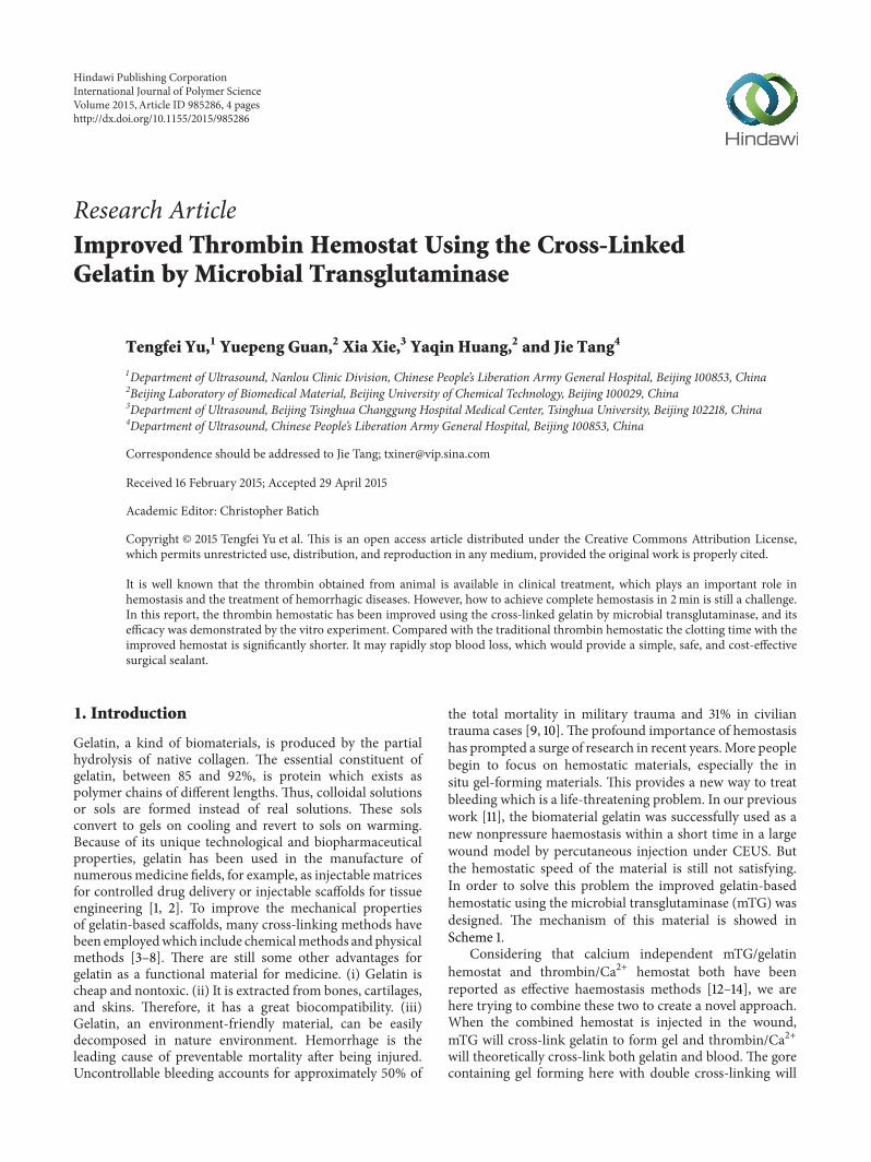
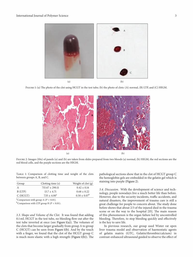
Figure 1: (a) The photo of the clot using HCGT in the test tube; (b) the photo of clots: (A) normal, (B) LTP, and (C) HIGM.
(a) (b)
Figure 2: Images (10x) of panels (a) and (b) are taken from slides prepared from two bloods (a) normal, (b) HIGM, the red sections are thered blood cells, and the purple sections are the HIGM.
Table 1: Comparison of clotting time and weight of the clotsbetween groups A, B, and C.
Group Clotting time (s) Weight of clot (g)A 715.67 ± 290.11 0.42 ± 0.14B (LTP) 13.7 ± 4.71 0.68 ± 0.22C (HCGT) 7.55 ± 4.00a 0.50 ± 0.07baComparison with group A (𝑃 < 0.01).bComparison with LTP group B (𝑃 < 0.01).
3.3. Shape and Volume of the Clot. It was found that adding0.1mL HCGT in the test tube, no bleeding flow out after thetest tube inverted at once (see Figure 1(a)). The volumes ofthe clots that become larger gradually from group A to groupC (HCGT) can be seen from Figure 1(b). And by the touchwith a finger, we found that the clot of the HCGT group Cis much more elastic with a high strength (Figure 1(b)). The
pathological sections show that in the clot of HCGT group Cthe hemoglobin gels are embedded in the gelatin gel which isstaining into purple (Figure 2).
3.4. Discussion. With the development of science and tech-nology, people nowadays live a much better life than before.However, due to the security incidents, traffic accidents, andnatural disasters, the improvement of trauma care is still agreat challenge for people to concern about. The study donebefore shows that about 2/3 of the injured died in the traumascene or on the way to the hospital [15]. The main reasonof this phenomenon is the organ failure led by uncontrolledbleeding. Therefore, to stop bleeding quickly and effectivelyis the key to save life.
In previous research, our group used Wister rat openliver trauma model and observation of haemostatic agentsof gelatin matrix (GTC, Gelatin/thrombin/calcium) incontrast-enhanced ultrasound guided to observe the effect of
4 International Journal of Polymer Science
interventional therapy in the treatment of trauma [16]. Theresults display, GTC, delivered by percutaneous injectionunder CEUS, may achieve haemostasis, especially in thecase of no pressure. In order to further accelerate hemostasisspeed, improved cross-linking hemostatic was designed.And the advantage was investigated through the test tubeexperiments. (i) High speed of hemostasis is achieved; bythis way the clot time can be limited in about 10 seconds.Moreover the clot time can be also controlled by regulatingthe reaction rate of thrombin. (ii) The elasticity and thestrength of the clot are improved owing to the reticularstructure in the gel formed by the improved cross-link.Since gelatin is a dispersant with good biocompatibility, thegels of hemoglobin can be dispersed in the gelatin systemuniformly. It also makes the clot become more elastic. (iii)Because gelatin has the ability of gel forming, it can increasethe volume of the clot through cross-link and make theprocess of hemostasis much easier. (iv) Gelatin is a kindof macromolecule produced by the partial hydrolysis ofnative collagen. When added in the bleeding wound, gelatinis conducive to gathering the blood coagulation factor sothat the hemostatic speed can be enhanced [17]. What else,gelatin would be able to be biodegradable and could promotethe repair and regeneration process of the damaged tissue[16]. So the improved cross-link hemostatic described in thisreport is a very promising product in hemositasis.
4. Conclusion
What we reported in this paper offers a simple, safe, and cost-effective surgical sealant, whichmay stop blood loss rapidly. Itwas provided that cross-linking gelation by microbial transg-lutaminase can enhance the thrombin hemostatic obviously.And the clotting time with the improved hemostat, measuredby vitro experiment, is significantly shorter compared withtraditional thrombin hemostatic.
Conflict of Interests
The authors declare that there is no conflict of interestsregarding the publication of this paper.
Authors’ Contribution
Tengfei Yu and Yuepeng Guan contributed equally to thiswork and should be considered co-first authors.
References
[1] K. M. Lewis, H. D. Atlee, A. J. Mannone et al., “Comparison oftwo gelatin and thrombin combination hemostats in a porcineliver abrasion model,” Journal of Investigative Surgery, vol. 26,no. 3, pp. 141–148, 2013.
[2] M. Kabiri, S. H. Emami, M. Rafinia, and M. Tahriri, “Prepara-tion and characterization of absorbable hemostat crosslinkedgelatin sponges for surgical applications,” Current AppliedPhysics, vol. 11, no. 3, pp. 457–461, 2011.
[3] S. R. Gomes, G. Rodrigues, G. G. Martins, C. M. R. Henriques,and J. C. Silva, “In vitro evaluation of crosslinked electrospun
fish gelatin scaffolds,”Materials Science and Engineering C, vol.33, no. 3, pp. 1219–1227, 2013.
[4] A. Saarai, V. Kasparkova, T. Sedlacek, and P. Saha, “On thedevelopment and characterisation of crosslinked sodium algi-nate/gelatine hydrogels,” Journal of the Mechanical Behavior ofBiomedical Materials, vol. 18, pp. 152–166, 2013.
[5] Z. Zhou, Y. Zhou, Y. Chen et al., “Bilayer porous scaffold basedon poly-(𝜀-caprolactone) nanofibrous membrane and gelatinsponge for favoring cell proliferation,” Applied Surface Science,vol. 258, no. 5, pp. 1670–1676, 2011.
[6] R. N. Kale and A. N. Bajaj, “Ultraviolet spectrophotometricmethod for determination of gelatin crosslinking in the pres-ence of amino groups,” Journal of Young Pharmacists, vol. 2, no.1, pp. 90–94, 2010.
[7] R. Elia, P.W. Fuegy, A. VanDelden,M.A. Firpo, G.D. Prestwich,and R. A. Peattie, “Stimulation of in vivo angiogenesis by insitu crosslinked, dual growth factor-loaded, glycosaminoglycanhydrogels,” Biomaterials, vol. 31, no. 17, pp. 4630–4638, 2010.
[8] T. Okamoto, I. Ishikawa, A. Kumasaka et al., “Blue-violet light-emitting diode irradiation in combination with hemostaticgelatin sponge (Spongel) application ameliorates immediatesocket bleeding in patients taking warfarin,” Oral Surgery, OralMedicine, Oral Pathology and Oral Radiology, vol. 117, no. 2, pp.170–177, 2014.
[9] A. Shukla, J. C. Fang, S. Puranam, and P. T. Hammond,“Release of vancomycin from multilayer coated absorbentgelatin sponges,” Journal of Controlled Release, vol. 157, no. 1, pp.64–71, 2012.
[10] T. Wang, X. K. Zhu, X. T. Xue, and D. Y. Wu, “Hydrogelsheets of chitosan, honey and gelatin as burn wound dressings,”Carbohydrate Polymers, vol. 88, no. 1, pp. 75–83, 2012.
[11] T.-F. Yu, F.-Q. Lu, Z.-Y. Li et al., “Haemostatic agents of thegelatin matrix for a large liver wound by percutaneous injectionwithout pressure under the guidance of contrast-enhancedultrasound,” Chinese Medical Journal, vol. 124, no. 9, pp. 1352–1356, 2011.
[12] D. Dimitroulis, E. Antoniou, N. P. Karidis, K. Kontzoglou, andG. Kouraklis, “Surgical control of life-threatening post-ERCPbleeding with a gelatin matrix-thrombin hemostatic agent,”International Journal of Surgery Case Reports, vol. 3, no. 9, pp.471–473, 2012.
[13] T. Chen, R. Janjua, M. K. McDermott, S. L. Bernstein, S. M.Steidl, and G. F. Payne, “Gelatin-based biomimetic tissue adhe-sive. Potential for retinal reattachment,” Journal of BiomedicalMaterials Research—Part B Applied Biomaterials, vol. 77, no. 2,pp. 416–422, 2006.
[14] M. Maegele, R. Lefering, N. Yucel et al., “Early coagulopathy inmultiple injury: an analysis from the German Trauma Registryon 8724 patients,” Injury, vol. 38, no. 3, pp. 298–304, 2007.
[15] Y. Liu, D. Kopelman, L.-Q.Wu et al., “Biomimetic sealant basedon gelatin and microbial transglutaminase: an initial in vivoinvestigation,” Journal of Biomedical Materials Research. Part BApplied Biomaterials, vol. 91, no. 1, pp. 5–16, 2009.
[16] Z. S. Patel, M. Yamamoto, H. Ueda, Y. Tabata, and A. G. Mikos,“Biodegradable gelatin microparticles as delivery systems forthe controlled release of bone morphogenetic protein-2,” ActaBiomaterialia, vol. 4, no. 5, pp. 1126–1138, 2008.
[17] R. Hajosch, M. Suckfuell, S. Oesser, M. Ahlers, K. Flechsenhar,and B. Schlosshauer, “A novel gelatin sponge for acceleratedhemostasis,” Journal of Biomedical Materials Research Part B:Applied Biomaterials, vol. 94, no. 2, pp. 372–379, 2010.
Submit your manuscripts athttp://www.hindawi.com
ScientificaHindawi Publishing Corporationhttp://www.hindawi.com Volume 2014
CorrosionInternational Journal of
Hindawi Publishing Corporationhttp://www.hindawi.com Volume 2014
Polymer ScienceInternational Journal of
Hindawi Publishing Corporationhttp://www.hindawi.com Volume 2014
Hindawi Publishing Corporationhttp://www.hindawi.com Volume 2014
CeramicsJournal of
Hindawi Publishing Corporationhttp://www.hindawi.com Volume 2014
CompositesJournal of
NanoparticlesJournal of
Hindawi Publishing Corporationhttp://www.hindawi.com Volume 2014
Hindawi Publishing Corporationhttp://www.hindawi.com Volume 2014
International Journal of
Biomaterials
Hindawi Publishing Corporationhttp://www.hindawi.com Volume 2014
NanoscienceJournal of
TextilesHindawi Publishing Corporation http://www.hindawi.com Volume 2014
Journal of
NanotechnologyHindawi Publishing Corporationhttp://www.hindawi.com Volume 2014
Journal of
CrystallographyJournal of
Hindawi Publishing Corporationhttp://www.hindawi.com Volume 2014
The Scientific World JournalHindawi Publishing Corporation http://www.hindawi.com Volume 2014
Hindawi Publishing Corporationhttp://www.hindawi.com Volume 2014
CoatingsJournal of
Advances in
Materials Science and EngineeringHindawi Publishing Corporationhttp://www.hindawi.com Volume 2014
Smart Materials Research
Hindawi Publishing Corporationhttp://www.hindawi.com Volume 2014
Hindawi Publishing Corporationhttp://www.hindawi.com Volume 2014
MetallurgyJournal of
Hindawi Publishing Corporationhttp://www.hindawi.com Volume 2014
BioMed Research International
MaterialsJournal of
Hindawi Publishing Corporationhttp://www.hindawi.com Volume 2014
Nano
materials
Hindawi Publishing Corporationhttp://www.hindawi.com Volume 2014
Journal ofNanomaterials