research article effect of cuo nanoparticles over...
TRANSCRIPT

Research ArticleEffect of CuO Nanoparticles over Isolated Bacterial Strains fromAgricultural Soil
Sandra I Concha-Guerrero1 Elcia Margareth Souza Brito2
Hilda A Pintildeoacuten-Castillo1 S H Tarango-Rivero3 Ceacutesar A Caretta4
Antonia Luna-Velasco1 Robert Duran5 and Erasmo Orrantia-Borunda1
1Center for Research in Advanced Materials Chihuahua CHIH Mexico2Ingenierıa Ambiental Departamento de Ingenierıa Civil DI-CGT Universidad de Guanajuato Guanajuato GTO Mexico3Campo Experimental Delicias INIFAP CHIH Mexico4Departamento de Astronomıa DCNyE-CGT Universidad de Guanajuato Guanajuato GTO Mexico5Equipe Environment et Microbiologie UMR IPREM5254 IBEAS Universite de Pau et des Pays de lrsquoAdour Pau France
Correspondence should be addressed to Sandra I Concha-Guerrero sandraconchacimavedumxand Elcia Margareth Souza Brito emsbritogmailcom
Received 26 June 2014 Accepted 4 December 2014 Published 31 December 2014
Academic Editor Mallikarjuna N Nadagouda
Copyright copy 2014 Sandra I Concha-Guerrero et al This is an open access article distributed under the Creative CommonsAttribution License which permits unrestricted use distribution and reproduction in any medium provided the original work isproperly cited
The increased use of the nanoparticles (NPs) on several processes is notorious In contrast the ecotoxicological effects of NPshave been scarcely studied The main current researches are related to the oxide metallic NPs In the present work fifty-sixbacterial strains were isolated from soil comprising 17 different OTUs distributed into 3 classes Bacilli (36 strains) Flavobacteria(2 strains) and Gammaproteobacteria (18 strains) Copper oxide nanoparticles (CuONPs) were synthesized using a process ofchemical precipitation The obtained CuONPs have a spherical shape and primary size less than 17 nm Twenty-one strains wereused to evaluate the cytotoxicity of CuONPs and 11 of these strains showed high sensibility Among those 11 strains 4 (Brevibacilluslaterosporus strain CSS8 Chryseobacterium indoltheticum strain CSA28 and Pantoea ananatis strains CSA34 and CSA35) wereselected to determine the kind of damage produced The CuONPs toxic effect was observed at expositions over 25mgsdotLminus1 andthe damage to cell membrane above 160mgsdotLminus1 The electron microscopy showed the formation of cavities holes membranedegradation blebs cellular collapse and lysis These toxic effects may probably be due to the ions interaction the oxide-reductionreactions and the generation of reactive species
1 Introduction
The use of nanoparticles (NPs) has largely increased inthe last years the expected projection for the engineerednanomaterials production reaching more than 58000 tonsfor 2012ndash2020 [1] The waste generated by the NPs industryhas been demonstrated to affect directly the environmentmainly soil ecosystems followed by water and air ones [2ndash6]Besides it was found that NPs contamination might result inadditive synergistic or antagonistic toxicity [7 8] affectingthe entire food chain
Although the environmental effect of somenanomaterialssuch as carbon nanotubes metal oxides and zero-valentmetal NPs has been well studied [9] information about itsinteraction and damage on native bacterial communities isstill scarce Soil microbial communities are involved in sev-eral biogeochemical cycles such as carbon nitrogen sulfurand phosphorus ones [9 10] Thus even a little perturbationin the structure and composition of microbial communitiesmay induce modifications in the surrounding environmentand vice versa It is known that the bactericidal efficiency ofNPs depends on themicroorganism type for instance copper
Hindawi Publishing CorporationJournal of NanomaterialsVolume 2014 Article ID 148743 13 pageshttpdxdoiorg1011552014148743
2 Journal of Nanomaterials
Delicias city
(a) (b)
(c) (d)
Figure 1 Location of Delicias city Chihuahua Mexico (a) aerial (b) and ground (c) views of the pecan orchard and a view of the sampledsoil (d)
nanoparticles (CuNPs) showed more inhibitory activity onbacterial than on fungal strains [11] Furthermore there arereports that the increased copper (Cu) concentrations affectthe soil microorganisms modifying their number biomassactivity and diversity [12ndash15] while the bactericidal effect ofcopper oxide nanoparticles (CuONPs) has just been reportedfor type strains [11 16ndash20] Moreover Kim et al (2007)[21] found that the biocide efficiency of silver nanoparticles(AgNPs) over microorganisms depends on cell wall com-position while Niazi and Gu [22] show that cell damagesuch as membrane disorganization generation of reactiveoxygen species (ROS) and DNA damages can be influencedby the surface area and composition of NPs Neverthelessdespite the importance of soil bacterial communities onhomeostasis of geochemical cycles and the intensive use ofthe NPs only few studies have been performed to elucidatethe impact of NPs on soil bacterial communities [6 22ndash25]The protection of soil microbial biomass and diversity is oneof themajor challenges for the future years especially becausethey play an important role on soil usage and maintenanceThe sustainable use of these resources may not be guaranteedif the nutrient cycles change their proper functioning [2627] It is thus urgent to obtain information on how NPscould affect bacterial communities in order to mitigate theirenvironmental impact
The aim of this work was to determine the mechanismsinvolved in the CuONP interaction with native bacterialstrains First we isolated wild bacterial strains from agri-cultural soil which were characterized by 16S rRNA genesequencing Then CuONPsbacterial cells interactions wereexamined using four of these bacterial strains which were
selected by their importance in the soil biogeochemical cyclesand also by their sensibility to CuONPs on exposition tests
2 Materials and Methods
21 Sampling Site In this work soil samples were collectedfrom a pecan agricultural soil at Delicias city Chihuahuastate Mexico (Figure 1) in July 2012 The superficial litterfallwas removed and 12 subsamples (maximum 10 cm depth)were obtained being 20m far from each other Afterwardsthey were mixed into a single composite sample sieved(gt2mm) and immediately conserved at 4∘C until use
22 Physicochemical Characterization of Soil Sample Thesoil characterization was performed in the Department ofAgrotechnological Sciences of the Chihuahua University(FACIATEC) The soil texture presented sand 71 silt 13and clay 16 Other characteristics are summarized inTable 1 The crystallinity of the soil was further characterizedby X-ray diffractometer (XRD MDP Phillips X1015840 pert PRO)The data was analyzed with HighScore softwareThe samplesfor morphology and soil structure analyses were preparedaccording to Maldonado et al [28] which were done using aJeol JSM 7401F field emission scanning electron microscope(SEM) while the elemental analysis was performed by energydispersive X-ray (EDX Oxford Inca PentaFETX3) coupled toSEM (see more details below)
23 Microbiological Characterization The microbial charac-terization was first performed with culture dependent tech-niques to determine the bacterial abundance by using colo-ny forming unit (CFU) counts and most probable number
Journal of Nanomaterials 3
Table 1 Pedologic and microbial soil characteristics
ParametersPhysical properties
sand 71 silt 13 clay 16pH 680 organic matter 029 CaCO3 154CH (cmsdothminus1) 571 saturation 425CE (mmhossdotcmminus1) 075
FertilityN-NO3 (kgsdotha
minus1) 216 times 103
P (kgsdothaminus1) 310K (mgsdotLminus1) 362 times 102
Ca (mgsdotLminus1) 1017 times 103
Mg (mgsdotLminus1) 962 times 102
Na (mgsdotLminus1) 2062 times 102
Cu (mgsdotLminus1) 060Fe (mgsdotLminus1) 180Mn (mgsdotLminus1) 522Zn (mgsdotLminus1) 7182
Microbiological propertiesCFUAGEL (Actinobacteria) (CFUsdotmLminus1) 53 times 102
M9 (coliform) (CFUsdotmLminus1) 86 times 104
YPS (aerobic bacteria Bacilli) (CFUsdotmLminus1) 52 times 104
LB (enteric bacteria) (CFUsdotmLminus1) 59 times 104
MPN (cellsdot[gsoil]minus1) 21 times 105
FDA ([120583gFDA]sdotmLminus1) 216 plusmn 0021
(MPN) as well as the total microbial activity and metabolicactivities
The CFU counts were performed in different culturemedia in order to obtain the largest possible number ofisolatesThe culturemedia used are listed in the following (1)the modified M9 minimal medium (47mM Na
2
HPO4
22mM KH
2
PO4
86mM NaCl 22 dextrose 15 gsdotLminus1 bac-teriological agar 16MgSO
4
02 vitamins and 02 traceelements) [29] (2) themodifiedAGELmedium (2 gsdotLminus1 yeastextract 1 gsdotLminus1 KH
2
PO4
5mL glycerol 15 gsdotLminus1 bacteriologi-cal agar and 5mL Crystal-violet 1) specific for Actinobac-teria (3) the nutritionally rich LB medium (Luria-Bertani10 gsdotLminus1 peptone from casein 5 gsdotLminus1 yeast extract 10 gsdotLminus1NaCl and 15 gsdotLminus1 bacteriological agar) for the growth ofenteric bacteria (4) the YPS medium (3 gLminus1 protease pep-tone 3 gLminus1 yeast extract 2mM CaCl
2
and 2mM MgSO4
)[30] used to isolate total aerobic bacteria and Bacilli (5)the MMB medium (37 gLminus1 Na
2
HPO4
098 gLminus1 KH2
PO4
05 gLminus1NH
4
Cl 003 gLminus1MgSO4
05 gLminus1 dextrose 04 gLminus1protease peptone 001M Tris-HCl 1mL vitamins and 002trace elements) [31] employed for isolating the heterotrophicbacteria (6) the soy medium (15 gsdotLminus1 peptone from casein
5 gsdotLminus1 peptone from soy 3 gsdotLminus1 yeast extract 5 gsdotLminus1 dex-trose 5 gsdotLminus1 NaCl and 15 gsdotLminus1 bacteriological agar) as anutritional rich medium for isolating aerobic and anaer-obic microorganisms and (7) the R2A medium (05 gsdotLminus1yeast extract 05 gsdotLminus1 protease peptone 05 gsdotLminus1 tryptone05 gsdotLminus1 dextrose 05 gsdotLminus1 starch 03 gsdotLminus1 sodiumpyruvate03 gsdotLminus1 K
2
HPO4
and 15 gsdotLminus1 bacteriological agar) [32]a medium with a low nutrient content appropriate forthe development of slow growing or stressed bacteria Allmorphologically distinct colonies observed in the solid plateswere selected and submitted to an isolation process using astreak plate traditional method
For MPN the MMB media 37 gLminus1 Na2
HPO4
098 gLminus1KH2
PO4
05 gLminus1 NH4
Cl 003 gLminus1 MgSO4
05 gLminus1 dex-trose 04 gLminus1 protease peptone 001M Tris-HCl 1mL vita-mins and 002 trace elements [31] were used
The total microbial activity was determined throughfluorescein diacetate (FDA) hydrolysis [33] The metabolicactivity of soil community was assessed by Biolog Ecoplates(Biolog Inc Hayward CA USA) [34] which will bedescribed below
24 DNA Extraction and Phylogenetic Analysis of IsolatesTotal genomic DNA extraction from each of the 56 isolatedbacteria was performed using the Wizard Genomic DNAPurification Kit (Promega Corporation) Bacterial 16S rRNAgene was PCR-amplified using the 8F (AGAGTTTGATCC-TGGCTAG) [35] and 1489R (TACCTTGTTACGACT-TCA) [36] primers The reaction was cycled with aninitial denaturation step (95∘C for 5min) followed by35 cycles of denaturation step (95∘C for 45 s) annealing(52∘C for 45 s) elongation (72∘C for 1min) and then afinal elongation step (72∘C for 10min) The PCR prod-ucts were purified with Illustra GFX PCR DNA and GelBand Purification Kit (GE Healthcare) The sequencing wascarried out by Research and Advanced Studies Center ofthe National Polytechnic Institute (CINVESTAV-IrapuatoMexico) Sequence data were analyzed with BioEdit pro-gram and compared with the online database NCBI (httpblastncbinlmnihgovBlastcgi) MAFFT software (multi-ple sequence alignment based on fast Fourier transform)was used for the alignment of sequences The phylogenetictree was constructed with Molecular Evolutionary GeneticsAnalysis v52 program [37] using the Jukes-Cantor modeland the neighbour-joining algorithm The significance ofbranching order was determined using bootstrap analysiswith 1000 resampling data sets
25 Synthesis and Characterization of CuONPs The synthesisof CuONPs was prepared according to Lanje et al [38]The size and morphology of nanoparticles were analyzedusing SEM (Jeol JSM 7401F) and field emission transmissionelectron microscopy (TEM) A suspension of the nanopar-ticles in ethanol (1mL) was sonicated for 15min 300 120583L ofthis solution was placed onto an aluminum support andthen analyzed for SEM operated at 50 kV TEM sampleswere prepared by putting one drop of the suspension ontonickel grids and the organic residues were cleaned using
4 Journal of Nanomaterials
plasma (5min) and finally were observed The nanoparticlescrystallinity was characterized by XRD The data was againanalyzed with HighScore software The absorption spectrumof the colloidal solutionwas determined onPerkinElmerUV-visible spectrophotometer
26 Antibacterial Activity The bactericidal effect of CuONPwas analyzed against 21 representative bacterial isolatesThe nanoparticles were dispersed in presterilized tridistilledwater by sonication Cells were grown and adjusted toOD620 nm = 01 Fresh axenic cultures were inoculated in
96-well plates (final volume 200 120583L) containing liquid cul-ture medium supplemented with various concentrations ofCuONP (0ndash100mgLminus1) After incubation (12ndash18 h at 37∘C)the bacterial growth was put in evidence according to thecolor developed using resazurin as indicator The shift fromblue to pink indicates the bacterial growth [39]
27 Carbon Source Utilization by the Four Bacterial IsolatesBacterial carbon utilization fingerprints were generated usingBiolog EcoPlates These 96-well microplates contain threereplicate wells of 31 carbon substrates 6 amino acids 10carbohydrates 7 carboxylic acids 2 amines 2 phenolic com-pounds and 4 polymers Each well also contains the redoxdye tetrazolium violet which turns from colorless to purplein the presence of respiration The four isolated bacterialstrains were isolated and selected from the 21 submittedto the toxicity test (above) according to their sensibilityto NPs exposition The CSS8 CSA28 CSA34 and CSA35strains were grown in LB broth at 37∘C for 12ndash18 h in ashaker incubator at 200 rpm The bacterial suspension wascentrifuged at 10000 rpm for 10min and resuspended in15mLof sterile 085NaCl solutionThebacterial suspensionwas then adjusted to OD
590 nm = 025 A 150 120583L aliquot wasinoculated into each well of the microplates Plates wereincubated at 27∘C for 24 h and 72 h
28 Bacterial Cells Exposition to Nanoparticles The strainsCSS8 CSA28 CSA34 and CSA35 were cultured on LB brothat 37∘C for 12ndash18 h in a shaker incubator at 200 rpm Thebacterial cells were recovered by centrifugation at 10000 rpmfor 10min and then resuspended in NaH
2
PO4
buffer solution(25mM pH 74) The absorbance of the bacterial suspen-sion was measured by a spectrophotometer and adjusted toOD620 nm = 01 The isolates were treated with various con-
centrations (0ndash160mgsdotLminus1) of CuONPs in NaH2
PO4
buffersolution (25mM pH 74) and incubated at 37∘Cwith shakingat 200 rpm After 05 1 3 8 and 24 h subsamples were takenand observed on scanning electronmicroscopy At the end ofexperiments (24 h incubation) the ROS was determined (seebelow) All assays were performed in triplicate in Erlenmeyerflasks with a total volume of 50mL
29 Biological Sample Preparation for SEM The culturesamples exposed to NPs and controls (not exposed) wereanalyzed by SEM The samples preparation method waspreviously described by Maldonado et al [28] Briefly sub-sample cultures were fixed in 3 glutaraldehyde Milloningbuffer phosphate (pH 73) for 4 h and washed two times in
the same buffer Afterwards they were filtered on polycar-bonate membrane (pore diameter 022120583m) fixed and dehy-drated increasing successively the ethanol concentration Allsamples were mounted on metal stubs and coated with gold(Denton Vacuum Desk II) A Jeol JSM 7401F microscopewas used to generate the imagesThe energy dispersive X-ray(EDX Oxford Inca PentaFETX3) coupled to SEM was usedfor the elementary analysis
210 Determination of Reactive Oxygen Species (ROS) Stocksolutions of 10 120583M2101584071015840-dichlorodihydrofluorescein diacetate(DCFH-DA) and 2101584071015840-dichlorodihydrofluorescein (DCFH)were prepared according to Luna-Velasco et al [40] A 2mLsubsample of the assay described above for bacterial cellexposition to nanoparticles was used for ROS test Culturesamples without exposure to NPs were used as controls Thebacterial cells of the subsample were recovered by cen-trifugation at 10000 rpm for 5min and then resuspendedin NaH
2
PO4
buffer solution (25mM pH 74) Aliquots of135 120583L were inoculated in black 96-well plates with 15 120583L of10 120583M DCF-DA The black 96-well plates were incubated indarkness for 30min finally the fluorescence wasmeasured atan excitation of 485 nm and emission wavelength of 535 nm
3 Results and Discussion
31 Chemical and Microbiological Characterization of SoilThe soil pHwas close to neutral with high salt concentrations(Table 1)The crystal composition analyzed by XRD showedthe presence of feldspars such as sanidine Bytownite andanorthoclase as well as silicates such as silicon oxide andmuscovite (Figure 2(a))
The soil structure and morphology were observed bySEMmicrograph the texture was rough forming flakes andporous (Figure 2(b)) The chemical EDX analysis showedthe presence of complementary elements such as Al SiO and H which form part of the components of feldspar(Figure 2(c)) The physicochemical characterization of thesoil showed contradictory results a low content of organicmatter and salts indicating that soil is poor but a highcationic interchange capacity (CIC) suggesting a fertile soil
The activity of the bacterial soil community was inferredfrom a shift of color media after an incubation time usingBiolog EcoPlates test showing the degradation of 2231carbon sources tested (Table 2) Additionally the hydrol-ysis of FDA was used to quantify this activity (216 plusmn0021 120583gFDAsdotmLminus1) A few compounds of Biolog EcoPlatestest were not used as carbon source 3 carbohydrates 1amino acids 2 carboxylic acids 1 phenolic compound and 2polymers Both the FDA hydrolysis and the Biolog EcoPlatesanalysis suggest that microbial populations have a highmetabolic capacity Thus although the soil conditions limitthe diversity of these samples the presentmicroorganisms arevery active explaining the soil fertility
The CFU and MPN were used to estimate the cultivablemicrobial richness using different culture media The highestbacterial counts were obtained with M9 medium (86 times104 CFUsdotmLminus1) followed by LB and YPS (59 times 104 and52 times 104 CFUsdotmLminus1 resp) below the values reported in
Journal of Nanomaterials 5
20000
10000
0
10 20 30 40 50 60 70 80
Position (2120579)
Relat
ive i
nten
sity
(au
)
SanidineMuscoviteBytownite
AnorthoclaseSilicon oxide
(a)
1120583m
(b)
Fe
FeFe
Na
Ca
Ca
Ca
K
C
O AlSi
MgK
K
0 1 2 3 4 5 6 7 8
(keV)
(c)
Figure 2 Characterization of soil (a) XRD spectra of the crystallinity (b) SEM image and (c) EDX of the soil sample
the literature for the bacterial recuperation by culture (108 to1010 bacterial per soil gram [41]) The bacterial populationsize was estimated to be 21 times 105 cellsdot(g soil)minus1 by MPN Thesoil microbial communities are usually described as pre-senting high heterogeneity which is usually related to thesoil physical-chemical characteristics [41]The results suggestthat the cultivable bacterial communities of the studied soildo not require specific or complex nutrientsTheutilization ofthe specific Actinobacteria media revealed that this bacterialgroup was scarcely represented in the studied soil (Table 1)
32 Bacterial Strains Isolation Fifty-six bacterial strains weresuccessfully isolated (Table 3) from this sample using 7different culture media The phylogenetic analysis basedon 16S rRNA gene sequences classified the isolated strainswithin 17 different OTUs (Figure 3) They were distributedinto 3 classes Bacilli (36 strains) Flavobacteria (2 strains)and Gammaproteobacteria (18 strains) According to their16S rRNA gene sequences these strains are similar to bac-terial strains associated with human disease like BacillusSerratia Enterobacter and Pantoea species or associatedwith
biocontrol agents such as Brevibacillus laterosporus Chry-seobacterium indoltheticum and Pantoea ananatis Howevermore studies are necessary to verify the real source of thesemicroorganisms In addition populations similar to Bacillusamyloliquefaciens and Pseudomonas putida were also foundSuch species are reported as natural antifungal agents whichalso can metabolize nitrogen and enhance the fertility
Taking into account that the recovery of cultivable bac-teria is usually very low we just obtained a little part of thetotal bacterial diversity Besides even when we use a specificmedia we are unable to isolate typical soil groups like Agro-bacteriumorActinobacteriaThis resultmay be influenced bythe soil characteristics such as the low and limited amountof organic matter and great quantity of sand (71) Theporosity and size of soil particles can produce constant shiftsin the soil moisture which does not allow growth or favorsstrong adhesion ofmicroorganisms over particles [42] Otherfactors that can inhibit or eliminate members of the origi-nal microflora are crop changes [43] the concentration ofsugar the osmolarity the water activity the pH and otherenvironmental factors [44ndash47] If any of these changes occurnaturally or anthropogenically the microorganisms that can
6 Journal of Nanomaterials
Table 2 Pattern of 31 carbon substratesrsquo utilization for sample soil and model strains CSS8 CSA28 CSA34 and CSA35
Enzyme activity Soil sample CSS8 CSA28 CSA34 CSA35Water minus minus minus minus minus
120573-Methyl-D-glucoside minus + + + +D-Galactonic acid 120574-lactone + + + + +L-Arginine + + + + +Pyruvic acid methyl ester + + + + +D-Xylose minus + + + +D-Galacturonic acid minus + + + +L-Asparagine + + + + +Tween 40 + + + + +I-Erythritol + + + + +2-Hydroxy benzoic acid minus minus + + minus
L-Phenylalanine minus + + + +Tween 80 + + + + +D-Mannitol + + + + +4-Hydroxy benzoic acid + minus + + +L-Serine + + + + +120572-Cyclodextrin minus + + + +N-Acetyl-D-glucosamine + + + + +120574-Hydroxybutyric acid + + + + +L-Threonine + + + + +Glycogen minus + + + +D-Glucoaminic acid + + + + +Itaconic acid + + + + +Glycil L glutamic acid + + + + +D-Cellobiose + + + + +Glucose-1-phosphate + + + + +120572-Ketobutyric acid minus + + + +Phenylethylamine + + + + +120572-D-Lactose + + + + +DL-120572-Glycerol phosphate minus + + + +D-Mallic acid + minus + + +Putrescine + + + + +
survive under these changes probably develop adaptive char-acteristics
33 Characterization of CuONPs The method used to syn-thesize the CuONPs was the same described by Lanje et al[38] with the difference that we obtained spherical insteadof rectangular NPs The SEM and TEM micrographs showspherical shape Differences in the morphology due to shiftin temperature were reported for CuO nanorods synthesisby Gao et al [48] They obtained two different structureswhen the nanorods are synthetized at room temperature or100∘C The particle sizes in SEM micrograph were in therange from 10 to 17 nm and the SEM-EDX profile indicatedthat the preparation did not contain synthesis waste andNPs composition is only of copper and oxygen (Figures4(a) and 4(b)) Zhang et al [49] reported that the initialconcentration might affect the NPs formation They proposethe formation of nanoparticles in solution in two stagesfirst the generation of copper nuclei and then the growing
up of these nuclei for the formation of NPs When thereactant concentration is high the number of nuclei increasesin the solution consequently the NPs sizes are incrementedresulting in agglomerates UV-Vis spectra of the synthesizedCuONPs showed the band-edge emission peak at 280 nm(Figure 4(c)) We also used X-ray analysis to identify thecrystalline phase this analysis revealed the presence of CuOonly (Figure 4(d))
34 Exposure of Bacterial Cells to CuONPs and ROS Genera-tion We used a liquid microculture (200120583L) for the screen-ing of bacterial strains sensitive to CuONPs (Table 4) Amongtwenty-one strains tested eleven showed no resistance toCuONPs while the strains CSR19A CSL10A and CSMB13Aexhibited the highest resistances (17 225 and 25mgsdotLminus1resp) Besides their sensibility toCuONPs exposition test andthe originality for their use as toxicity model to the studiedNP we also considered their importance in the contributionin soil microbial community and biogeochemical cycles
Journal of Nanomaterials 7
Bacillus amyloliquefaciens NBRC
Baci
lliFl
avob
acte
riaG
amm
apro
teob
acte
ria
15535T (NR 0414551)75
74
99
95 82
19 96
99
93
47
99
99
59
29
100
100
100
100
100
78
72
99
99
99
9869
100
100
100
005
100
69
30
100
100
34
CSM39 (6)CSY20 (19)CSMB12 (3)CSM
CSS12
CSY17
CSM18
CSS15
CSS14
CSS8
CSY5
CSL10A
CSA27
4 (2)Bacillus subtilis subsp subtilis DSM 10
T (NR 0275521)
Bacillus stratosphericus 41KF2aT (NR 0423361)
DM 122T (NR 0369561)
Staphylococcus aureus subsp aureus N315T (NR 0750001)
Enterococcus faecium R-10T (KF3184001)
Bacillus marisflavi TF-11T (NR 0252401)
Bacillus endophyticus 2DTT (NR 0251221)
Pyrococcus furiosus DSM 3638T
CSR19A (2)Enterobacter ludwigii EN-119 DSMZ 16688 CIP 108491
T (NR 0423491)
Erwinia piriflorinigrans T (JX8677591)5ndash8CSA35 (3)Pantoea ananatis 1846T (NR 0260451)
CSMB13A (8)Serratia marcescens WW4
T (NR 1025091)Aeromonas caviae VITKPAI-N1
T (JX3076861)CSM2 (2)
Brevibacillus laterosporus IAM 12465T (NR 0370051)
Paenibacillus sophorae S2T (GQ9853952)
Empedobacter brevis Y7DT (EU2931541)Flavobacterium cucumis R2A45-3T (NR 0441071)
Cellulophaga sp N5-2T (GU1299781)
Stenotrophomonas maltophilia IAM 12423T (NR 0415771)
Lysobacter koreensis Dae16T (NR 0410141)Pseudomonas putida KT2440T (NR 0745961)
Chryseobacterium indoltheticum LMG 4025 ATCC27950T (NR 0429261)CSA28 (2)
Staphylococcus hominis subsp hominis
Figure 3 Phylogenetic tree based on 16S rRNA gene sequences of isolated strains showing the cultivable bacterial diversity of the soil
The CSS8 (99 similarity to Brevibacillus laterosporus)CSA28 (99 similarity to Chryseobacterium indoltheticum)CSA34 andCSA35 (bothwith 99 similarity to Pantoea ana-natis) were selected as bacterial models for the determinationof damages caused byCuONPsTheirmetabolic capacities arepresented Table 2 The CSS8 strain was capable to use 2831(except 2 phenolic compounds and 1 carboxylic acid) theCSA28 and CSA34 strains were able to use all carbon sources
and the CSA35 strain degraded 3031 substrates (except 2-hydroxy benzoic acid) These strains showed higher capac-ities for carbon source utilization than that observed for thewhole bacterial communityThis differencemay be explainedby the fact that bacterial expression depends on complexbacterialbacterial interactions in microbial communities
The selected strains were individually exposed to CuONPsin phosphate buffer (25mM pH 74) and subsamples were
8 Journal of Nanomaterials
(a)
0 1 2 3 4 5 6 7 8
(keV)
Cu
Cu
O
(b)
3002826242220181614121008060402000
Abso
rban
ce
1900
250
300
350
400
450
500
550
600
650
700
750
800
850
900
950
1000
1050
1100
Wavelength (nm)
(c)
3000
2000
1000
0
Relat
ive i
nten
sity
(au
)
10 20 30 40 50 60 70
Position (2120579)
CuO
(d)
Figure 4 Characterization of CuONPs (a) TEM micrograph (b) SEM micrograph showing the shape and the particle size estimation withthe superposed SEM-EDX spectrum indicating the composition (c) UV-visible spectrum and (d) X-ray spectra
observed by SEM The SEM micrographs showed differentkind of damage on the cell membrane in the 4 strains whenthey were exposed above 160mgsdotLminus1 of CuONPs Changes onthe shape were observed from normal shape rod to biggerstretched and apparently empty cells and even to lysed cells(Figures 5(b) and 5(k)) Other observed effects were thecavities and pits formation over cells membrane (Figures 5(e)and 5(h)) These modifications were not observed in controlrod cells (not exposed to CuONPs) (Figures 5(a) 5(d) 5(g)and 5(j)) Although the exact mechanisms of toxicity arenot completely understood it is probable that the toxicitygenerated by CuONPs on CSA28 and CSA34 strains is due toNPs solubilization that produce Cu2+ ions At low concentra-tions copper is a micronutrient essential for cell metabolismIt is required as a cofactor in multiple proteins it takespart in redox reactions such as electron transport oxidativerespiration or the denitrification process and in some casesit is also a structural element [50] However despite thecritical role of Cu in a wide array of biological processesthe excess of Cu could be toxic [51] We also observed theadhesion of Cu over cellsmembrane in the SEM-EDXprofiles
(Figures 5(c) 5(f) 5(i) and 5(l)) suggesting some interactionsof Cu directly to the cell membrane The CuONPs-cellinteractions due to opposite electrical charges carry out areduction reaction in the bacterial cell wall which causesadhesion and bioactivity conducted by electrostatic forces[52ndash55] The wall cell of Gram-negative bacteria is mademainly of polysaccharides and the negative charge resultingfromgroups such as carboxylate phosphate hydroxyl amineand sulfhydryl has an important role as membrane activesites [56 57]The interactions of these groups with Cu2+ ionsproduce denaturation and alteration of membrane proteins[58]
Oxidative stress is normally used to evaluate the toxicityof several compounds including those caused by metallicnanoparticles [8] Diverse reactive molecules can be pro-duced during the oxidative stress such as nitric oxide (NO)peroxynitrite (ONOOminus) superoxide (O
2
∙minus) hydrogen perox-ide (H
2
O2
) hypochlorous acid (HOCl) and hydroxyl radical(OH∙) The fluorescent probe traditionally used to identifyROS is 2101584071015840-dichlorodihydrofluorescein diacetate (DCFH-DA) which reflects the overall oxidative status of the cell
Journal of Nanomaterials 9
Table 3 GenBank accession numbers of bacterial strains isolatedfrom agricultural soil
Isolated strain GenBank accession numberCSM39 KM091676CSL6 KM091677CSL21 KM091678CSM40 KM091679CSM42 KM091680CSL44 KM091681CSY20 KM091682CSM30 KM091683CSM31 KM091684CSA25 KM091685CSA26 KM091686CSL22 KM091687CSA23 KM091688CSY19 KM091689CSL16 KM091690CSS13 KM091691CSM11 KM091692CSS9 KM091693CSS7 KM091694CSM38 KM091695CSM32 KM091696CSA36 KM091697CSM33 KM091698CSM41 KM091699CSMB12 KM091700CSY1 KM091701CSMB13B KM091702CSM4 KM091703CSM5 KM091704CSS12 KM091705CSY17 KM091706CSM18 KM091707CSS15 KM091708CSS14 KM091709CSS8 KM091710CSL43 KM091711CSY5 KM091712CSL10A KM091713CSM2 KM091714CSM10 KM091715CSMB13A KM091716CSY6A KM091717CSMB16 KM091718CSMB14A KM091719CSY2 KM091720CSY6B KM091721CSY7 KM091722CSL11 KM091723CSA35 KM091724CSA34 KM091725
Table 3 Continued
Isolated strain GenBank accession numberCSA37 KM091726CSA27 KM091727CSR19A KM091728CSR19B KM091729
Table 4 Maximum concentration of CuONPs in which eachbacterial isolate can survive
ConcentrationCuONPs(mgsdotLminus1)
Strains
0 CSY17 CSM18 CSS15 CSS14 CSS8lowast CSA28lowastCSY5 CSM2 CSA34lowast CSA35lowast CSM39B
1 CSS122 CSMB12 CSM4 CSA274 CSY2017 CSR19A225 CSL10A25 CSMB13AlowastStrains selected for evaluating the specific damage caused by CuONPsexposition
However it is not applicable to individual ROS compoundsdetection By using this compound we did not observe anyincrease in the ROS production comparing the control strains(without NPs) with the strains exposed to CuONPs In facta slight decrease in the ROS activities is observed for CSS8CSA34 and CSA35 strains whereas for CSA28 strain thedecrease is until 6-fold less (Figure 6)
Ours results are comparable to those obtained by Cuiet al [59] working with Escherichia coli and gold NPs(AuNPs)They found that the AuNPs did not induce increasein cellular ROS but instead led to a decrease after 4 h ofexposition Meanwhile there are same controversial resultsin the scientific literature about the mechanism of oxidativestress generated by NPs For example Dasari et al [60]evaluated the toxicity mechanism of several NPs with E coliFor titanium NPs (TiO
2
NPs) and zinc NPs (ZnONPs) theyfound an increase in the amount of ROS while for CuONPsand cobalt oxide NPs (Co
3
O4
NPs) any effect was observedIn other work Dimkpa et al [53] evaluated the toxicityof commercial CuONPs in the pathogenic bacteria Pseu-domonas chlororaphisO6They observed an accumulation ofintracellular ROS with concentrations higher to 500 ppm ofCuONPs whereas lower concentrations did not induce ROSgeneration Furthermore several studies have reported theincreased of ROS activities when cells are exposed to NPs forexample Lee et al [61] Gunawan et al [62] and Rastogi etal [63] The ROS levels increment was produced when E coliwas exposed to CuONPs [62] and AgNPs [61] Similar effectsare shown for Staphylococcus aureus 49834 E coli 25922 andPseudomonas aeruginosa 27853 in presence of AgNPs [61 63]
It is known that denitrifying bacteria can produce NOendogenously as an intermediate from the sequential reaction
10 Journal of Nanomaterials
1120583m
Control
(a)
1120583m
CuONPs
(b)
Cu
CuCu
PNaN
Cl
Cl
CO
0 1 2 3 4 5 6 7 8 9 10
(keV)
(c)
1120583m
(d)
1120583m
(e)
Cu
CuCu
Na
N
Cl
Cl
CO
0 1 2 3 4 5 6 7 8 9 10
(keV)
(f)
1120583m
(g)
1120583m
(h)
Cu
Cu Cu
O
0 1 2 3 4 5 6 7 8 9
(keV)
(i)
1120583m
(j)
1120583m
(k)
Ca
CuNa
C
O
P
S CaCa
N
0 1 2 3 4 5 6 7
(keV)
(l)
Figure 5 Scanning electronmicroscopy (SEM) of CSS8 CSA28 CSA34 and CSA35 cells treated with CuONPs (b e h and k) and respectivecontrols (a d g and j) SEM-EDX spectrum for the surface of a cell (c f i and l)
Journal of Nanomaterials 11
008
007
006
005
004
003
002
001
00
DFC
conc
entr
atio
n (120583
M)
CSS8
cont
rol
CSS8
CuO
NPs
CSA28
cont
rol
CSA28
CuO
NPs
CSA34
cont
rol
CSA34
CuO
NPs
CSA35
cont
rol
CSA35
CuO
NPs
Figure 6 ROS activities in CSS8 CSA28 CSA34 and CSA35modelstrains
of nitrate to dinitrogen [64] The SEM micrographs of CSS8and CSA35 strains show different damage generated fromCuONPs such as membrane collapse bleb formation andcellular debris leading to the loss of the bacterial membraneintegrity irreversible cell damage or cell death (Figure 5)Considering that the model bacterial strains selected on thisstudy are phylogenetically related to denitrifying bacteriait is possible that the membrane damage observed by SEMcould be caused by the presence of nitrogen reactive speciessuch as nitric oxide (NO) In the case of our selected strainsthe cytotoxicity could be associated with damage in proteinsthat use copper like cofactor such as nitrous oxide reductase(N2
OR) nitric oxide reductase (NOR) and cytochrome CThese proteins are also involved in the denitrification processresulting in overproduction of reactive nitrogen and oxygenspecies Several reports have associated the presence of NPswith the increase of expression of these proteins and themalfunction of metal binding enzymes [65] Besides otherreports analyzing the effect produced over the cell membraneby the exposition to NO in P aeruginosa and E coli strains[65ndash69] showed similar damage to those observed in thisstudy However further studies are required to verify thecytotoxic mechanism
4 Conclusions
Several pure culture studies have shown that the bacterialinteraction with NPs may produce cytotoxicity in differentparts of the cell such as membrane disorganization denatu-ration of thiol containing membrane proteins DNA damage[70ndash72] and oxidation beyond ROS production [25] Weshowed here that CuONPs are very toxic for native soilbacteria The CuONPs interaction with cell wall componentsmodified cell morphology and affected the function ofmembrane proteins We thus considered that the cytotoxicityagainst the model strains could be attributed mainly tooxide-reduction reactions over the cell membrane and thegeneration of nitrogen reactive species These results pointout that wild strains like those isolated here can indeed beaffected by nanocontaminants Thus it is pivotal to intensify
the studies on the damage and toxicity of nanomaterials toliving cells and microbial communities in order to establishfair regulations for the discharge of NPs in the environmentdirectly or through the waste of products containing them
Conflict of Interests
The authors declare that there is no conflict of interestsregarding the publication of this paper
Acknowledgments
This work was supported by grants from CONACyT-CNPq MODORD442012 (42-205000-CB3O040113) Uni-versidad de Guanajuato-DAIP (01952013) and Sandra IConcha-Guerrero received a fellowship fromCONACyTTheauthors acknowledge the Regional Platform for Environ-mental Microbiology PREMICE supported by the AquitaineRegional Government Council and the urban commu-nity of Pau-Pyrenees (France) They are also indebted toWilber Antunez Flores (Department of NanotechnologyNANOTECH CIMAV Chihuahua CHIH Mexico) andLuis Antonio Soto Plascencia (CIMAV Chihuahua CHIHMexico)
References
[1] ADMaynardNanotechnology AResearch Strategy forAddress-ing Risk Woodrow Wilson International Center for ScholarsWashington DC USA 2006
[2] F Gottschalk T Sonderer R W Scholz and B Nowack ldquoMod-eled environmental concentrations of engineered nanomate-rials (TiO
2
ZnO Ag CNT fullerenes) for different regionsrdquoEnvironmental Science and Technology vol 43 no 24 pp 9216ndash9222 2009
[3] S J Klaine P J J Alvarez G E Batley et al ldquoNanomaterialsin the environment behavior fate bioavailability and effectsrdquoEnvironmental Toxicology andChemistry vol 27 no 9 pp 1825ndash1851 2008
[4] P A Maurice andM F Hochella ldquoNanoscale particles and pro-cesses a new dimension in soil sciencerdquoAdvances in Agronomyvol 100 pp 123ndash153 2009
[5] K Tiede M Hassellov E Breitbarth Q Chaudhry and AB A Boxall ldquoConsiderations for environmental fate and eco-toxicity testing to support environmental risk assessments forengineered nanoparticlesrdquo Journal of Chromatography A vol1216 no 3 pp 503ndash509 2009
[6] L Vittori Antisari S Carbone A Gatti G Vianello andP Nannipieri ldquoToxicity of metal oxide (CeO
2
Fe3
O4
SnO2
)engineered nanoparticles on soil microbial biomass and theirdistribution in soilrdquo Soil Biology and Biochemistry vol 60 pp87ndash94 2013
[7] M C Newman and M A Unger Fundamentals of Ecotoxicol-ogy Lewis Publishing Albany Ga USA 2nd edition 2003
[8] O Choi and Z Hu ldquoRole of reactive oxygen species in deter-mining nitrification inhibition bymetallicoxide nanoparticlesrdquoJournal of Environmental Engineering vol 135 no 12 pp 1365ndash1370 2009
[9] H Schwegmann and F H Frimmel ldquoNanoparticles interactionwithmicroorganismsrdquo inNanoparticles in theWater Cycle F HFrimmel and R Niessner Eds Springer Berlin Germany 2010
12 Journal of Nanomaterials
[10] D Y Lyon A Thill J Rose and P J J Alvarez ldquoAlvarezecotoxicological impact of nanomaterialsrdquo in EnvironmentalNanotechnology Application and Impacts of Nanomaterials MRWiesner and J Y Bottero Eds McGraw-Hill New York NYUSA 2007
[11] J Ramyadevi K Jeyasubramanian A Marikani G Rajakumarand A A Rahuman ldquoSynthesis and antimicrobial activity ofcopper nanoparticlesrdquo Materials Letters vol 71 pp 114ndash1162012
[12] E Baath ldquoEffects of heavy metals in soil on microbial processesand populationsrdquoWater Air and Soil Pollution vol 47 no 3-4pp 335ndash379 1989
[13] P C Brookes ldquoThe use of microbial parameters in monitoringsoil pollution by heavymetalsrdquo Biology and Fertility of Soils vol19 no 4 pp 269ndash279 1995
[14] C Viti D Quaranta R de Philippis et al ldquoCharacterizingcultivable soil microbial communities from copper fungicide-amended olive orchard and vineyard soilsrdquo World Journal ofMicrobiology and Biotechnology vol 24 no 3 pp 309ndash3182008
[15] D Fernandez-Calvino A Martın M Arias-Estevez E Baathand M Dıaz-Ravina ldquoMicrobial community structure of vine-yard soils with different pH and copper contentrdquo Applied SoilEcology vol 46 no 2 pp 276ndash282 2010
[16] G Ren D Hu E W C Cheng M A Vargas-Reus P Reip andR P Allaker ldquoCharacterization of copper oxide nanoparticlesfor antimicrobial applicationsrdquo International Journal of Antimi-crobial Agents vol 33 pp 587ndash590 2009
[17] Y-W Baek and Y-J An ldquoMicrobial toxicity of metal oxidenanoparticles (CuO NiO ZnO and Sb
2
O3
) to Escherichia coliBacillus subtilis and Streptococcus aureusrdquo Science of the TotalEnvironment vol 409 no 8 pp 1603ndash1608 2011
[18] O Bondarenko A Ivask A Kakinen and A Kahru ldquoSub-toxic effects of CuO nanoparticles on bacteria kinetics roleof Cu ions and possible mechanisms of actionrdquo EnvironmentalPollution vol 169 pp 81ndash89 2012
[19] S Jadhav S GaikwadMNimse andA Rajbhoj ldquoCopper oxidenanoparticles synthesis characterization and their antibacte-rial activityrdquo Journal of Cluster Science vol 22 no 2 pp 121ndash1292011
[20] A M Studer L K Limbach L Van Duc et al ldquoNanoparticlecytotoxicity depends on intracellular solubility comparison ofstabilized copper metal and degradable copper oxide nanopar-ticlesrdquo Toxicology Letters vol 197 no 3 pp 169ndash174 2010
[21] J S Kim E Kuk K N Yu et al ldquoAntimicrobial effects of silvernanoparticlesrdquo Nanomedicine vol 3 no 1 pp 95ndash101 2007
[22] J H Niazi and M B Gu ldquoToxicity of metallic nanoparticles inmicroorganismmdasha reviewrdquo in Atmospheric and Biological Envi-ronmentalMonitoring Y J Kim Ed Springer Science+BusinessMedia BV 2009
[23] S He Y Feng H Ren Y Zhang N Gu and X Lin ldquoThe impactof iron oxide magnetic nanoparticles on the soil bacterialcommunityrdquo Journal of Soils and Sediments vol 11 no 8 pp1408ndash1417 2011
[24] C O Dimkpa J E McLean D E Latta et al ldquoCuO and ZnOnanoparticles phytotoxicity metal speciation and induction ofoxidative stress in sand-grown wheatrdquo Journal of NanoparticleResearch vol 14 no 9 article 1125 2012
[25] Y Ge J P Schimel and P A Holdena ldquoIdentification of soilbacteria susceptible to TiO
2
and ZnO nanoparticlesrdquo Appliedand Environmental Microbiology vol 78 no 18 pp 6749ndash67582012
[26] V Torsvik and L Oslashvreas ldquoMicrobial diversity and function insoil from genes to ecosystemsrdquo Current Opinion in Microbiol-ogy vol 5 no 3 pp 240ndash245 2002
[27] R Dinesh M Anandaraj V Srinivasan and S Hamza ldquoEngi-neered nanoparticles in the soil and their potential implicationsto microbial activityrdquo Geoderma vol 173-174 pp 19ndash27 2012
[28] J Maldonado A Sole Z M Puyen and I Esteve ldquoSelection ofbioindicators to detect lead pollution in Ebro delta microbialmats using high-resolution microscopic techniquesrdquo AquaticToxicology vol 104 no 1-2 pp 135ndash144 2011
[29] C-N Lok C-M Ho R Chen et al ldquoProteomic analysis of themode of antibacterial action of silver nanoparticlesrdquo Journal ofProteome Research vol 5 no 4 pp 916ndash924 2006
[30] P F Weaver J D Wall and H Gest ldquoCharacterization ofRhodopseudomonas capsulatardquo Archives of Microbiology vol105 no 3 pp 207ndash216 1975
[31] J L Oblinger and J A Koburger ldquoUnderstanding and teachingthe most probable number technquerdquo Journal of Milk and FoodTechnology vol 38 no 9 pp 540ndash545 1975
[32] D J Reasoner and E E Geldreich ldquoA new medium for theenumeration and subculture of bacteria from potable waterrdquoApplied and Environmental Microbiology vol 49 no 1 pp 1ndash71985
[33] G Adam andHDuncan ldquoDevelopment of a sensitive and rapidmethod for the measurement of total microbial activity usingfluorescein diacetate (FDA) in a range of soilsrdquo Soil Biology andBiochemistry vol 33 no 7-8 pp 943ndash951 2001
[34] C Floch A-C Chevremont K Joanico Y Capowiez and SCriquet ldquoIndicators of pesticide contamination soil enzymecompared to functional diversity of bacterial communities viaBiolog Ecoplatesrdquo European Journal of Soil Biology vol 47 no4 pp 256ndash263 2011
[35] D J Lane ldquorRNA sequencingrdquo in Nucleic Acid Techniques inBacterial Systematic G M E Stachenbradt Ed pp 115ndash175Wiley Chichester UK 1991
[36] W GWeisburg S M Barns D A Pelletier and D J Lane ldquo16Sribosomal DNA amplification for phylogenetic studyrdquo Journalof Bacteriology vol 173 no 2 pp 697ndash703 1991
[37] S Kumar K Tamura andMNei ldquoMEGA3 integrated softwarefor molecular evolutionary genetics analysis and sequencealignmentrdquo Briefings in Bioinformatics vol 5 no 2 pp 150ndash1632004
[38] A S Lanje S J Sharma R B Pode and R S NingthoujamldquoSynthesis and optical characterization of copper oxide nano-particlesrdquo Advances in Applied Science Research vol 1 no 2 pp36ndash40 2010
[39] J-C Palomino A Martin M Camacho H Guerra JSwings and F Portaels ldquoResazurin microtiter assay platesimple and inexpensive method for detection of drug resis-tance inMycobacterium tuberculosisrdquo Antimicrobial Agents andChemotherapy vol 46 no 8 pp 2720ndash2722 2002
[40] A Luna-Velasco J A Field A Cobo-Curiel and R Sierra-Alvarez ldquoInorganic nanoparticles enhance the production ofreactive oxygen species (ROS) during the autoxidation of l-34-dihydroxyphenylalanine (l-dopa)rdquo Chemosphere vol 85 no 1pp 19ndash25 2011
[41] R L Tate Soil Microbiology JohnWiley amp Sons New York NYUSA 2000
[42] H Hohl and A Varma ldquoSoil the living matrixrdquo in Soil HeavyMetals vol 19 of Soil Biology pp 1ndash18 Springer BerlinGermany 2010
Journal of Nanomaterials 13
[43] G Chaer M Fernandes D Myrold and P Bottomley ldquoCom-parative resistance and resilience of soil microbial communitiesand enzyme activities in adjacent native forest and agriculturalsoilsrdquoMicrobial Ecology vol 58 no 2 pp 414ndash424 2009
[44] A D Brown Microbial Water Strees Physiology-Principles andPerspectives John Wiley amp Sons Chichester UK 1990
[45] J E Hallsworth Y Nomura andM Iwahara ldquoEthanol-inducedwater stress and fungal growthrdquo Journal of Fermentation andBioengineering vol 86 no 5 pp 451ndash456 1998
[46] P Bhaganna R J M Volkers A NW Bell et al ldquoHydrophobicsubstances induce water stress in microbial cellsrdquo MicrobialBiotechnology vol 3 no 6 pp 701ndash716 2010
[47] J A Cray A N W Bell P Bhaganna A Y Mswaka D J Tim-son and J E Hallsworth ldquoThe biology of habitat dominancecanmicrobes behave as weedsrdquoMicrobial Biotechnology vol 6no 5 pp 453ndash492 2013
[48] X P Gao J L Bao G L Pan et al ldquoPreparation and electro-chemical performance of polycrystalline and single crystallineCuO nanorods as anode materials for Li ion batteryrdquo Journal ofPhysical Chemistry B vol 108 no 18 pp 5547ndash5551 2004
[49] Q-L Zhang Z-M Yang B-J Ding X-Z Lan and Y-J GuoldquoPreparation of copper nanoparticles by chemical reductionmethod using potassium borohydriderdquo Transactions of Nonfer-rous Metals Society of China vol 20 no 1 pp s240ndashs244 2010
[50] J M Arguello D Raimunda and T Padilla-Benavides ldquoMech-anism of copper homeostasis in bacteriardquo Cellular and InfectionMicrobiology vol 3 no 73 pp 1ndash14 2013
[51] M I Samanovic C Ding D J Thiele and K H DarwinldquoCopper in microbial pathogenesis meddling with the metalrdquoCell Host and Microbe vol 11 no 2 pp 106ndash115 2012
[52] M Raffi S Mehrwan T M Bhatti et al ldquoInvestigationsinto the antibacterial behavior of copper nanoparticles againstEscherichia colirdquoAnnals ofMicrobiology vol 60 no 1 pp 75ndash802010
[53] C O Dimkpa A Calder D W Britt J E McLean and A JAnderson ldquoResponses of a soil bacterium Pseudomonas chloro-raphis O6 to commercial metal oxide nanoparticles comparedwith responses to metal ionsrdquo Environmental Pollution vol 159no 7 pp 1749ndash1756 2011
[54] J P Ruparelia A K Chatterjee S P Duttagupta and S Muk-herji ldquoStrain specificity in antimicrobial activity of silver andcopper nanoparticlesrdquoActa Biomaterialia vol 4 no 3 pp 707ndash716 2008
[55] F Rispoli A Angelov D Badia A Kumar S Seal and V ShahldquoUnderstanding the toxicity of aggregated zero valent coppernanoparticles against Escherichia colirdquo Journal of HazardousMaterials vol 180 no 1-3 pp 212ndash216 2010
[56] S Vinopal T Ruml and P Kotrba ldquoBiosorption of Cd2+and Zn2+ by cell surface-engineered Saccharomyces cerevisiaerdquoInternational Biodeterioration amp Biodegradation vol 60 no 2pp 96ndash102 2007
[57] E Navarro A Baun R Behra et al ldquoEnvironmental behaviorand ecotoxicity of engineered nanoparticles to algae plants andfungirdquo Ecotoxicology vol 17 no 5 pp 372ndash386 2008
[58] A Orell C A Navarro R Arancibia J C Mobarec and CA Jerez ldquoLife in blue copper resistancemechanisms of bacteriaand Archaea used in industrial biomining of mineralsrdquo Biotech-nology Advances vol 28 no 6 pp 839ndash848 2010
[59] Y Cui Y Zhao Y Tian W Zhang X Lu and X Jiang ldquoThemolecular mechanism of action of bactericidal gold nano-particles on Escherichia colirdquo Biomaterials vol 33 no 7 pp2327ndash2333 2012
[60] T P Dasari K Pathakoti andH-MHwang ldquoDetermination ofthemechanism of photoinduced toxicity of selectedmetal oxidenanoparticles (ZnO CuO Co
3
O4
and TiO2
) to E coli bacteriardquoJournal of Environmental Sciences vol 25 no 5 pp 882ndash8882013
[61] W Lee K-J Kim and D G Lee ldquoA novel mechanism for theantibacterial effect of silver nanoparticles on Escherichia colirdquoBioMetals vol 27 no 6 pp 1191ndash1201 2014
[62] C GunawanW Y Teoh C PMarquis and R Amal ldquoCytotoxicorigin of copper(II) oxide nanoparticles comparative studieswith micron-sized particles leachate and metal saltsrdquo ACSNano vol 5 no 9 pp 7214ndash7225 2011
[63] L Rastogi A J Kora and R B Sashidhar ldquoAntibacterial effectsof gum kondagogu reducedstabilized silver nanoparticles incombination with various antibiotics a mechanistic approachrdquoApplied Nanoscience 2014
[64] N J Watmough G Butland M R Cheesman J W B Moir DJ Richardson and S Spiro ldquoNitric oxide in bacteria synthesisand consumptionrdquo Biochimica et Biophysica Acta vol 1411 no2-3 pp 456ndash474 1999
[65] EMHetrick JH ShinNA Stasko et al ldquoBactericidal efficacyof nitric oxide-releasing silica nanoparticlesrdquo ACS Nano vol 2no 2 pp 235ndash246 2008
[66] S M Deupree and M H Schoenfisch ldquoMorphological analysisof the antimicrobial action of nitric oxide on Gram-negativepathogens using atomic force microscopyrdquo Acta Biomaterialiavol 5 no 5 pp 1405ndash1415 2009
[67] F Mirzajani H Askari S Hamzelou et al ldquoProteomics studyof silver nanoparticles toxicity on Bacillus thuringiensisrdquo Eco-toxicology and Environmental Safety vol 100 no 1 pp 122ndash1302014
[68] I Manconi P van der Maas and P Lens ldquoEffect of copperdosing on sulfide inhibited reduction of nitric and nitrousoxiderdquo Nitric Oxide vol 15 no 4 pp 400ndash407 2006
[69] K Bondarczuk and Z Piotrowska-Seget ldquoMolecular basis ofactive copper resistance mechanisms in Gram-negative bacte-riardquoCell Biology and Toxicology vol 29 no 6 pp 397ndash405 2013
[70] I Sondi and B Salopek-Sondi ldquoSilver nanoparticles as antimi-crobial agent a case study on E coli as a model for Gram-negative bacteriardquo Journal of Colloid and Interface Science vol275 no 1 pp 177ndash182 2004
[71] R M Amin M B Mohamed M A Ramadan T Verwangerand B Krammer ldquoRapid and sensitive microplate assay forscreening the effect of silver and gold nanoparticles on bacteriardquoNanomedicine vol 4 no 6 pp 637ndash643 2009
[72] H K Daima P Selvakannan Z Homan S K Bhargava andV Bansal ldquoTyrosine mediated gold silver and their alloynanoparticles synthesis antibacterial activity toward gram pos-itive and gram negative bacterial strainsrdquo in Proceedings ofthe International Conference on Nanoscience Technology andSocietal Implications (NSTSI rsquo11) pp 6ndash11 Bhubaneshwar IndiaDecember 2011
Submit your manuscripts athttpwwwhindawicom
ScientificaHindawi Publishing Corporationhttpwwwhindawicom Volume 2014
CorrosionInternational Journal of
Hindawi Publishing Corporationhttpwwwhindawicom Volume 2014
Polymer ScienceInternational Journal of
Hindawi Publishing Corporationhttpwwwhindawicom Volume 2014
Hindawi Publishing Corporationhttpwwwhindawicom Volume 2014
CeramicsJournal of
Hindawi Publishing Corporationhttpwwwhindawicom Volume 2014
CompositesJournal of
NanoparticlesJournal of
Hindawi Publishing Corporationhttpwwwhindawicom Volume 2014
Hindawi Publishing Corporationhttpwwwhindawicom Volume 2014
International Journal of
Biomaterials
Hindawi Publishing Corporationhttpwwwhindawicom Volume 2014
NanoscienceJournal of
TextilesHindawi Publishing Corporation httpwwwhindawicom Volume 2014
Journal of
NanotechnologyHindawi Publishing Corporationhttpwwwhindawicom Volume 2014
Journal of
CrystallographyJournal of
Hindawi Publishing Corporationhttpwwwhindawicom Volume 2014
The Scientific World JournalHindawi Publishing Corporation httpwwwhindawicom Volume 2014
Hindawi Publishing Corporationhttpwwwhindawicom Volume 2014
CoatingsJournal of
Advances in
Materials Science and EngineeringHindawi Publishing Corporationhttpwwwhindawicom Volume 2014
Smart Materials Research
Hindawi Publishing Corporationhttpwwwhindawicom Volume 2014
Hindawi Publishing Corporationhttpwwwhindawicom Volume 2014
MetallurgyJournal of
Hindawi Publishing Corporationhttpwwwhindawicom Volume 2014
BioMed Research International
MaterialsJournal of
Hindawi Publishing Corporationhttpwwwhindawicom Volume 2014
Nano
materials
Hindawi Publishing Corporationhttpwwwhindawicom Volume 2014
Journal ofNanomaterials
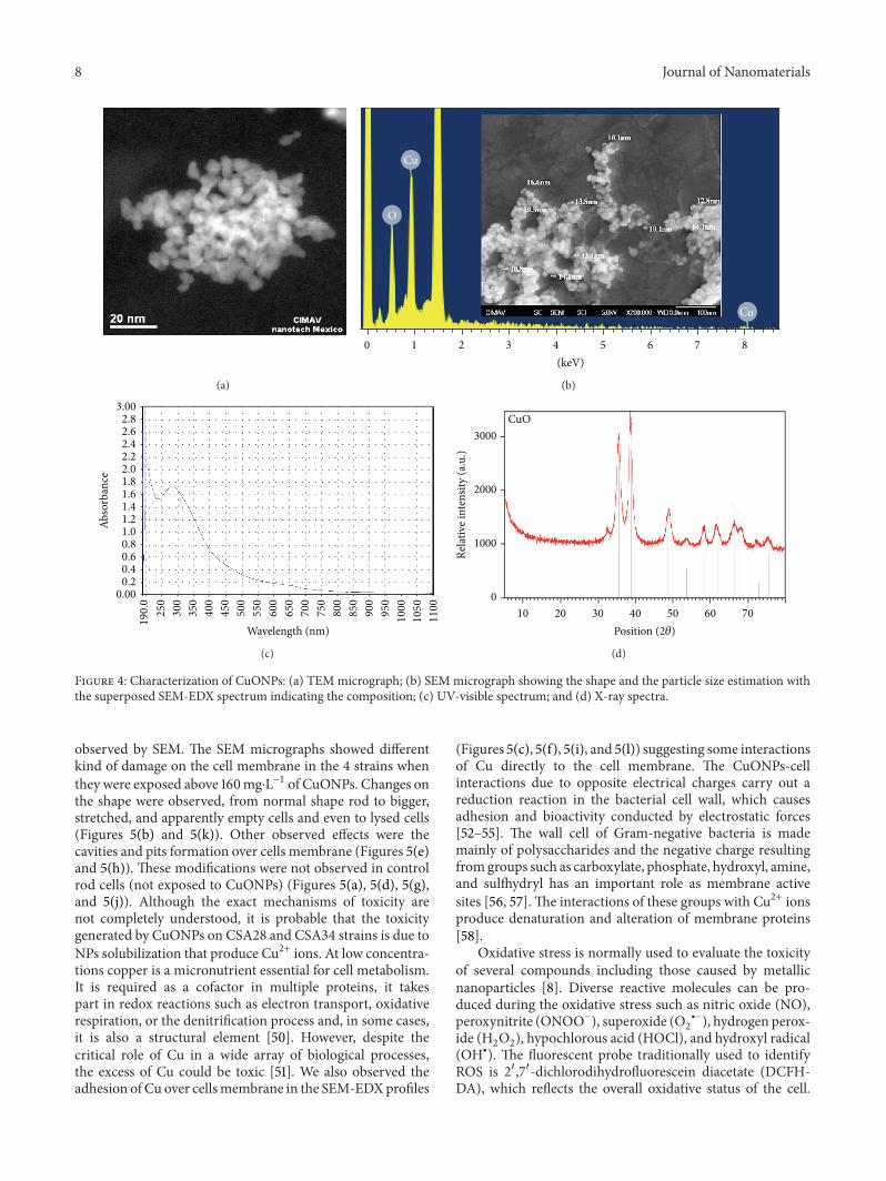