research article - asian pacific journal of cancer...
TRANSCRIPT

Asian Pacific Journal of Cancer Prevention, Vol 13, 2012 95
DOI:http://dx.doi.org/10.7314/APJCP.2012.13.KKSuppl.95Aberrant Expression of CD44 in Bile Duct Cancer Correlates with Poor Prognosis
Asian Pacific J Cancer Prev, 13, 95-99
Introduction
CD44 is a single pass transmembrane glycoprotein and known as a receptor of hyaluronan (Culty et al., 1990). CD44 expresses in many isoforms according to the cell type and organs. The smallest isoform, is referred as “standard” form (CD44s) and the variant isoforms which are the combination of different alternatively spliced exons, is referred as CD44v (Tolg et al., 1993). CD44 is widely distributed on many cell types and participates in many cellular processes, including the regulation of cell growth, survival, differentiation and motility (Ponta et al., 2003). Alteration of CD44 expression or dysfunction contributes to numerous pathological conditions, including cancers. However, the association of CD44 and its variants with tumor behavior varies depending on tumor type and the organ site of tumor. Bile duct cancer is a relatively uncommon and poorly understood cancer. There are several types of cancers arise from epithelial cell lining of bile ducts. It can be found as a cystic lesion as cystadenocarcinoma (CAC) or as a more aggressive cancer, cholangiocarcinoma (CCA). A cystic tumor, either benign (cystadenoma) or malignant
1Department of Biochemistry, 2Department of Pathology, 3Department of Surgery, 4Liver Fluke and Cholangiocarcinoma Research Center, Faculty of Medicine, Khon Kaen University, Thailand *For correspondence: [email protected]
Abstract
CCD44,atransmembraneglycoproteinreceptor,playssignificantrolesincellmigration,differentiation,andsurvivalsignalingwhichareimportantforbothnormalandcancercells.Inthisstudy,weexaminedtheexpressionofallisoformsofCD44byimmunohistochemistryin3casesofbiliarycystadenoma,15casesofnon-invasivecystadenocarcinoma(CAC)bileducttumors,and67casesoftheaggressivebileducttumor,cholangiocarcinoma(CCA).NormalbileductepitheliaatdifferentsegmentsalongthebiliarytreedidnotexpressCD44.However,normalbiliarycellsofthelargebileductadjacenttotumorareasanddysplasticbiliarycellsinCCAtissueswerepositive.CD44wasnotexpressedincystadenomasandthemajorityofCACs.TwoCACcaseswithshortsurvivalandthemajorityofCCAaberrantlyexpressedCD44.TheseobservationssuggestimportantrolesforCD44intheearlystageofcarcinogenesisandprogressionofbileductcancer.Regardlessofthetypeofbileducttumor,CACorCCApatientswithpositiveCD44expressioninbiliaryepitheliahadsignificantshortersurvivalthanthosewithnegativeCD44.AberrantexpressionofCD44inCACorCCAtissuesmayindicateanunfavorablepatientoutcomeandmayserveasausefulpracticaladjuncttoconventionalprognosticindicatorsforbileductcancer. Keywords: CD44, isoform - variant - cholangiocarcinoma - prognosis
RESEARCH ARTICLE
AberrantExpressionofCD44inBileDuctCancerCorrelateswith Poor Prognosis Kunlathida Kunlabut1,4, Kulthida Vaeteewoottacharn1,4, Chaisiri Wongkham1,4, NarongKhuntikeo3,4, Sakda Waraasawapati2,4,Chawalit Pairojkul2,4, Sopit Wongkham1,4*
CAC, is lined by the epithelium with papillary infoldings and is usually multilocular and contains mucoid fluid. On the other hand, CCA arises from any portion of the intra- or extra-hepatic bile duct epithelium and is classified anatomically into intrahepatic CCA and extrahepatic CCA. The intrahepatic CCA can be further classified macroscopically into mass forming, periductal infiltrating and intraductal growth types; which are believed to have different nature and gene expression patterns. CAC and CCA are relatively rare tumors in most populations. CAC accounts for less than 5% of the reported cystic lesions of the liver (Wheeler and Edmondson, 1985; Devaney et al., 1994), whereas CCA is the second among primary malignant liver tumors; about 15% of liver cancers are estimated to be CCA (Anon., 1990; Parkin et al., 1993; Yamanaka et al., 1995). At the present, detection of bile duct carcinomas is based on clinical examination and imaging techniques; and complete surgical resection is the mainstay of curative treatment with good prognosis. However, because of their rarity and nonspecific presentations, the diagnosis is often delayed resulting in inappropriate treatment modalities, morbidity and mortality (Lempinen et al., 2005; Seidel

Kunlathida Kunlabut et al
Asian Pacific Journal of Cancer Prevention, Vol 13, 201296
et al., 2007). Therefore, it is important to search for a prognostic marker which supports the clinicians for an appropriate and effective judgment of treatment to improve the survival of patients with this malignancy. There are a few studies on CD44 expression in bile duct cancer. As a general agreement, CD44 is not expressed in normal bile duct epithelium (Ashida et al., 1998; Sato et al., 2006; Pongcharoen et al., 2011). The over-expressions of CD44s and various CD44v have been pronounced. CD44v3, v6, and v5 expression was reported to be associated with tumor progression in different CCA types (Ashida et al., 1998; Mikami et al., 2001; Sato et al., 2006). Silencing of CD44 expression resulted in the decreased invasiveness of CCA cell lines in vitro (Pongcharoen et al., 2011). In this study, we examined the expression of CD44 in two different types of bile duct cancers, CAC, a rare but good prognosis type and CCA, a frequently found with poor prognosis. In addition, 3 cases of biliary cystadenoma were also examined. The results show that regardless of the nature of the tumors, CD44 expression of tumors indicates aggressiveness and poor outcome of patients.
Materials and Methods
Tissue specimens Formalin-fixed paraffin embedded tissues were obtained from the tissue bank of the Liver Fluke and Cholangiocarcinoma Research Center, Faculty of Medicine, Khon Kaen University, Thailand. The study was approved by the Khon Kaen University Ethics Committee for Human Research, and the informed consent was obtained from each subject. Tissues included in this study were histologically proved biliary cystadenoma (n=3), CAC (n=15) and CCA (n=67). Tumor staging was judged according to TNM classification by the American Joint Committee 6th edition in the year 2006 (Greene and American Joint Committee on Cancer., 2006). Histopathological grading of CCA was done according to the WHO classification (Bosman et al. ,2000) as papillary and non-papillary type. As a representative of normal bile duct, haemangioma liver (n =10) sections were used to examine the CD44 expression in the biliary epithelium along the biliary tree. The 3 cystadenoma cases were all males with median age of 62 years (ranged 52-66 years); CAC cases were 2 males and 13 females (male:female=0.15:1) with median age of 51 years (ranged 36-70 years); and CCA were 41 males and 26 females (male:female=1.6:1) with median age of 56 years (ranged 39-75 years). Clinical data were reviewed from the medical records and the overall survivals were recorded from the date of operation till 25 April 2011. Survivals of all patients were more than 60 days and hence no peri-operative death was observed in our series.
Immunohistochemistry The immunohistochemical staining of CD44 was performed according to the standard protocol. The expression of CD44 was examined using monoclonal antibody (IM7; Biolegend, San Diego, CA) which detects
all isoforms of CD44. Briefly, the deparaffinized tissue sections were treated with 0.3% H2O2 for 30 min to block endogenous peroxidase activity and heated with 10 mM Tris-EDTA buffer, pH 9.0 in a pressure cooker for 3 min to enhance antigen expression. The slides were incubated with 1:25 diluted rat anti-CD44 monoclonal antibody at room temperature, overnight and with 1:100 diluted HRP-conjugated goat anti-rat IgG (Zymed Laboratories; San Francisco, CA) at room temperature, for 2 h. The peroxidase activity was visualized using diaminobenzidine tetrahydroxychloride solution (DAB; Dako; Glostrup, Denmark). The expression of CD44 was evaluated independently by two observers who were blinded to the clinical outcome and the results were graded into either the negative group with no CD44 expression or the positive group with various expression of CD44.
Statistical analysis The statistical significance between the presence of CD44 and the clinicopathological factors was determined using the chi-square test. The overall survival was calculated according to the Kaplan-Meier method and the log-rank test was used for statistical comparisons between curves. Multivariate analysis was performed using Cox proportional hazard model. P value<0.05 was considered statistically significant.
Results
CD44 expression in cystadenoma and CAC We first examined the CD44 expression in the biliary
Figure1.ImmunohistochemistryofCD44inNormalBileDuctEpithelia.(A) interlobular bile ducts, (B) septal bile ducts, (C) Large bile duct. Arrows indicate bile duct epithelium. Original magnification 200x.
Figure2.TheimmunohistochemistryStainingofCD44inBiliaryCysticTumors.(A) Biliary cystadenoma (200x) and (B) most of the of cystadenocarcinoma (100x) showed negative staining for CD44; (C, D) focal staining of CD44 was found in 2 cases of biliary cystadenocarcinoma (200x, 100x).
Asian Pacific Journal of Cancer Prevention, Vol 13, 2012 97
DOI:http://dx.doi.org/10.7314/APJCP.2012.13.KKSuppl.95Aberrant Expression of CD44 in Bile Duct Cancer Correlates with Poor Prognosis
0
25.0
50.0
75.0
100.0
New
ly d
iagn
osed
with
out
trea
tmen
t
New
ly d
iagn
osed
with
tre
atm
ent
Pers
iste
nce
or r
ecur
renc
e
Rem
issi
on
Non
e
Chem
othe
rapy
Radi
othe
rapy
Conc
urre
nt c
hem
orad
iatio
n
10.3
0
12.8
30.025.0
20.310.16.3
51.7
75.051.1
30.031.354.2
46.856.3
27.625.033.130.031.3
23.738.0
31.3
0
25.0
50.0
75.0
100.0
New
ly d
iagn
osed
with
out
trea
tmen
t
New
ly d
iagn
osed
with
tre
atm
ent
Pers
iste
nce
or r
ecur
renc
e
Rem
issi
on
Non
e
Chem
othe
rapy
Radi
othe
rapy
Conc
urre
nt c
hem
orad
iatio
n
10.3
0
12.8
30.025.0
20.310.16.3
51.7
75.051.1
30.031.354.2
46.856.3
27.625.033.130.031.3
23.738.0
31.3
Table1.CorrelationbetweenCD44ExpressionandClinicopathologicalFactorsofCCAPatientsVariables N CD44 expression Ve- Ve+ P
Age ≤ 56 years 37 16 21 0.779 > 56 years 30 14 16 Gender Female 26 11 15 0.746 Male 41 19 22 Tumor location ECC 13 4 9 0.258 ICC 54 26 28 Gross type MF 53 22 31 0.295 others 14 8 6 Tumor size ≤ 5 cm. 21 12 9 0.148 > 5 cm. 46 18 28 Histological type Papillary 27 10 17 0.295 Non-papillary 40 20 20 Tumor stage I-IIIC 33 14 19 0.148 IV 11 2 9 Lymph node present 30 12 18 0.622invasion absent 37 18 19 Vascular invasion present 15 5 10 0.385 absent 52 25 27 Neural invasion present 1 0 1 1 absent 66 30 36 Survival < 2 years 55 21 34 0.027 ≥ 2 years 12 9 3
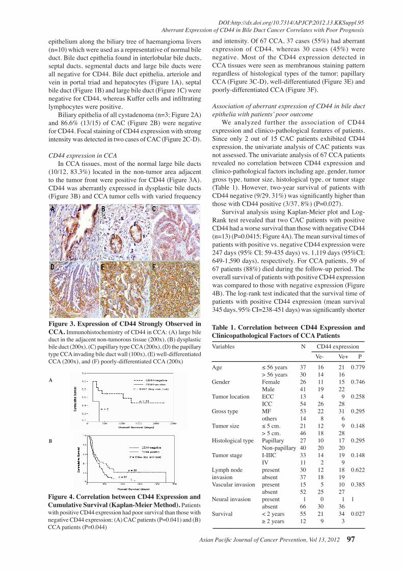
Figure3.ExpressionofCD44StronglyObservedinCCA.Immunohistochemistry of CD44 in CCA: (A) large bile duct in the adjacent non-tumorous tissue (200x), (B) dysplastic bile duct (200x), (C) papillary type CCA (200x), (D) the papillary type CCA invading bile duct wall (100x), (E) well-differentiated CCA (200x), and (F) poorly-differentiated CCA (200x)
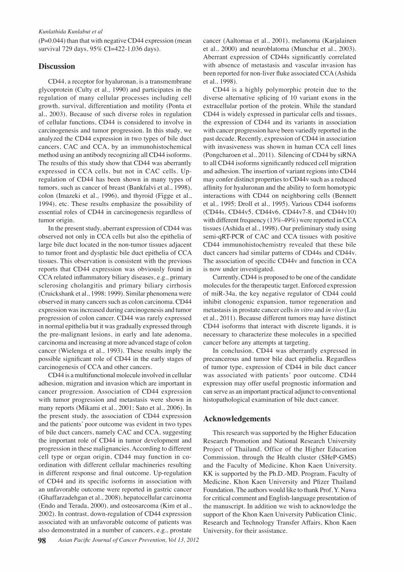
Figure4.CorrelationbetweenCD44ExpressionandCumulativeSurvival(Kaplan-MeierMethod).Patients with positive CD44 expression had poor survival than those with negative CD44 expression; (A) CAC patients (P=0.041) and (B) CCA patients (P=0.044)
and intensity. Of 67 CCA, 37 cases (55%) had aberrant expression of CD44, whereas 30 cases (45%) were negative. Most of the CD44 expression detected in CCA tissues were seen as membranous staining pattern regardless of histological types of the tumor; papillary CCA (Figure 3C-D), well-differentiated (Figure 3E) and poorly-differentiated CCA (Figure 3F).
Association of aberrant expression of CD44 in bile duct epithelia with patients’ poor outcome We analyzed further the association of CD44 expression and clinico-pathological features of patients. Since only 2 out of 15 CAC patients exhibited CD44 expression, the univariate analysis of CAC patients was not assessed. The univariate analysis of 67 CCA patients revealed no correlation between CD44 expression and clinico-pathological factors including age, gender, tumor gross type, tumor size, histological type, or tumor stage (Table 1). However, two-year survival of patients with CD44 negative (9/29, 31%) was significantly higher than those with CD44 positive (3/37, 8%) (P=0.027). Survival analysis using Kaplan-Meier plot and Log-Rank test revealed that two CAC patients with positive CD44 had a worse survival than those with negative CD44 (n=13) (P=0.0415; Figure 4A). The mean survival times of patients with positive vs. negative CD44 expression were 247 days (95% CI; 59-435 days) vs. 1,119 days (95%CI; 649-1,590 days), respectively. For CCA patients, 59 of 67 patients (88%) died during the follow-up period. The overall survival of patients with positive CD44 expression was compared to those with negative expression (Figure 4B). The log-rank test indicated that the survival time of patients with positive CD44 expression (mean survival 345 days, 95% CI=238-451 days) was significantly shorter
epithelium along the biliary tree of haemangioma livers (n=10) which were used as a representative of normal bile duct. Bile duct epithelia found in interlobular bile ducts, septal ducts, segmental ducts and large bile ducts were all negative for CD44. Bile duct epithelia, arteriole and vein in portal triad and hepatocytes (Figure 1A), septal bile duct (Figure 1B) and large bile duct (Figure 1C) were negative for CD44, whereas Kuffer cells and infiltrating lymphocytes were positive. Biliary epithelia of all cystadenoma (n=3; Figure 2A) and 86.6% (13/15) of CAC (Figure 2B) were negative for CD44. Focal staining of CD44 expression with strong intensity was detected in two cases of CAC (Figure 2C-D).
CD44 expression in CCA In CCA tissues, most of the normal large bile ducts (10/12, 83.3%) located in the non-tumor area adjacent to the tumor front were positive for CD44 (Figure 3A). CD44 was aberrantly expressed in dysplastic bile ducts (Figure 3B) and CCA tumor cells with varied frequency
A
B
Kunlathida Kunlabut et al
Asian Pacific Journal of Cancer Prevention, Vol 13, 201298
(P=0.044) than that with negative CD44 expression (mean survival 729 days, 95% CI=422-1,036 days).
Discussion
CD44, a receptor for hyaluronan, is a transmembrane glycoprotein (Culty et al., 1990) and participates in the regulation of many cellular processes including cell growth, survival, differentiation and motility (Ponta et al., 2003). Because of such diverse roles in regulation of cellular functions, CD44 is considered to involve in carcinogenesis and tumor progression. In this study, we analyzed the CD44 expression in two types of bile duct cancers, CAC and CCA, by an immunohistochemical method using an antibody recognizing all CD44 isoforms. The results of this study show that CD44 was aberrantly expressed in CCA cells, but not in CAC cells. Up-regulation of CD44 has been shown in many types of tumors, such as cancer of breast (Bankfalvi et al., 1998), colon (Imazeki et al., 1996), and thyroid (Figge et al., 1994), etc. These results emphasize the possibility of essential roles of CD44 in carcinogenesis regardless of tumor origin.
In the present study, aberrant expression of CD44 was observed not only in CCA cells but also the epithelia of large bile duct located in the non-tumor tissues adjacent to tumor front and dysplastic bile duct epithelia of CCA tissues. This observation is consistent with the previous reports that CD44 expression was obviously found in CCA related inflammatory biliary diseases, e.g., primary sclerosing cholangitis and primary biliary cirrhosis (Cruickshank et al., 1998; 1999). Similar phenomena were observed in many cancers such as colon carcinoma. CD44 expression was increased during carcinogenesis and tumor progression of colon cancer. CD44 was rarely expressed in normal epithelia but it was gradually expressed through the pre-malignant lesions, in early and late adenoma, carcinoma and increasing at more advanced stage of colon cancer (Wielenga et al., 1993). These results imply the possible significant role of CD44 in the early stages of carcinogenesis of CCA and other cancers.
CD44 is a multifunctional molecule involved in cellular adhesion, migration and invasion which are important in cancer progression. Association of CD44 expression with tumor progression and metastasis were shown in many reports (Mikami et al., 2001; Sato et al., 2006). In the present study, the association of CD44 expression and the patients’ poor outcome was evident in two types of bile duct cancers, namely CAC and CCA, suggesting the important role of CD44 in tumor development and progression in these malignancies. According to different cell type or organ origin, CD44 may function in co-ordination with different cellular machineries resulting in different response and final outcome. Up-regulation of CD44 and its specific isoforms in association with an unfavorable outcome were reported in gastric cancer (Ghaffarzadehgan et al., 2008), hepatocellular carcinoma (Endo and Terada, 2000), and osteosarcoma (Kim et al., 2002). In contrast, down-regulation of CD44 expression associated with an unfavorable outcome of patients was also demonstrated in a number of cancers, e.g., prostate
cancer (Aaltomaa et al., 2001), melanoma (Karjalainen et al., 2000) and neuroblatoma (Munchar et al., 2003). Aberrant expression of CD44s significantly correlated with absence of metastasis and vascular invasion has been reported for non-liver fluke associated CCA (Ashida et al., 1998).
CD44 is a highly polymorphic protein due to the diverse alternative splicing of 10 variant exons in the extracellular portion of the protein. While the standard CD44 is widely expressed in particular cells and tissues, the expression of CD44 and its variants in association with cancer progression have been variedly reported in the past decade. Recently, expression of CD44 in association with invasiveness was shown in human CCA cell lines (Pongcharoen et al., 2011). Silencing of CD44 by siRNA to all CD44 isoforms significantly reduced cell migration and adhesion. The insertion of variant regions into CD44 may confer distinct properties to CD44v such as a reduced affinity for hyaluronan and the ability to form homotypic interactions with CD44 on neighboring cells (Bennett et al., 1995; Droll et al., 1995). Various CD44 isoforms (CD44s, CD44v5, CD44v6, CD44v7-8, and CD44v10) with different frequency (13%-49%) were reported in CCA tissues (Ashida et al., 1998). Our preliminary study using semi-qRT-PCR of CAC and CCA tissues with positive CD44 immunohistochemistry revealed that these bile duct cancers had similar patterns of CD44s and CD44v. The association of specific CD44v and function in CCA is now under investigated.
Currently, CD44 is proposed to be one of the candidate molecules for the therapeutic target. Enforced expression of miR-34a, the key negative regulator of CD44 could inhibit clonogenic expansion, tumor regeneration and metastasis in prostate cancer cells in vitro and in vivo (Liu et al., 2011). Because different tumors may have distinct CD44 isoforms that interact with discrete ligands, it is necessary to characterize these molecules in a specified cancer before any attempts at targeting.
In conclusion, CD44 was aberrantly expressed in precancerous and tumor bile duct epithelia. Regardless of tumor type, expression of CD44 in bile duct cancer was associated with patients’ poor outcome. CD44 expression may offer useful prognostic information and can serve as an important practical adjunct to conventional histopathological examination of bile duct cancer.
Acknowledgements This research was supported by the Higher Education
Research Promotion and National Research University Project of Thailand, Office of the Higher Education Commission, through the Health cluster (SHeP-GMS) and the Faculty of Medicine, Khon Kaen University. KK is supported by the Ph.D.-MD. Program, Faculty of Medicine, Khon Kaen University and Pfizer Thailand Foundation. The authors would like to thank Prof. Y. Nawa for critical comment and English-language presentation of the manuscript. In addition we wish to acknowledge the support of the Khon Kaen University Publication Clinic, Research and Technology Transfer Affairs, Khon Kaen University, for their assistance.

Asian Pacific Journal of Cancer Prevention, Vol 13, 2012 99
DOI:http://dx.doi.org/10.7314/APJCP.2012.13.KKSuppl.95Aberrant Expression of CD44 in Bile Duct Cancer Correlates with Poor Prognosis
References
Aaltomaa S, Lipponen P, Ala-Opas M, Kosma VM (2001). Expression and prognostic value of CD44 standard and variant v3 and v6 isoforms in prostate cancer. Eur Urol, 39, 138-44.
Anon. (1990). Primary liver cancer in Japan. Clinicopathologic features and results of surgical treatment. Liver Cancer Study Group of Japan. Ann Surg, 211, 277-87.
Ashida K, Terada T, Kitamura Y, Kaibara N (1998). Expression of E-cadherin, alpha-catenin, beta-catenin, and CD44 (standard and variant isoforms) in human cholangiocarcinoma: an immunohistochemical study. Hepatology, 27, 974-82.
Bankfalvi A, Terpe HJ, Breukelmann D, et al (1998). Gains and losses of CD44 expression during breast carcinogenesis and tumour progression. Histopathology, 33, 107-16.
Bennett KL, Jackson DG, Simon JC, et al (1995). CD44 isoforms containing exon V3 are responsible for the presentation of heparin-binding growth factor. J Cell Biol, 128, 687-98.
Bosman FT, World Health Organization., International Agency for Research on Cancer. WHO classification of tumours of the digestive system. IARC Press, Lyon.
Cruickshank SM, Southgate J, Selby PJ, Trejdosiewicz LK (1998). Expression and cytokine regulation of immune recognition elements by normal human biliary epithelial and established liver cell lines in vitro. J Hepatol, 29, 550-8.
Cruickshank SM, Southgate J, Wyatt JI, Selby PJ, Trejdosiewicz LK (1999). Expression of CD44 on bile ducts in primary sclerosing cholangitis and primary biliary cirrhosis. J Clin Pathol, 52, 730-4.
Culty M, Miyake K, Kincade PW, Sikorski E, Butcher EC, et al (1990). The hyaluronate receptor is a member of the CD44 (H-CAM) family of cell surface glycoproteins. J Cell Biol, 111, 2765-74.
Devaney K, Goodman ZD, Ishak KG (1994). Hepatobiliary cystadenoma and cystadenocarcinoma. A light microscopic and immunohistochemical study of 70 patients. Am J Surg Pathol, 18, 1078-91.
Droll A, Dougherty ST, Chiu RK, Dirks JF, McBride WH, et al (1995). Adhesive interactions between alternatively spliced CD44 isoforms. J Biol Chem, 270, 11567-73.
Endo K, Terada T (2000). Protein expression of CD44 (standard and variant isoforms) in hepatocellular carcinoma: relationships with tumor grade, clinicopathologic parameters, p53 expression, and patient survival. J Hepatol, 32, 78-84.
Figge J, del Rosario AD, Gerasimov G, Dedov I, Bronstein M, et al (1994). Preferential expression of the cell adhesion molecule CD44 in papillary thyroid carcinoma. Exp Mol Pathol, 61, 203-11.
Ghaffarzadehgan K, Jafarzadeh M, Raziee HR, Sima HR, Esmaili-Shandiz E, et al (2008). Expression of cell adhesion molecule CD44 in gastric adenocarcinoma and its prognostic importance. World J Gastroenterol, 14, 6376-81.
Greene FL, American Joint Committee on Cancer. (2006). AJCC cancer staging atlas. Springer, New York, NY.
Imazeki F, Yokosuka O, Yamaguchi T, Ohto M, Isono K, et al (1996). Expression of variant CD44-messenger RNA in colorectal adenocarcinomas and adenomatous polyps in humans. Gastroenterology, 110, 362-8.
Karjalainen JM, Tammi RH, Tammi MI, et al (2000). Reduced level of CD44 and hyaluronan associated with unfavorable prognosis in clinical stage I cutaneous melanoma. Am J Pathol, 157, 957-65.
Kim HS, Park YB, Oh JH, et al (2002). Expression of CD44 isoforms correlates with the metastatic potential of osteosarcoma. Clin Orthop Relat Res, 184-90.
Lempinen M, Halme L, Numminen K, Arola J, Nordin A, et al
(2005). Spontaneous rupture of a hepatic cystadenoma and cystadenocarcinoma: report of two cases. J Hepatobiliary Pancreat Surg, 12, 409-14.
Liu C, Kelnar K, Liu B, et al (2011). The microRNA miR-34a inhibits prostate cancer stem cells and metastasis by directly repressing CD44. Nat Med, 17, 211-5.
Mikami T, Saegusa M, Mitomi H, Yanagisawa N, Ichinoe M, et al (2001). Significant correlations of E-cadherin, catenin, and CD44 variant form expression with carcinoma cell differentiation and prognosis of extrahepatic bile duct carcinomas. Am J Clin Pathol, 116, 369-76.
Munchar MJ, Sharifah NA, Jamal R, Looi LM (2003). CD44s expression correlated with the International Neuroblastoma Pathology Classification (Shimada system) for neuroblastic tumours. Pathology, 35, 125-9.
Naor D, Nedvetzki S, Golan I, Melnik L, Faitelson Y (2002). CD44 in cancer. Crit Rev Clin Lab Sci, 39, 527-79.
Parkin DM, Ohshima H, Srivatanakul P, Vatanasapt V (1993). Cholangiocarcinoma: epidemiology, mechanisms of carcinogenesis and prevention. Cancer Epidemiol Biomarkers Prev, 2, 537-44.
Picker LJ, Nakache M, Butcher EC (1989). Monoclonal antibodies to human lymphocyte homing receptors define a novel class of adhesion molecules on diverse cell types. J Cell Biol, 109, 927-37.
Pongcharoen P, Jinawath A, Tohtong R (2011). Silencing of CD44 by siRNA suppressed invasion, migration and adhesion to matrix, but not secretion of MMPs, of cholangiocarcinoma cells. Clin Exp Metastasis, 28, 827-39.
Ponta H, Sherman L, Herrlich PA (2003). CD44: from adhesion molecules to signalling regulators. Nat Rev Mol Cell Biol, 4, 33-45.
Sato K, Murai H, Ueda Y, Katsuda S (2006). Intrahepatic sarcomatoid cholangiocarcinoma of round cell variant: a case report and immunohistochemical studies. Virchows Arch, 449, 585-90.
Seidel R, Weinrich M, Pistorius G, Fries P, Schneider G (2007). Biliary cystadenoma of the left intrahepatic duct (2007: 2b). Eur Radiol, 17, 1380-3.
Tolg C, Hofmann M, Herrlich P, Ponta H (1993). Splicing choice from ten variant exons establishes CD44 variability. Nucleic Acids Res, 21, 1225-9.
Wheeler DA, Edmondson HA (1985). Cystadenoma with mesenchymal stroma (CMS) in the liver and bile ducts. A clinicopathologic study of 17 cases, 4 with malignant change. Cancer, 56, 1434-45.
Wielenga VJ, Heider KH, Offerhaus GJ, Adolf GR, van den Berg FM, et al (1993). Expression of CD44 variant proteins in human colorectal cancer is related to tumor progression. Cancer Res, 53, 4754-6.
Yamanaka N, Okamoto E, Ando T, Oriyama T, Fujimoto J, et al (1995). Clinicopathologic spectrum of resected extraductal mass-forming intrahepatic cholangiocarcinoma. Cancer, 76, 2449-56.