research ar ticle - rp2u.unsyiah.ac.id
TRANSCRIPT
/
Research Article
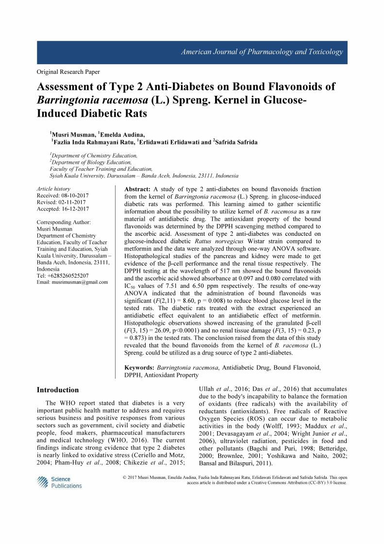
Assessment of Type 2 Anti-Diabetes on Bound Flavonoids of Barringtonia racemosa (L.) Spreng. Kernel in
Glucose-Induced Diabetic Rats (https://thescipub.com/abstract/10.3844/ajptsp.2017.48.61)
Musri Musman, Emelda Audina, Fazlia Inda Rahmayani Ratu, Erlidawati Erlidawati and Safrida Safrida
Pages :
48-61
DOI :
10.3844/ajptsp.2017.48.61
Published On :
December 16, 2017
Read more (https://thescipub.com/abstract/10.3844/ajptsp.2017.48.61) Download PDF (https://thescipub.com/pdf/10.3844/ajptsp.2017.48.61)
Modulatory Effects of Bryophyllum Pinnatum Leaves Extract on Peroxidation Indices of CCl Induced-
Hepatotoxicity in Wistar Albino Rats (https://thescipub.com/abstract/10.3844/ajptsp.2017.62.67)
Chioma Assumpta Anosike, Sophia Uyo Mokwunye, Victor Eshu Okpashi and Obiorah Abonyi
Pages :
62-67
DOI :
10.3844/ajptsp.2017.62.67
Published On :
December 17, 2017
Read more (https://thescipub.com/abstract/10.3844/ajptsp.2017.62.67) Download PDF (https://thescipub.com/pdf/10.3844/ajptsp.2017.62.67)
SIGN IN
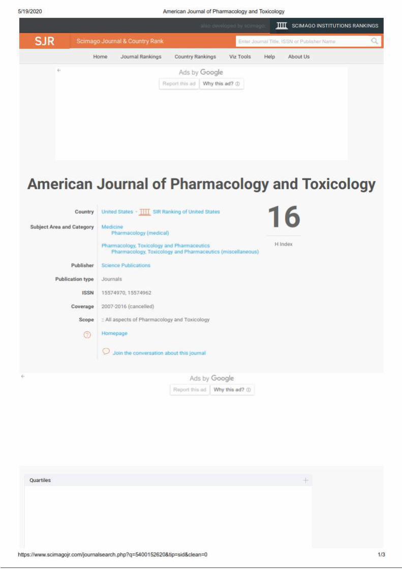
American Journal of Pharmacology and Toxicology
4

/
Most Recent Most Cited Most Downloaded Most Viewed
ISSN Print: 1557-4962
ISSN Online: 1557-4970
Journal Home (https://thescipub.com/journals/ajpt/)
Continuous Publication (https://thescipub.com/journals/ajpt/continuous_publication)
Abstracting and Indexing (https://thescipub.com/journals/ajpt/indexing)
Online First (https://thescipub.com/journals/ajpt/aof)
Archive (https://thescipub.com/journals/ajpt/archive)
Editorial Board (https://thescipub.com/journals/ajpt/editors)
Instructions to Authors (https://thescipub.com/journals/ajpt/instructions)
Publication Ethics (https://thescipub.com/journals/ajpt/ethics)
Editorial Work�ow (https://thescipub.com/journals/ajpt/work�ow)
Publication Charges (https://thescipub.com/journals/ajpt/apc)
Open Special Issues (https://thescipub.com/journals/ajpt/osi)
Published Special Issues (https://thescipub.com/journals/ajpt/psi)
Submit an Article (https://thescipub.com/es)
Copyright 2020 © Science Publications
Twitter (https://twitter.com/scipub) Facebook (https://www.facebook.com/SciPub)
/
Edward. J. Calabrese
University of Massachusetts Amherst
United States
Expertise: toxicology, risk assessment, environmental health, dose response, hormesis
SIGN IN
Associate Editors
American Journal of Pharmacology and Toxicology

/
/
Expertise: Formulation science, pharmaceutics, drug delivery, transdermal systems, microneedles, photodynamic therapy
�
/
Editorial Board Members
/
/ Stringer
/
/

/
/
/
Vincenzo Calderone

© 2017 Musri Musman, Emelda Audina, Fazlia Inda Rahmayani Ratu, Erlidawati Erlidawati and Safrida Safrida. This open
access article is distributed under a Creative Commons Attribution (CC-BY) 3.0 license.
American Journal of Pharmacology and Toxicology
Original Research Paper
Assessment of Type 2 Anti-Diabetes on Bound Flavonoids of
Barringtonia racemosa (L.) Spreng. Kernel in Glucose-
Induced Diabetic Rats
1Musri Musman,
1Emelda Audina,
1Fazlia Inda Rahmayani Ratu,
1Erlidawati Erlidawati and
2Safrida Safrida
1Department of Chemistry Education, 2Department of Biology Education,
Faculty of Teacher Training and Education,
Syiah Kuala University, Darussalam – Banda Aceh, Indonesia, 23111, Indonesia
Article history
Received: 08-10-2017
Revised: 02-11-2017
Accepted: 16-12-2017
Corresponding Author:
Musri Musman
Department of Chemistry
Education, Faculty of Teacher
Training and Education, Syiah
Kuala University, Darussalam –
Banda Aceh, Indonesia, 23111,
Indonesia
Tel: +6285260525207 Email: [email protected]
Abstract: A study of type 2 anti-diabetes on bound flavonoids fraction
from the kernel of Barringtonia racemosa (L.) Spreng. in glucose-induced
diabetic rats was performed. This learning aimed to gather scientific
information about the possibility to utilize kernel of B. racemosa as a raw
material of antidiabetic drug. The antioxidant property of the bound
flavonoids was determined by the DPPH scavenging method compared to
the ascorbic acid. Assessment of type 2 anti-diabetes was conducted on
glucose-induced diabetic Rattus norvegicus Wistar strain compared to
metformin and the data were analyzed through one-way ANOVA software.
Histopathological studies of the pancreas and kidney were made to get
evidence of the β-cell performance and the renal tissue respectively. The
DPPH testing at the wavelength of 517 nm showed the bound flavonoids
and the ascorbic acid showed absorbance at 0.097 and 0.080 correlated with
IC50 values of 7.51 and 6.50 ppm respectively. The results of one-way
ANOVA indicated that the administration of bound flavonoids was
significant (F(2,11) = 8.60, p = 0.008) to reduce blood glucose level in the
tested rats. The diabetic rats treated with the extract experienced an
antidiabetic effect equivalent to an antidiabetic effect of metformin.
Histopathologic observations showed increasing of the granulated β-cell
(F(3, 15) = 26.09, p<0.0001) and no renal tissue damage (F(3, 15) = 0.23, p
= 0.873) in the tested rats. The conclusion raised from the data of this study
revealed that the bound flavonoids from the kernel of B. racemosa (L.)
Spreng. could be utilized as a drug source of type 2 anti-diabetes.
Keywords: Barringtonia racemosa, Antidiabetic Drug, Bound Flavonoid,
DPPH, Antioxidant Property
Introduction
The WHO report stated that diabetes is a very
important public health matter to address and requires
serious business and positive responses from various
sectors such as government, civil society and diabetic
people, food makers, pharmaceutical manufacturers
and medical technology (WHO, 2016). The current
findings indicate strong evidence that type 2 diabetes
is nearly linked to oxidative stress (Ceriello and Motz,
2004; Pham-Huy et al., 2008; Chikezie et al., 2015;
Ullah et al., 2016; Das et al., 2016) that accumulates
due to the body's incapability to balance the formation
of oxidants (free radicals) with the availability of
reductants (antioxidants). Free radicals of Reactive
Oxygen Species (ROS) can occur due to metabolic
activities in the body (Wolff, 1993; Maddux et al.,
2001; Devasagayam et al., 2004; Wright Junior et al.,
2006), ultraviolet radiation, pesticides in food and
other pollutants (Bagchi and Puri, 1998; Betteridge,
2000; Brownlee, 2001; Yoshikawa and Naito, 2002;
Bansal and Bilaspuri, 2011).
Musri Musman et al. / American Journal of Pharmacology and Toxicology 2017, 12 (3): 48.61
DOI: 10.3844/ajptsp.2017.48.61
49
ROS are formed in the nucleus and also in the cell
membrane where it destroys biologically relevant
molecules such as DNA, proteins, sugars and lipids
(Young and Woodside, 2001). ROS have been
concerned with the initiation and complications of
diabetes mellitus (Martin et al., 2003; Yung et al., 2006;
Iqbal et al., 2016). Excessive ROS production causes
damage to cells and cell tissues. To halt the
production of ROS, a compound that has the property
of free radical deactivation is required. The amount of
ROS (oxidants) formed in cell tissues must be
balanced with the availability of antioxidants.
Therefore, the administration of external sources of
antioxidants can be applied in managing the ROS
(Halliwell, 1995; Laight et al., 2000; Kangralkar et al.,
2012; Santos-Buelga and Feliciano, 2017). One of the secondary metabolites that have antioxidant
property is flavonoid (Pietta, 2000; Rice-Evans, 2001;
Heim et al., 2002). Flavonoids are phenolic glycoside
compounds widely found in plants (Hahlbrock, 1981,
Ferreyra et al., 2012) and microorganisms (Das and
Rosazza, 2006; Wang et al., 2011). Flavonoids have the
ability to reduce the formation of free radicals and to
scavenge free radicals (Rice-Evans et al., 1996; 1997;
Amić et al., 2003; Ganesan et al., 2016). Consequently,
the exploration for phyto-nutraceutical substances with
antioxidative activity has been exaggerated in recent
years (Lobo et al., 2010; Pandeya et al., 2013) mainly in
connection with type 2 diabetes (Jakus, 2000;
Montonen et al., 2004; Kamalakkannan and Prince,
2006; Pandey and Rizvi, 2009; Dewanjee et al., 2011;
Wedick et al., 2012; Babu et al., 2013; Kan et al., 2015;
Li et al., 2016).
Nature has provided medicinal materials in its
surroundings. Humans have and will exploit the
medicinal plants to cope with the illness, i.e.,
Barringtonia racemosa (L.) Spreng. The plant is an
evergreens mangrove association that has been used as
an ethnomedicinal agent to treat a number of illnesses as
shown in Table 1. Outstanding to its wide range of
ethnopharmacological applications, researchers have
devoted their attention to finding out the
pharmacological activities of the plant as revealed in
Table 2 which may be used as a source of medicinal
substances. Considering the presence of secondary
metabolites in B. racemosa seeds as disclosed in Table 2,
the exploitation of the bound flavonoids to manage type
2 diabetes mellitus interest to be investigated. The bound
flavonoids have demonstrated very strong antioxidant
activity, high bioavailability and more ready absorbed in
metabolism (Nijveldt et al., 2001; Kumar and Pandey,
2013). Based on data searching via the internet,
information concerning to bioactive property as type 2
anti-diabetes originating from B. racemosa can not be
found (Hasan et al., 2000; Sun et al., 2006; Gowri et al.,
2009; Lim, 2012; Osman et al., 2015; Nazaruk and
Borzym-Kluczyk, 2015; Das et al., 2016; RIRDC, 2017).
Therefore, this study was the first investigation of type 2
antidiabetic property derived from the plant.
Table 1: Ethnopharmacological uses of B. racemosa
Part of the plant used Treatment Reference
leaves high blood pressure, itchiness, chickenpox Kabir et al. (2013; Osman et al., 2015).
leaves itch, chickenpox, rheumatism febrifuge Lim (2012).
seeds Tumors Thomas et al. (2002).
seeds, barks fish poison Manjunath (1948).
seeds colic, parturition, vermifuge, febrifuge Jayaweera (1981).
fruits poison wild pigs Manjunath (1948).
fruits hemicrania, ophthalmia, coughs, asthma, diarrhea Nadkarni (1976).
fruits coughs, asthma, diarrhea, eczema Jayaweera (1981).
fruits, barks fish poison Giesen et al. (2007).
barks Insecticide Manjunath (1948).
barks fish poison, skin diseases Jayaweera (1981).
roots deobstruent, relief in stomachache. Jayaweera (1981).
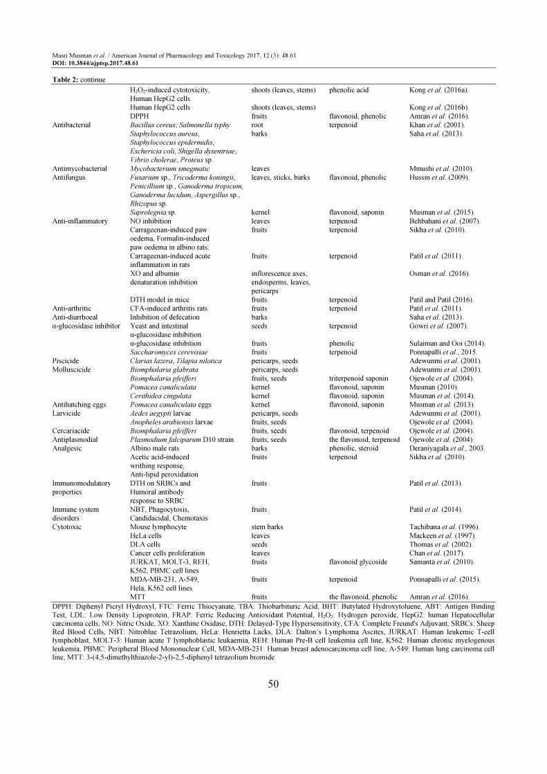
Table 2: Pharmacological activity of B. racemosa
Bioactive property Assay Part of the plant used Secondary metabolite Reference
Antioxidant DPPH, FTC, TBA leaves terpenoid Behbahani et al. (2007). BHT, Ascorbic Acid, α-tocopherol leaves, sticks, barks terpenoid Nurul-Mariam et al. (2008). DPPH, FTC, TBA leaves Zawawi et al. (2011). ABT, DPPH, Superoxide shoots (leaves, stems) flavonoid, terpenoid, Kong et al. (2012). anion radicals phenolic Inhibition of LDL, serum shoot (leaves, stems) phenolic Kong et al. (2014). and haemoglobin oxidation DPPH, FRAP fruits phenolic acid Sulaiman and Ooi (2014). DPPH, FTC, TBA leaves Dalila et al. (2015).
Musri Musman et al. / American Journal of Pharmacology and Toxicology 2017, 12 (3): 48.61
DOI: 10.3844/ajptsp.2017.48.61
50
Table 2: continue
H2O2-induced cytotoxicity, shoots (leaves, stems) phenolic acid Kong et al. (2016a).
Human HepG2 cells
Human HepG2 cells shoots (leaves, stems) Kong et al. (2016b).
DPPH fruits flavonoid, phenolic Amran et al. (2016).
Antibacterial Bacillus cereus; Salmonella typhy root terpenoid Khan et al. (2001).
Staphylococcus aureus, barks Saha et al. (2013).
Staphylococcus epidermidis,
Eschericia coli, Shigella dysentriae,
Vibrio cholerae, Proteus sp.
Antimycobacterial Mycobacterium smegmatic leaves Mmushi et al. (2010).
Antifungus Fusarium sp., Tricoderma koningii, leaves, sticks, barks flavonoid, phenolic Hussin et al. (2009).
Penicillium sp., Ganoderma tropicum,
Ganoderma lucidum, Aspergillus sp.,
Rhizopus sp.
Saprolegnia sp. kernel flavonoid, saponin Musman et al. (2015).
Anti-inflammatory NO inhibition leaves terpenoid Behbahani et al. (2007).
Carrageenan-induced paw fruits terpenoid Sikha et al. (2010).
oedema, Formalin-induced
paw oedema in albino rats.
Carrageenan-induced acute fruits terpenoid Patil et al. (2011).
inflammation in rats
XO and albumin inflorescence axes, Osman et al. (2016).
denaturation inhibition endosperms, leaves,
pericarps
DTH model in mice fruits terpenoid Patil and Patil (2016).
Anti-arthritic CFA-induced arthritis rats fruits terpenoid Patil et al. (2011).
Anti-diarrhoeal Inhibition of defecation barks Saha et al. (2013).
α-glucosidase inhibitor Yeast and intestinal seeds terpenoid Gowri et al. (2007).
α-glucosidase inhibition
α-glucosidase inhibition fruits phenolic Sulaiman and Ooi (2014).
Saccharomyces cerevisiae fruits terpenoid Ponnapalli et al., 2015.
Piscicide Clarias lazera, Tilapia nilotica pericarps, seeds Adewunmi et al. (2001).
Molluscicide Biomphalaria glabrata pericarps, seeds Adewunmi et al. (2001).
Biomphalaria pfeifferi fruits, seeds triterpenoid saponin Ojewole et al. (2004).
Pomacea canaliculata kernel flavonoid, saponin Musman (2010).
Cerithidea cingulata kernel flavonoid, saponin Musman et al. (2014).
Antihatching eggs Pomacea canaliculata eggs kernel flavonoid, saponin Musman et al. (2013).
Larvicide Aedes aegypti larvae pericarps, seeds Adewunmi et al. (2001).
Anopheles arabiensis larvae fruits, seeds Ojewole et al. (2004).
Cercariacide Biomphalaria pfeifferi fruits, seeds flavonoid, terpenoid Ojewole et al. (2004).
Antiplasmodial Plasmodium falciparum D10 strain fruits, seeds the flavonoid, terpenoid Ojewole et al. (2004).
Analgesic Albino male rats barks phenolic, steroid Deraniyagala et al., 2003.
Acetic acid-induced fruits terpenoid Sikha et al. (2010).
writhing response,
Anti-lipid peroxidation
Immunomodulatory DTH on SRBCs and fruits Patil et al. (2013).
properties Humoral antibody
response to SRBC
Immune system NBT, Phagocytosis, fruits Patil et al. (2014).
disorders Candidacidal, Chemotaxis
Cytotoxic Mouse lymphocyte stem barks Tachibana et al. (1996).
HeLa cells leaves Mackeen et al. (1997).
DLA cells seeds Thomas et al. (2002).
Cancer cells proliferation leaves Chan et al. (2017).
JURKAT, MOLT-3, REH, fruits flavonoid glycoside Samanta et al. (2010).
K562, PBMC cell lines
MDA-MB-231, A-549, fruits terpenoid Ponnapalli et al. (2015).
Hela, K562 cell lines
MTT fruits the flavonoid, phenolic Amran et al. (2016).
DPPH: Diphenyl Picryl Hydroxyl, FTC: Ferric Thiocyanate, TBA: Thiobarbituric Acid, BHT: Butylated Hydroxytoluene, ABT: Antigen Binding Test, LDL: Low Density Lipoprotein, FRAP: Ferric Reducing Antioxidant Potential, H2O2: Hydrogen peroxide, HepG2: human Hepatocellular carcinoma cells, NO: Nitric Oxide, XO: Xanthine Oxidase, DTH: Delayed-Type Hypersensitivity, CFA: Complete Freund's Adjuvant, SRBCs: Sheep Red Blood Cells, NBT: Nitroblue Tetrazolium, HeLa: Henrietta Lacks, DLA: Dalton’s Lymphoma Ascites, JURKAT: Human leukemic T-cell lymphoblast, MOLT-3: Human acute T lymphoblastic leukaemia, REH: Human Pre-B cell leukemia cell line, K562: Human chronic myelogenous leukemia, PBMC: Peripheral Blood Mononuclear Cell, MDA-MB-231: Human breast adenocarcinoma cell line, A-549: Human lung carcinoma cell line, MTT: 3-(4,5-dimethylthiazole-2-yl)-2,5-diphenyl tetrazolium bromide
Musri Musman et al. / American Journal of Pharmacology and Toxicology 2017, 12 (3): 48.61
DOI: 10.3844/ajptsp.2017.48.61
51
Materials and Methods
Chemicals, Drug and Kit
All of the analytical grade chemicals, drug and the kit
were procured commercially. The metformin
hydrochloride (C4H11N5, Dexa Medica, Indonesia) was
decided as a positive control of the antidiabetic drug.
The tested-diabetic rats were induced by the glucose
monohydrate (C6H12O6, Merck, Germany). The Nesco
Multicheck (Gesunde Medical, Indonesia) was operated
to measure the blood glucose level of the tested rats.
Sample Collection
The old fruit that has been loose from its stem was
collected from Lampuuk (5° 31’ 56” N 95° 24’ 00” E)
village of Kuta Baro SubDistrict, Aceh Besar District of
Aceh Province on October 15th, 2015. The specimen
was authenticated by a plant taxonomist of Syiah Kuala
University under code MM-015102015.
Preparation of Fruit Sample
The collected fruits (1.50 kg, gross weight) were decorticated to pick kernels up. The kernels (0.65 kg, gross weight) were cut into thin slices and then air dried under shade for seven days. The thin slices of a dried kernel (0.20 kg, dry weight) were ground with an electric blender and sieved with 40 mm mesh sieve to get a fine powder. The powder was stored in a dark bottle at room temperature until used.
Extraction of Bound Flavonoids
The procedure of Subramanian and Nagarajan (1969) was applied in order to obtain the bound flavonoid substances. The kernel powder was Soxhlet extracted with 96% (v/v) ethanol (EtOH, 100 mL g
−1 dry weight)
for 24 h and then concentrated under vacuum at 45°C. The concentrated extract was further fractioned in series petroleum ether (pet ether), diethyl ether (Et2O) and ethyl acetate (EtOAc). The ethyl acetate fraction was hydrolyzed by refluxing with 7% sulphuric acid (H2SO4, 10 mL g
−1 residue) for two hours and then the filtrate
was extracted with the ethyl acetate solvent. The obtained fraction was washed with distilled water to neutrality and dried by laying in a vacuum desiccator. The bound flavonoids extract was stored in the labeled bottle for the next step.
Phytochemical Analysis of Bound Flavonoids
The secondary metabolites of the bound flavonoids extract were carried out by means of standard laboratory for phytochemical screening (Banu and Cathrine, 2015). The alkaloids were examined through the Dragendorff’s tests, the Mayer’s and the Wagner’s (Evans, 2009), the flavonoid constituents were investigated by the Shinoda’s test (Raaman, 2006), the phenolic components were evaluated by the ferric chloride test (Sangeetha et al.,
2014), the saponin constituents were noticed via the frothing test (Evans, 2009), the tannins were assessed through the ferric chloride and the alkaline tests (Evans, 2009) and the terpenoids were studied over the Liebermann-Burchard’s test (Harborne, 1998).
DPPH Assay
The DPPH (2,2-diphenyl-1-picrylhydrazyl, C18H12N5O6) procedure (Huang et al., 2005) was applied to evaluate the antioxidant activity of the bounded flavonoids extract. The ascorbic acid was preferred as the standard of the antioxidant and the trial was set up in triplicate. A 10 mL of 0.1 mM methanolic DPPH solution was prepared. A control solution was made by adding 3.5 mL of 96% methanol (MeOH) to 0.5 mL of the DPPH solution. The tested extract was dissolved in the 96% methanol at five different concentrations, e.g., 2, 4, 6, 8 and 10 ppm. A three mL of each the methanolic tested extract solution was mixed with one mL of the DPPH solution. The mixture was homogenized and kept standing at room temperature for 30 min. The wavelength of 517 nm was set to measure absorbance by using UV-Vis Spectronik 20D
+ single-beam
Spectrophotometer (Thermo Fisher Scientific, USA). The percentage inhibition of antioxidant activity was designed through the formula: Inhibition (%) = {(A0-A1)/A0} x 100, where A0 was the absorbance of the ascorbic acid and A1 was the absorbance of the extract (Hossain et al., 2016). The IC50 value of the tested extract was calculated through the log dose inhibition curve.
In Vivo Experiment
The healthy adult Rattus norvegicus (200-250 g body
weight) Wistar fatty strain (Abdul-Ghani and DeFronzo, 2010) were conditioned in a cage (Alexandru, 2011; Fawcett, 2012) for a week. After a week adaptation, a dozen rat was separated into four groups by setting: The negative control group (marked as NC group), the positive control group (marked as PC group), the dose of
100 mg kg−1
body weight group (marked as BF1 stands for the bound flavonoids extract at a dose of 100 mg kg
−1
b.wt.) and the dose of 200 mg kg−1
body weight group (marked as BF2 stands for the bound flavonoids extract at a dose of 200 mg kg
−1 b.wt.). Individual rat in each
group was collected its blood on the 7th day and marked
as a pre-treatment blood. The diabetic rat was generated by giving orally one mL of 50% (w/v) aqueous glucose monohydrate to each rat in each group (Arul et al., 2006) on the 8th and the 11th days. After a week since the glucose given, the blood was collected from individual rat to check the diabetic rat according to the value blood
glucose level ≥200 mg dL−1
(ADA, 2015). This blood was noticeable as the blood obtained before treatment. After finding out the diabetic rat, all rats were given orally: The aqueous metformin of 65 mg kg
−1 body
weight in PC group, the aqueous tested extract of 100 mg kg
−1 body weight in BF1 group and the aqueous
Musri Musman et al. / American Journal of Pharmacology and Toxicology 2017, 12 (3): 48.61
DOI: 10.3844/ajptsp.2017.48.61
52
tested extract of 200 mg kg−1
body weight in BF2 group respectively every day at 10 a.m. for seven days. Later on this point, the individual rat in each group was collected its blood. The blood was noticeable as the
blood obtained after treatment. One day later, a rat in each group was selected to be sacrificed for histopathological observation on the kidney and pancreas organs. The difference of blood glucose level was stated as an antidiabetic effect. The percentage of antidiabetic effect was calculated by the formula:
Antidiabetic effect (%) = {(a-b)/a} x 100, where a was blood glucose level of rat obtained before treatment and b was blood glucose level of rat obtained after treatment (Candasamy et al., 2014).
Histopathological Study
The kidney and pancreas organs were submerged in
Neutral Buffered Formalin for a week and then
histopathological investigations were performed
(Spitalnik, 2016). The slices were tainted with
Hematoxylin Eosin (HE) and studied under DP12
Olympus binocular research microscope.
Statistical Analysis
One-way ANOVA was performed using the SPSS
software version 24 (IBM Corp., Armonk, New York,
USA) and the SAS software version 9.1.3 (SAS Institute
Inc., Cary, NC, USA) to assess the effect of bound
flavonoids of B. racemosa kernel on blood glucose level
of glucose-induced diabetic rats. The values were stated
statistically significant difference when the p value <
0.05 following Duncan's post hoc test for comparing the
treatments (Steel et al., 1997).
Results
Phytochemical Analysis
The results of the phytochemical analysis of the
bound flavonoid extract showed only flavonoid
components as disclosed in Table 3.
Antioxidant Evaluation
The bounded flavonoids extract was run to
antioxidant evaluation over DPPH radical scavenging
method. At the wavelength of 517 nm, the absorbances
of 0.080 and 0.097 for the ascorbic acid and the extract
respectively were observed as shown in Table 4.
In Vivo Experiment
The in vivo experiment on the glucose-induced diabetic
rats shown decreasing the blood glucose level along with
increasing dose of the extract as shown in Table 5.
Table 3: Phytochemical screening of the bound flavonoids of B. racemosa kernel
Secondary metabolite
-------------------------------------------------------------------------------------------------------------------
Extract Alkaloid flavonoid phenolic saponin tannin terpenoid
Bound flavonoids fraction - +++ - - - -
Note: - stands for absent, +++ stands for present in a high levels Table 4: The IC50 value of the bound flavonoids of B. racemosa kernel with reference to ascorbic acid
Ascorbic acid Inhibition (%) IC50 (ppm)
--------------------------------- ----------------------------------- ---------------------------------
Control Concentration Ascorbic Bound Ascorbic Bound Ascorbic Bound
(A) (ppm) Acid flavonoids acid flavonoids acid flavonoids
2 0.323 0.332 10.53 8.03
4 0.274 0.299 24.10 17.17
0.361 6 0.196 0.252 45.71 30.19 6.50 7.51
8 0.111 0.154 69.25 57.34
10 0.080 0.097 77.84 73.13
Table 5: Effect of the bound flavonoids of B. racemosa kernel on blood glucose level of glucose-induced diabetic rats
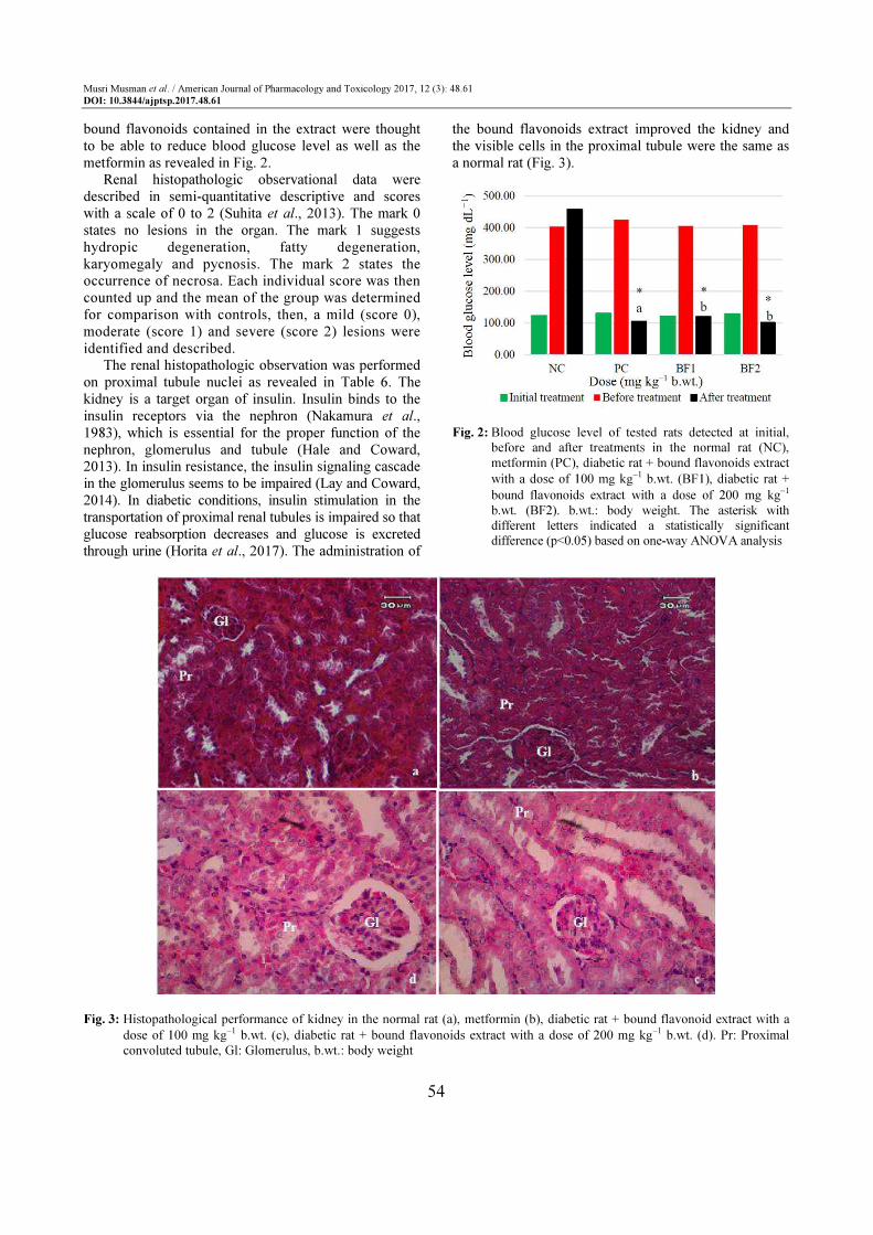
Blood glucose level (Mean ± SD mg dL−1)
Dose (mg kg−1 -------------------------------------------------------------------------------- Antidiabetic
Group b.wt.) Initial Before After effect (%)
NC - 122.67 402.67 457.67 -
PC 65 129.33 424.67* a104.00* 75.51
BF1 100 121.33 405.00* b119.33* 70.53
BF2 200 128.67 408.33* b101.33* 75.18
NC: Negative Control, PC: Positive Control, BF1: Bound Flavonoids with a dose of 100 mg kg−1 b.wt., BF2: Bound Flavonoids with
a dose of 200 mg kg−1 b.wt., b.wt.: body weight. Different letters indicated statistically significant differences (*) in blood glucose
level among the treatments (p<0.05, Duncan’s post hoc following one-way ANOVA)
Musri Musman et al. / American Journal of Pharmacology and Toxicology 2017, 12 (3): 48.61
DOI: 10.3844/ajptsp.2017.48.61
53
Table 6: Proximal renal tubule cell scores in the rats’ kidney at various treatments
Group Dose (mg kg−1 b.wt.) Proximal convoluted tubule score (Mean ± SD)
NC - 0.25±0.50a
PC 65 0.50±0.57a
BF1 100 0.25±0.50a
BF2 200 0.25±0.50a
NC: Negative Control, PC: Positive Control, BF1: Bound Flavonoids with a dose of 100 mg kg−1 b.wt., BF2: Bound Flavonoids with
a dose of 200 mg kg−1 b.wt., b.wt.: body weight. The Same letter indicated statistically insignificant differences (F(3, 15) = 0.23, p =
0.873) in proximal renal tubule cell count among the treatments based on one-way ANOVA analysis
Table 7: Granulation of pancreatic β-cells at various treatments
Group Dose (mg kg−1 b.wt.) Pancreatic β-cell (cell, Mean ± SD)
NC - 454.50±20.82a
PC 65 384.00±30.53b
BF1 100 391.25±4.57b
BF2 200 437.00±12.05a
NC: Negative Control, PC: Positive Control, BF1: Bound Flavonoids with a dose of 100 mg kg−1 b.wt., BF2: Bound Flavonoids with
a dose of 200 mg kg−1 b.wt., b.wt.: Body weight. Different letters indicated statistically significant differences (F(3, 15) = 26.09, p <
0.0001) in pancreatic cell count among the treatments based on one-way ANOVA analysis
Histopathological Study
The histopathologic observations of the kidney and
pancreas images showed results in renal damage within
the normal range (Table 6) and an increase in β-cell
granulation (Table 7) respectively.
Discussion
The bound flavonoids extract shown an intense red
color on Shinoda’s test (Raaman, 2006) based on the
phytochemical analysis. In this extract, the alkaloid,
phenolic, saponin, tannin and terpenoid components did
not detect according to the standard procedures. This
indicated that only bound flavonoids were existing in the
extract. The bound flavonoids containing extract had the
ability to turn deep violet color to pale yellow color in
ethanolic DPPH solution. The magnitude of the
reduction strength of the extract to neutralize the DPPH
free radical was not much different when compared to
the ability of ascorbic acid to neutralize the DPPH in
terms of inhibition as shown in Table 4. The IC50 values
for the ascorbic acid and the extract were in the amount
of 6.50 and 7.51 ppm respectively based on the log
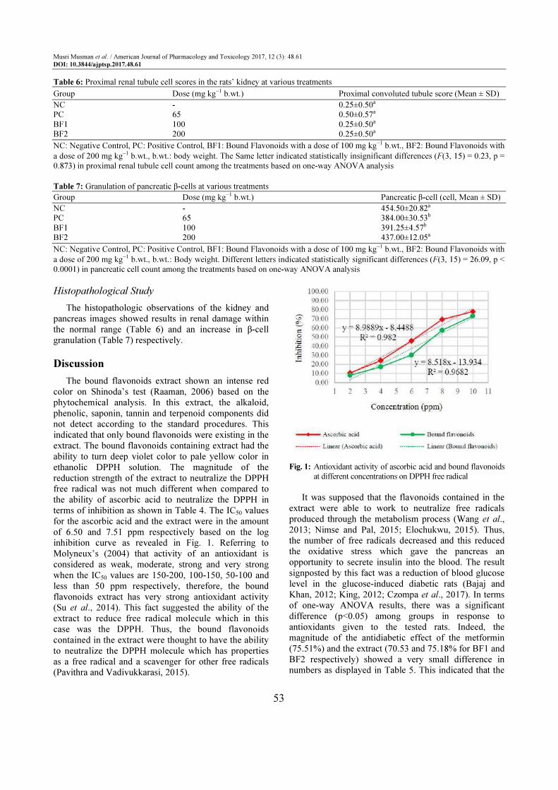
inhibition curve as revealed in Fig. 1. Referring to
Molyneux’s (2004) that activity of an antioxidant is
considered as weak, moderate, strong and very strong
when the IC50 values are 150-200, 100-150, 50-100 and
less than 50 ppm respectively, therefore, the bound
flavonoids extract has very strong antioxidant activity
(Su et al., 2014). This fact suggested the ability of the
extract to reduce free radical molecule which in this
case was the DPPH. Thus, the bound flavonoids
contained in the extract were thought to have the ability
to neutralize the DPPH molecule which has properties
as a free radical and a scavenger for other free radicals
(Pavithra and Vadivukkarasi, 2015).
Fig. 1: Antioxidant activity of ascorbic acid and bound flavonoids
at different concentrations on DPPH free radical
It was supposed that the flavonoids contained in the
extract were able to work to neutralize free radicals
produced through the metabolism process (Wang et al.,
2013; Nimse and Pal, 2015; Elochukwu, 2015). Thus,
the number of free radicals decreased and this reduced
the oxidative stress which gave the pancreas an
opportunity to secrete insulin into the blood. The result
signposted by this fact was a reduction of blood glucose
level in the glucose-induced diabetic rats (Bajaj and
Khan, 2012; King, 2012; Czompa et al., 2017). In terms
of one-way ANOVA results, there was a significant
difference (p<0.05) among groups in response to
antioxidants given to the tested rats. Indeed, the
magnitude of the antidiabetic effect of the metformin
(75.51%) and the extract (70.53 and 75.18% for BF1 and
BF2 respectively) showed a very small difference in
numbers as displayed in Table 5. This indicated that the
Musri Musman et al. / American Journal of Pharmacology and Toxicology 2017, 12 (3): 48.61
DOI: 10.3844/ajptsp.2017.48.61
54
bound flavonoids contained in the extract were thought
to be able to reduce blood glucose level as well as the
metformin as revealed in Fig. 2.
Renal histopathologic observational data were
described in semi-quantitative descriptive and scores
with a scale of 0 to 2 (Suhita et al., 2013). The mark 0
states no lesions in the organ. The mark 1 suggests
hydropic degeneration, fatty degeneration,
karyomegaly and pycnosis. The mark 2 states the
occurrence of necrosa. Each individual score was then
counted up and the mean of the group was determined
for comparison with controls, then, a mild (score 0),
moderate (score 1) and severe (score 2) lesions were
identified and described.
The renal histopathologic observation was performed
on proximal tubule nuclei as revealed in Table 6. The
kidney is a target organ of insulin. Insulin binds to the
insulin receptors via the nephron (Nakamura et al.,
1983), which is essential for the proper function of the
nephron, glomerulus and tubule (Hale and Coward,
2013). In insulin resistance, the insulin signaling cascade
in the glomerulus seems to be impaired (Lay and Coward,
2014). In diabetic conditions, insulin stimulation in the
transportation of proximal renal tubules is impaired so that
glucose reabsorption decreases and glucose is excreted
through urine (Horita et al., 2017). The administration of
the bound flavonoids extract improved the kidney and
the visible cells in the proximal tubule were the same as
a normal rat (Fig. 3).
Fig. 2: Blood glucose level of tested rats detected at initial,
before and after treatments in the normal rat (NC),
metformin (PC), diabetic rat + bound flavonoids extract
with a dose of 100 mg kg−1 b.wt. (BF1), diabetic rat +
bound flavonoids extract with a dose of 200 mg kg−1
b.wt. (BF2). b.wt.: body weight. The asterisk with
different letters indicated a statistically significant
difference (p<0.05) based on one-way ANOVA analysis
Fig. 3: Histopathological performance of kidney in the normal rat (a), metformin (b), diabetic rat + bound flavonoid extract with a
dose of 100 mg kg−1 b.wt. (c), diabetic rat + bound flavonoids extract with a dose of 200 mg kg−1 b.wt. (d). Pr: Proximal
convoluted tubule, Gl: Glomerulus, b.wt.: body weight
Musri Musman et al. / American Journal of Pharmacology and Toxicology 2017, 12 (3): 48.61
DOI: 10.3844/ajptsp.2017.48.61
55
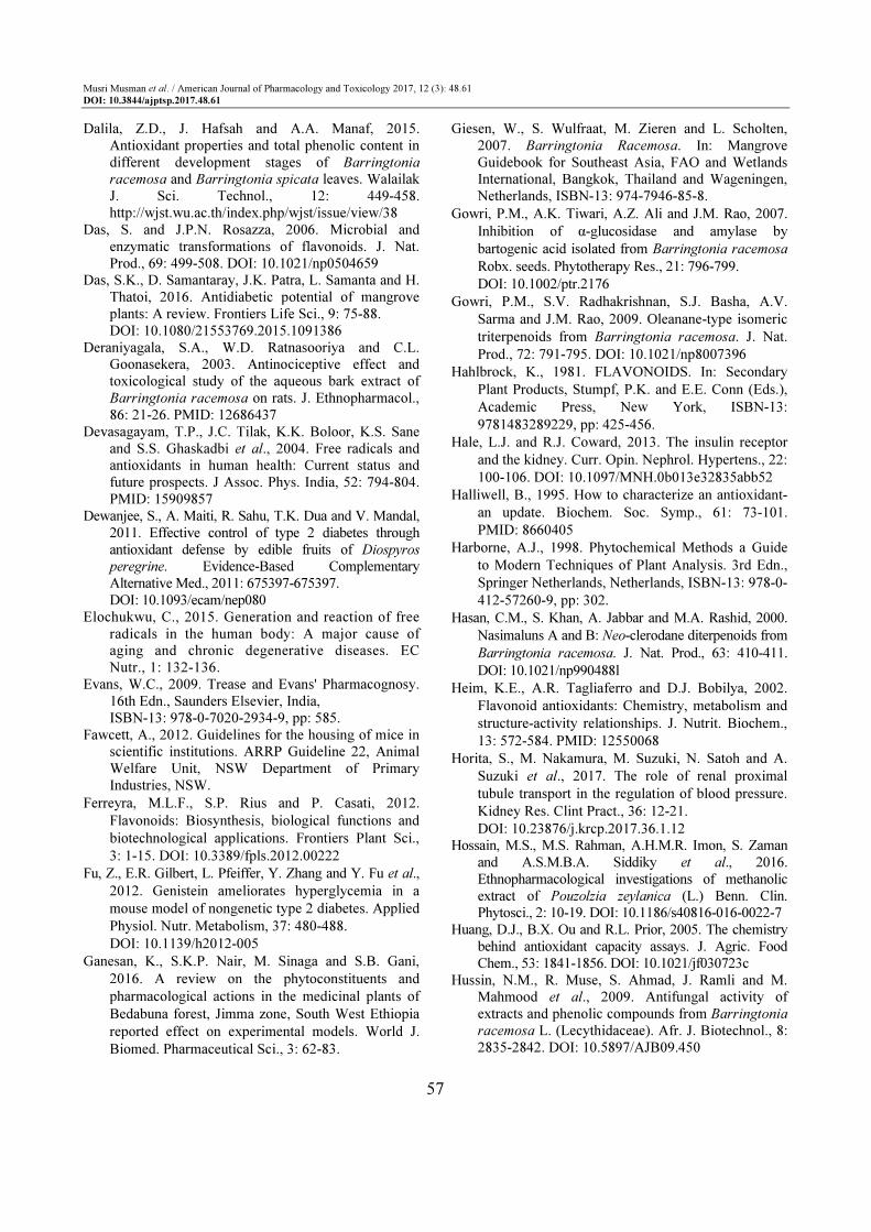
Fig. 4: Histopathological observation of pancreatic β-cell as indicated by the white arrow in the normal rat (1), metformin (2),
diabetic rat + bound flavonoids extract with a dose of 100 mg kg−1 b.wt. (3), diabetic rat + bound flavonoids extract with a
dose of 200 mg kg−1 b.wt. (4). b.wt.: body weight
The score value of renal proximal tubule cells in diabetic rats given metformin by 0.50 expressed damage conditions in the normal range as shown in Fig. 3. The metformin could reduce hyperglycemia in the blood so it could reduce damage to proximal renal tubular cells. The one-way ANOVA results showed that the bound flavonoid extract given to diabetic rats did not cause any significant change in the histologic structure of the kidney (F(3, 15) = 0.23, p = 0.873). Indeed, the result of scores on a proximal tubular cell in diabetic rats given the extract displayed 0.25. This result indicated that the physiological functions of renal cells worked within the range of normal changes (Khoshnoud et al., 2017). This suggested that administration of the bound flavonoids extract of B. racemosa kernel in diabetic rats did not show specific damage to proximal renal tubular cells. Thus, the bound flavonoids contained in the extract did not cause damage to the kidney organs in the tested rats when applied as a controlling agent for type 2 diabetes.
The histopathologic images demonstrated that the
pancreatic β-cell granulation was directly proportional to
the given extract dose as shown in Table 7. The number
of β-cells enhancement (F(3, 15) = 26.09, p<0.0001) for
each treatment stated that the bound flavonoids extract
administered to hyperglycemic rats could improve
pancreatic β-cells and depresses necrosis or apoptosis of
pancreatic β-cells compared to metformin as shown in
Figure 4. It was assumed that the modulatory effects of
bound flavonoid constituents on the blood glucose
transporter by increasing insulin secretion, decreasing
apoptosis and stimulating proliferation of pancreatic
β-cells (Fu et al., 2012; Vinayagam and Xu, 2015;
Zheng et al., 2016).
Conclusion
The bound flavonoids extract of B. racemosa kernel showed the strong antioxidant power and it displayed the type 2 anti-diabetes property. Administration of the extract with doses of 100 mg kg
−1 b.wt. and 200 mg kg
−1 b.wt. orally for 14 days
was not causing the histopathologic disturbance on the tested rat kidney organ.
Acknowledgement
Authors are thankful to Prof. Dr. Djufri, M.Si., the
plant taxonomist of Teacher Teaching and Education

Musri Musman et al. / American Journal of Pharmacology and Toxicology 2017, 12 (3): 48.61
DOI: 10.3844/ajptsp.2017.48.61
56
Faculty of Syiah Kuala University, for identification and
authentication of Barringtonia racemosa (L.) Spreng.
Research Funding
The research was supported by Syiah Kuala
University funding (grant number:
33/UN11.2/PP/PNBP/SP3/2017).
Conflict of Interest
The authors declare that they do not have any conflict
of interests.
Author’s Contribution
Musri Musman: Conceived, designed the
experiments and wrote the paper.
Emelda Audina: Performed the experiments.
Fazlia I. R. Ratu: Experimental tools analyses.
Erlidawati Erlidawati: Provided reagents and
materials.
Safrida Safrida: Analyzed the data.
Ethics
This original article contains unpublish material. The
corresponding author states that all of the other authors
have read and agreed to the manuscript and no ethical
issues are involved.
References
Abdul-Ghani, M. and R. DeFronzo, 2010. Pathogenesis
of insulin resistance in skeletal muscle. J. Biomed.
Biotechnol., 2010: 1-19. DOI: 10.1155/2010/476279
Adewunmi, C.O., A.J. Aladesanmi, F.B. Adewoyin,
J.A.O. Ojewole and N. Naido, 2001. Molluscicidal,
insecticidal and piscicidal activities of Barringtonia
racemosa. Nig. J. Nat. Prod. Med., 5: 56-58.
DOI: 10.4314/NJNPM.V5I1.11727
Alexandru, I., 2011. Experimental use of animal in
research spa. Balneo Res. J., 2: 65-69.
DOI: 10.12680/balneo.2011.1014
ADA, 2015. Classification and diagnosis of diabetes.
Diabetes Care, 38: 58-516. DOI: 10.2337/dc15-S005
Amić, D., D. Davidović-Amić, D. Bešlo and N.
Trinajstić, 2003. Structure-radical scavenging
activity relationship of flavonoids. Croatia Chem.
Acta, 76: 55-61.
Amran, N., A.N.A. Rani, R. Mahmud and K.B. Yin,
2016. Antioxidant and cytotoxic effect of
Barringtonia racemosa and Hibiscus sabdariffa fruit
extracts in MCF-7 human breast cancer cell line.
Pharmacognosy Res., 8: 66-70.
DOI: 10.4103/0974-8490.171104
Arul, B., R. Kothai and A.J.M. Christina, 2006.
Antihyperglycemic and hypoglycemic effect of
Bougainvillea spectabilis Linn. In normal and glucose-
induced diabetic rats. Hamdard Med., 49: 18-21.
Babu, P.V.A., D. Liu and E.R. Gilbert, 2013. Recent
advances in understanding the anti-diabetic
actions of dietary flavonoids. J. Nutr. Biochem., 24:
1777-1789. DOI: 10.1016/j.jnutbio.2013.06.003
Bagchi, K. and S. Puri, 1998. Free radicals and
antioxidants in health and disease. East
Mediterranean Health Jr., 4: 350-60.
Bajaj, S. and A. Khan, 2012. Antioxidants and diabetes.
Ind. J. Endocrinol Metab., 16: S267-S271.
DOI: 10.4103/2230-8210.104057
Bansal, A.K. and G.S. Bilaspuri, 2011. Impacts of
oxidative stress and antioxidants on semen
functions. Vet. Med. Int. 2011: 686137-686137.
DOI: 10.4061/2011/686137
Banu, K.S. and L. Cathrine, 2015. General techniques
involved in phytochemical analysis. IJARCS, 2: 25-32.
Behbahani, M., A.M. Ali, R. Muse and N.B.
Mohammad, 2007. Anti-oxidant and anti-
inflammatory activities of leaves of Barringtonia
racemosa. J. Med. Plants Res., 1: 95-102.
Betteridge, D.J., 2000. What is oxidative stress?
Metabolism, 49: 3-8. PMID: 10693912
Brownlee, M., 2001. Biochemistry and molecular cell
biology of diabetic complications. Nature, 414:
813-20. PMID: 11742414
Candasamy, M., T.E.G.K. Murthy, K.S. Gubiyappa,
D.K. Chellappan and G. Gupta, 2014. Alteration of
glucose lowering effect of glibenclamide on single
and multiple treatments with fenofibrate in
experimental rats and rabbit models. J. Basic Clin.
Pharmacy, 5: 62-67.
DOI: 10.4103/0976-0105.139728 Ceriello, A. and E. Motz, 2004. Is oxidative stress the
pathogenic mechanism underlying insulin resistance, diabetes and cardiovascular disease? The common soil hypothesis revisited. Arterioscler Thromb Vasc Biol., 24: 816-23.
DOI: 10.1161/01.ATV.0000122852.22604.78 Chan, E.W.C., S. Baba, H.T. Chan, M. Kainuma and T.
Inoue et al., 2017. Ulam herbs: A review on the medicinal properties of Anacardium occidentale and Barringtonia racemosa. J. Applied Pharmaceutical Sci., 7: 241-247. DOI: 10.7324/JAPS.2017.70235
Chikezie, P.C., O.A. Ojiako and A.C. Ogbuji, 2015. Oxidative stress in diabetes mellitus. Integr Obesity Diabetes. DOI: 10.15761/IOD.1000116
Czompa, A., A. Gyongyosi, K. Szoke, I. Bak and E. Csepanyi et al., 2017. Effects of Momordica charantia (Bitter Melon) on ischemic diabetic myocardium. Molecules, 22: 488-502.
DOI: 10.3390/molecules22030488
Musri Musman et al. / American Journal of Pharmacology and Toxicology 2017, 12 (3): 48.61
DOI: 10.3844/ajptsp.2017.48.61
57
Dalila, Z.D., J. Hafsah and A.A. Manaf, 2015.
Antioxidant properties and total phenolic content in
different development stages of Barringtonia
racemosa and Barringtonia spicata leaves. Walailak
J. Sci. Technol., 12: 449-458.
http://wjst.wu.ac.th/index.php/wjst/issue/view/38
Das, S. and J.P.N. Rosazza, 2006. Microbial and
enzymatic transformations of flavonoids. J. Nat.
Prod., 69: 499-508. DOI: 10.1021/np0504659
Das, S.K., D. Samantaray, J.K. Patra, L. Samanta and H.
Thatoi, 2016. Antidiabetic potential of mangrove
plants: A review. Frontiers Life Sci., 9: 75-88.
DOI: 10.1080/21553769.2015.1091386
Deraniyagala, S.A., W.D. Ratnasooriya and C.L.
Goonasekera, 2003. Antinociceptive effect and
toxicological study of the aqueous bark extract of
Barringtonia racemosa on rats. J. Ethnopharmacol.,
86: 21-26. PMID: 12686437
Devasagayam, T.P., J.C. Tilak, K.K. Boloor, K.S. Sane
and S.S. Ghaskadbi et al., 2004. Free radicals and
antioxidants in human health: Current status and
future prospects. J Assoc. Phys. India, 52: 794-804.
PMID: 15909857
Dewanjee, S., A. Maiti, R. Sahu, T.K. Dua and V. Mandal,
2011. Effective control of type 2 diabetes through
antioxidant defense by edible fruits of Diospyros
peregrine. Evidence-Based Complementary
Alternative Med., 2011: 675397-675397.
DOI: 10.1093/ecam/nep080
Elochukwu, C., 2015. Generation and reaction of free
radicals in the human body: A major cause of
aging and chronic degenerative diseases. EC
Nutr., 1: 132-136.
Evans, W.C., 2009. Trease and Evans' Pharmacognosy.
16th Edn., Saunders Elsevier, India,
ISBN-13: 978-0-7020-2934-9, pp: 585.
Fawcett, A., 2012. Guidelines for the housing of mice in
scientific institutions. ARRP Guideline 22, Animal
Welfare Unit, NSW Department of Primary
Industries, NSW.
Ferreyra, M.L.F., S.P. Rius and P. Casati, 2012.
Flavonoids: Biosynthesis, biological functions and
biotechnological applications. Frontiers Plant Sci.,
3: 1-15. DOI: 10.3389/fpls.2012.00222
Fu, Z., E.R. Gilbert, L. Pfeiffer, Y. Zhang and Y. Fu et al.,
2012. Genistein ameliorates hyperglycemia in a
mouse model of nongenetic type 2 diabetes. Applied
Physiol. Nutr. Metabolism, 37: 480-488.
DOI: 10.1139/h2012-005
Ganesan, K., S.K.P. Nair, M. Sinaga and S.B. Gani,
2016. A review on the phytoconstituents and
pharmacological actions in the medicinal plants of
Bedabuna forest, Jimma zone, South West Ethiopia
reported effect on experimental models. World J.
Biomed. Pharmaceutical Sci., 3: 62-83.
Giesen, W., S. Wulfraat, M. Zieren and L. Scholten,
2007. Barringtonia Racemosa. In: Mangrove
Guidebook for Southeast Asia, FAO and Wetlands
International, Bangkok, Thailand and Wageningen,
Netherlands, ISBN-13: 974-7946-85-8.
Gowri, P.M., A.K. Tiwari, A.Z. Ali and J.M. Rao, 2007.
Inhibition of α-glucosidase and amylase by
bartogenic acid isolated from Barringtonia racemosa
Robx. seeds. Phytotherapy Res., 21: 796-799.
DOI: 10.1002/ptr.2176
Gowri, P.M., S.V. Radhakrishnan, S.J. Basha, A.V.
Sarma and J.M. Rao, 2009. Oleanane-type isomeric
triterpenoids from Barringtonia racemosa. J. Nat.
Prod., 72: 791-795. DOI: 10.1021/np8007396
Hahlbrock, K., 1981. FLAVONOIDS. In: Secondary
Plant Products, Stumpf, P.K. and E.E. Conn (Eds.),
Academic Press, New York, ISBN-13:
9781483289229, pp: 425-456.
Hale, L.J. and R.J. Coward, 2013. The insulin receptor
and the kidney. Curr. Opin. Nephrol. Hypertens., 22:
100-106. DOI: 10.1097/MNH.0b013e32835abb52
Halliwell, B., 1995. How to characterize an antioxidant-
an update. Biochem. Soc. Symp., 61: 73-101.
PMID: 8660405
Harborne, A.J., 1998. Phytochemical Methods a Guide
to Modern Techniques of Plant Analysis. 3rd Edn.,
Springer Netherlands, Netherlands, ISBN-13: 978-0-
412-57260-9, pp: 302.
Hasan, C.M., S. Khan, A. Jabbar and M.A. Rashid, 2000.
Nasimaluns A and B: Neo-clerodane diterpenoids from
Barringtonia racemosa. J. Nat. Prod., 63: 410-411.
DOI: 10.1021/np990488l
Heim, K.E., A.R. Tagliaferro and D.J. Bobilya, 2002.
Flavonoid antioxidants: Chemistry, metabolism and
structure-activity relationships. J. Nutrit. Biochem.,
13: 572-584. PMID: 12550068
Horita, S., M. Nakamura, M. Suzuki, N. Satoh and A.
Suzuki et al., 2017. The role of renal proximal
tubule transport in the regulation of blood pressure.
Kidney Res. Clint Pract., 36: 12-21.
DOI: 10.23876/j.krcp.2017.36.1.12
Hossain, M.S., M.S. Rahman, A.H.M.R. Imon, S. Zaman
and A.S.M.B.A. Siddiky et al., 2016.
Ethnopharmacological investigations of methanolic
extract of Pouzolzia zeylanica (L.) Benn. Clin.
Phytosci., 2: 10-19. DOI: 10.1186/s40816-016-0022-7
Huang, D.J., B.X. Ou and R.L. Prior, 2005. The chemistry
behind antioxidant capacity assays. J. Agric. Food
Chem., 53: 1841-1856. DOI: 10.1021/jf030723c
Hussin, N.M., R. Muse, S. Ahmad, J. Ramli and M.
Mahmood et al., 2009. Antifungal activity of
extracts and phenolic compounds from Barringtonia
racemosa L. (Lecythidaceae). Afr. J. Biotechnol., 8:
2835-2842. DOI: 10.5897/AJB09.450
Musri Musman et al. / American Journal of Pharmacology and Toxicology 2017, 12 (3): 48.61
DOI: 10.3844/ajptsp.2017.48.61
58
Iqbal, Z., A. Ashraf, A. Touseef, F. Farman and A. Asghar et al., 2016. Antioxidant activity of essential oil of mature bulbof Allium cepa L. from Pakistan. World J. Pharmaceutical Res., 5: 1959-1965.
DOI: 10.20959/wjpr20166-6378 Jakus, V., 2000. The role of free radicals, oxidative stress
and antioxidant systems in diabetic vascular disease. Bratisl Lek Listy, 101: 541-51. PMID: 11218944
Jayaweera, D.M.A., 1981. Medicinal plants (indigenous and exotic) used in Ceylon, part III. The National Science Council of Sri Lanka, Colombo.
Kabir, M.Z., S.M. Rahman, M.R. Islam, P.K. Paul and S. Rahman et al., 2013. A review on a mangrove species from the Sunderbans, Bangladesh: Barringtonia racemosa (L.) roxb. Am. Eurasian J. Sustainable Agric., 7: 356-372.
Kamalakkannan, N. and P.S. Prince, 2006.
Antihyperglycaemic and antioxidant effect of rutin,
a polyphenolic flavonoid, in streptozotocin-induced
diabetic wistar rats. Basic Clin. Pharmacol. Toxicol.,
98: 97-103.
DOI: 10.1111/j.1742-7843.2006.pto_241.x Kan, E., E. Kiliçkan, A. Ayar and R. Colak, 2015.
Effects of two antioxidants; α-lipoic acid and fisetin against diabetic cataract in mice. Int Ophthalmol., 35: 115-120. DOI: 10.1007/s10792-014-0029-3
Kangralkar, V.A., S.D. Patil and R.M. Bandivadekar, 2012. Oxidative stress and diabetes: A review. Int. J. Pharma Appl., 1: 38-45.
Khan, S., A. Jabbar, C.M. Hasan and M.A. Rashid, 2001. Antibacterial activity of Barringtonia racemosa. Fitoterapia, 72: 162-164.
DOI: 10.1016/S0367-326X(00)00264-1 Khoshnoud, S., H.M. Kouchesfahani and M. Nabiuni,
2017. Evaluation of the protective effect of hydro-
alcoholic extract of raspberry fruit on aquaporin1
expression in rats kidney treated by methotrexate.
Cell J., 19: 306-313. DOI: 10.22074/cellj.2016.3957
King, A.J.F., 2012. The use of animal models in diabetes
research. Br J. Pharmacol., 166: 877-894.
DOI: 10.1111/j.1476-5381.2012.01911.x Kong, K.W., S. Mat-Junit, N. Aminudin, F.A. Hassan
and A. Ismail et al., 2016a. Protective effects of the extracts of Barringtonia racemosa shoots against oxidative damage in HepG2 cells. Peer J., 4: e1628-e1628. DOI: 10.7717/peerj.1628
Kong, K.W., A. Abdul-Aziz, N. Razali, N. Aminuddin and S. Mat-Junit, 2016b. Antioxidant-rich leaf extract of Barringtonia racemosa significantly alters the in vitro expression of genes encoding enzymes that are involved in methylglyoxal degradation III. Peer J., 4: e2379-e2379. DOI: 10.7717/peerj.2379
Kong, K.W., S. Mat-Junit, A. Ismail, N. Aminudin and
A. Abdul-Aziz, 2014. Polyphenols in Barringtonia
racemosa and their protection against oxidation of
LDL, serum and haemoglobin. Food Chem., 146:
85-93. DOI: 10.1016/j.foodchem.2013.09.012
Kong, K.W., S. Mat-Junit, N. Aminudin, A. Ismail and A. Abdul-Aziz, 2012. Antioxidant activities and polyphenolics from the shoots of Barringtonia racemosa (L.) Spreng in a polar to apolar medium system. Food Chem., 134: 324-332.
DOI: 10.1016/j.foodchem.2012.02.150 Kumar, S. and A.K. Pandey, 2013. Chemistry and
biological activities of flavonoids: An overview.
Scientific World J., 2013: 162750-162750.
DOI: 10.1155/2013/162750
Laight, D.W., M.J. Carrier and E.E. Anggard, 2000.
Antioxidants, diabetes and endothelial dysfunction.
Cardiovasc. Res., 47: 457-64. PMID: 10963719 Lay, A. and R.J. Coward, 2014. Recent advances in our
understanding of insulin signaling to the podocyte. Nephrol. Dial Transplant. 29: 1127-1133.
DOI: 10.1093/ndt/gft471 Li, J., F. Gong and F. Li, 2016. Hypoglycemic and
hypolipidemic effects of flavonoids from tatary buckwheat in type 2 diabetic rats. Biomed. Res., 27: 132-137.
Lim, T.K., 2012. Barringtonia racemosa. In: Edible Medicinal and Non-Medicinal Plants, Lim T.K. (Ed.), Springer Science and Business Media BV, Dordrecht, Heidelberg, London and New York, ISBN-13: 978-94-007-2534-8, pp: 114-121.
Lobo, V., A. Patil, A. Phatak and N. Chandra, 2010. Free radicals, antioxidants and functional foods: Impact on human health. Pharmacogn. Rev., 4: 118-126. DOI: 10.4103/0973-7847.70902
Mackeen, M.M., A.M. Ali, S.H. El-Sharkawi, M.Y. Manap and K.M. Salleh et al., 1997. Antimicrobial and cytotoxic properties of some Malaysian traditional vegetables (Ulam). Int. J. Pharmacog., 35: 174-178. DOI: 10.1076/phbi.35.3.174.13294
Maddux, B.A., W. See, J.C. Lawrence, Jr., A.L.
Goldfine and I.D. Goldfine et al., 2001. Protection
against oxidative stress-induced insulin resistance in
rat L6 muscle cells by micromolar concentrations of
alpha-lipoic acid. Diabetes, 50: 404-10. Manjunath, B.L., 1948. The Wealth of India: A
Dictionary of Indian Raw Materials and Industrial Products. 1st Edn., Council of Scientific and Industrial Research, New Delhi, ISBN-10: 81-85038-00-7, pp: 159.
Martin, A.C., R.A. Sanders and J.B. Watkins, 2003. Diabetes, oxidative stress and antioxidants: A review. J. Biochem. Mol. Toxicol., 17: 24-38.
DOI: 10.1002/jbt.10058
Mmushi, T.J., P. Masoko, L.K. Mdee, M.P. Mokgotho and
L.J. Mampuru et al., 2010. Anti-mycobacterial
evaluation of fifteen medicinal plants in South Africa.
Afr. J. Tradit. Complement Altern. Med., 7: 34-39.
Molyneux, P., 2004. The use of the stable free radical
Diphenylpicrylhydrazyl (DPPH) for estimating
antioxidant activity. Songklanakarin J. Sci.
Technol., 26: 211-219.
Musri Musman et al. / American Journal of Pharmacology and Toxicology 2017, 12 (3): 48.61
DOI: 10.3844/ajptsp.2017.48.61
59
Montonen, J., P. Knekt, R. Jarvinen and A. Reunanen,
2004. Dietary antioxidant intake and risk of type 2
diabetes. Diabetes Care, 27: 362-366.
PMID: 14747214
Musman, M., 2010. Toxicity of Barringtonia racemosa
(L.) Kernel extract on Pomacea canaliculata
(Ampullariidae). Tropical Life Sci. Res., 21: 41-50.
Musman, M., S. Kamaruzzaman, S. Karina, R. Rizqi and F.
Arisca, 2013. A preliminary study on the antihatching
of freshwater golden apple snail Pomacea canaliculata
(Gastropoda: Ampullariidae) eggs from Barringtonia
racemosa (Magnoliopsida: Lecythidaceae) seeds
extract. AACL Bioflux, 6: 394-398.
Musman, M., S. Karina and F. Rizki, 2014. Saponins
extract from Barringtonia racemosa as molluscicide
to brackishwater pond snails (Cerithidea cingulata).
Int. J. Applied Res. Technol., 3: 92-97.
Musman, M., S. Karina, C.N. Defira, N. Fadhillah and
A. Kayana et al., 2015. Phytofungitoxic agent from
wild plants. IJSBAR, 21: 78-85.
Nadkarni, A.K., 1976. Dr. KM Nadakarni’s Indian
Materia Medica. 3rd Ed., Popular Prakashan Ltd.,
Bombay, pp: 177.
Nakamura, R., D.S. Emmanuel and A.I. Katz, 1983.
Insulin binding sites in various segments of the
rabbit nephron. J. Clin. Invest., 72: 388-392.
DOI: 10.1172/JCI110979
Nazaruk, J. and M. Borzym-Kluczyk, 2015. The role of
triterpenes in the management of diabetes mellitus
and its complications. Phytochem. Rev., 14: 675-690.
DOI: 10.1007/s11101-014-9369-x
Nijveldt, R.J., E. van Nood, D.E.C. van Hoorn, P.G.
Boelens and K. van Norren et al., 2001. Flavonoids:
A review of probable mechanisms of action and
potential application. Am. J. Clin. Nutr., 74: 418-25.
PMID: 11566638
Nimse, S.B. and D.K. Pal, 2015. Free radicals, natural
antioxidants and their reaction mechanisms. R.
Society Chem. Adv., 5: 27986-28006.
DOI: 10.1039/C4RA13315C
Nurul-Mariam, H., M. Radzali, R. Johari, A. Syahida and
M. Maziah, 2008. Antioxidant activities of different
aerial parts of Putat (Barringtonia racemosa L.).
Malaysian J. Biochem. Mol. Biol., 16: 15-24.
Ojewole, J.A.O., N. Nundkumar and C.O. Adewunmi,
2004. Molluscicidal, cercariacidal, larvacidal and
antiplasmodial properties of Barringtonia racemosa
fruit and seed extracts. BLACPMA, 3: 88-92.
Osman, N.I., N.J. Sidik and A. Awal, 2015.
Pharmacological activities of Barringtonia
racemosa L. (Putat), a tropical medicinal plant
species. J. Pharm. Sci. Res., 7: 185-188.
Osman, N.I., N.J. Sidik, A. Awal, N.A.M. Adam and
N.I. Rezali, 2016. In vitro xanthine oxidase and
albumin denaturation inhibition assay of
Barringtonia racemosa L. and total phenolic content
analysis for potential anti-inflammatory use in gouty
arthritis. J. Intercult. Ethnopharmacol., 5: 343-349.
DOI: 10.5455/jice.20160731025522
Pandey, K.B. and S.I. Rizvi, 2009. Plant polyphenols as
dietary antioxidants in human health and disease.
Oxid. Med. Cell Longev., 2: 270-278.
DOI: 10.4161/oxim.2.5.9498
Pandeya, K.B., I.P. Tripathi, M.K. Mishra, N. Dwivedi
and Y. Pardhi et al., 2013. A critical review on
traditional herbal drugs: An emerging alternative
drug for diabetes. Int. J. Organic Chem., 3: 1-22.
DOI: 10.4236/ijoc.2013.31001
Patil, K.R. and C.R. Patil, 2016. Anti-inflammatory
activity of bartogenic acid containing fraction of
fruits of Barringtonia racemosa Roxb. In acute and
chronic animal models of inflammation. J. Tradit.
Complementary Med., 7: 86-93.
DOI: 10.1016/j.jtcme.2016.02.001
Patil, K.R., C.R. Patil, R.B. Jadhav, V.K. Mahajan and P.
Raosaheb et al., 2011. Anti-arthritic activity of
bartogenic acid isolated from fruits of Barringtonia
racemosa Roxb. (Lecythidaceae). Evidence-Based
Complementary Alternative Med., 2011: 1-7.
DOI: 10.1093/ecam/nep148
Patil, P.R., M.R. Patil, A. Mane and S. Patil, 2013.
Immunomodulatory effects of fruits of Barringtonia
racemosa Linn. Int. J. Basic Clin. Pharmacol., 2:
216-219. DOI: 10.5455/2319-2003.ijbcp20130318
Patil, S., M.R. Patil, P. Patil, R. Dixit and R.C. Sharma,
2014. Effects of fruits of Barringtonia racemosa
linn. on human polymorphonuclear cell. Int. J. Anat.
Res., 2: 806-809. DOI: 10.16965/ijar.2014.553
Pavithra, K. and S. Vadivukkarasi, 2015. Evaluation of
free radical scavenging activity of various extracts
of leaves from Kedrostis foetidissima (Jacq.) Cogn.
Food Sci. Human Wellness, 4: 42-46.
DOI: 10.1016/j.fshw.2015.02.001
Pham-Huy, L.A., H. He and C. Pham-Huy, 2008. Free
radicals, antioxidants in disease and health. IJBS, 4:
89-96.
Pietta, P.G., 2000. Flavonoids as antioxidants. J. Nat
Prod., 63: 1035-42. PMID: 10924197
Ponnapalli, M.G., S. Sukki, S.C.V.A.R. Annam, M.
Ankireddy and H. Tirunagari et al., 2015.
α‐Glucosidase inhibitory monoacylated
polyhydroxytriterpenoids from the fruits of
Barringtonia racemosa. Tetrahedron Lett., 56:
1570-1574. DOI: 10.1016/j.tetlet.2015.01.193
Musri Musman et al. / American Journal of Pharmacology and Toxicology 2017, 12 (3): 48.61
DOI: 10.3844/ajptsp.2017.48.61
60
Raaman, N., 2006. Phytochemical Techniques. 1st Edn.,
New Indian Publishing Agency, New Delhi, ISBN-
10: 8189422308, pp: 318.
Rice-Evans, C.A., N.J. Miller and G. Paganga, 1996.
Structure-antioxidant activity relationships of
flavonoids and phenolic acids. Free Rad. Biol. Med.,
20: 933-956. PMID: 8743980
Rice-Evans, C., 2001. Flavonoid antioxidants. Curr.
Med. Chem., 8: 797-807. PMID: 11375750
Rice-Evans, C., N. Miller and G. Paganga, 1997.
Antioxidant properties of phenolic compounds.
Trends Plant Sci., 2: 152-159.
DOI: 10.1016/S1360-1385(97)01018-2
RIRDC, 2017. Listing of interesting plants of the world:
Barringtonia racemosa. Australian New Crops Info
2016, Rural Industries Research and Development
Corporation.
Saha, S., K.K. Sarkar, M.L. Hossain, A. Hossin and A.K.
Barman et al., 2013. Bioactivity studies on
Barringtonia racemosa (lam.) Bark. Pharmacol.
OnLine, 1: 93-100. Samanta, S.K., K. Bhattacharya, C. Mandal and B.C.
Pal, 2010. Identification and quantification of the active component quercetin 3-O-rutinoside from Barringtonia racemosa targets mitochondrial apoptotic pathway in acute lymphoblastic leukemia. J. Asian Nat. Prod. Res., 12: 639-648.
DOI: 10.1080/10286020.2010.489040
Sangeetha, V.S., M. Babu and B. Lawrence, 2014. Phytochemical analysis of Annona reticulata L. leaf extracts. Int. Res. J. Pharm. Applied Sci., 4: 4-8.
Santos-Buelga, C. and A.S. Feliciano, 2017.
Flavonoids: From structure to health issues. Molecules, 22: 477-482.
DOI: 10.3390/molecules22030477
Sikha, P., P.G. Latha, S.R. Suja, G.I. Anuja and S.
Shyamal et al., 2010. Anti-inflammatory and
analgesic activity of Barringtonia racemosa Roxb.
Fruits. Ind. J. Nat. Prod. Resour., 1: 356-361.
Spitalnik, P.F., 2016. Histology laboratory manual 2016-
2017. College of Physicians and Surgeons,
Columbia University, New York.
Steel, R.G.D., J.H. Torrie and D.A. Dickey, 1997.
Principles and Procedures of Statistics: A
Biometrical Approach. 3rd Edn., McGraw-Hill,
New York, ISBN-10: 0070610282, pp: 666.
Su, D., R. Zhang, F. Hou, M. Zhang and J. Guo et al.,
2014. Comparison of the free and bound phenolic
profiles and cellular antioxidants of litchi pulp
extracts from different solvents. BMC
Complement Altern. Med., 14: 9-9.
DOI: 10.1186/1472-6882-14-9
Subramanian, S.S. and S. Nagarajan, 1969. Flavonoids
of the seeds of Crotolaria retusa and C.striata. Curr.
Sci., 38: 65-68.
Suhita, N.L. P.R., I.W. Sudira and I.B.O. Winaya, 2013.
Histopathological kidney of rat white the effect of
the pegagan (Centella asiatica) extract against
peroral. Buletin Veteriner Udayana, 5: 71-78.
Sulaiman, S.F. and K.L. Ooi, 2014. Antioxidant and α-
glucosidase inhibitory activities of 40 tropical juices
from Malaysia and identification of phenolics from
the bioactive fruit juices of Barringtonia racemosa
and Phyllanthus acidus. J. Agric. Food Chem., 62:
9576-9585. DOI: 10.1021/jf502912t
Sun, H.Y., L.J. Long and J. Wu, 2006. Chemical
constituents of mangrove plant Barringtonia
racemosa. Zhong Yao Cai, 29: 671-672. PMID:
17059003
Tachibana, Y., A. Kato, Y. Nishiyama, M. Ikemi and K.
Ohoka et al., 1996. Mitogenic activity in African
traditional herbal medicines (Part II).
Phytomedicine, 2: 335-339.
DOI: 10.1016/S0944-7113(96)80078-X
Thomas, T.J., B. Panikkar, A. Subramanian, M.K. Nair
and K.R. Panikkar, 2002. Antitumour property and
toxicity of Barringtonia racemosa Roxb seed extract
in mice. J. Ethnopharmacol., 82: 223-227.
DOI: 10.1016/S0378-8741(02)00074-0
Ullah, A., A. Khan and I. Khan, 2016. Diabetes mellitus
and oxidative stress-A concise review. Saudi
Pharmaceutical J., 24: 547-553.
DOI: 10.1016/j.jsps.2015.03.013
Vinayagam, R. and B. Xu, 2015. Antidiabetic properties
of dietary flavonoids: A cellular mechanism review.
Nutr. Metabolism, 12: 1-20.
DOI: 10.1186/s12986-015-0057-7
Wang, Y., S. Chen and O. Yu, 2011. Metabolic
engineering of flavonoids in plants and
microorganisms. Applied Microbiol. Biotechnol.,
91: 949-956. DOI: 10.1007/s00253-011-3449-2
Wang, Z., J. Wang and P. Chan, 2013. Treating type 2
diabetes mellitus with traditional chinese and indian
medicinal herbs. Evidence-Based Complementary
Alternative Med., 2013: 343594-343594.
DOI: 10.1155/2013/343594
Wedick, N.M., A. Pan, A. Cassidy, E.B. Rimm and L.
Sampson et al., 2012. Dietary flavonoid intakes and
risk of type 2 diabetes in US men and women. Am.
J. Clin. Nutr., 95: 925-933.
DOI: 10.3945/ajcn.111.028894
Wolff, S.P., 1993. Diabetes mellitus and free radicals. Free
radicals, transition metals and oxidative stress in the
aetiology of diabetes mellitus and complications. Br.
Med. Bull., 49: 642-652. PMID: 8221029
WHO, 2016. Global report on diabetes. World Health
Organization, France.
Musri Musman et al. / American Journal of Pharmacology and Toxicology 2017, 12 (3): 48.61
DOI: 10.3844/ajptsp.2017.48.61
61
Wright Junior, E., J.L. Scism-Bacon and L.C. Glass,
2006. Oxidative stress in type 2 diabetes: The role of
fasting and postprandial glycaemia. Int. J. Clin.
Pract., 60: 308-314.
DOI: 10.1111/j.1368-5031.2006.00825.x
Yoshikawa, T. and Y. Naito, 2002. What is oxidative
stress? JMAJ, 45: 271-276.
Young, I.S. and J.V. Woodside, 2001. Antioxidants in
health and disease. J. Clin. Pathol., 54: 176-86.
PMID: 11253127
Yung, L.M., F.P. Leung, X. Yao, Z.Y. Chen and Y.
Huang, 2006. Reactive oxygen species in vascular
wall. Cardiovasc Hematol. Disord. Drug Targets, 6:
1-19. PMID: 16724932
Zawawi, D.D., H. Ja’afar and A.M. Ali, 2011. Total
phenolic compounds and antioxidant properties in
different stage of B. racemosa and B. spicata leaf.
Proceeding of the International Conference on
Biology, Environment and Chemistry, (BEC’ 11), IACSIT Press, Singapoore, pp: 100-105.
Zheng, S., M. Zhao, Y. Wu, Z. Wang and Y. Ren, 2016.
Suppression of pancreatic beta cell apoptosis by
Danzhi Jiangtang capsule contributes to the
attenuation of type 1 diabetes in rats. BMC
Complement Altern. Med., 16: 31-41.
DOI: 10.1186/s12906-016-0993-4