requirement of myosin vb rab11a rab11-fip2 complex in
TRANSCRIPT

Requirement of Myosin Vb�Rab11a�Rab11-FIP2 Complexin Cholesterol-regulated Translocation of NPC1L1 to theCell Surface*□S
Received for publication, June 16, 2009 Published, JBC Papers in Press, June 19, 2009, DOI 10.1074/jbc.M109.034355
Bei-Bei Chu1, Liang Ge1, Chang Xie, Yang Zhao, Hong-Hua Miao, Jing Wang, Bo-Liang Li, and Bao-Liang Song2
From The State Key Laboratory of Molecular Biology, Institute of Biochemistry and Cell Biology, Shanghai Institutes for BiologicalSciences, Chinese Academy of Sciences, 320 Yue-Yang Road, Shanghai 200031, China
Niemann-Pick C1-like 1 (NPC1L1) plays a critical role in theenterohepatic absorption of free cholesterol. Cellular choles-terol depletion induces the transport ofNPC1L1 from the endo-cytic recycling compartment to the plasma membrane (PM),and cholesterol replenishment causes the internalization ofNPC1L1 together with cholesterol via clathrin-mediated endo-cytosis. Although NPC1L1 has been characterized, the otherproteins involved in cholesterol absorption and the endocyticrecycling of NPC1L1 are largely unknown.Most of the vesiculartrafficking events are dependent on the cytoskeleton andmotorproteins. Here, we investigated the roles of the microfilamentandmicrofilament-associated triple complex composed ofmyo-sin Vb, Rab11a, and Rab11-FIP2 in the transport of NPC1L1from the endocytic recycling compartment to the PM. Interfer-ing with the dynamics of the microfilament by pharmacologicaltreatment delayed the transport of NPC1L1 to the cell surface.Meanwhile, inactivation of any component of the myosinVb�Rab11a�Rab11-FIP2 triple complex inhibited the export ofNPC1L1. Expression of the dominant-negativemutants ofmyosinVb, Rab11a, or Rab11-FIP2 decreased the cellular cholesteroluptake by blocking the transport of NPC1L1 to the PM. Theseresults suggest that the efficient transport of NPC1L1 to the PMis dependent on the microfilament-associated myosinVb�Rab11a�Rab11-FIP2 triple complex.
Cholesterol homeostasis in human bodies is maintainedthrough regulated cholesterol synthesis, absorption, and excre-tion. Intestinal cholesterol absorption is one of the major path-ways to maintain cholesterol balance. NPC1L1 (Niemann-PickC1-like protein 1), a polytopic transmembrane protein highlyexpressed in the intestine and liver, is required for dietary cho-lesterol uptake and biliary cholesterol reabsorption (1–4).Genetic or pharmaceutical inactivation of NPC1L1 signifi-cantly inhibits cholesterol absorption and confers the resist-ance to diet-induced hypercholesterolemia (1, 2, 4). Ezetimibe,
an NPC1L1-specific inhibitor, is currently used to prevent andtreat cardiovascular diseases (5).Human NPC1L1 contains 1,332 residues with 13 transmem-
brane domains (6). The third to seventh transmembrane helicesconstitute a conserved sterol-sensing domain (4, 7). NPC1L1recycles between the endocytic recycling compartment (ERC)3and the plasma membrane (PM) in response to the changes ofcholesterol level (8). ERC is a part of early endosomes that isinvolved in the recycling ofmany transmembrane proteins. It isalso reported that ERC is a pool for free cholesterol storage (9).When cellular cholesterol concentration is low,NPC1L1movesfrom the ERC to the PM (8, 10). Under cholesterol-replenishingconditions, NPC1L1 and cholesterol are internalized togetherand transported to the ERC (8). Disruption of microfilament,depletion of the clathrin�AP2 complex, or ezetimibe treatmentcan impede the endocytosis of NPC1L1, thereby decreasingcholesterol internalization (8, 10, 11).The microfilament (MF) system, part of the cytoskeleton
network, is required for multiple cellular functions such as cellshape maintenance, cell motility, mitosis, protein secretion,and endocytosis (12, 13). The major players in the microfila-ment system are actin fibers and motor proteins (14). Actinfibers form a network that serves as the tracks for vesiculartransport (15, 16). Meanwhile, the dynamic assembly and dis-assembly of actin fibers and the motor proteins provides thedriving force for a multitude of membrane dynamics includingendocytosis, exocytosis, and vesicular trafficking between com-partments (15, 16).Myosins are a large family of motor proteins that are respon-
sible for actin-based mobility (14). Class V myosins (17, 18),comprisingmyosin Va, Vb, andVc, are involved in a wide rangeof vesicular trafficking events in different mammalian tissues.Myosin Va is expressed mainly in neuronal tissues (19, 20),whereas myosins Vb and Vc are universally expressed withenrichment in epithelial cells (21, 22). Class V myosins arerecruited to their targeting vesicles by small GTPase proteins(Rab) (23). Rab11a and Rab11 family-interacting protein 2(Rab11-FIP2) facilitate the binding of myosin Vb to the cargoproteins of endocytic recycling vesicles (24–28).* This work was supported by Grants 2009CB919000 and 2007AA09Z402
from the Ministry of Science and Technology of China, 30670432 and90713025 from the National Natural Science Foundation of China, and08JC1421300 and 08431900500 from the Shanghai Science and Technol-ogy Committee.
□S The on-line version of this article (available at http://www.jbc.org) containssupplemental Figs. S1–S4.
1 Both authors contributed equally to this work.2 To whom correspondence should be addressed. E-mail: [email protected].
3 The abbreviations used are: ERC, endocytic recycling compartment; MF,microfilament; NPC1L1, Niemann-Pick C1-like 1; PM, plasma membrane;WT, wild type; CT, C-terminal tail; MVID, microvillus inclusion disease; CDX,methyl-�-cyclodextrin; siRNA, small interfering RNA; EGFP, enhancedgreen fluorescent protein; PBS, phosphate-buffered saline; RFP, red fluo-rescent protein.
THE JOURNAL OF BIOLOGICAL CHEMISTRY VOL. 284, NO. 33, pp. 22481–22490, August 14, 2009© 2009 by The American Society for Biochemistry and Molecular Biology, Inc. Printed in the U.S.A.
AUGUST 14, 2009 • VOLUME 284 • NUMBER 33 JOURNAL OF BIOLOGICAL CHEMISTRY 22481
by guest on April 6, 2018
http://ww
w.jbc.org/
Dow
nloaded from

Myosin Vb binds Rab11a and Rab11-FIP2 through the C-ter-minal tail (CT) domain. The triple complex of myosin Vb,Rab11a, and Rab11-FIP2 is critical for endocytic vesiculartransport and the recycling of many proteins including trans-ferrin receptor (29), AMPA receptors (30), CFTR (28), GLUT4(31, 32), aquaporin-2 (26), and �2-adrenergic receptors (33).The myosin Vb-CT domain (24) competes for binding toRab11a and Rab11-FIP2 and functions as a dominant-negativeform. Expression of the CT domain substantially impairs thetransport of vesicles. Deficient endocytic trafficking is alsoobserved in cells expressing the GDP-locked form of Rab11a(S25N) (34) or a truncated Rab11-FIP2, which competes for therab11a binding (35).Here we investigated the roles of actin fibers and motor pro-
teins in the cholesterol-regulated endocytic recycling ofNPC1L1. Using pharmaceutical inactivation, dominant-nega-tive forms, and an siRNA technique, we demonstrated thatactin fibers and myosin Vb�Rab11a�Rab11-FIP2 triple complexare involved in the export of NPC1L1 to the PM and that thisintact MF-associated triple complex is required for efficientcholesterol uptake. Characterization of the molecules involvedin the recycling of NPC1L1may shed new light upon themech-anism of cholesterol absorption.
EXPERIMENTAL PROCEDURES
Materials—The reagents used include horseradish peroxi-dase-conjugated donkey anti-mouse and anti-rabbit IgG (fromJackson ImmunoResearch Laboratories); filipin, cytochalasinD, latrunculin B, jasplakinolide, nocodazole, and colchicine(from Sigma-Aldrich); sulfosuccinimidyl 6-(biotinamido)hex-anoate and NeutrAvidin-agarose (from Pierce); methyl-�-cy-clodextrin (CDX) (from Cyclodextrin Technologies Develop-ment, Inc.); and other reagents from previously describedsources (8).Plasmids—The plasmids expressing the EGFP-tagged myo-
sin Vb full-length, myosin Va, andmyosin Vb C-terminal dom-inant-negative tails were generously provided by Dr. AlaaEl-Husseini (University of British Columbia, Canada). MyosinVb-CT�RBD was generated as described previously (25).Human myosin Vc-CT (residues 912–1742), Rab11a, andRab11-FIP2 were PCR-amplified from the cDNA of HEK293cells and inserted in-frame into pEGFP-C1 or other vectorswith the indicated tags described in the text. The truncatedRab11-FIP2 encoding residues 446–511 was subcloned fromthe plasmids containing the full-length Rab11-FIP2 andinserted into pEGFP-C1.Cell Culture—CRL-1601 (McArdle RH7777 rat hepatoma
cell), and CRL-1601/NPC1L1-EGFP, which stably expressesNPC1L1-EGFP, were grown in amonolayer at 37 °C in 5%CO2.The cells were maintained in medium A (Dulbecco’s modifiedEagle’s medium containing 100 units/ml penicillin and 100�g/ml streptomycin sulfate) supplemented with 10% fetalbovine serum. For CRL-1601/NPC1L1-EGFP, an additional200 �g/ml G418 was supplemented. The cholesterol-depletingmedium was medium A supplemented with 5% lipoprotein-deficient serum, 10�M compactin, 50�Mmevalonate, and 1.5%CDX. Cholesterol-replenishing medium contained medium Asupplemented with 5% lipoprotein-deficient serum, 10 �M
compactin, 50 �M mevalonate, and different concentrations ofcholesterol-CDX. The cholesterol�CDX inclusion complexeswere prepared as described previously (36).Transfection, Co-immunoprecipitation, and Immunoblotting—
Transfection of cells with FuGENE HD (Roche Applied Sci-ence) was performed according to the manufacturer’s manual.Conditions of the drug incubations are described in the legendfor Fig.6. At the end of incubation, triplicate samples for eachtreatmentwere harvested and lysed in lysis buffer (PBS contain-ing 5 mM EGTA, 5 mM EDTA, 0.5% digitonin, and proteaseinhibitors). Whole cell lysate was incubated with anti-EGFPbeads and rotated at 4 °C for 2 h. The agarose beads were thenwashed five times with lysis buffer. Proteins bound to the beadswere eluted by elution buffer (0.2 M glycine, pH 2.9), and theeluentwasmixedwith SDS-PAGE loading buffer and incubatedat 37 °C for 30 min before being analyzed on SDS-PAGE.Immunoblotting was carried out as described previously (8).Cell Surface Biotinylation Assay—The cells were washed
twice with ice-cold PBS and treated with 1 mg/ml sulfosuccin-imidyl 6-(biotinamido)hexanoate in PBS for 40min at 4 °C. Thecells were washed with ice-cold buffer A (20 mM Tris-HCl, pH8.0, and 150 mM NaCl) and incubated in buffer A for 15 min atroom temperature. Then, cells were collected by scraping andlysed in buffer B (10mMTris-HCl, pH8.0, 150mMNaCl, and 1%Nonidet P-40). Each sample of cell lysates was incubated with100 �l of 50% (v/v) NeutrAvidin-agarose and rotated overnightat 4 °C. The agarose beads were then washed three times withbuffer B. Biotinylated proteins binding to the beads were elutedby incubation with SDS-PAGE sample buffer at 37 °C for 30min and analyzed by SDS-PAGE followed by Western blot.RNA Interference—Duplexes of siRNA were synthesized by
GenePharma (Shanghai, China). The sequences of the siRNAstargeting rat myosin Vb, Rab11a, and Rab11-FIP2 were5�-GGTCCAGGACTTAGAAGCT-3�, 5�-GCAAGAGTACC3ATTGGAGT-3�, and 5�-GGTGGCAATCAATCTCAAT-3�,respectively. The siRNA against VSV-G was described previ-ously and used as a negative control. Transfection of siRNAwascarried out as described previously (37).Filipin Staining andFluorescenceQuantification—Cellswere
fixed with 4% paraformaldehyde for 30 min at room tempera-ture and washed three times with PBS. A fresh 5 mg/ml filipinstock solution prepared in ethanol was diluted to a concentra-tion of 50 �g/ml with PBS containing 10% fetal bovine serum.Cells were stained with this diluted filipin solution in the darkfor 30 min at room temperature followed by three washes withPBS. Filipin signals of stained cells were analyzed with a LeicaTCS SP5 confocal microscope equipped with a two-photonlaser using an excitation wavelength of 720 nm. In each exper-iment, images of the same channel were acquired at identicallaser output, gain, and offset. Fluorescence quantification wascarried out as described previously (8). In brief, two circles, oneoutlining thewhole cell and the other beneath the plasmamem-brane, were drawn manually. After subtraction of the back-ground, the fluorescence intensities in each circle were meas-ured by Image-Pro Plus, version 5.02, and taken as the total andintracellular fluorescence intensity. The intensity of each cellwas arbitrarily defined as 1, against which the intracellular sig-
Myosin Vb�Rab11a�Rab11-FIP2 Mediates NPC1L1 Transport to PM
22482 JOURNAL OF BIOLOGICAL CHEMISTRY VOLUME 284 • NUMBER 33 • AUGUST 14, 2009
by guest on April 6, 2018
http://ww
w.jbc.org/
Dow
nloaded from

nals were normalized. For every time point, 50 cells were ran-domly selected and calculated.
RESULTS
MF Is Required for the Cholesterol-regulated Endocytic Recy-cling of NPC1L1—Our previous studies have shown that dis-ruption of MF by cytochalasin D inhibits the endocytic recy-cling of NPC1L1 between the PM and the ERC, whereasdisruption of the microtubules by nocodazole shows no effect(8). Because the transport of intracellular vesicles is a dynamicprocess that requires the polymerization and depolymerizationof MF, we further investigated the roles of MF in the vesiculartransport of NPC1L1. Cytochalasin D (38) (to depolymerizeMF), latrunculin B (39) (to inhibit actin polymerization), andjasplakinolide (40) (to stabilize the MF) affected MF with dif-ferent mechanisms; they were all tested in our experiments toexclude nonspecific effects. Both nocodazole (41) and colchi-cine (42) depolymerized the microtubules and were used ascontrols here.We first tested how the translocation of NPC1L1to the PM upon cholesterol depletion was affected by differentdrugs. CRL1601/NPC1L1-EGFP cells, which stably express
NPC1L1-EGFP fusion proteins,were pretreated with the indicatedcompounds to disturb the cytoskel-eton. Then the cells were incubatedin cholesterol-depleting mediumalong with the indicated drugs, andthe localization of EGFP-NPC1L1was analyzed at different timepoints. In control cells, NPC1L1-EGFP gradually moved to theplasma membrane (Fig. 1A, Ctr (toprow)). However, in the cells treatedwith MF inhibitors, the transloca-tion of NPC1L1-EGFP to PM wasreduced (Fig. 1A, rows 2–4). Quan-titative analysis confirmed thatNPC1L1 nearly completely relocal-ized from intracellular vesicles tothe PM in control cells after 90 minof cholesterol depletion, whereasmore than 50% of NPC1L1 re-mained in the ERC of cytochalasinD-, latrunculin B-, and jasplakinol-ide-treated cells (Fig. 1B). In cellstreated with nocodazole or colchi-cine, the perinuclear localization ofNPC1L1-EGFP was dispersed. Thiseffect was probably due to the dis-ruption of ERC structure by thesedrugs, as ERC has been reported toassociate with the microtubules anddisruption of the microtubulesinterrupts the organization of ERC(9, 24, 43, 44). However, NPC1L1still efficiently relocated to the PMafter cholesterol-depletion (Fig. 1,A, rows 5 and 6, and B). The effects
of different compounds on the transport of NPC1L1 were alsovalidated by surface biotinylation assay (Fig. 1C).We next tested the MF dependence of cholesterol-induced
NPC1L1-EGFP endocytosis. Nocodazole and colchicine had noobvious effect on the internalization of NPC1L1-EGFP, althoughthe intracellular distribution of NPC1L1-EGFP was dispersed(supplemental Fig. S1B, compare rows 5 and 6 with row 1). How-ever, cytochalasinD, latrunculin B, and jasplakinolide reduced theendocytosis of NPC1L1-EGFP (supplemental Fig. S1B, comparerows 2–4with row 1). These results indicate that the cholesterol-regulated recycling of NPC1L1 is dependent on the integrity aswell as the dynamics of theMF network.Expression of the Myosin Vb-CT Domain Inhibits Transloca-
tion of NPC1L1 to the PM—Myosins are the mechanoenzy-matic proteins that move along the MF powered by ATPhydrolysis (45). Themyosin V class, comprisingmyosin Va, Vb,and Vc, was recently shown to be required for the endocyticrecycling of a series of receptors (17, 18, 21–27, 30). To testwhether the class Vmyosins are involved in the vesicular trans-port of NPC1L1, we co-expressed the EGFP-tagged CTdomains of myosins Va, Vb, and Vc with RFP-tagged NPC1L1.
FIGURE 1. The transport of NPC1L1 to the PM requires microfilament integrity and dynamics. A, CRL-1601/NPC1L1-EGFP cells were pretreated with the indicated drugs for 30 min to disturb the cytoskeleton.These compounds were present throughout all time durations. The cells were then incubated in cholesterol-depleting medium to reduce cellular cholesterol for various time durations. At the indicated time points, thecells were fixed and examined by confocal microscopy. Images show the cells representative of the whole cellpopulation at each indicated time point. Ctr, control; Cyt D, cytochalasin D; Lat B, latrunculin B; Jasp, jasplakino-lide; Noco, nocodazle; Colc, colchicine. Bar, 10 �m. B, quantification of the intracellular localization of NPC1L1-EGFP shown in A. Error bars represent standard deviations. C, CRL-1601/NPC1L1-EGFP cells were treated asshown in A. After depletion of cholesterol for 30 min, the cells were collected and subjected to a surfacebiotinylation assay. Immunoblot analysis was carried out using the indicated antibodies. Results shown arerepresentative of three independent experiments.
Myosin Vb�Rab11a�Rab11-FIP2 Mediates NPC1L1 Transport to PM
AUGUST 14, 2009 • VOLUME 284 • NUMBER 33 JOURNAL OF BIOLOGICAL CHEMISTRY 22483
by guest on April 6, 2018
http://ww
w.jbc.org/
Dow
nloaded from

The myosin V-CT domains lack the motor domain and func-tion as dominant-negative mutants. Western blotting showedthat similar levels of myosin V-CTs (supplemental Fig. S2,
upper row) and NPC1L1-RFP (sup-plemental Fig. S2,middle row) wereexpressed. We then examined thelocalization and recycling of NPC1L1in the presence of different myosinV-CTs. In control cells, NPC1L1localized in the ERC at steady state(Fig. 2A, first set of rows, Ctr). Whencells were depleted of cholesterol for60 min, NPC1L1 moved to the PM(Fig. 2A, first set, Chol-Dep). Then,replenishment of cholesterol pro-moted the endocytosis of NPC1L1 tointracellular vesicles (Fig. 2A, first set,Chol-Rep). Expression of the myosinVb-CTled toacondensedERCwhereit co-localized with NPC1L1 (Fig. 2A,third set, Ctr). Furthermore, myosinVb-CT inhibited the transport ofNPC1L1 to the PM when cells weredepleted of cholesterol (Fig. 2A, thirdset, Chol-Dep). On the contrary,expressionof eithermyosinVa-CTormyosin Vc-CT had no dramaticeffects on the localization and recy-cling of NPC1L1 (Fig. 2A, second andfourth sets), although myosin Va-CTco-localized with NPC1L1 at steadystate (Fig. 2A, second set, Ctr). Fluo-rescence quantification showed thatonly approximately less than 20% ofthe NPC1L1 localized in the intracel-lular vesicles in the control ormyosinVa-CT- or Vc-CT-expressing cellsafter cholesterol depletion (Fig. 2B,Chol-Dep). However, myosin Vb-CTcausedmore than80%of theNPC1L1to be retained within the cells whencholesterol was depleted (Fig. 2B,Chol-Dep). Co-immunoprecipitationexperiments showed that NPC1L1interacted with both myosin Vb-CT(Fig. 2,C andD) and full-lengthmyo-sin Vb (Fig. 2D, Vb-FL), whereas nointeraction was detected betweenNPC1L1 and myosin Va-CT orVc-CT (Fig. 2C). Taken together,these results suggested that myosinVb is specifically involved in thetransport of NPC1L1 to the PMinduced by cholesterol depletion.Involvement of Rab11a and
Rab11-FIP2 in Translocation ofNPC1L1 toward the PM—MyosinVb associates with Rab11a and
Rab11-FIP2 through the CT domain (24, 26), and Rab11a/Rab11-FIP2 facilitates the loading of myosin Vb to cargo vesi-cles. Deletion of the Rab11a binding site inmyosinVb-CT abol-
FIGURE 2. The C-terminal tail of Myosin Vb inhibits the transport of NPC1L1 to PM. A, the EGFP-tagged CTs ofmyosin Va, Vb, and Vc were co-expressed with NPC1L1-RFP in CRL-1601 cells. 48 h after transfection, the cells wereeither depleted of cholesterol for 60 min (Chol-Dep) or replenished with cholesterol for 60 min (Chol-Rep) followingcholesterol depletion. Then the cells were fixed and examined by confocal microscopy. Images show the cellsrepresentative of the whole cell population at each indicated time point. Ctr, control; Bar, 10 �m. B, quantification ofthe intracellular localization of NPC1L1-RFP shown in A. Error bars represent standard deviations. C, EGFP-tagged CTsof myosin Va, Vb, and Vc were co-expressed individually with NPC1L1-T7 in CRL-1601 cells. 48 h after transfection,the cells were collected, and immunoprecipitation (IP) was performed by pulling down EGFP-tagged myosin V-CTvariants with anti-EGFP-agarose. IB, immunoblot. D, EGFP-tagged myosin Vb-CT and full-length (FL) myosin Vb wereco-expressed individually with NPC1L1-T7 in CRL-1601 cells. 48 h after transfection, the cells were collected, andEGFP-tagged myosin Vb variants were immunoprecipitated by using anti-EGFP-agarose. Immunoblot analysis wascarried out using the indicated antibodies. Results shown are representative of four independent experiments.
Myosin Vb�Rab11a�Rab11-FIP2 Mediates NPC1L1 Transport to PM
22484 JOURNAL OF BIOLOGICAL CHEMISTRY VOLUME 284 • NUMBER 33 • AUGUST 14, 2009
by guest on April 6, 2018
http://ww
w.jbc.org/
Dow
nloaded from
ishes its dominant-negative effect on the export of GluR1 to thecell surface (25, 39). NPC1L1 co-localized with Rab11a in theERC and deletion of the Rab11a binding site in myosin Vb-CT(named myosin Vb-CT�RBD) neutralized its dominant-nega-tive effect on the translocation of NPC1L1 to the PM (supple-mental Fig. S3A, third set of rows), suggesting that Rab11a and
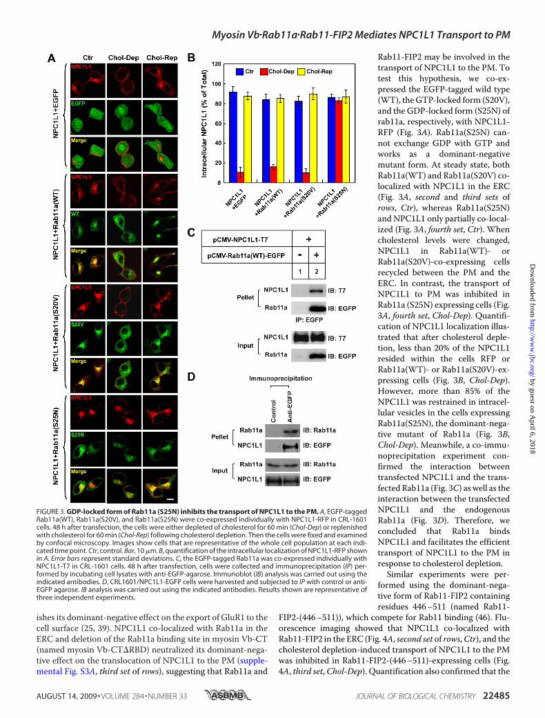
Rab11-FIP2 may be involved in thetransport of NPC1L1 to the PM. Totest this hypothesis, we co-ex-pressed the EGFP-tagged wild type(WT), theGTP-locked form (S20V),and the GDP-locked form (S25N) ofrab11a, respectively, with NPC1L1-RFP (Fig. 3A). Rab11a(S25N) can-not exchange GDP with GTP andworks as a dominant-negativemutant form. At steady state, bothRab11a(WT) and Rab11a(S20V) co-localized with NPC1L1 in the ERC(Fig. 3A, second and third sets ofrows, Ctr), whereas Rab11a(S25N)andNPC1L1 only partially co-local-ized (Fig. 3A, fourth set, Ctr). Whencholesterol levels were changed,NPC1L1 in Rab11a(WT)- orRab11a(S20V)-co-expressing cellsrecycled between the PM and theERC. In contrast, the transport ofNPC1L1 to PM was inhibited inRab11a (S25N) expressing cells (Fig.3A, fourth set, Chol-Dep). Quantifi-cation of NPC1L1 localization illus-trated that after cholesterol deple-tion, less than 20% of the NPC1L1resided within the cells RFP orRab11a(WT)- or Rab11a(S20V)-ex-pressing cells (Fig. 3B, Chol-Dep).However, more than 85% of theNPC1L1 was restrained in intracel-lular vesicles in the cells expressingRab11a(S25N), the dominant-nega-tive mutant of Rab11a (Fig. 3B,Chol-Dep). Meanwhile, a co-immu-noprecipitation experiment con-firmed the interaction betweentransfected NPC1L1 and the trans-fectedRab11a (Fig. 3C) aswell as theinteraction between the transfectedNPC1L1 and the endogenousRab11a (Fig. 3D). Therefore, weconcluded that Rab11a bindsNPC1L1 and facilitates the efficienttransport of NPC1L1 to the PM inresponse to cholesterol depletion.Similar experiments were per-
formed using the dominant-nega-tive form of Rab11-FIP2 containingresidues 446–511 (named Rab11-
FIP2-(446–511)), which compete for Rab11 binding (46). Flu-orescence imaging showed that NPC1L1 co-localized withRab11-FIP2 in the ERC (Fig. 4A, second set of rows,Ctr), and thecholesterol depletion-induced transport of NPC1L1 to the PMwas inhibited in Rab11-FIP2-(446–511)-expressing cells (Fig.4A, third set,Chol-Dep). Quantification also confirmed that the
FIGURE 3. GDP-locked form of Rab11a (S25N) inhibits the transport of NPC1L1 to the PM. A, EGFP-taggedRab11a(WT), Rab11a(S20V), and Rab11a(S25N) were co-expressed individually with NPC1L1-RFP in CRL-1601cells. 48 h after transfection, the cells were either depleted of cholesterol for 60 min (Chol-Dep) or replenishedwith cholesterol for 60 min (Chol-Rep) following cholesterol depletion. Then the cells were fixed and examinedby confocal microscopy. Images show cells that are representative of the whole cell population at each indi-cated time point. Ctr, control. Bar, 10 �m. B, quantification of the intracellular localization of NPC1L1-RFP shownin A. Error bars represent standard deviations. C, the EGFP-tagged Rab11a was co-expressed individually withNPC1L1-T7 in CRL-1601 cells. 48 h after transfection, cells were collected and immunoprecipitation (IP) per-formed by incubating cell lysates with anti-EGFP-agarose. Immunoblot (IB) analysis was carried out using theindicated antibodies. D, CRL1601/NPC1L1-EGFP cells were harvested and subjected to IP with control or anti-EGFP agarose. IB analysis was carried out using the indicated antibodies. Results shown are representative ofthree independent experiments.
Myosin Vb�Rab11a�Rab11-FIP2 Mediates NPC1L1 Transport to PM
AUGUST 14, 2009 • VOLUME 284 • NUMBER 33 JOURNAL OF BIOLOGICAL CHEMISTRY 22485
by guest on April 6, 2018
http://ww
w.jbc.org/
Dow
nloaded from

intracellularly localizedNPC1L1 inRab11-FIP2-(446–511)-ex-pressing cells (�82%) was significantly higher than that of thecontrol (�4%) or Rab11-FIP2-expressing cells (�5%) (Fig. 4B).Subsequent co-immunoprecipitation experiments verified theassociation between NPC1L1 and Rab11-FIP2 (Fig. 4C).Myosin Vb has been reported to associate with Rab8a-posi-
tive vesicles (31, 47). Therefore, Rab8a was tested similarly byco-transfection and imaging analysis. However, none of theWT or GTP- or GDP-locked forms of Rab8a had an obviouseffect on the recycling of NPC1L1 protein (supplemental Fig.S4). Therefore, it is unlikely that Rab8a is involved in the cho-lesterol-regulated recycling of NPC1L1.The Endocytic Recycling Triple Complex Is Required for
Translocation of NPC1L1 to the PM—Next, we examined therequirement of endogenous myosin Vb, Rab11a, and Rab11-
FIP2 in the transport of NPC1L1 tothe PMusing siRNA-mediated genesilencing. Real-time PCR analysisshowed that the mRNA levels ofmyosin Vb, Rab11a, and Rab11-FIP2 were reduced by �60, �75,and �80%, respectively (Fig. 5A).Then we analyzed the transport ofNPC1L1-EGFP to the PM inducedby cholesterol depletion followingRNA interference. As shown in Fig.5, B and C, knockdown of myosinVb, Rab11a, or Rab11-FIP2 causedretention of�50%of theNPC1L1 inintracellular vesicles comparedwithcontrol cells after cholesterol deple-tion for 60 min. To rule out the pos-sible off-target effect of siRNAs, twoadditional siRNAs against eachcomponent of the triple complexwere tested, and consistent resultswere obtained (data not shown).Thus, the transport of NPC1L1 tothe PM relies on the myosinVb�Rab11a�Rab11-FIP2 endocyticrecycling triple complex, and deple-tion of any one of the componentsinhibits the transport of NPC1L1to PM.Cholesterol-regulated Binding
of NPC1L1 to Rab11a and MyosinVb—Because the myosin Vb�Rab11a�Rab11-FIP2 complex facilitated thecholesterol depletion-induced trans-port of NPC1L1 to the PM, we thentested whether the interactionbetween NPC1L1 and the triplecomplex was regulated by choles-terol. Consistent with the earlierresults (Fig. 3C),NPC1L1 associatedwith Rab11a at steady state (Fig. 6A,lane 3). After 10 min of CDX treat-ment, the interaction between
NPC1L1 and Rab11a gradually decreased (Fig. 6A, lanes 4–8),suggesting that Rab11a gradually dissociated from NPC1L1during cholesterol depletion. On the other hand, the binding ofNPC1L1 to myosin Vb did not change during the first 20 minafter CDX treatment (Fig. 6B, lanes 3–5) but was drasticallyreduced following the 20-min time point (Fig. 6B, lanes 6–8).Our data indicate that Rab11a and myosin Vb dissociatedsequentially from NPC1L1.Disturbing the Endocytic Recycling Triple Complex Inhibits
NPC1L1-mediated Cholesterol Uptake—Our previous studiesshowed that NPC1L1 facilitates cholesterol uptake via its vesic-ular endocytosis from the PM (8). Therefore, localization ofNPC1L1 on the PM is a prerequisite for cholesterol uptake. Tofurther elucidate the role of the endocytic recycling triple com-plex in NPC1L1-mediated cholesterol uptake, NPC1L1-RFP
FIGURE 4. Rab11-FIP2-(446 –511) blocks the transport of NPC1L1 to PM. A, NPC1L1-RFP were co-expressedwith EGFP-tagged Rab11-FIP2 and Rab11-FIP2-(446 –511) individually in CRL-1601 cells. 48 h after transfection,the cells were either depleted of cholesterol for 60 min (Chol-Dep) or replenished with cholesterol for 60 min(Chol-Rep) following cholesterol depletion. Then the cells were fixed and examined by confocal microscopy.Images show cells that are representative of the whole cell population at each indicated time point. Ctr, control.Bar, 10 �m. B, quantification of the intracellular localization of NPC1L1-RFP shown in A. Error bars representstandard deviations. C, EGFP-tagged Rab11-FIP2 and Rab11-FIP2-(446 –511) were co-expressed individuallywith NPC1L1-T7 in CRL-1601 cells. 48 h after transfection, EGFP-tagged Rab11-FIP2 variants were immunopre-cipitated (IP) with anti-EGFP antibody-coupled agarose. Immunoblot (IB) analysis was carried out using theindicated antibodies. Results shown are representative of three independent experiments.
Myosin Vb�Rab11a�Rab11-FIP2 Mediates NPC1L1 Transport to PM
22486 JOURNAL OF BIOLOGICAL CHEMISTRY VOLUME 284 • NUMBER 33 • AUGUST 14, 2009
by guest on April 6, 2018
http://ww
w.jbc.org/
Dow
nloaded from

was co-expressed with EGFP-tagged myosin Vb-CT,Rab11a(S25N), or Rab11-FIP2-(446–511) in rat hepatocyteCRL1601 cells. After transfection, the cells were depletedof cholesterol followed by cholesterol replenishment. Then thecells were fixed, stained with filipin, and imaged by two-photonconfocal microscopy to analyze cholesterol uptake. As
shown in Fig. 7A, cells expressing NPC1L1-RFP and EGFP(indicated by arrowheads) took up much more cholesterolthan the untransfected control cells (indicated by arrows).However, in myosin Vb-CT-, Rab11a(S25N)-, and Rab11-FIP2(446–511)-expressing cells, the uptake of cholesterolwas similar to the untransfected cells. Quantification showed thatafter cholesterol replenishment, the cholesterol internalization inEGFP-expressing cells (�60%)was 4 times that in the control cells(�15%),whereas expressionofmyosinVb-CT,Rab11a(S25N), orRab11-FIP2-(446–511) reduced cholesterol internalizationmediated by NPC1L1 (Fig. 7B). These data indicate that inhib-iting the myosin Vb�Rab11a�Rab11-FIP2 triple complex blocks
FIGURE 5. siRNA-mediated silencing of endogenous myosin Vb, Rab11a,or Rab11-FIP2 attenuates the transport of NPC1L1 to PM. A, real-time PCRshowing the knockdown efficiency of myosin Vb, Rab11a, or Rab11-FIP2.B, CRL1601/NPC1L1-EGFP cells were transfected with the indicated siRNAs.72 h after transfection, the cells were depleted of cholesterol for differentdurations of time. At each indicated time point, the cells were fixed and exam-ined by confocal microscopy. Images show the cells representative of thewhole cell population at each indicated time point. Bar, 10 �m. C, quantifica-tion of the intracellular localization of NPC1L1-EGFP shown in B. Error barsrepresent standard deviations. Results shown are representative of threeindependent experiments.
FIGURE 6. The binding of NPC1L1 to Rab11a and myosin Vb is regulatedby cholesterol depletion. A, plasmids encoding NPC1L1-EGFP andRab11a-T7 were co-transfected into CRL-1601 cells. 48 h later, the cells weredepleted of cholesterol by treatment with 1% CDX for various time durations.Then the cells were harvested, and immunoprecipitation (IP) was performedby pulling down EGFP-tagged NPC1L1 with anti-EGFP antibody-coupled aga-rose. Immunoblot (IB) analysis was carried out using the indicated antibodies.B, plasmids encoding NPC1L1-T7 and EGFP-myosin Vb were co-transfectedinto CRL-1601 cells. 48 h later, the cells were depleted of cholesterol by treat-ment 1% CDX for various durations of time. Then the cells were harvested,and immunoprecipitation was performed by pulling down EGFP-taggedmyosin Vb with anti-EGFP antibody-coupled agarose. Immunoblot analysiswas carried out using the indicated antibodies. Results shown are represent-ative of three independent experiments.
Myosin Vb�Rab11a�Rab11-FIP2 Mediates NPC1L1 Transport to PM
AUGUST 14, 2009 • VOLUME 284 • NUMBER 33 JOURNAL OF BIOLOGICAL CHEMISTRY 22487
by guest on April 6, 2018
http://ww
w.jbc.org/
Dow
nloaded from

the transport of NPC1L1 to the PM and decreases NPC1L1-mediated cholesterol uptake.
DISCUSSION
Here we investigated the role of the MF and the myosinVb�Rab11a�Rab11-FIP2 triple complex in cholesterol-regu-lated endocytic recycling of NPC1L1. First, the integrity anddynamics of the MF are critical for the transport of NPC1L1.Second, the endocytic recycling triple complex, consisting ofthe MF-interacting motor myosin Vb, the small GTPaseRab11a, and the adaptor Rab11-FIP2, mediates the transportof NPC1L1 to the PM. Inactivation of the triple complexinhibits the export of NPC1L1 and NPC1L1-mediated cho-lesterol uptake.
Interestingly, our data support that not only the integrity butalso the dynamics of actin fibers are important for the transportof NPC1L1, suggesting that the MFmay function more than asthe tracks for cargomotility. TheMFnetwork is highly dynamicand may participate in multiple steps of vesicle traffickingincluding scission, transport, and fusion. The polymerizationand depolymerization of the MF may provide the mechanicalforce for each step. Meanwhile, many proteins are involved inthe remodeling of the MF, such as the ARP2/3 (48) complexthat initiates MF nucleation and facilitates vesicle transport.Based on our previous result that ARP2/3 is associated withNPC1L1, it is very likely that MF remodeling factors such asARP2/3 also play a role in NPC1L1 recycling and cholesteroluptake (8).Myosin Vb has been found to be responsible for the endo-
cytic shuttling and sorting of proteins including transferrinreceptor, CFTR, AMPA receptors, BSEP, GLUT4, aquaporin-2,and �2-adrenergic receptor (18, 24–33). Our finding that themyosin Vb�Rab11a�Rab11-FIP2 complex is also required forNPC1L1 recycling further supports the hypothesis that myosinVb and its effectors function mainly in the traffic of transmem-brane proteins between the PM and recycling endosomes. Inaddition, myosin Vb and Rab11a have been shown to be criticalfor the bile canaliculus formation in polarized hepatocytes (49).In fact, human NPC1L1 is also highly expressed in the liver inaddition to the intestine. We and others have found thatNPC1L1 localizes on the apical membrane of cultured hepa-tocytes, which resemble the canalicular membrane (data notshown) (8, 50). It is very likely that in human liver, myosinVb, together with Rab11a and Rab11-FIP2, targets NPC1L1to the canalicular membrane to reabsorb the free cholesterolfrom bile.Although the MF system is required for the recycling of
NPC1L1 between the PM and ERC, the role of the myosinVb�Rab11a�Rab11-FIP2 triple complex in the internalization ofNPC1L1 has not been determined here. Upon disrupting themyosin Vb�Rab11a�Rab11-FIP2 complex, NPC1L1 did not relo-calize to the PM after cholesterol depletion in our experimentalsystem, and therefore we could not study the internalization ofNPC1L1 under cholesterol repletion conditions. Because it hasbeen shown that the triple complex is localized in endocyticrecycling endosomes, whereas NPC1L1 presents on the PMafter cholesterol depletion, it is unlikely that the myosinVb�Rab11a�Rab11-FIP2 triple complex is involved directlyin the endocytosis of NPC1L1.Loss-of-function mutations of myosin Vb can cause
microvillus inclusion disease (MVID) (51), which is charac-terized by lack of microvilli on the surface of intestinalenterocytes and accumulation of microvilli in intracellularvacuolar structures. The vesicles that are otherwise targetedto the apical or basolateral membranes become aberrantlylocalized in the patient’s enterocytes, suggesting the impor-tance of myosin Vb in the correct targeting of endocyticvesicles in the intestine. Although the distribution ofNPC1L1 in the intestinal cells of MVID patients has not beenstudied, it is likely that the localization of NPC1L1 and cho-lesterol absorption are both disrupted in MVID enterocytes,as myosin Vb is required for the targeting of NPC1L1. More
FIGURE 7. Myosin Vb-CT, Rab11a(S25N), or Rab11-FIP2-(446 –511)impairs the NPC1L1-mediated cholesterol uptake. A, NPC1L1-RFP was co-expressed with EGFP-tagged myosin Vb-CT, Rab11a(S25N), and Rab11-FIP2-(446 –511) individually in CRL-1601 cells. 48 h after transfection, the cells weredepleted of cholesterol followed by cholesterol replenishment for 60 min.Then the cells were fixed, stained with filipin, and examined by two-photonconfocal microscopy. Images show the cells representative of the whole cellpopulation at each indicated time point. Bar, 10 �m. Arrows indicate the cellswith no expression of the transfected genes. Arrowheads indicate the cellsexpressing the transfected genes. B, quantification of the intracellular choles-terol of the cells shown in A. Error bars represent standard deviations. Resultsshown are representative of two independent experiments.
Myosin Vb�Rab11a�Rab11-FIP2 Mediates NPC1L1 Transport to PM
22488 JOURNAL OF BIOLOGICAL CHEMISTRY VOLUME 284 • NUMBER 33 • AUGUST 14, 2009
by guest on April 6, 2018
http://ww
w.jbc.org/
Dow
nloaded from
work is required to address the physiological role of the myo-sin Vb�Rab11a�Rab11-FIP2 triple complex in cholesterolabsorption in animals.Myosin Vb and Rab11a showed different trends when they
dissociated from NPC1L1 during cholesterol depletion (Fig.6), implying that Rab11a and myosin Vb may associate withNPC1L1 in different ways during the translocation ofNPC1L1 to the PM. We also found that the Rab11a bindingdomain in myosin Vb-CT is required for the dominant-neg-ative effect (supplemental Fig. S3). Taking all of our findingstogether, the following model is conceivable. Rab11a andRab11-FIP2 binds to NPC1L1 at the steady state and bridgesthe interaction between NPC1L1 and myosin Vb. When thecellular cholesterol level drops, Rab11a/Rab11-FIP2 dissoci-ates from the NPC1L1�myosin Vb complex, which allows theexport of NPC1L1�myosin Vb toward the PM in a MF-de-pendent manner. After arriving at the PM, myosin Vb isseparated and released. The interaction between NPC1L1and myosin Vb or Rab11a/Rab11-FIP2 may be direct or maybe mediated by other factors. Additional studies are neededto address this question.In summary, our data provide evidence showing that the
myosin Vb�Rab11a�Rab11-FIP2 triple complex is requiredfor the transport of NPC1L1 to the PM and plays an impor-tant role in cholesterol uptake. This finding enriches ourknowledge of the molecular pathway of cholesterol absorp-tion and provides a basis for developing novel cholesterolabsorption inhibitors.
Acknowledgments—We greatly appreciate the gift of myosin Va andVb plasmids fromDr. Alaa El-Husseini (University of British Colum-bia, Canada).We thank Yu-Xiu Qu, Su-Zhe Pan, Qing Li, and Chun-Geng Yi for technical assistance and Dr. Wei Qi for critical reading ofthe manuscript.
REFERENCES1. Davies, J. P., Scott, C., Oishi, K., Liapis, A., and Ioannou, Y. A. (2005) J. Biol.
Chem. 280, 12710–127202. Davis, H. R., Jr., Zhu, L. J., Hoos, L. M., Tetzloff, G., Maguire, M., Liu, J.,
Yao, X., Iyer, S. P., Lam, M. H., Lund, E. G., Detmers, P. A., Graziano,M. P., and Altmann, S. W. (2004) J. Biol. Chem. 279, 33586–33592
3. Temel, R. E., Tang, W., Ma, Y., Rudel, L. L., Willingham, M. C., Ioannou,Y. A., Davies, J. P., Nilsson, L. M., and Yu, L. (2007) J. Clin. Invest. 117,1968–1978
4. Altmann, S. W., Davis, H. R., Jr., Zhu, L. J., Yao, X., Hoos, L. M., Tetzloff,G., Iyer, S. P., Maguire, M., Golovko, A., Zeng, M., Wang, L., Murgolo, N.,and Graziano, M. P. (2004) Science 303, 1201–1204
5. Davis, H. R., and Veltri, E. P. (2007) J. Atheroscler. Thromb. 14, 99–1086. Wang, J., Chu, B. B., Ge, L., Li, B. L., Yan, Y., and Song, B. L. (2009) J. Lipid
Res, in press7. Davies, J. P., Levy, B., and Ioannou, Y. A. (2000) Genomics 65, 137–1458. Ge, L., Wang, J., Qi, W., Miao, H. H., Cao, J., Qu, Y. X., Li, B. L., and Song,
B. L. (2008) Cell Metab. 7, 508–5199. Maxfield, F. R., and McGraw, T. E. (2004) Nat. Rev. Mol. Cell Biol. 5,
121–13210. Yu, L., Bharadwaj, S., Brown, J. M.,Ma, Y., Du,W., Davis, M. A., Michaely,
P., Liu, P., Willingham, M. C., and Rudel, L. L. (2006) J. Biol. Chem. 281,6616–6624
11. Chang, T. Y., and Chang, C. (2008) Cell Metab. 7, 469–47112. Prentki,M., Chaponnier, C., Jeanrenaud, B., andGabbiani, G. (1979) J. Cell
Biol. 81, 592–60713. Segawa, A., and Yamashina, S. (1989) Cell Struct. Funct. 14, 531–54414. Dantzig, J. A., Liu, T. Y., and Goldman, Y. E. (2006) Ann. N.Y. Acad. Sci.
1080, 1–1815. Apodaca, G. (2001) Traffic 2, 149–15916. Schafer, D. A. (2002) Curr. Opin. Cell Biol. 14, 76–8117. Bridgman, P. C. (2004) J. Neurobiol. 58, 164–17418. Langford, G. M. (2002) Traffic 3, 859–86519. Tilelli, C. Q., Martins, A. R., Larson, R. E., and Garcia-Cairasco, N. (2003)
Neuroscience 121, 573–58620. Walikonis, R. S., Jensen, O. N., Mann, M., Provance, D. W., Jr., Mercer,
J. A., and Kennedy, M. B. (2000) J. Neurosci. 20, 4069–408021. Rodriguez, O. C., and Cheney, R. E. (2002) J. Cell Sci. 115, 991–100422. Zhao, L. P., Koslovsky, J. S., Reinhard, J., Bahler, M., Witt, A. E., Provance,
D. W., Jr., and Mercer, J. A. (1996) Proc. Natl. Acad. Sci. U.S.A. 93,10826–10831
23. Passeron, T., Bahadoran, P., Bertolotto, C., Chiaverini, C., Busca, R., Va-lony, G., Bille, K., Ortonne, J. P., and Ballotti, R. (2004) FASEB J. 18,989–991
24. Lapierre, L. A., Kumar, R., Hales, C. M., Navarre, J., Bhartur, S. G., Bur-nette, J. O., Provance, D.W., Jr., Mercer, J. A., Bahler, M., and Goldenring,J. R. (2001)Mol. Biol. Cell 12, 1843–1857
25. Lise, M. F., Wong, T. P., Trinh, A., Hines, R. M., Liu, L., Kang, R., Hines,D. J., Lu, J., Goldenring, J. R.,Wang, Y. T., and El-Husseini, A. (2006) J. Biol.Chem. 281, 3669–3678
26. Nedvetsky, P. I., Stefan, E., Frische, S., Santamaria, K.,Wiesner, B., Valenti,G., Hammer, J. A., 3rd, Nielsen, S., Goldenring, J. R., Rosenthal, W., andKlussmann, E. (2007) Traffic 8, 110–123
27. Volpicelli, L. A., Lah, J. J., Fang, G., Goldenring, J. R., and Levey, A. I. (2002)J. Neurosci. 22, 9776–9784
28. Swiatecka-Urban, A., Talebian, L., Kanno, E., Moreau-Marquis, S., Couter-marsh, B., Hansen, K., Karlson, K. H., Barnaby, R., Cheney, R. E., Langford,G.M., Fukuda,M., and Stanton, B.A. (2007) J. Biol. Chem.282, 23725–23736
29. Provance, D. W., Jr., Addison, E. J., Wood, P. R., Chen, D. Z., Silan, C. M.,and Mercer, J. A. (2008) BMC Cell Biol. 9, 44
30. Wang, Z., Edwards, J. G., Riley, N., Provance, D. W., Jr., Karcher, R., Li,X. D., Davison, I. G., Ikebe,M.,Mercer, J. A., Kauer, J. A., and Ehlers,M. D.(2008) Cell 135, 535–548
31. Ishikura, S., and Klip, A. (2008) Am. J. Physiol. Cell Physiol. 295,C1016–C1025
32. Kessler, A., Tomas, E., Immler, D., Meyer, H. E., Zorzano, A., and Eckel, J.(2000) Diabetologia 43, 1518–1527
33. Millman, E. E., Zhang, H., Zhang, H., Godines, V., Bean, A. J., Knoll, B. J.,and Moore, R. H. (2008) Traffic 9, 1958–1971
34. Ullrich, O., Reinsch, S., Urbe, S., Zerial, M., and Parton, R. G. (1996) J. CellBiol. 135, 913–924
35. Jagoe, W. N., Lindsay, A. J., Read, R. J., McCoy, A. J., McCaffrey, M. W.,and Khan, A. R. (2006) Structure 14, 1273–1283
36. Cao, J., Wang, J., Qi, W., Miao, H. H., Wang, J., Ge, L., DeBose-Boyd,R. A., Tang, J. J., Li, B. L., and Song, B. L. (2007) Cell Metab. 6, 115–128
37. Song, B. L., Sever, N., and DeBose-Boyd, R. A. (2005) Mol. Cell 19,829–840
38. Dubinsky, W. P., Mayorga-Wark, O., and Schultz, S. G. (1999) Proc. Natl.Acad. Sci. U.S.A. 96, 9421–9426
39. Gibbon, B. C., Kovar, D. R., and Staiger, C. J. (1999) Plant Cell 11,2349–2363
40. Xie, L., and Forer, A. (2008) Cell Motil. Cytoskeleton 65, 876–88941. Lee, J. C., Field, D. J., and Lee, L. L. (1980) Biochemistry 19, 6209–621542. Caperta, A. D., Delgado, M., Ressurreiçcao, F., Meister, A., Jones, R. N.,
Viegas, W., and Houben, A. (2006) Protoplasma 227, 147–15343. Yamashiro, D. J., Tycko, B., Fluss, S. R., and Maxfield, F. R. (1984) Cell 37,
789–80044. Vossenkamper, A., Nedvetsky, P. I., Wiesner, B., Furkert, J., Rosenthal,
W., and Klussmann, E. (2007) Am. J. Physiol. Cell Physiol. 293,C1129–C1138
45. Taft, M. H., Hartmann, F. K., Rump, A., Keller, H., Chizhov, I., Manstein,D. J., and Tsiavaliaris, G. (2008) J. Biol. Chem. 283, 26902–26910
46. Lindsay, A. J., and McCaffrey, M. W. (2002) J. Biol. Chem. 277,
Myosin Vb�Rab11a�Rab11-FIP2 Mediates NPC1L1 Transport to PM
AUGUST 14, 2009 • VOLUME 284 • NUMBER 33 JOURNAL OF BIOLOGICAL CHEMISTRY 22489
by guest on April 6, 2018
http://ww
w.jbc.org/
Dow
nloaded from

27193–2719947. Roland, J. T., Kenworthy, A. K., Peranen, J., Caplan, S., and Goldenring,
J. R. (2007)Mol. Biol. Cell 18, 2828–283748. Pollard, T. D. (2007) Annu. Rev. Biophys. Biomol. Struct. 36, 451–47749. Wakabayashi, Y., Dutt, P., Lippincott-Schwartz, J., and Arias, I. M. (2005)
Proc. Natl. Acad. Sci. U.S.A. 102, 15087–15092
50. Yu, L. (2008) Curr. Opin. Lipidol. 19, 263–26951. Muller, T., Hess, M. W., Schiefermeier, N., Pfaller, K., Ebner, H. L.,
Heinz-Erian, P., Ponstingl, H., Partsch, J., Rollinghoff, B., Kohler, H.,Berger, T., Lenhartz, H., Schlenck, B., Houwen, R. J., Taylor, C. J.,Zoller, H., Lechner, S., Goulet, O., Utermann, G., Ruemmele, F. M.,Huber, L. A., and Janecke, A. R. (2008) Nat. Genet. 40, 1163–1165
Myosin Vb�Rab11a�Rab11-FIP2 Mediates NPC1L1 Transport to PM
22490 JOURNAL OF BIOLOGICAL CHEMISTRY VOLUME 284 • NUMBER 33 • AUGUST 14, 2009
by guest on April 6, 2018
http://ww
w.jbc.org/
Dow
nloaded from

Li and Bao-Liang SongBei-Bei Chu, Liang Ge, Chang Xie, Yang Zhao, Hong-Hua Miao, Jing Wang, Bo-Liang
Translocation of NPC1L1 to the Cell SurfaceRequirement of Myosin Vb·Rab11a·Rab11-FIP2 Complex in Cholesterol-regulated
doi: 10.1074/jbc.M109.034355 originally published online June 19, 20092009, 284:22481-22490.J. Biol. Chem.
10.1074/jbc.M109.034355Access the most updated version of this article at doi:
Alerts:
When a correction for this article is posted•
When this article is cited•
to choose from all of JBC's e-mail alertsClick here
Supplemental material:
http://www.jbc.org/content/suppl/2009/06/17/M109.034355.DC1
http://www.jbc.org/content/284/33/22481.full.html#ref-list-1
This article cites 50 references, 20 of which can be accessed free at
by guest on April 6, 2018
http://ww
w.jbc.org/
Dow
nloaded from