reprinted from annals of the new york academy of sciences
TRANSCRIPT

Reprinted from Annals of The New York Academy of Sciences Volume 66, Article 3, Pages 435-444
March 14. 1957
THE DISTRIBUTION AND METABOLISM OF LYSERGIC ACID DIETHYLAMIDE
By Julius Axelrod,* Roscoe 0. Brady,+ Bernard Witkop,$ and Edward V. Evarts*
National Institutes of Health, Public Health Service, Department of Health, Education, and Wel/are, Bethesda, Md.
Although lysergic acid diethylamide (LSD), the hallucinogenic agent, has been the subject of numerous investigations, little is known about its biologic fate. In recent studies Boyd et al.’ and St011 el ~1.2 have examined the tissue distribution of CL4-labeled LSD in mice. Stoll and his co-workers2 have shown that the Cr4-labeled material measured in tissues included LSD, as well as transformation products of the drug. Lanz, Cerletti, and Roth1in3 have also measured the tissue levels of LSD in mice by the use of a bioassay procedure based on the antagonism of LSD to serotonin-induced contraction of the uterus of the rat. These investigators, however, did not show that possible bio- logically active transformation products of LSD were excluded from the assay procedure.
The development of a specific and sensitive method for the estimation of LSD in biological materials has enabled us to study the tissue distribution, excretion, rate of biotransformation, and metabolism of the drug, as well as the subcellular processes involved in its transformation.
Methods and Materials
Eslimatiorz of LSD content. LSD was isolated from sodium chloride-satu- rated biological material at an alkaline pH by extraction into heptane. The LSD in the heptane extract was returned to dilute hydrochloric acid and its quantity was estimated spectrofluorometrically.
Procedure. Biological material (up to 5 ml.) was added to a 60-ml. glass- stoppered bottle that held 25 ml. of n-heptane containing 2 per cent isoamyl alcohol,§ 0.5 ml. of 1 N NaOH, and about 3 gm. of sodium chloride. The bottle was shaken for 30 minutes and then centrifuged. Twenty ml. of the heptane phase were transferred to a 40-ml. glass-stoppered centrifuge tube containing 3 ml. of 0.004 N HCl, and the tube was shaken for 10 minutes. An aliquot of the acid phase was transferred to a quartz cuvette, and the LSD content was determined by measuring its fluorescence in a Farrand or Bowman4 spectrofluorophotometer at 445 mp after activation at 325 mp.T As little as 0.001 pg. per ml. of LSD could be determined by this procedure. Concen- trations of the drug greater than 10 pg. may be measured spectrophoto- metrically at 310 ml.c.
l National Institute of Menial Health. t National Institute of Neurological Diseases and Blindness. 1 National Institute of Arthritis and Metabolic Diseases. 5 All solvents were purified by successive washings with 1 N N&H, 1 N HCI, and water. II When LSD in dilute hydrochloric acid was exposed to light at 325 mr for mcvre than 2
of the fluorescence of the compound gradually diminished. seconds the intensity
the intensity of fluorescence returned to its initial value. Upon removal from the light scwce for 5 minutes
435

436 Annals New York Academy of Sciences TABLE 1
DISTRIBUTION OF LSD AND APPARENT LSD BETWEEN HEPTANE AND WATER AT VARIOUS PH VALUES*
PI3 I
Authentic LSD Apparent LSD from plasma
5.0 6.3 7.0
*Theapparent LSD from the plasma,of a cat that had received the drug intravenously wasextractedintoa heptane-isovyl alcohol mixture BS descrtbed under the heading Y&od~ and Molevials. Abquots of this solution and a solution of authentic LSD in heptane (containing 2 per cent isoamyl alcohol) were shaken with one- quarter volumes of sodium-chloride-saturated bu5ers at various pH values. The fraction of the compound ex- tmcted WBS expressed as the ratio of the amount of compound in the organic phase to total compound.
Tissues were prepared for analysis by homogenization in a Waring blender with 5 volumes of water.
LSD added to biological material was recovered with adequate precision (95 f 6 per cent).
Assay of speci@ly. In order to determine whether closely related trans- formation products of LSD were included in the measurement by the method described above, the specificity was assayed by means of the technique of comparative distribution ratios (Brodie, Udenfriend, and Baer).6 To escape detection by this technique, metabolic products must simulate LSD to an im- probable degree, in that they would have identical solubility characteristics in 2 solvents and similar dissociation constants. The distribution ratios of ap- parent LSD extracted from the plasma of a cat that had received the drug were compared with those of authentic LSD in a series of 2-phase systems consist- ing of heptane containing 2 per cent isoamyl alcohol and water at various pH values. The results shown in TABLE 1 indicate that the 2 compounds had the same solubility at various pH values and were presumably the same.
Preparation of tissue samples for enzyme studies. Preparation of all tissue samples was carried out at 0 to 3” C. Animals were stunned and exsanguinated, and the tissues were immediately removed and chilled. Tissue slices were prepared with a Stadie-Riggs* slicer. Liver preparations free of nuclei and mitochondria, but containing microsomes, were prepared by homogenizing the tissue with 4 volumes of isotonic KC1 with a Potter-Elvehjem-type homoge- nizer* followed by centrifugation at 10,000 g for 15 minutes. Nuclei, mi- tochondria, microsomes, and soluble fractions were separated by fractional centrifugation (Schneider).6
Measurement of the enzymic corrzjersio?l of LSD. In most studies on the en- zymic conversion of LSD, 1 ml. of enzyme preparation, obtained from 200 mg. of tissue, was incubated in a 20 ml. beaker at 3i”C. with 0.66 pmol of LSD, 0.2 lmol of triphosphopyridine nucleotide (TPN+), 25 pmol of nico- tinamide, 25 pmol of magnesium chloride, 500 pmol of potassium phosphate buffer (pH 7.9), and water to make a final volume of 5 ml. The mixture was shaken in a Dubnoff metabolic apparatus for 120 minutes in an atmosphere of
l Manufactured by the Arthur H. Thomas Co., Phila., Pa.

Axelrod et al.: Metabolism of Lysergic Acid Diethylamide 437 TABLE 2
TISSUE DISTRIBUTION OF LSD IN THE CAT* ----- Tissue LSD
Plasma............. ,.. ..,_...,._ Cerebrospinal fluid. Brain.... .._ Liver. . Kidney.. . . . . . ...‘...:::‘:::.:‘:::::‘:::::::::::::: Muscle. Heart................::::‘:::::‘::’:.::::.:::::.‘~...... Lung.. Spleen..... I’ I.:. ~::::::I~:‘:: II” :I:“’ Y:::::‘:: Intestine.......::....:‘.. . . . . . . . . . .._.... . ..I...._.._,. Fat. . . Bile..........::~~.::::::::::::......... ::::::::::::::::
mz.lkg. 1.75 0.36 0.52 0.67 0.53 0.20
8% 0:3s 0.39 0.20 1.85
l The studies were made PO minutes after the intravenous administration of 1 mg./kg. of LSD.
9.5 per cent 02 to 5 per cent CO2 . At the end of the incubation period an aliquot of the reaction mixture was immediately taken for LSD analysis. Enzyme activity was expressed as pmol of LSD, which disappeared on in- cubation.
Materials. Triphosphopyridine nucleotide (TPN+) of about 95 per cent purity and glucosed-phosphate were obtained from Sigma Chemical Co., St. Louis, MO. Glucose-6-phosphate dehydrogenase was prepared by the procedure of Kornberg.’ Lysergic acid diethylamide was obtained from the Sandoz Chemical Works, Hanover, N. J.
Results and Discussion
The distribution of LSD in tissues. The distribution of LSD was examined in representative tissues of a cat that had received 1 mg./kg. of the drug in- travenously. Ninety minutes after administration of the drug, the animal was sacrificed and the tissues sampled immediately afterward (TABLE 2). LSD was found to be localized in the bile and plasma to a considerable extent, whereas only negligible amounts were present in the fat. The high concen- tration of LSD in the bile and the negligible amounts of the drug found in the feces (see below) would suggest that it was secreted into the intestines and then reabsorbed. In the order of decreasing magnitude, the concentration of LSD in the organ tissues was as follows: lung, liver, brain, intestines,. spleen, cerebrospinal fluid, muscle, and fat. The presence of considerable amounts of the drug in the brain and cerebrospinal fluid indicates that the substance can pass the blood-brain barrier. No differences in the concentration of LSD in the cortex, midbrain, spinal cord, or hypothalamus were found. From the amount of LSD found in the brain of a cat after the administration of 1 mg./ kg., it can be calculated that the drug exerts its psychological effect in man (given 1 pg. per kg.) at a concentration of 0.0005 fig. per gm. of brain tissue.
Since considerable amounts of LSD were found in the plasma, the extent to which the drug was bound to plasma proteins was determined by dialysis at

438 Annals New York Academy of Sciences
0.307
123456 TIME IN HOURS
Frcnnk 1. Plasma (solid line) and cerebrospinal &lid (dashed line) 1 evels of LSD after the intravenous ad- ministration of 0.2 mg./kg. of the drug to a monkey.
37°C. for 18 hours against isotonic phosphate buffer at $H 7.4. Visking membranes* were used as dialysis bags. At plasma concentrations of 0.1 and 20 mg. per liter, 90 and 6.5 per cent of LSD, respectively, was found to be bound to the nondiffusable constituents of the plasma.
Relationship between plasma and cerebrospinal-&id levels of LSD. Monkeys (Macaca mulatta) were given LSD (0.2 mg./kg.) intravenously. Blood and cerebrospinal fluid from the lumbar subarachnoid space were taken at various time intervals and assayed for LSD (FIGURE 1). The rapid decline of the plasma level of the drug in the first hour was presumbaly due to a shift of LSD from plasma to tissues. After the first hour, the slower disappearance was a reflection of the rate of metabolism of the drug. The biologic half life of LSD in the monkey, that is, the time required for the plasma level to fall to half its value, was found to be about 100 minutes.
Maximum concentration of LSD in the cerebrospinal fluid was reached within 10 minutes, and then the amount declined slowly. Since the amount of drug present in the cerebrospinal fluid was about the same as that present in the unbound form in the plasma, it can be concluded that there was little hindrance to the passage of LSD across the blood-brain barrier.
Species diferences in the rate of metabolism of LSD. Considerable species differences in the rate of metabolism of LSD were found (TABLE 3). In mon-
’ Manufactured by The Visking Corporation, New York, N. Y.

Axelrod et al.: Metabolism of Lysergic Acid Diethylamide 439 TABLE 3
THE RATE OF METABOLISM OF LSD IN A NUMBER OF SPECIES
SPWkS I Biologic half life
Monkey*................................................ Cat*.................................................... Mouset........... . . . . . .
100
‘7 ---
* Three monkeys (Mococa mulatta) and 3 cats were given 0.2 mg./kg. LSD intravenously and the i-iologic half life was determined by the plasma-decay curve of the drug as described previously (Axelrod. Ud.: friend, and Brodieg).
t Ten mice W.I.H. stock) were given 2 mg./kg. LSD intraperitoneally. The mice were sacri td 20 minutes after the administration of the drug, and the LSD concentration was determined after homoge.n&g the whole animal with 4 volumes of water in a Waring blender. The biologic half life of LSD was determined graphically by plotting the concentration of the compound at eem time and at 20 minutes on semilog paper.
keys and cats the drug was transformed at about the same rate, whereas in mice the LSD was metabolized very rapidly. In view of the speed with which LSD was transformed in mice, studies that require prolonged action by the drug in mice would presumably require its repeated administration.
Excretion of LSD. Three monkeys were given LSD (0.2 mg./kg.) intra- venously, and then urine was collected for 24 hours, and feces were collected for 48 hours. Less than 1.0 per cent of the administered LSD was found in the urine or feces. Previous experiments had shown that the drug was not destroyed in stool suspensions after incubation for 18 hours at 37“C. These observations suggested that LSD underwent almost complete metabolic change in the body.
The site of metabolic transforiation of LSD. Guinea pig liver, brain, kidney, spleen, and muscle (diaphragm) slices were incubated at 37°C. with 0.6 pmol of LSD in a Krebs-Ringer bicarbonate buffer in an atmosphere of 95 per cent 02 to 5 per cent CO2 . At the end of 2 hours the incubation mixtures were homogenized and examined for the amount of LSD remaining. Liver tissue was the only tissue that metabolized the drug.
Requirementsfor in vitro transformation of LSD. The requirements for the enzymic transformation of LSD were determined in guinea pig liver micro- somes and dialyzed soluble fractions. Maximal enzyme activity was obtained in the presence of TPNf and nicotinamide (TABLE 4). When DPN+ was
TABLE 4 REQUIREMENTS FOR THE ENZYMIC TRANSFORMATION OF LSD*
*
I LSD metabolized
Complete system.. . 0.46 TPN+omitted............................................ 0.14 Nicotinamide omitted. 0.10 DPN+ substituted for TPN+. 0.23 Oxygen omitted _.... ..,_ ._.._ 0.00 --- -____ --z-z - - -. -
*The complete system contained microsomes and the dialyzed soluble fraction obtained irom ZOO mg. guinea I)ig liver 0.2 rmol TPh’+, So pmol nicotinamide, 25 rmol MgClz , So0 qml potassium phosphate buffer (pH 7.9) and k6 ,umol LSD. 5 per cent CO*.
The incubation lime wns 120 minutes at 3i’C. in nn atmosphere of 95 per cent Oz to
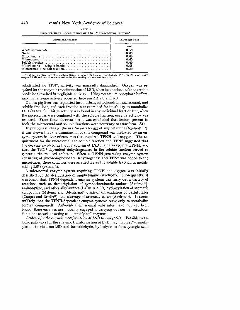
440 Annals New York Academy of Sciences TABLE 5
INTRACELLULAR LOCALIZATION OF LSD-METABOLIZING ENZYKE*
Intracellular fraction LSD m&b&fed
PnWl Wholehomogenate........................................ 0.50 Nuclei................................................... 0.00 Mitochondria. . . . . . . . . . . 0.00 Microsomes.............................................. Soluble fraction.. . . 8.: Mitochondria + soluble fraction.. . 0:os Microsomes + soluble fraction. 0.50
l Intracellular fractions obtained from 200 mg. of guinea pig liver were incubated at 37°C. for 120 minutes with 0.6 rmol LSD and cofactors described under the heading ddelhods oud Molericlls.
substituted for TPN+, activity was markedly diminished. Oxygen was re- quired for the enzymic transformation of LSD, since incubation under anaerobic conditions resulted in negligible activity. Using potassium phosphate buffers, maximal enzyme activity occurred between $H 7.0 and 8.0.
Guinea pig liver was separated into nuclear, mitochondrial, microsomal, and soluble fractions, and each fraction was examined for its ability to metabolize LSD (TABLE 5). Little activity was found in any individual fraction but, when the microsomes were combined with the soluble fraction, enzyme activity was restored. From these observations it was concluded that factors present in both the microsomal and soluble fractions were necessary to transform LSD.
In previous studies on the in o&o metabolism of amphetamine (AxelrodQ* lo), it was shown that the deamination of this compound was mediated by an en- zyme system in liver microsomes that required TPNH and oxygen. The re- quirement for the microsomal and soluble fraction and TPNf suggested that the enzyme involved in the metabolism of LSD may also require TPNH, and that the TPN+-dependent dehydrogenases in the soluble fraction served to generate the reduced cofactor. When a TPNH-generating enzyme system consisting of glucose-6-phosphate dehydrogenase and TPN+ was added to the microsomes, these cofactors were as effective as the soluble fraction in metab- olizing LSD (TABLE 6).
A microsomal enzyme system requiring TPNH and oxygen was initially described for the deamination of amphetamine (Axelrods). Subsequently, it was found that TPNH-dependent enzyme systems can carry out a variety of reactions such as demethylation of sympathomimetic amines (Axelrodllj, aminopyrine, and other alkylamines (LaDu et uZ.‘~), hydroxylation of aromatic compounds (Mitoma and Udenfriend13), side-chain oxidation of barbiturates (Cooper and Brodie14), and cleavage of aromatic ethers (Axelrod16). It seems unlikely that the TPNH-dependent enzyme systems serve only to metabolize foreign compounds. Although their normal substrates have not yet been found, these enzymes are probably engaged in carrying out normal metabolic functions as well as acting as “detoxifying” enzymes.
Evidencefor the enzymic transformation of LSD to 2-0xyLSD. Possible meta- bolic pathways for the enzymic transformation of LSD may involve N-demeth- ylation to yield norLSD and formaldehyde, hydrolysis to form lysergic acid,

Axelrod et al.: Metabolism of Lysergic Acid Diethylamide 441 TABLE 6
REQUIREMENT FOR TPNH*
Additions LSD metabolized
Pm1 Microsomes + soluble fraction. . . . . . . . . . . 0.53 Soluble fraction.. . . . . . . . . . . . Microsomes.............................................. X:E Microsomes + 35 pmol of glucose-6-POh. 0.04 Microsomes + 35 pmol of glucose-6-PO1 + 1 mg. of glucose-
6-PO,dehydrogenaset................................... 0.48
*The microsomes and the soluble fraction were prepared by fractional centrifugation of B homogenate of 200 mg. of guinea pig liver in 5 volumes of isotonic KCl. The reaction mixtures contained 5001rmol of phate buffer (pH 7.9), 25 ~01 of MgClz , 1 run01 TPN+, 25 rmol of nicotinamide, 0.66 @noI of f?
tassium phos- to make a final volume of 5.0 ml.
SD, and water 5 per cent co1 .
Incubation time 120 minutes at 37°C. in an atmosphere of 95 per cent 01 to t 2.4 Komberg units per mg. of protein.’
or oxidation of the indole ring. The extent of the enzymic demethylation of n’-methyl-substituted compounds can be determined by measuring the for- maldehyde formed (Axelrodl”) . When 5 pmol LSD were incubated with guinea pig microsomes, soluble fraction, cofactors described under the heading Methods and Materials, and 100 pmol of semicarbazide to trap formaldehyde, no formaldehyde was found.
The formation of lysergic acid or oxidized LSD was examined after incubat- ing 30 Hmol of LSD with microsomes and a soluble fraction obtained from 20 gm. of guinea pig liver, 10 pmol of TPN +, 500 pmol of nicotinamide, 250 pm01 of MgClz , and 50 ml. of pH 7.9 phosphate buffer (0.2M) at 37°C. After 3 hours of incubation, the reaction mixture was saturated with NaCl, made alkaline, and the residual LSD was removed by shaking with 3 portions of a heptane-2 per cent isoamyl-alcohol mixture. An aliquot of the aqueous phase was adjusted to pH 6.0 and shaken with 4 volumes of isoamyl alcohol. The isoamyl-alcohol extract was then shaken with one-fifth volume of 0.1 N NaOH. The alkaline extract showed negligible absorption at 310 rnp. Lysergic acid has an absorption peak at 310 rnp and can be quantitatively extracted under the conditions described above.
Another aliquot of the reaction mixture that was washed free of residual LSD was adjusted to pH 9.0 and then extracted with 5 volumes of n-butanol. Five volumes of n-heptane were added to an aliquot of the butanol extract and the mixture was shaken with one-tenth volume of 0.1 N HCl. An aliquot of the acid extract gave a Folin-Ciocalteun reaction, indicating the presence of a* reducing indole. Unlike LSD, the metabolite gave no reaction with Hopkins- Cole or Van Urk’sr* dimethylaminobenzaldehyde reagents. These observa- tions suggested that substitution had occurred on position 2 of the indole ring. Since alkaline hydrolysis is known to cleave oxindoles at the amide linkage to a salt of an amino acid containing a diazotizable amino group (Witkoplg), the LSD metabolite was heated at 100°C. in 2 N NaOH for 5 minutes, cooled, and subsequently treated with sodium nitrite in an acid solution. An intense wine color appeared when the diazotization mixture was added to an alkaline solution of &naphthol. These observations indicated that LSD was converted

Annals New York Academy of Sciences TABLE 7
BALANCE STUDY ON THE ENZYMIC CONVERSION* OF LSD TO Z-OXYLSD
Experiment No. LSD metabolized 2-0xyLSD formed
pm01 pm01
: 0.61 0.52 0.64 0.62
3 0.68 0.64
* Guinea pig liver microsomes and the soluble fraction obtained from 200 mg. of liver were incubated at 37°C. for 120 minutes with 0.7 wnol LSD and cofactors described under the heading Melhods and .Maferiols. At the end of the incubation period the reaction mixture was assayed for LSD and I-oxyLSD.
to 2-0xyLSD by the TPNH-dependent microsomal enzyme system. Enzy- matically formed 2-0xyLSD and synthetic 2-0~~ LSD (synthesized by K. Freter, J. Axelrod, and B. Witkop, of the National Institutes of Health) were found to be the same with respect to their ultraviolet-absorption and ultra- violet-fluorescence spectra, distribution coefficients in various organic solvents, Rf values on paper chromatograms and chemical tests. Preliminary studies have shown that ergotamine may undergo a similar transformation.
A balance study on the enzymic conversion of LSD to 2-0xyLSD was made by measuring the disappearance of LSD and the formation of 2-0xyLSD in the microsomal preparations. The formation of 2-0xyLSD was determined in the following manner:
Two ml. of the incubated reaction mixture were made alkaline, saturated with sodium chloride, and the unreacted LSD was removed by extraction with 2 portions of n-heptane containing 2 per cent of isoamyl alcohol. The aqueous residue was adjusted to pH 9, and the 2-0xyLSD was extracted into 15 ml. of It-butanol. Ten ml. of the butanol extract were transferred to a 40 ml. glass- stoppered centrifuge tube containing 20 ml. of n-heptane and 1.5 ml. of 0.1 N HCI and the contents of the tube were shaken for 10 minutes. To 1 ml. of the acid extract, 0.1 ml. of Folin-Ciocalteu reagent17 and 0.2 ml. Na&Oa (20 per cent solution) were added. The solution was heated for 1 minute in a boiling water bath, cooled, and its optical density at 630 rnp was determined in a Beckman* spectrophotometer adapted for small volumes. A known amount of 2-oxyLSD,t which was incubated and extracted in the same manner as described above, served as a standard. From the results shown in TABLE 7, it appeared that approximately 1 mol of 2-0xyLSD was formed for each mol of LSD that was metabolized.
Activity of 2-oxyLSD in !he central nervous system. Previous studies havg shown that LSD blocks synaptic transmission in the lateral geniculate nucleus and reduces spontaneous cortical activity (Evarts et a1.20). An intracarotid administration of 0.4 mg./kg. of 2-0xyLSD in cats that had received Nembutal did not alter the postsynaptic response to optic-nerve shock, whereas 0.03 mg./kg. of LSD resulted in an 80 per cent decrease in the amplitude of the postsynaptic response. We also found that 2-0xyLSD was without effect on the spontaneous cortical activity recorded during barbiturate anesthesia. The
* Manufactured by the Pyrocell Manufacturing Co., New York, N. Y. t On this occasion, P-oxyLSD used as a standard was previous1
r isolated from a microsomal preparation in-
cubated with LSD. The concentration of the standard solution o Z-oxyLSD was determined by comparing its optical density at 630 rnp with that of LSD after treatment with Folin-Ciocalteu reagent.” It was assumed that both LSD and 2-0xyLSD have the same molecular extinction at 630 wafter treatment with this reagent.

Axelrod et al.: Metabolism of Lysergic Acid Diethylamide 443 TABLE 8
INHIBITION OF LSD METABOLISM IN Vrmo*
Additions I Per cent inhibition
Serotonin 1 X 10eaM.. . . , . . . . . Reserpine 1 X 10maM . . . . . Chlorpromazine 1 X lo-‘M. . SKF 525 1 X 10⁻⁴M. . .
40 20
ii
* Guinea pig microsomes and the soluble fraction were incubated at 37°C. for 60 minutes with 6 X 10-S M LSD, inhibitors and cofactors as described under the heading Methods md MofcrMls.
oral administration of 300 pg. of 2-0xyLSD did not produce any psychological effects in a human subject who responded to 30 pg. of LSD. It may be con- cluded on the basis of this evidence that 2-0xyLSD does not possess LSD-like activity in the central nervous system.
Inhibitors of the mefabolism of LSD. Since other investigators have demon- strated biologic interactions between LSD, serotonin, and tranquilizing agents ( Gaddum,21 Wooley,22 and Shore, Silver, and Brodie23), we examined the effect of serotonin, reserpine, and chlorpromazine on the metabolism of LSD by the microsomal preparation (TABLE 8). It was found that chlorpromazine mark- edly inhibited the enzymic oxidation of LSD at relatively low concentrations (1 X 10⁻⁴RI), while serotonin and reserpine inhibited at higher concentrations (1 X 10⁻³M). Since chlorpromazine exerted marked inhibitory action on the metabolism of LSD in vitro, the effect of this compound on the in tivo metab- olism of LSD was examined in mice. The biologic half life of LSD (2 mg./kg. administered intraperitoneally) was 7 minutes. When chlorpromazine (100 mg./kg.) was administered intraperitoneally 5 minutes prior to the injection of LSD, the biologic half life was increased to 15 minutes.
The inhibitory action of SKF 525 (@-diethylaminoethyl diphenylpropyl acetate) on the enzymic oxidation of LSD was also examined. This compound has been shown to inhibit many TPNH-dependent microsomal enzymes both in aizlo (Axelrod, Rkichenthal, and Brodie24) and in vitro (Cooper, Axelrod, and Brodie?5). SKF 525 at a concentration of 1 X 10⁻⁴M almost completely blocked the enzymic transformation of LSD (TABLE 8).
Summary (1) A specific and sensitive method for the estimation of LSD in biological
material is described. (2) After administration of LSD to the cat, the drug is found in all tissu&
in the following order of decreasing concentrations: bile, plasma, lung, liver, kidney, brain, intestines, spleen, cerebrospinal fluid, muscle, and fat. The drug is extensively bound to plasma proteins, and there is no hindrance in the passage of the drug across the blood-brain harrier.
(3) LSD is almost completely metabolized in the body, only negligible amounts of the drug being excreted in the urine or the stools. The liver is the major site of metabolism of the drug.
(4) There are considerable species differences in the rate of biotransforma- tion of LSD.

444 Annals New York Academy of Sciences
(5) LSD is transformed to 2-0xyLSD by an enzyme system present in liver microsomes that requires oxygen and a reduced-triphosphopyridine nucleotide generating system. The new compound, 2-oxyLSD, does not possess LSD-like activity in the central nervous system.
(6) Chlorpromazine (1 X 10P4M) and SKF 525 (1 X 10⁻⁴M) markedly inhibit the in z&o metabolism of LSD, while serotonin (1 X 1W3) and reser- pine (1 X 1W3M) inhibit the in oitro metabolism moderately.
1.
2.
3.
4.
5.
6. 7.
8.
Y.
10.
11.
12.
13.
14.
15. 16.
17.
18.
19.
20.
21.
22.
23.
24.
25.
References BOYD, E. S., E. ROTHLIN, J. F. BONNER, I. H. SLATER & H. C. HODGE. 1955. Pre-
luminary studies on the metabolism of lysergic acid diethylamide. J. Pharmacol. Exptl. Therap. 113: 6.
STOLL, A., E. ROTHLIN,, J. RUTSCHMANN & W. R. SCHALCH. 1955. Distribution and fate ;: r4F9fbeled lysergtc acid diethylamide (LSD 25) in the animal body. Experientia.
LAN; U.. A. CERLETTI & E. ROTHLIN. diaihyiamids im Organismus.
1955. Uber die Verteilung des Lysergsaure- Helv. Physiol. et Pharmacol. Acta. 13: 207.
BOWMAN, R. L., P. A. CAULFIELD & S. UDENFRIEND. in the ultraviolet. Science. 122: 32.
19.55. Spectrofluorometric assay
BRODIE, B. B., S. UDENFRIEND & J. J. BAER. 1947. Estimation of basic organic com- pounds. I. General principles. J. Biol. Chem. 168: 299.
SCHNEIDER, W. C. 1948. Intracellular distribution of enzymes. J. Biol. Chem. 176: 259. KORNBERG, A. 19.50. Enzymatic synthesis of triphosphopyridine nucleotide. J. Biol.
Chem. 182: 805. AXELROD, J., S. UDENFRIEND & B. B. BRODIE. 1954. Eriect of ascorbic acid on hy-
cllq$mn of acetanilide, aniline and antipyrine in V&J. J. Pharmacol. Exptl. Therap.
AxEL:OD, j. 1954. A new enzyme for the deamination of sympathomimetic amines. J. Pharmacol. Exptl. Therap. 110: 2.
A~l~~~3 J. 1955. The enzymatic deamination of amphetamine. J. Biol. Chem. : .
AXELROD, J. 1954. The enzymatic demethylation of sympathomimetic amines. Fed- eration Proc. 13: 332.
LADu, B. W., L. GAUDETTE, N. TRAUSOF & B. B. BRODIE. 1955. The enzymatic dealkylation of aminopyrine and other alkylamines. J. Biol. Chem. 214: 741.
MITOMA, C. & S. UDENFRIEND. 1955. J. Pharmacol. Exptl. Therap. 113: 40.
The enzymatic hydroxylation of aromatic drugs.
COOPER, J. R. & B. B. BRODIE. 1955. The enzymatic metabolism of hexobarbital. J. Pharmacol. Exptl. Therap. 114: 409.
AXELROD, J. 1956. The enzymic cleavage of aromatic ethers. Biochem. J. 63: 634. AXELROD, J. 1955. The enzymatic demethylation of ephedrine. J. Pharmacol. Exptl.
Therap. 114: 430. FOLIN, 0. & V. CIOCALTEU. 1927. On tyrosine and tryptophane determinations in
proteins. J. Biol. Chem. 73: 627. VAN URK, H. W. 1929. Ein nieuve gevoelige reactie op de moederkoornalkaloi’den.
Pharm. Weekblad. 66: 473. WITKOP, B. 1947. Gelenkte Oxydationen in der Indol-Reihe. Ann. Chem. Justus
Liebigs. 668: 98. EVARTS, E. V., W. LANDAU, W. FREYCANC & W. H. MARSIIALL. 1955. Some effects
of lysergic acid diethylamide and bufotenine on electrical activity in the cat’s visual system. Am. J. Physiol. 182: 594.
GADDUM, J. H. 1953. Antagonism between lysergic acid diethylamide and 5-hydroxy- wz;[aunn. wJ. Physiol. 121: 15P.
. . 1955. Production of abnormal behavior in mice and its partial pre- vention’with cholinergic drugs and serotonin. Proc. Natl. Acad. Sci. U. S. 41: 338.
SHORE, P. A:, S. L. SILVER & B. B. BRODIE. 19.55. Interaction of reserpine, serotonin and lysergtc acid diethylamide in brain. Science. 122: 284.
AXELROD, J,, J. REICHENTHAL & B. B. BRODIE. 1954. Mechanism of the potentiating action of P-diethylaminoethyl diphenylpropylacetate (SKF 52.5). J. Pharmacol. Exptl. Therap. 112: 49.
COOPER, J. R., J. AXELROD & B. B. BRODIE. 1954. Inhibitory effects of SKF 525 on a variety of drug metabolic pathways in z&o. J. Pharmacol. Exptl. Therap. 112: 55.