reply
TRANSCRIPT

letters
Alternative to the Bimanual Technique
The article about a bimanual technique to manage subincisional cortex! illustrates a difficulty that
has confronted all extracapsular cataract extraction
(ECCE) surgeons since ECCE became the most popu
lar surgical procedure. Dr. Colvard suggests a bimanual technique to
overcome this difficulty. I have been suggesting a way to
overcome it for the past 15 years. Since I routinely use separate inflow and aspiration cannulas, I am able to
perform manual aspiration with a 5.0 cc syringe and a cannula attached to any location in the capsular bag
from two paracenteses located at 11 and/or 14 degrees.
All the advantages Dr. Colvard lists are provided
with the 5.0 cc syringe aspiration. There is more
flexibility and no need to touch the posterior capsule;
rather, the water jet technique can be used to clean up any remnants of cortical material on the posterior
capsule or at the bag periphery. fu permanent inflow is advantageous in many
other stages of the surgery, its presence would solve Dr. Colvard's problem without a two-handed approach but
with immediate availability of the aspiration technique
any time he chose to perform it.
Reference
MICHAEL BLUMENTHAL, MD Tel Aviv, Israel
1. Colvard DM. Bimanual technique to manage subincisional cortical material. ] Cataract Refract Surg 1997; 23:707-709
Reply: I would like to thank Dr. Blumenthal for his comments, which highlight his contributions to the management of cortex in extracapsular surgery. Most of us are, of course, very aware of Dr. Blumenthal's work. His efforts to champion the use of a static side-port chamber maintainer in extracapsular surgery are well known and were described and referenced in my recent article.
Dr. Blumenthal advocates the use of a 5.0 cc syringe to remove cortex during extracapsular surgery. In a recent eye surgical expedition to an underdeveloped area of Africa, I had the opportunity to remind myself how effective this technique can be. It is a very practical method if automated equipment is not available.
My article was written specifically for the phacoemulsification surgeon who is comfortable with a bimanual
technique. In small incision surgery, it is often difficult to access subincisional cortex with the standard, automated, single-handpiece irrigation/aspiration (I/A) device. Dr. Peter Brauweiler of Germany and Dr. Lucio Buratto of Italy among others have described the use of two separate aspiration and irrigating hand pieces introduced through small side-port incisions. My article details the advantages of this approach in small incision surgery. These advantages include better access to subincisional cortex, better stabilization of the globe, and improved chamber maintenance during cortical removal (since the primary phaco incision is self-sealing). The bimanual I/A system facilitates removal of viscoelastic material from behind a posterior chamber intraocular lens. The devices are useful in polishing the capsule, and the cannulas can be used to provide improved visualization by retracting the iris when pupillary constriction has occurred. I have been impressed by the usefulness and simplicity of this technique and would like to encourage other bimanual phacoemulsification surgeons to try it.-o. Michael Colvard, MO
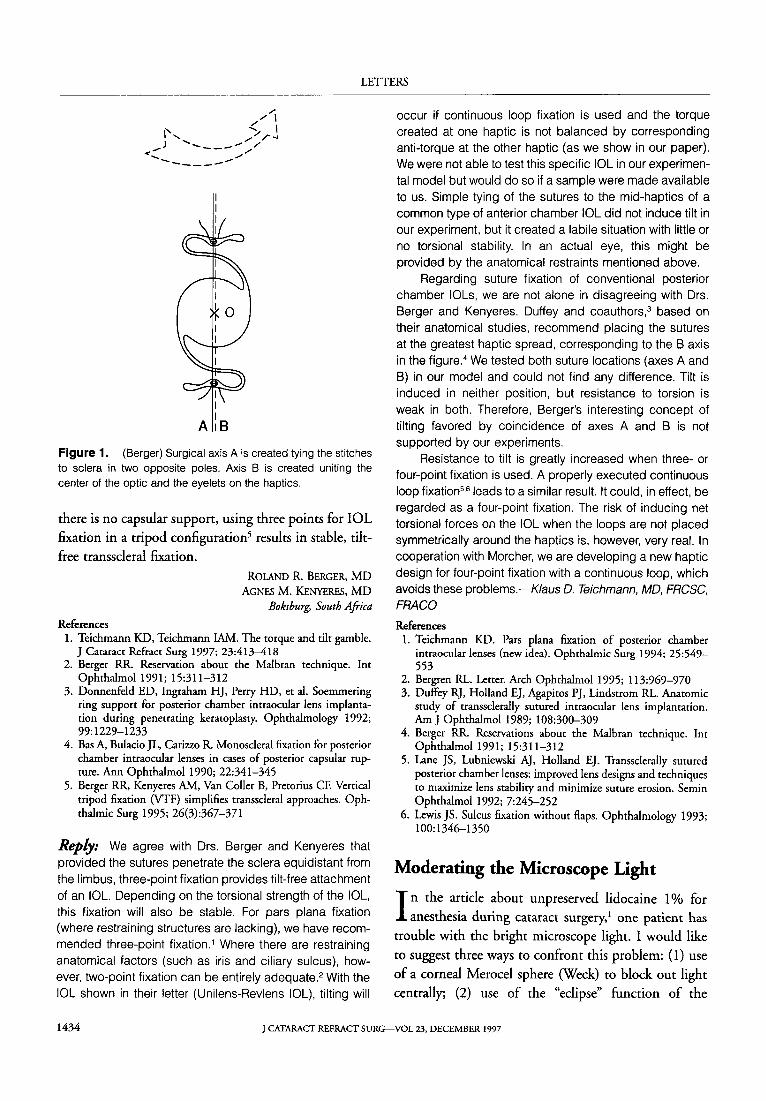
Aware that tilt is a common problem with scleralfixated intraocular lenses (IOLs), the authors of
"The Torque and Tilt Gamble"! have analyzed various
possible suture configurations and conclude that sur
geons should aim for symmetrical suture configuration at the two haptics.
From our experience, as well as from discussions
with colleagues, we believe that the main cause of 10L tilt during transscleral fixation is the use of two sym
metrical opposite points of fixation to the scleral wall, especially if the 10rs optic and haptic are concentric.2
In our opinion, using two opposite points creates a surgical axis (axis A in Figure O. If this axis coincides with the axis uniting the center of the optic and the haptics, i.e., if they are concentric (axis B), tilt will be
generated when the stitches are pulled through the
sclera.
However, the surgeon should not waste valuable
surgical time analyzing each 10L before transscleral
fixation. For practical reasons, the best way to avoid tilt
is to avoid the use of two opposite points for fixation. If
there is enough posterior capsular rim or if a Soem
mering's ring is present, there is no need to suture the 10L.3 When enough torn posterior capsule is present, monoscleral fixation4 is an excellent solution. When
J CATARACT REFRACT SURG-VOL 23, DECEMBER 1997 1433
LETTERS
/' /'/' \
1'-. ..... \ \ .... ",,/,-4
<..,.,J .............. ___ ,."., // ..... ,-
....... _----.".,
I I I
AlB
Figure 1. (Berger) Surgical axis A is created tying the stitches to sclera in two opposite poles. Axis B is created uniting the center of the optic and the eyelets on the haptics.
there is no capsular support, using three points for IOL fixation in a tripod configuration5 results in stable, tilt
free transscleral fixation.
References
ROLAND R. BERGER, MD AGNES M. KENYERES, MD
Boksburg, South Africa
1. Teichmann KD, Teichmann lAM. The torque and tilt gamble. J Cataract Refract Surg 1997; 23:413-418
2. Berger RR. Reservation about the Malbran technique. Int Ophthalmol 1991; 15:311-312
3. Donnenfeld ED, Ingraham HJ, Perry HD, et aI. Soemmering ring support for posterior chamber intraocular lens implantation during penetrating keratoplasty. Ophthalmology 1992; 99:1229-1233
4. Bas A, Bulacio JL, Carizw R. Monoscleral fixation for posterior chamber intraocular lenses in cases of posterior capsular rupture. Ann Ophthalmol1990; 22:341-345
5. Berger RR, Kenyeres AM, Van Coller B, Pretorius CF. Vertical tripod fixation (VTF) simplifies transscleral approaches. Ophthalmic Surg 1995; 26(3):367-371
Reply: We agree with Drs. Berger and Kenyeres that provided the sutures penetrate the sclera equidistant from the limbus, three-point fixation provides tilt-free attachment of an IOL. Depending on the torsional strength of the IOl, this fixation will also be stable. For pars plana fixation (where restraining structures are lacking), we have recommended three-point fixation. 1 Where there are restraining anatomical factors (such as iris and ciliary sulcus), however, two-point fixation can be entirely adequate.2 With the IOl shown in their letter (Unilens-Revlens IOl), tilting will
occur if continuous loop fixation is used and the torque created at one haptic is not balanced by corresponding anti-torque at the other haptic (as we show in our paper). We were not able to test this specific IOl in our experimental model but would do so if a sample were made available to us. Simple tying of the sutures to the mid-haptics of a common type of anterior chamber IOl did not induce tilt in our experiment, but it created a labile situation with little or no torsional stability. In an actual eye, this might be provided by the anatomical restraints mentioned above.
Regarding suture fixation of conventional posterior chamber IOls, we are not alone in disagreeing with Drs. Berger and Kenyeres. Duffey and coauthors,3 based on their anatomical studies, recommend placing the sutures at the greatest haptic spread, corresponding to the B axis in the figure. 4 We tested both suture locations (axes A and B) in our model and could not find any difference. Tilt is induced in neither position, but resistance to torsion is weak in both. Therefore, Berger's interesting concept of tilting favored by coincidence of axes A and B is not supported by our experiments.
Resistance to tilt is greatly increased when three- or four-point fixation is used. A properly executed continuous loop fixation5.6 leads to a similar result. It could, in effect, be regarded as a four-point fixation. The risk of inducing net torsional forces on the IOl when the loops are not placed symmetrically around the haptics is, however, very real. In cooperation with Morcher, we are developing a new haptic design for four-point fixation with a continuous loop, which avoids these problems.-Klaus D. Teichmann, MD, FRCSC, FRACa
References 1. Teichmann KD. Pars plana fixation of posterior chamber
intraocular lenses (new idea). Ophthalmic Surg 1994; 25:549-553
2. Bergren RL. Letter. Arch Ophthalmol 1995; 113:969-970 3. Duffey RJ, Holland EJ, Agapitos pJ, Lindstrom RL. Anatomic
study of transsclerally sutured intraocular lens implantation. Am J Ophthalmol 1989; 108:300-309
4. Berger RR. Reservations about the Malbran technique. Int Ophthalmol 1991; 15:311-312
5. Lane JS, Lubniewski AJ, Holland EJ. Transsclerally sutured posterior chamber lenses: improved lens designs and techniques to maximize lens stability and minimize suture erosion. Semin Ophthalmol 1992; 7:245-252
6. Lewis JS. Sulcus fixation without flaps. Ophthalmology 1993; 100:1346-1350
Moderating the Microscope Light
I n the article about unpreserved lidocaine 1 % for anesthesia during cataract surgery, 1 one patient has
trouble with the bright microscope light. I would like
to suggest three ways to confront this problem: (1) use
of a corneal Merocd sphere (Week) to block out light centrally; (2) use of the "eclipse" function of the
1434 J CATARACT REFRACT SURG-VOL 23. DECEMBER 1997