replication and control of circular bacterial plasmidsmmbr.asm.org/content/62/2/434.full.pdf ·...
TRANSCRIPT
MICROBIOLOGY AND MOLECULAR BIOLOGY REVIEWS,1092-2172/98/$04.0010
June 1998, p. 434–464 Vol. 62, No. 2
Copyright © 1998, American Society for Microbiology
Replication and Control of Circular Bacterial PlasmidsGLORIA DEL SOLAR, RAFAEL GIRALDO, MARIA JESUS RUIZ-ECHEVARRIA,
MANUEL ESPINOSA, AND RAMON DIAZ-OREJAS*
Centro de Investigaciones Biologicas, CSIC, E-28006 Madrid, Spain
INTRODUCTION .......................................................................................................................................................434PLASMID REPLICATION MECHANISMS...........................................................................................................435
Replication by the Theta Mechanism ..................................................................................................................435Origins of replication .........................................................................................................................................435
(i) General features ........................................................................................................................................435(ii) Iteron-containing origins ........................................................................................................................436(iii) Other origin configurations...................................................................................................................437
Rep proteins ........................................................................................................................................................437(i) Protein-protein interactions: the leucine zipper-like motif .................................................................438(ii) Specific binding of Rep proteins to DNA: the helix-turn-helix motif ...............................................440
Initiation and elongation of replication...........................................................................................................441(i) DNA replication dependent on plasmid initiators ...............................................................................441(ii) Replication independent of plasmid-encoded initiator proteins .......................................................442
Termination of replication.................................................................................................................................443Synopsis................................................................................................................................................................444
Strand Displacement Replication.........................................................................................................................444Origins of replication .........................................................................................................................................444Rep proteins ........................................................................................................................................................445Replication mechanism ......................................................................................................................................445Synopsis................................................................................................................................................................446
Rolling-Circle Replication .....................................................................................................................................446Origins of leading-strand synthesis .................................................................................................................446Rep proteins ........................................................................................................................................................448Initiation and elongation of leading-strand synthesis ...................................................................................450Termination of leading-strand synthesis .........................................................................................................450Replication of the lagging strand .....................................................................................................................451Synopsis................................................................................................................................................................452
CONTROL OF PLASMID REPLICATION............................................................................................................452Control by Antisense RNA.....................................................................................................................................453
Control of primer RNA processing: plasmid ColE1 ......................................................................................453Copy number control of plasmid R1................................................................................................................453Other instances of control by antisense RNAs...............................................................................................453Direct inhibition of Rep synthesis: blocking rep translation........................................................................455Transcriptional attenuation: the pT181 paradigm.........................................................................................455
Control by both Transcriptional Repressor and Antisense RNA ....................................................................455Control by Iterons...................................................................................................................................................456Hemimethylation and Regulation of Plasmid Replication................................................................................457Synopsis....................................................................................................................................................................457
SUMMING UP: DIFFERENCES AND SIMILARITIES IN PLASMID REPLICATION MECHANISMS...457CONCLUDING REMARKS AND PROSPECTS ...................................................................................................458ACKNOWLEDGMENTS ...........................................................................................................................................458REFERENCES ............................................................................................................................................................458
INTRODUCTION
Plasmids are extrachromosomal DNA elements with char-acteristic copy numbers within the host. These replicons havebeen found in species from the three representatives of theliving world, namely, the domains Archaea, Bacteria, and Eu-karya (318). Plasmids may constitute a substantial amount ofthe total genetic content of an organism, representing more
than 25% of the genetic material of the cell in some membersof the Archaea (127, 331). They can incorporate and delivergenes by recombination or transposition, thus favoring geneticexchanges in bacterial populations. Since plasmids can be in-troduced into new hosts by a variety of mechanisms, they canbe considered to be a pool of extrachromosomal DNA which isshared among populations. The wealth of genetic informationcarried by plasmids, their impact in the microbial communities,and the potential of these elements to act as natural cloningvectors have stimulated research into plasmids not only fromthe fundamental but also from the clinical, biotechnological,and environmental points of view. Three main factors have
* Corresponding author. Mailing address: Centro de InvestigacionesBiologicas, C.S.I.C., Velazquez 144, E-28006 Madrid, Spain. Phone:34-91-5611800. Fax: 34-91-5627518. E-mail: [email protected].
434
on June 13, 2018 by guesthttp://m
mbr.asm
.org/D
ownloaded from

contributed to the development of plasmid research: (i) thegenetic organization of these elements is apparently simple,(ii) they can be easily isolated and manipulated in vitro, and(iii) since plasmids are dispensable, their manipulation doesnot appear, in principle, to have adverse consequences to thehosts.
The feature that better defines plasmids is that they replicatein an autonomous and self-controlled way. The analysis ofplasmid replication and its control has led to milestone discov-eries, such as the existence of antisense RNAs, and has con-tributed to the unraveling of mechanisms of DNA replication,macromolecular interactions, and control of gene expression.The ability of some plasmids to pass across the so-called ge-netic barriers among different living organisms has posed ques-tions about general mechanisms governing replication andabout the communication between plasmid replication compo-nents and the host machinery involved in DNA replication.This plasmid-host communication has attracted the attentionof researchers working in environmental and in evolutionaryfields. Plasmid host range studies also have clear implicationsin clinical microbiology and in biotechnology. Despite theirautonomous replication, plasmids extensively use the replica-tion machinery of the host, and therefore plasmid replicationstudies facilitate the exploration of the mechanisms involved inchromosome replication.
PLASMID REPLICATION MECHANISMS
There are three general replication mechanisms for circularplasmids, namely, theta type, strand displacement, and rollingcircle (RC). Historical development of research on plasmidshas led to the idea that theta replication is more frequent inreplicons from gram-negative than from gram-positive bacteriawhereas the opposite is found for plasmids replicating by theRC mode. This belief is probably wrong. It is true, however,that present knowledge on theta-replicating plasmids stemsfrom replicons from gram-negative bacteria and that on RC-replicating plasmids derives from replicons from gram-positivehosts. Strand displacement replication has been associatedwith broad-host-range plasmids from the IncQ family. Themolecular interactions and the functional relationships thattake place in these three types of replication mechanisms arethe focus of this review. Linear plasmids have been found inboth gram-positive and gram-negative bacteria, and their struc-ture can be of two types: those having a hairpin at each end,and those having a protein covalently bound at their 59 ends.Linear plasmids of the first group replicate via concatemericintermediates, whereas those of the second group seem toreplicate by a protein-priming mechanism, similar to that ofbacteriophage f29 (264). However, initiation of replicationfrom an internal origin in a plasmid with a terminal protein hasbeen reported (48). Linear plasmids have been reviewed pre-viously (123), and they will not be discussed here. Replicationof plasmids from gram-negative bacteria has been specificallyaddressed (168a).
Concerning their genetic structure, plasmids have an essen-tial region which contains the genes or loci involved in repli-cation and its control. The organization of this essential regioncorresponds, in general, to the one described in the repliconmodel. In addition, plasmids may bear genes that could beconsidered dispensable, although they could actually play animportant role for the plasmid itself and/or for the host. Someof these so-called dispensable genes are involved in processessuch as plasmid transfer and spread among bacteria, resistanceto antibiotics and heavy metals, resistance to radiation, andtransfer of DNA to higher eukaryotes. Within the plasmid
essential region, several genes and sequences can be consid-ered. (i) The first is the origin(s) of replication (genericallytermed ori), which is characteristic of each replicon. (ii)Although this is not a general feature, many plasmids encodea protein involved in the initiation of replication, usuallytermed Rep protein. (iii) The third is the plasmid-borne genesinvolved in the control of replication. The requirement of aplasmid-encoded initiator is reflected by the presence of DNAcognate sites in the origin of replication, where protein-DNAinteractions take place. These specific sites are the hallmark ofa class of replicons that are different from replicons that do notrequire specific initiators.
Replication by the Theta Mechanism
Replication by the theta-type mechanism has been mostextensively studied among the prototype circular plasmids ofgram-negative bacteria, although this replication mode hasalso been described for plasmids isolated from gram-positivebacteria, namely, the streptococcal/enterococcal Inc18 group(40), some lactococcal replicons (152), and at least one Bacillussubtilis plasmid (192). DNA replication through the thetamechanism involves melting of the parental strands, synthesisof a primer RNA (pRNA), and initiation of DNA synthesis bycovalent extension of the pRNA (163). DNA synthesis is con-tinuous on one of the strands (leading strand) and discontin-uous on the other (lagging strand), although synthesis of thetwo strands seems to be coupled (reviewed in references 148and 326). Theta-type DNA synthesis can start from one orfrom several origins, and replication can be either uni- or bi-directional. Under electron microscopy (EM), the replicationintermediates are seen as typical Q (“theta”)-shaped moleculesthat, when digested with enzymes that cleave within the rep-licated region, yield Y-shaped molecules (“forks”). The rep-lication intermediates can also be monitored by one- ortwo-dimensional electrophoresis. These analyses provide in-formation on the nature of the replication intermediates, di-rection of replication, location of the origin and terminus, anddegree of coupling between leading- and lagging-strand syn-thesis.
With some exceptions, plasmids using the theta mechanismof replication require a plasmid-encoded Rep initiator protein.Some replicons may require the host DNA polymerase I (DNAPol I) during the early stages of leading-strand synthesis. Somefeatures of various well-known replicons which are describedhere are depicted in Fig. 1.
Origins of replication. Plasmid origins of replication can bedefined as (i) the minimal cis-acting region that can supportautonomous replication of the plasmid; (ii) the region whereDNA strands are melted to initiate the replication process, or(iii) the base(s) at which leading-strand synthesis starts. Rep-lication origins contain sites that are required for interactionsof plasmid-encoded and/or host-encoded proteins.
(i) General features. With some exceptions, initiation ofplasmid DNA replication requires a specific plasmid-encodedRep initiator protein. This is reflected by the presence, at theorigin of replication, of specific sequences with which the Repprotein interacts. Additional features found in many origins oftheta-replicating plasmids are (i) an adjacent AT-rich regioncontaining sequence repeats, where opening of the strands andassembly of host initiation factors occur, and (ii) one or moresites (dnaA boxes) where the host DnaA initiator protein binds(30, 163). Multiple Dam methylation sequences, which arepresent in the origin of replication of the Escherichia coli chro-mosome, oriC, can also be found at the origin of replication ofplasmids such as P1 (36, 38) and pSC101 (30). Methylation is
VOL. 62, 1998 REPLICATION OF CIRCULAR BACTERIAL PLASMIDS 435
on June 13, 2018 by guesthttp://m
mbr.asm
.org/D
ownloaded from
not essential for replication, its role being primarily in postrep-lication (3). Dam methylation sequences are not present inother plasmid replicons.
Comparative analysis of the structural organization of thePol I-independent origins of replication predicts that althoughthe Rep-binding site is located within a potentially curvedDNA region, the DNA within the repeats of the AT-rich re-gion is essentially straight (81). Intrinsic DNA bends at theRep-binding sites would favor additional curvatures of theorigin induced by Rep proteins. The origins of replication canalso contain sites for factors (e.g., the integration host factor,IHF, or the factor for inversion stimulation, FIS) that play anarchitectural role. These host-encoded proteins favor a topo-logical proximity between different ori regions or even betweendifferent origins present in the same plasmid (as in plasmidR6K [see below]). The plasmid DNA sites are essential com-ponents of the origin of replication since they are required toorganize a functional replisome (61, 62, 282). The presence ofDNA sites for the binding of structural factors, found at theorigin of replication of several plasmids (see below), resemblesthe situation found in oriC (317).
(ii) Iteron-containing origins. In many cases, the origin ofreplication contains directly repeated sequences, termed iter-ons, which are the binding sites for the plasmid-encoded Repproteins and which have control properties. As discussed be-low, iterons not only are essential for replication but also arekey elements for the control of plasmid replication (reviewedin references 51, 87, 155, and 223). Among plasmids whichrestrict their establishment to a single or a few species ofenterobacteria, iterons have been described for several repli-cons like P1 (5), F (209, 295), pSC101 (52), R6K (97, 98, 277,278), Rts1 (144), and pColIV-K30 (247). Iterons are also foundin theta-replicating broad-host-range plasmids such as RK2/RP4 (241, 279), pCU1 (164) and pSa (286), as well as inconditional broad-host-range plasmids such as pPS10 (85, 104,215). It should be noted that the presence of directly repeatedsequences to which Rep proteins bind is not restricted to plas-mids replicating by the theta mechanism, since these sequenceshave been reported for plasmids using the strand-displacementmechanism or the RC mechanism (171, 176, 267). Iterons canalso be found outside the origin region in some plasmids (P1,F, RK2, R6K, Rts1, and pColIV-K30). These iterons, unlike
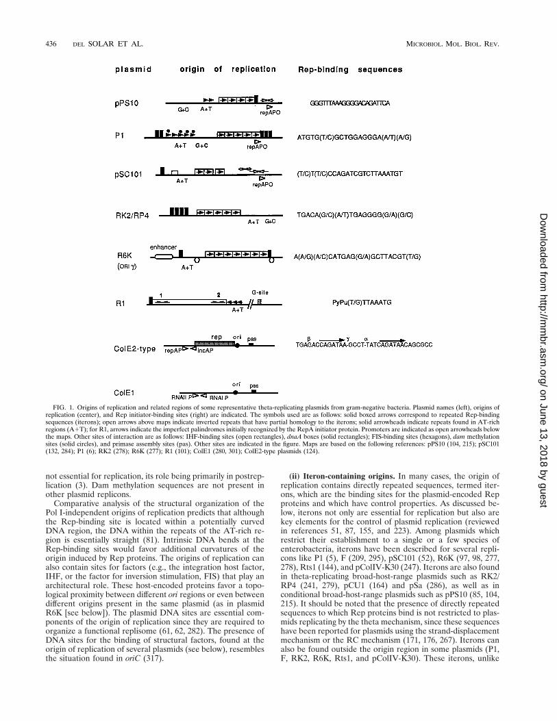
FIG. 1. Origins of replication and related regions of some representative theta-replicating plasmids from gram-negative bacteria. Plasmid names (left), origins ofreplication (center), and Rep initiator-binding sites (right) are indicated. The symbols used are as follows: solid boxed arrows correspond to repeated Rep-bindingsequences (iterons); open arrows above maps indicate inverted repeats that have partial homology to the iterons; solid arrowheads indicate repeats found in AT-richregions (A1T); for R1, arrows indicate the imperfect palindromes initially recognized by the RepA initiator protein. Promoters are indicated as open arrowheads belowthe maps. Other sites of interaction are as follows: IHF-binding sites (open rectangles), dnaA boxes (solid rectangles); FIS-binding sites (hexagons), dam methylationsites (solid circles), and primase assembly sites (pas). Other sites are indicated in the figure. Maps are based on the following references: pPS10 (104, 215); pSC101(132, 284); P1 (6); RK2 (278); R6K (277); R1 (101); ColE1 (280, 301); ColE2-type plasmids (124).
436 DEL SOLAR ET AL. MICROBIOL. MOL. BIOL. REV.
on June 13, 2018 by guesthttp://m
mbr.asm
.org/D
ownloaded from

the origin iterons, are not required for initiation but play animportant role in the control of replication, as the origin iter-ons do. In plasmids that do not have auxiliary iterons, theorigin iterons are the only locus involved in control (see be-low).
Iterons can be adjacent or separated by intervening se-quences. Iterons found in the origin region tend to be arrangedas tandem repeats situated at a distance that is, in general, amultiple of 11 bp, i.e., close to the helical periodicity of theDNA double helix. This implies that the Rep-iteron nucleo-protein complexes roughly place the Rep molecules aligned onthe same face of the DNA. In general, for a particular origin,the sequences of the different iterons are not identical, al-though they adjust to a consensus motif that defines the essen-tial features of these sequences. However, the four 22-bp iter-ons of plasmid pPS10 are identical (215). Statistical analysis ofthe frequency of base changes within the iterons of plasmid P1,combined with the available footprinting data, have been per-formed (242). Three highly conserved sequence patches arefound within the iterons of this replicon. The two outer patchesare separated by one helix turn. Protection experiments indi-cated that the major groove sides of those patches are con-tacted by the RepA initiator protein of P1. The function of themiddle patch is less clear, but it may contribute to a properconformation of the RepA-binding site. It is remarkable thatthis pattern resembles the DNA-binding patterns of dimericproteins, some of which are transcriptional repressors. Takinginto account that some of these iterons are contacted by mo-nomeric forms of the initiator proteins, this may reflect thepresence of two DNA-binding domains in RepA (discussed inreference 51), a feature that may be extended to other plasmid-encoded Rep proteins (100). Alignment of iterons present inthe origin of replication of different plasmids showed the con-servation of the hexanucleotide TCAGPuG (86), which is di-rectly involved in the binding of the p initiator protein to theori-g region of plasmid R6K (97, 98).
Multiple iterons are required for origin activity, although notall iterons present in a given origin have to be essential. Forinstance, removal of one of the seven iterons from ori-g ofplasmid R6K has no effect but deletion of two reduces theefficiency of replication and deletion of three or more abolishesplasmid replication (160). Interestingly, the deletions makeori-g replication independent of DnaA (16a). In the case of P1,all five iterons seem to be required for replication in vivo, butdeletion of one can be tolerated in an in vitro replicationsystem (314).
Single iterons are present in the ori-a and ori-b origins ofplasmid R6K and in the minimal origins of plasmids ColE2 andColE3. In R6K, ori-a and ori-b contain just one iteron and halfan iteron, respectively (87). This situation is compensated forby the presence of a cis-acting sequence (enhancer), which islocated in a third origin (ori-g) that contains seven iterons. Theenhancer facilitates the transfer of the initiator p protein,assembled at the seven iterons of ori-g, to ori-a and ori-b, andleads to initiation of DNA replication (see below). The small-est of all the prokaryotic origins described so far have beenfound in the ColE2 and ColE3 replicons (322). They consist ofa stretch of 47 bp (ColE2) or 33 bp (ColE3) and contain twomajor directly repeated sequences.
(iii) Other origin configurations. Origins of replication with-out iterons can be found in other well studied theta-replicatingplasmids like R1 and ColE1, as well as in plasmid pLS20 fromB. subtilis.
(a) Plasmid R1. Initiation of replication of R1 is dependenton a plasmid-encoded initiator protein, RepA. The minimalregion required for RepA-dependent replication (oriR) is in-
cluded within a 188-bp DNA region (183) and comprises (i) a9-bp dnaA box, (ii) a contiguous 100-bp region where RepAinteracts, and (iii) an adjacent AT-rich region containing three9-mers. A detailed study of the site of RepA interaction re-vealed two RepA-binding sites: a preferential RepA site,termed site 1 (59-CAGTTAAATG-39), which is adjacent to thednaA box, and a related RepA binding sequence, site 2 (59-TGTTTAAAAG-39), for which the protein has a lower affinity.This second site is contiguous to the AT-rich region. Sites 1and 2 share a core sequence (g/tTTAAA) that is an imperfectpalindrome (101). The intervening sequence between the sitesshows potential intrinsic curvature. The presence of the dnaAbox optimizes the action of the DnaA protein at the origin,both in vivo and in vitro, but it is not absolutely required for theDnaA-dependent replication of R1 (233). EM of replicatingintermediates obtained in vivo and in vitro shows that initiationof R1 replication occurs in a locus that is separated from theminimal origin region (78). A G-type priming signal, located400 bp downstream of the RepA-binding sequences, has beenidentified as the site where initiation of the leading strand,primed by DnaG, occurs (186).
(b) Plasmid ColE1. ColE1 is the prototype of a class of smallmulticopy plasmids that replicate by a theta-type mechanism.Unlike R1, ColE1 does not require a plasmid initiator proteinbut requires DNA Pol I to initiate replication. The origin ofColE1 replication spans a region of about 1 kb that includes (i)sequences promoting the synthesis of RNA II, the primer ofthe leading strand (298, 299); (ii) sequences that allow a stablehybridization of RNA II to DNA (139, 189); (iii) sequencesthat favor specific processing of this coupled complex byRNase H, which generates the 39 end needed to prime leading-strand synthesis (122, 139); (iv) a primosome assembly site (pasor ssiA) that allows the loading of the DnaB helicase andDnaG primase to initiate the discontinuous priming of thelagging strand (28, 189, 220) (a dnaA box that is close to pascan be used as a DnaA-dependent DnaB-DnaC assembly site[269, 270]); and (v) a sequence for termination of lagging-strand synthesis, terH, which determines unidirectional repli-cation (57, 198). The first two sequences are the most relevant,since they are required for ColE1 replication in the presence orabsence of DNA Pol I and RNase H (57, 158, 211). The originof ColE1 replication, defined as the transition point betweenRNA II primer and DNA synthesized by DNA Pol I, has beenpositioned 555 bp downstream of the start point of RNA II (24,300). This transition point corresponds with data obtained invivo for plasmid pMB1 (closely related to ColE1) (29). Anal-ysis of replication intermediates of ColE1 by EM, located asingle origin and showed that replication is unidirectional. Atan early stage, leading-strand synthesis proceeds in the absenceof lagging-strand synthesis (297, 298).
(c) Plasmid pLS20. An interesting example of plasmid fromgram-positive bacteria is the B. subtilis plasmid pLS20, forwhich a preliminary characterization has been reported (192).This plasmid replicates by the theta mechanism, and its repli-cation is independent of DNA Pol I and of a Rep initiatorprotein. Several palindromes flanking a putative dnaA box arelocated within the origin of replication of pLS20.
Rep proteins. Up to now, dozens of plasmids have beenisolated from most bacteria, but not many of them have hadtheir basic replicons dissected and characterized to the level oftheir nucleotide sequence, and even fewer replicons have beengenetically and biochemically studied in detail. The classic wayof classifying plasmids is to distribute them among incompat-ibility groups, whose members have very similar origin se-quences and replication control mechanisms. However, due tothe difficulty to cope with a complex experimentally based
VOL. 62, 1998 REPLICATION OF CIRCULAR BACTERIAL PLASMIDS 437
on June 13, 2018 by guesthttp://m
mbr.asm
.org/D
ownloaded from
classification of each newly isolated replicon, a criterion basedon sequence comparisons appears to be much more practical.Such a criterion could be the comparison of the amino acidsequences of Rep initiator proteins, since they are encoded bymost of the plasmids and they share common functions. Repproteins recognize specific sequences at the origin of replica-tion, similar to the DnaA initiator protein in bacterial chro-mosomal replication, and they generate a nucleoprotein initi-ation complex in which essential macromolecular interactionstake place (Rep-DNA, Rep-Rep, and Rep with other initiationproteins of the host) (30). In addition, many Rep proteins cangenerate complexes that negatively regulate their synthesis andthe frequency of initiation.
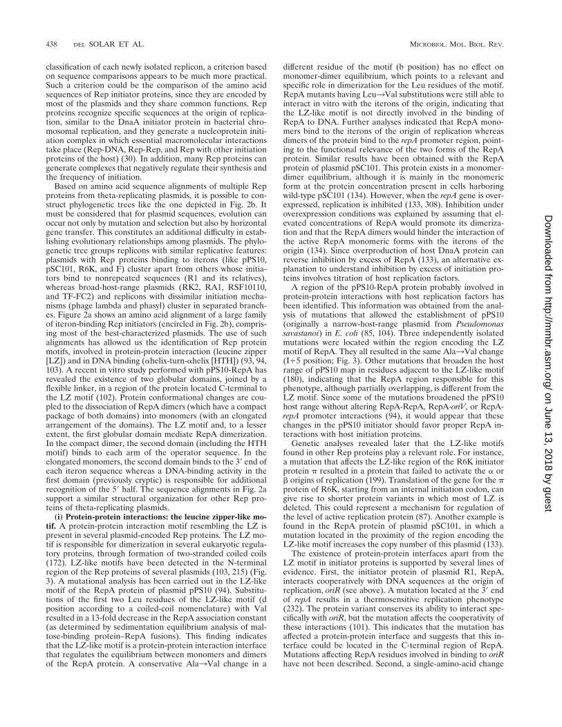
Based on amino acid sequence alignments of multiple Repproteins from theta-replicating plasmids, it is possible to con-struct phylogenetic trees like the one depicted in Fig. 2b. Itmust be considered that for plasmid sequences, evolution canoccur not only by mutation and selection but also by horizontalgene transfer. This constitutes an additional difficulty in estab-lishing evolutionary relationships among plasmids. The phylo-genetic tree groups replicons with similar replicative features:plasmids with Rep proteins binding to iterons (like pPS10,pSC101, R6K, and F) cluster apart from others whose initia-tors bind to nonrepeated sequences (R1 and its relatives),whereas broad-host-range plasmids (RK2, RA1, RSF10110,and TF-FC2) and replicons with dissimilar initiation mecha-nisms (phage lambda and phasyl) cluster in separated branch-es. Figure 2a shows an amino acid alignment of a large familyof iteron-binding Rep initiators (encircled in Fig. 2b), compris-ing most of the best-characterized plasmids. The use of suchalignments has allowed us the identification of Rep proteinmotifs, involved in protein-protein interaction (leucine zipper[LZ]) and in DNA binding (ahelix-turn-ahelix [HTH]) (93, 94,103). A recent in vitro study performed with pPS10-RepA hasrevealed the existence of two globular domains, joined by aflexible linker, in a region of the protein located C-terminal tothe LZ motif (102). Protein conformational changes are cou-pled to the dissociation of RepA dimers (which have a compactpackage of both domains) into monomers (with an elongatedarrangement of the domains). The LZ motif and, to a lesserextent, the first globular domain mediate RepA dimerization.In the compact dimer, the second domain (including the HTHmotif) binds to each arm of the operator sequence. In theelongated monomers, the second domain binds to the 39 end ofeach iteron sequence whereas a DNA-binding activity in thefirst domain (previously cryptic) is responsible for additionalrecognition of the 59 half. The sequence alignments in Fig. 2asupport a similar structural organization for other Rep pro-teins of theta-replicating plasmids.
(i) Protein-protein interactions: the leucine zipper-like mo-tif. A protein-protein interaction motif resembling the LZ ispresent in several plasmid-encoded Rep proteins. The LZ mo-tif is responsible for dimerization in several eukaryotic regula-tory proteins, through formation of two-stranded coiled coils(172). LZ-like motifs have been detected in the N-terminalregion of the Rep proteins of several plasmids (103, 215) (Fig.3). A mutational analysis has been carried out in the LZ-likemotif of the RepA protein of plasmid pPS10 (94). Substitu-tions of the first two Leu residues of the LZ-like motif (dposition according to a coiled-coil nomenclature) with Valresulted in a 13-fold decrease in the RepA association constant(as determined by sedimentation equilibrium analysis of mal-tose-binding protein–RepA fusions). This finding indicatesthat the LZ-like motif is a protein-protein interaction interfacethat regulates the equilibrium between monomers and dimersof the RepA protein. A conservative Ala3Val change in a
different residue of the motif (b position) has no effect onmonomer-dimer equilibrium, which points to a relevant andspecific role in dimerization for the Leu residues of the motif.RepA mutants having Leu3Val substitutions were still able tointeract in vitro with the iterons of the origin, indicating thatthe LZ-like motif is not directly involved in the binding ofRepA to DNA. Further analyses indicated that RepA mono-mers bind to the iterons of the origin of replication whereasdimers of the protein bind to the repA promoter region, point-ing to the functional relevance of the two forms of the RepAprotein. Similar results have been obtained with the RepAprotein of plasmid pSC101. This protein exists in a monomer-dimer equilibrium, although it is mainly in the monomericform at the protein concentration present in cells harboringwild-type pSC101 (134). However, when the repA gene is over-expressed, replication is inhibited (133, 308). Inhibition underoverexpression conditions was explained by assuming that el-evated concentrations of RepA would promote its dimeriza-tion and that the RepA dimers would hinder the interaction ofthe active RepA monomeric forms with the iterons of theorigin (134). Since overproduction of host DnaA protein canreverse inhibition by excess of RepA (133), an alternative ex-planation to understand inhibition by excess of initiation pro-teins involves titration of host replication factors.
A region of the pPS10-RepA protein probably involved inprotein-protein interactions with host replication factors hasbeen identified. This information was obtained from the anal-ysis of mutations that allowed the establishment of pPS10(originally a narrow-host-range plasmid from Pseudomonassavastanoi) in E. coli (85, 104). Three independently isolatedmutations were located within the region encoding the LZmotif of RepA. They all resulted in the same Ala3Val change(I15 position; Fig. 3). Other mutations that broaden the hostrange of pPS10 map in residues adjacent to the LZ-like motif(180), indicating that the RepA region responsible for thisphenotype, although partially overlapping, is different from theLZ motif. Since some of the mutations broadened the pPS10host range without altering RepA-RepA, RepA-oriV, or RepA-repA promoter interactions (94), it would appear that thesechanges in the pPS10 initiator should favor proper RepA in-teractions with host initiation proteins.
Genetic analyses revealed later that the LZ-like motifsfound in other Rep proteins play a relevant role. For instance,a mutation that affects the LZ-like region of the R6K initiatorprotein p resulted in a protein that failed to activate the a orb origins of replication (199). Translation of the gene for the pprotein of R6K, starting from an internal initiation codon, cangive rise to shorter protein variants in which most of LZ isdeleted. This could represent a mechanism for regulation ofthe level of active replication protein (87). Another example isfound in the RepA protein of plasmid pSC101, in which amutation located in the proximity of the region encoding theLZ-like motif increases the copy number of this plasmid (133).
The existence of protein-protein interfaces apart from theLZ motif in initiator proteins is supported by several lines ofevidence. First, the initiator protein of plasmid R1, RepA,interacts cooperatively with DNA sequences at the origin ofreplication, oriR (see above). A mutation located at the 39 endof repA results in a thermosensitive replication phenotype(232). The protein variant conserves its ability to interact spe-cifically with oriR, but the mutation affects the cooperativity ofthese interactions (101). This indicates that the mutation hasaffected a protein-protein interface and suggests that this in-terface could be located in the C-terminal region of RepA.Mutations affecting RepA residues involved in binding to oriRhave not been described. Second, a single-amino-acid change
438 DEL SOLAR ET AL. MICROBIOL. MOL. BIOL. REV.
on June 13, 2018 by guesthttp://m
mbr.asm
.org/D
ownloaded from

FIG. 2. Sequence alignment and phylogenetic tree of Rep initiator proteinsfrom theta-replicating plasmids. (a) Sequence alignment of the Rep initiatorproteins of nine related plasmids of the iteron-containing class. Sequences werealigned with the program CLUSTAL W (version 1.5) by using, for pairwisealignment, gap opening and extension penalties of 10.0 and 0.1, respectively, andthe protein weight matrix BLOSUM30. For multiple alignment, the delay fordivergent sequences was set to 40% (294). The degree of sequence identity to thepPS10 initiator sequence for conserved residues is shown: p, identical residues ineight or nine of the total sequences; ●, identical residues in five to seven se-quences. Over the sequences is shown a secondary-structure prediction per-formed by the neural network algorithm PHD (260) on the output fromCLUSTAL W: predicted a-helical regions (boxes) and b-strands (arrows). Thetwo characteristic motifs found in the Rep initiators, LZ (hydrophobic heptadresidues pointed to by open arrowheads) and HTH, are indicated. The EMBLdatabase accession numbers are as follows: pPS10, X58896; pECB2, Y10829;pRO1614, L30112; pCM1, X86092; pFA3, M31727; pSC101, K00828; pCU1,Z11775; RepFIA, Y00547; R6K, M65025. (b) Phylogenetic tree for theta-typereplicons from gram-negative bacteria, based on sequence alignments of theirRep proteins (such as the one shown in panel a for the pPS10 family, encircledin the tree with a dashed line). The sequences for 35 initiators were retrievedfrom databases, and a preliminary alignment was performed with CLUSTAL W(data not shown). Sequences that were virtually identical (pairwise scores, $90)were discarded, and a refined alignment was used as input data for the DIS-TANCES program of the Genetics Computer Group software package (95).Distance matrixes were corrected for multiple substitutions by the method ofKimura. The phylogenetic tree was built up according to the UPGMA methodwith the program GROWTREE (95). For further discussion, see the text.
VOL. 62, 1998 REPLICATION OF CIRCULAR BACTERIAL PLASMIDS 439
on June 13, 2018 by guesthttp://m
mbr.asm
.org/D
ownloaded from

at the N-terminal end of the initiator protein p of plasmid R6Kallows this protein to discriminate between palindromic andnonpalindromic binding sites (325). It has been proposed thatthe change alters a protein-protein interface which modulatesinteractions of p protein with differently arranged DNA targetsequences. Third, RK2 is a broad-host-range plasmid, a char-acteristic that is related to the existence of two forms of thereplication protein, TrfA-44 and TrfA-33. The larger form,TrfA-44, is required for replication in P. aeruginosa (80, 274).The shorter version, TrfA-33, starts in an internal initiationcodon of trfA and promotes the establishment of RK2 in mostof its hosts, including E. coli and P. putida. In addition, theorigin requirements are different in the two cases (56, 142, 213,241). A mutation at the 39 end of the trfA gene (affecting thetwo versions of the protein) modifies the host range of RK2without altering the binding of the protein to DNA (45, 175;also see reference 241). These results suggest that the C ter-minus of TrfA plays an important role in the interactions of theinitiator protein(s) with host replication factors. Interactions ofplasmid initiator proteins with host replication factors havebeen reported in different systems: (i) the DnaJ protein inter-acts with the initiation protein of plasmid P1 (312a) and withother chaperones, promoting the efficient binding of this initi-ator to the origin of replication; (ii) the DnaA protein requiresa functional interaction with the RepA protein of plasmid R1to enter the DnaA box present in the origin of replication(184) (this protein interaction seems to be sufficient to pro-mote DNA replication in the absence of the DnaA box [233]);and (iii) most interestingly, the DnaA, DnaB, and DnaG pro-
teins of the host interact with the p protein of plasmid R6K(16a, 258a) (mutations in the p protein that disrupt the inter-action with the DnaA protein are defective in R6K replication[16a]; the specific regions of DnaB and p proteins involved intheir interaction have been defined [258a]).
(ii) Specific binding of Rep proteins to DNA: the helix-turn-helix motif. As mentioned above, Rep proteins are able tospecifically recognize DNA sequences in the origin region. Inaddition to this, some of the Rep proteins autoregulate theirown synthesis at the transcriptional level by binding to se-quences in the rep promoter (operator) which show some de-gree of homology to those present in the origin region. Whenautoregulation exists, either a single species of the protein isinvolved in both regulation and replication, or different speciesof the protein, monomeric and dimeric, recognize the originand the regulatory regions, respectively (discussed in reference51). rep mutants leading to impaired Rep protein-DNA inter-actions have been found in various plasmids.
The Pseudomonas plasmid pPS10 contains four identicaliterons in its origin of replication and an inverted repeat (IR)in the repA promoter region. The iterons and IR have partialsequence similarity (92). RepA variants that fail to repress therepA promoter had amino acid changes within or in the vicinityof an HTH motif located at the C-terminal end of the protein(93). This motif has been described in many prokaryotic DNA-binding proteins, where it is involved in binding to specificregulatory DNA regions (39, 235). The RepA proteins affectedin the HTH motif failed to interact with both the repA pro-moter and the oriV, indicating that the motif is involved in
FIG. 3. The theta-type replicon of the Pseudomonas plasmid pPS10. The iteron-containing origin (oriV) and motifs found in the replication initiator protein (RepA)are depicted. The minimal origin (oriV) of pPS10 plasmid is a good example for a “canonical” iteron-containing origin. It is composed of four contiguous and identical22-bp iterons arranged as direct repeats (half arrows), flanked by a 9-bp dnaA box (hatched) and AT- and GC-rich sequences (215). The pPS10 replicon also containsthe repA gene, encoding the RepA initiator protein (shadowed ovals). RepA is under a monomer-to-dimer equilibrium, which has consequences for protein activity:RepA dimers bind to an inverted repeat (with a sequence partially homologous to that of the iterons) that overlaps with the repA promoter (P), acting as self-repressorof repA expression, whereas RepA monomers bind to the iterons to form the initiation complex (94). Protein motifs found in RepA are depicted under the protein gene.The LZ motif, responsible for protein dimerization, is represented as a helical wheel projection, in which the hydrophobic spine of Leu residues and the chemical natureof the other displayed residues is indicated (103). For the HTH motif, involved in binding to DNA, the two proposed a-helices are underlined and a stretch of basicresidues at the C end of the DNA recognition helix is indicated (1), whereas an arrow points to the Gly residue thought to start the turn (93).
440 DEL SOLAR ET AL. MICROBIOL. MOL. BIOL. REV.
on June 13, 2018 by guesthttp://m
mbr.asm
.org/D
ownloaded from

interactions with both the DNA regions. A working modelproposes that the RepA protein contacts the inverted repeatsof the repA promoter region as a dimer, using the HTH motif(92, 93). This HTH motif also binds to a conserved 39 region inthe iterons, which in their 59 ends would be bound by anotherregion of the RepA protein (102). A similar model has beenpostulated for other plasmid Rep proteins, in which mono-meric and dimeric forms of the protein are involved in repli-cation and autoregulation, respectively (discussed in reference51).
In plasmid RK2, mutations that lead to TrfA protein vari-ants affected in binding to the origin were scattered over a trfAgene region encoding the 162-amino-acid C-terminal moiety ofthe protein (46). In plasmid pSC101, the last third of the RepAprotein is not needed for binding to specific DNA sites (181),which contrasts with the role of the C-terminal region in otherinitiators.
Initiation and elongation of replication. (i) DNA replicationdependent on plasmid initiators. Initiation of replication re-quires the assembly of the complete replication machineryincluding DNA polymerase III holoenzyme (DNA Pol III-HE), DnaB helicase, and primase at the plasmid origin. Oncethe checkpoint corresponding to the initiation of leading-strand synthesis is past, replication continues until completion,following a process catalyzed by DNA Pol III and other hostproteins. Most of the theta-type replicons require, at least, aplasmid-encoded Rep protein and the host DnaA protein forthe initiation step. The general organization of the origin re-gion in these plasmids resembles the arrangement found inoriC (30). The plasmid ori includes not only the specific se-quences where the Rep and DnaA proteins interact but also anAT-rich region containing direct repeats, analogous to the13-mers in oriC, where the DNA strands are melted. TheAT-rich repeats have also been involved in the transfer of theDnaB-DnaC complex to oriC. In the theta-replicating plas-mids, the Rep protein binds to specific sequences in the origin,forming a nucleoprotein preinitiation complex analogous tothe one formed by DnaA at oriC. The Rep-DNA complex, incombination with DnaA, facilitates the transfer of the DnaB-DnaC complex to the origin and the opening of the strands inthe AT-rich region. The structural organization of the initia-tion complex could be facilitated by host factors such as HU,IHF, or FIS. The assembly of the preinitiation complex anddetails of the molecular interactions leading to the initiation ofreplication are well documented for a few theta-replicatingplasmids (described below).
(a) Plasmid pSC101. RepA, the initiator protein of plasmidpSC101, exists in a monomer-dimer equilibrium, which deter-mines the efficiency of RepA in replication (134). Monomersand dimers of RepA are both functional, but they play differentroles: monomers bind to the iterons at the origin, promotinginitiation, whereas dimers bind to the adjacent inverted repeat,repressing transcription of the repA gene (181). However, in-teractions of the RepA dimers with the inverted repeat alsoplay a role in replication in the absence of the par locus (197).Initiation of pSC101 replication requires, in addition to RepA(308), the DnaA host replication initiator (113), and IHF pro-teins (91, 283). Binding of IHF to its target, within the AT-richregion, leads to DNA bending (283), which promotes interac-tions between DnaA molecules bound to dnaA boxes sepa-rated by some 200 bp (282). Binding of RepA to the originregion further stabilizes DnaA contacts with the distant dnaAboxes (282). The RepA-DNA-DnaA complex plays a role inreplication but also in partitioning of the plasmid (54). Stableplasmid inheritance requires the par locus, which is close to theorigin region: this locus contains a site for DNA topoisomerase
II and also determines the proper supercoiling at the originregion needed for initiation (53, 132).
(b) Plasmid P1. Plasmid P1 replication is dependent, both invivo and in vitro, on the specific initiator protein RepA (6, 313)and on the host DnaA protein (110, 313). Formation of theinitiation complex requires the monomeric form of RepA(315), and RepA-DNA binding is stimulated by heat shockchaperones. The latter proteins could contribute to the disso-ciation of the RepA dimers into monomers, which is the formof the RepA protein that recognizes the five iterons of theorigin (58, 315). However, growing evidence indicates that thechaperones are required to activate the monomers of RepA(50, 78a, 236). Binding of the activated RepA monomers to thefive iterons of the origin results in wrapping of the DNAaround RepA, presumably due to in-phase bending of DNA(206). RepA monomers contact each iteron through two con-secutive major grooves on the same face of the DNA helix(242). RepA alone is unable to melt the origin; this role isperformed or favored by DnaA, which also stimulates theDNA-binding activity of RepA (206). There is a set of twotandem dnaA boxes at one end and a set of three tandem dnaAboxes at the other end of the P1 origin. Although either of thesets, or even just one dnaA box that conforms exactly to theconsensus, is sufficient to support DnaA-dependent replication(4, 36, 38), melting of the origin region by DnaA is maximallyefficient when both sets are present, probably due to DNAlooping mediated by DnaA bound to the two sets (206). Theorientation of the dnaA boxes and the different sensitivity ofthe two strands to reagents specific for single-stranded DNAsuggest that DnaA-dependent loading of DnaB preferentiallyoccurs in one of the strands, which can account for the unidi-rectional mode of P1 replication (206). Efficient replication ofP1 requires adenine methylation of the five GATC sites of theorigin. These GATC sites are clustered in direct heptamerrepeats which are separated from the RepA-binding site by aGC-rich spacer (1, 2, 37).
(c) Plasmid RK2. Important information on the initial eventsof replication of plasmid RK2 has been obtained (161a). TheClpX chaperone yields monomers of the plasmid initiationprotein, TrfA, which is the form that is active in binding to thefive 17-bp iterons of the origin (161b). This binding promotes,in the presence of HU protein, local strand opening within theAT-rich region of the origin. Interactions of the DnaA proteinof the host with four DnaA boxes present in this region are alsorequired for initiation of plasmid replication. These interac-tions increase, but are not strictly required for, the opening ofthe strands. DnaA is required for the delivery of the DnaBhelicase to the origin region and both DnaA and TrfA arerequired for DnaB-induced template unwinding. This suggestsa role of TrfA in the repositioning and activation of the DnaBactivity (79b). The requirement of particular DnaA boxes ishost-dependent (79a). This is consistent with the plasticity ofthe RK2 origin with respect to structural requirements forreplication in different bacterial hosts.
(d) Plasmid R6K. As stated above, replication from the g oriof R6K requires p protein (96, 160, 272, 278). This protein canrecognize different types of DNA sequences: iterons, enhanc-er, inverted repeats in the promoter of the pir gene (encodingthe p protein), and the AT-rich segment of the origin (174).The p initiator promotes the initiation of replication fromthree origins of replication: a, b, and g (reviewed in reference87). ori-g is in a central position, separated by 3 and 1.2 kb,respectively, from ori-a and ori-b. ori-g contains seven 22-bpiterons, flanked by two IHF-binding sites and two dnaA boxes.Contiguous to the iterons is an AT-rich region which containsone of the dnaA boxes and one of the IHF-binding sites (62).
VOL. 62, 1998 REPLICATION OF CIRCULAR BACTERIAL PLASMIDS 441
on June 13, 2018 by guesthttp://m
mbr.asm
.org/D
ownloaded from

ori-b contains half an iteron, and ori-a one complete iteronthat are essential for function. Under standard conditions,ori-a and ori-b are more active than ori-g, but they depend ona distant enhancer for activity (146, 147). This enhancer par-tially overlaps ori-g, but its activity and the origin function havebeen distinguished by mutational analysis.
A dimer of p seems to bind to each of the seven 22-bpiterons present in ori-g (86). Protein p binds preferentially toone of the strands of the ori-g (98) and bends the DNA,generating a wrapped nucleoprotein structure (205). DnaAprotein is required for replication from this origin, and it canbind the two dnaA boxes that flank the iterons (146, 147).Although two IHF-binding sites are flanking the iterons ofori-g, the preferential or unique binding site(s) is the one lo-cated within the AT-rich region (62, 145a). IHF protein bind-ing to these sequences induces conformational changes thatare important in the regulation of replication initiation (145a).This binding could also affect the interaction of the p proteinwith the DnaA initiation protein of the host (16a). In thepresence of normal levels of p, IHF is required for replicationfrom ori-g (61). An active ori-g requires the binding sites for p,DnaA, and IHF proteins in the correct geometrical alignment(147). Protein p binds efficiently to the iterons of the ori-g butnot to ori-b or ori-a. However, the enhancer favors the long-range activation of ori-b and ori-a by transfer of the initiatorprotein, and possibly other initiation factors, from ori-g (199,203, 204). The activation of ori-b, unlike ori-g and ori-a, doesnot require DnaA protein (147). Three new R6K gene prod-ucts that distort essential sequences of ori-a and ori-b havebeen described (89). However, these proteins have been iden-tified as proteins needed for conjugative transfer rather thanfor plasmid replication (230a).
(e) Plasmid R1. Plasmid R1 is the most extensively studiedmember of the IncFII family of plasmids. In vivo and in vitroreplication of R1 requires the initiator protein, RepA (77, 159,183, 305). oriR, defined as the minimal region required forRepA-dependent replication of R1 in vitro, is bound specifi-cally by RepA (101, 183, 184). Unlike the above cases, oriRdoes not contain iterons. RepA protein, probably as a dimer,recognizes sequentially (albeit with different affinities) thecores of two partially palindromic sequences (Fig. 4) (101).These sequences are located on the same face of the DNAhelix, within a region of potential curvature, and are 8 helicalturns apart (79). Interactions between RepA molecules boundto each of the sites could be responsible for DNA looping atthe ends of a 100-bp region within oriR that is protected byRepA against DNase I cleavage (101). Following formation ofthe initial complex, additional RepA molecules could bind tothe intermediate region by cooperative protein-protein inter-actions, generating a high-order complex. These interactionsare needed for replication, as indicated by the defective repli-cation phenotype associated with a repA mutant that failedto generate high-order RepA-oriR complexes (101, 232). TheRepA-DNA complex seems to melt the DNA strands in theAT-rich region (185). In vitro replication of R1 requires DnaAprotein (184, 231). DnaA binds to a dnaA box that is adjacentto the RepA-binding region, but this binding does not occur, oris very inefficient, in the absence of RepA (184, 233). It is likelythat RepA-DnaA contacts guide the entrance of the DnaAprotein in oriR, because the dnaA box is not absolutely neces-sary for the DnaA-dependent replication of R1 (233). Surpris-ingly, although in vitro replication of R1 is dependent onDnaA, this protein is dispensable for the replication of IncFIIplasmids in vivo (20, 290). In vivo replication in the absence ofDnaA is inefficient, but plasmid copy mutants that increase thelevels of RepA protein improve the efficiency of replication
(20). These results show the essential role of RepA in originactivation and imply that RepA could promote melting of theDNA strands at the origin and loading of the DnaB-DnaCcomplexes.
Interactions of RepA protein with the oriR region promote,both in vitro and in vivo, initiation of leading-strand synthesisat a DnaG-priming site (the G site, resembling the bacterio-phage G4 origin for complementary strand synthesis) (21, 186)that is located 400 bp downstream of oriR. It has been pro-posed that the G site is activated when synthesis of the laggingstrand, which initiates at the AT-rich region, reaches the G4-like site. Initiation at the G site, promoted by DnaG, cannotprogress toward the origin. This leaves a gap that is filled laterin the replication cycle (186). The relevant role played byDnaG in initiation of R1 replication is consistent with thecomplete inhibition of the in vitro replication of the plasmid byanti-DnaG antibodies (231). Antibodies against the E. colisingle-stranded DNA-binding protein (SSB) also block in vitroR1 replication (231), indicating the essential role of this pro-tein at an early stage in R1 replication. Finally, it has beenestablished, both in vivo and in vitro, that replication of R1requires DnaK protein (105). RepA protein of R1 seems tointeract initially with the two partially palindromic sequencesat the oriR region as a dimer, but further binding to the inter-mediate region could be by monomers, since there are nosymmetrical sequences (102). This could support the notionthat DnaK plays a role in the activation of R1 replicationsimilar to that proposed for P1 replication (58, 315).
(f) Plasmids ColE2 and ColE3. As mentioned above, thesmallest of all the prokaryotic origins described so far havebeen defined in the ColE2 and ColE3 replicons (322). Theseplasmids require for replication a plasmid-specific initiator,but, like ColE1 (see below), they also require DNA Pol I toinitiate leading-strand synthesis. Initiation of ColE2 and ColE3replication is dependent on the synthesis of specific primer bytheir Rep proteins (288). A single-strand initiation site (ssi) forthe priming of DNA replication has been located near the oriof ColE2 (219).
(g) Plasmids of the pAMb1 family. Replication of the pAMb1/pIP501 broad-host-range plasmid family from gram-positivebacteria (32, 41, 42) requires, like ColE2 and ColE3, DNA PolI and Rep protein. A model based on the synthesis of a primerRNA catalyzed by the host RNA polymerase (RNAP), thespecific cleavage of the primer at the origin region (perhapsmediated by the Rep protein), and the extension of the 39 endcatalyzed by DNA Pol I has been proposed (41).
(ii) Replication independent of plasmid-encoded initiatorproteins. The best-characterized replicon that is independentof plasmid-encoded initiator proteins is ColE1. Initiation ofColE1 replication involves the consecutive activities of RNAP,RNase H, DNA Pol I, and DNA Pol III-HE (reviewed inreferences 163, 182, 248, 280, and 301). Transcription medi-ated by the host RNAP is required to synthesize the preprimerfor leading-strand synthesis. Specific cleavage by RNase H ofthe preprimer (termed RNA II) annealed to DNA provides a39 end that defines the starting point for leading-strand syn-thesis. This synthesis is initially carried out by DNA Pol I.Steric hindrance of the bulky DNA Pol III-HE by the foldedRNA II (upstream of the RNA-to-DNA transition point)probably prevents extension of the primer by this polymerase(188). DNA Pol I synthesizes about 400 nucleotides of theleading strand, exposing, on the displaced strand, a primosomeassembly site (pas). Once the primosome is assembled at thepas site, it translocates in the 59339 direction, unwinding thehelix and priming the discontinuous DNA synthesis. At thispoint, DNA Pol I is replaced by the highly processive DNA Pol
442 DEL SOLAR ET AL. MICROBIOL. MOL. BIOL. REV.
on June 13, 2018 by guesthttp://m
mbr.asm
.org/D
ownloaded from

III-HE. The switch between DNA Pol I and DNA Pol III-HEcould be favored by helix-destabilizing proteins bound to thetemplate of the leading strand, which has to be exposed by theDnaB helicase (discussed in reference 280). Leading-strandsynthesis can occur uncoupled from lagging-strand synthesis,and DnaG, but not DnaB, is dispensable for this uncoupledsynthesis (281). Lagging-strand synthesis, initiated at the passite, extends toward the promoter of RNA II but is arrested ata site (terH) 17 bp upstream of the leading-strand initiation site(57). The mechanism of this arrest is unknown. These eventsdetermine the unidirectional pattern of ColE1 replication.
Termination of replication. The points at which theta-typereplication terminates can be actively determined by molecularinteractions at particular sequences. The first replication arrestsequence, ter, was identified in plasmid R6K as a barrier to the
unidirectional replication initiated in either ori-a or ori-b ofthis plasmid. Replication starts then from the initial origin inthe opposite direction and progresses to completion (177). TheR6K terminus acts as a temporal barrier to replication initiatedin other replicons (160). The nucleotide sequence of the terregion was determined, and the replication terminus of R6Kwas cloned (17, 18). The organization of this sequence as twoseparable and polar terminus sites was recognized and verified(130). The recognition of the essential features of the ter siteallowed the identification of similar sites in plasmids of theIncFII (R1 and R100) and IncFI (repFIC) groups, as well as inthe chromosome of E. coli. The ter sequence is the binding siteof Tus, a monomeric protein of E. coli that promotes thetermination of plasmid replication (121, 275). The identifica-tion of ter homologous sites in the chromosome of E. coli
FIG. 4. RepA-oriR complexes in initiation of R1 plasmid replication. A 100-bp region in the oriR replication origin is continuously bound by the plasmid RepAprotein to form the initiation complex. There are no iterons in oriR, but two partially palindromic 10-bp sequences (sites 1 and 2) are found at the ends of that 100-bpregion. They are flanked, respectively, by a consensus dnaA box and by three AT-rich sequences. These three sequences are believed to be melted to allow the DnaBCcomplex access to the open origin. A RepA initiator (hatched oval) dimer binds with high affinity to site 1, and then, in a second binding event, a different RepA dimerwould bind with lower affinity to the distal site 2 sequence. The DNA of the oriR region could be bent to facilitate the topological proximity of sites 1 and 2, which aredisposed on the same face of DNA double helix. Binding of RepA dimers to sites 1 and 2 would generate a small DNA loop, held together by protein-proteininteractions. The DNA loop would be filled afterwards with more RepA molecules that are brought to the complex mainly by protein-protein interactions. This modelis based on experimental gel mobility shift assays and footprinting data with both wild-type and mutant RepA proteins and oriR sequences (101). Arrowheads indicateDNase I-hypersensitive sites (the size is proportional to the intensity of cleavage), whereas arrows point to strong cleavage sites for hydroxyl radicals. The interactionof DnaA host initiator with its DNA-binding site is dependent on the previous formation of the RepA-oriR nucleoprotein complex (233). A requirement for a DnaKchaperone has been described for R1 DNA replication (105). A hypothetical role for DnaK in modulating the aggregation and activation state of RepA dimers, inspiredby the findings for P1 plasmid RepA initiator (316), is also shown.
VOL. 62, 1998 REPLICATION OF CIRCULAR BACTERIAL PLASMIDS 443
on June 13, 2018 by guesthttp://m
mbr.asm
.org/D
ownloaded from

triggered replication termination studies in this bacteria andalso in B. subtilis, where DNA-arresting sequences (IR-I andIR-II) and a dimeric protein that promotes the termination ofDNA replication, RTP, have been identified (reviewed in ref-erences 14, 19, and 120). Unlike Tus, which acts as a monomer,RTP acts as a tetramer of two dimers (261, 262).
A major step in understanding active termination of DNAreplication was the determination of the three-dimensionalstructure of the RTP dimer at the atomic level by crystallo-graphic methods (43). This determination allowed the identi-fication of protein-protein interfaces in RTP and opened theway to comparative structural analyses which suggested thatRTP folding is similar to the “winged-helix” domain found ina family of DNA-binding proteins (43, 285). The polar arrest ofthe replication fork caused by RTP protein has been proposedto be due to specific interactions between the terminator pro-tein and the DnaB helicase (116, 151, 172a, 261). Like the B.subtilis RTP-IR complex, the E. coli Tus-ter complex interfereswith the helicase activity of the replisome complex in an ori-entation-dependent manner (151). Following the determina-tion of the RTP structure, the structure of the Tus-ter complexwas also solved by X-ray crystallography (143). This structureprovided information on the singular architecture of the Tusprotein and on the Tus-ter protein-DNA interactions involved.In addition, these studies support the speculation that thepolar arrest of the replication fork, occurring at the Tus-tercomplex, could be due to the polar inaccessibility of the heli-case to the protein DNA-binding site. The termination ofDNA replication is a regulated process, as indicated by theidentification of a small protein of E. coli that binds to aterminator site and prevents replication fork arrest mediatedby the Tus protein (214).
Sequences arresting lagging-strand synthesis, called terH,have been found upstream of and close to the pas site ofColE1; the arrest seems to be caused by the nonhybridizedportion of RNA II (57, 212). It has been found that in mul-timers containing ColE1-type replicons oriented head-to-head,one origin of replication acts as a polar pausing site for repli-cation initiated in the other origin (307). It is possible that thispausing is due to the stalling of the replication fork by theunhybridized portion of RNA II. Alternatively, the replisomecould be transiently stalled at a protein-DNA complex, such asthe pas site (discussed in reference 307). The potential role ofan initiator protein to arrest replication progressing toward theorigin has been reported in plasmid R1 (168). Active stalling ofreplication forks could be important for determining the di-rection of replication and for accurate termination and maymodulate the efficiency of replication or the coupling betweenreplication and cell division.
During the final stages of plasmid replication, catenates con-taining gaps in both daughter strands can be originated (212).These catenates can be resolved by either type I or type IItopoisomerases. Genetic analysis revealed the involvement of aspecific type II topoisomerase, Topo IV, in the segregation ofplasmids and bacterial nucleoids (145). A two-stage model forthe segregation of the replication products has been proposed(8, 9), with DNA gyrase reducing the linking number duringelongation of DNA synthesis (stage I) and Topo IV resolvingthe supercoiled catenates which are the products of replication(stage II). Although cross-activities of DNA gyrase in decat-enation and of Topo IV in supporting fork progression can bedetected, in vivo and in vitro data confirm the specialized roleof Topo IV in unlinking daughter replicons (117, 245, 246,327). Maturation of the open-circular forms arising from thedecatenation by Topo IV into supercoiled molecules can beefficiently carried out by DNA gyrase. Data obtained with
plasmid R1 indicate that maturation of newly replicated DNAmolecules is a slow process, which prevents rapid reutilizationof the last replicated molecules (221). As the result of an oddnumber of homologous recombination events, dimers canarise. Replication intermediates can provide ideal substratesfor these homologous recombination events. The resolution ofDNA dimers by specialized systems can also be consideredpart of the replication termination process (discussed in refer-ence 19).
Synopsis. Replication by the theta-type mechanism is wide-spread among plasmids from gram-negative bacteria and hasalso been reported in plasmids from gram-positive bacteria.EM shows that replicating intermediates appear as bubbles(early stages) that, when they increase in size, result in theta-shaped molecules. Two early events in this mode of replicationare the opening of the strands at specific sequences (the originof replication) and the synthesis of RNA primers. Opening ofthe strands is catalyzed by specific initiators (Rep and DnaAproteins) and/or by transcription by RNAP. Initiation proteinspromote, at the origin of replication, the sequential assemblyof components of the replisome complex. The main replicativehelicase of the cell catalyzes further unwinding of the strands.RNA primers are synthesized either by RNAP or by bacterialor plasmid primases. DNA synthesis of both strands is coupledand occurs continuously on one of them (leading strand) anddiscontinuously on the other (lagging strand). DNA Pol III isrequired for elongation of plasmid DNA replication. In addi-tion, DNA Pol I can participate in the early synthesis of theleading strand (ColE1 and pAMb1). Theta-type replication is,in most cases, unidirectional. Topoisomers are originated attermination (right-handed catenates), and their resolution re-quires the participation of Topo IV. Termination of DNAreplication is determined in some plasmids by specific protein-DNA complexes.
Strand Displacement Replication
The best-known examples of plasmids replicating by thestrand displacement mechanism are the promiscuous plasmidsof the IncQ family, whose prototype is RSF1010. Members ofthis family require three plasmid-encoded proteins for initia-tion of DNA replication. These proteins promote initiation ata complex origin region, and replication then proceeds in ei-ther direction by a strand displacement mechanism (266; re-viewed in reference 263).
Origins of replication. The origin of replication of plasmidRSF1010 has been defined as the minimal region able to sup-port bidirectional replication when the RSF1010 replicationproteins (RepA, RepB, and RepC) are supplied in trans by asecond plasmid (266). This region also contains the origin ofreplication, as defined by EM analysis of replication interme-diates obtained in vivo (59) and in vitro (266). The minimal oriregion includes three identical 20-bp iterons plus a 174-bpregion that contains a GC-rich stretch (28 bp) and an AT-richsegment (31 bp). The origin extends further with a nonessen-tial region and two small palindromic sequences containing thessiA and ssiB sites located in opposite strands (Fig. 5a). Iteronsare the RepC-binding sites (111, 112). The inverted repeats atthe ssi sites could favor the formation of hairpins. In thesehairpins, base complementarity in the upper part of the puta-tive stem is essential for replication, while base complementa-rity and sequence specificity in the lower part of the stem areimportant for primer synthesis (195). The ssiA and ssiB se-quences are specifically recognized by the plasmid-encodedRepB primase, which primes continuous replication fromthese sequences (111, 128, 129). Genetic analysis indicated that
444 DEL SOLAR ET AL. MICROBIOL. MOL. BIOL. REV.
on June 13, 2018 by guesthttp://m
mbr.asm
.org/D
ownloaded from

a single ssi, in an orientation that favors priming and chainelongation away from the iterons, is sufficient for RSF1010replication (118). This organization suggests that the origin ofreplication of RSF1010 can be separated into three functionalloci: the iterons, the ssiA region and the ssiB region. Theiterons and the adjacent AT-rich region function as a duplex-opening region, and the ssiA and ssiB sites form a primingregion (263).
Rep proteins. As indicated above, replication of RSF1010 ispromoted by the joint activity of three plasmid-encoded pro-teins, RepA, RepB, and RepC, that have, respectively, 59339helicase, primase, and initiator activity (111, 263). The RepCprotein, a dimer of 31-kDa subunits, interacts specifically withthe iterons of the origin (111, 112) and probably with the RepAhelicase, promoting the exposure of the ssi sites in a single-stranded DNA (ssDNA) configuration (129, 266, 289). TheRepA protein is a hexamer of 30-kDa subunits, and it containstwo activities: an ssDNA-dependent ATPase and a 59339DNA helicase. Expression of repB from two in-frame alterna-tive start codons results in two polypeptides of 36 and 38 kDa,
which correspond to two functional forms of the RepB pri-mase: RepB and RepB9 (111, 267). The 38-kDa RepB9 wasshown to be identical to the RSF1010-encoded MobA protein(involved in conjugative mobilization) (266).
Replication mechanism. Replication of RSF1010 DNA isindependent of the host-encoded DnaA, DnaB, DnaC, andDnaG proteins, whose roles are played by the combined actionof the plasmid-encoded RepA, RepB, and RepC proteins (90,111, 265). The template for initiation of RSF1010 replication issupercoiled plasmid DNA (78, 266). DNA Pol III-HE and SSBare required for replication. Figure 5b outlines a model forinitiation of RSF1010 replication, proposed by Scherzinger etal. (266). The first stage of this process involves the binding ofthe RepC protein to the iterons of the origin. It is assumed thatthe RepA helicase binds to both DNA strands in the AT-richregion, close to the site of interaction of RepC. Subsequenttranslocation in the 59339 direction of the RepA helicasebound to the L strand (the DNA strand which has the samesequence as the mRNAs coding for 10 of the 11 knownRSF1010 proteins) (267) melts the duplex, exposing and acti-
FIG. 5. Replication of plasmid RSF1010 by the strand displacement mechanism. (a) Origin of replication and related regions. The interaction sites of RepB(inverted repeat [convergent arrows]) and of RepC (iterons [boxed arrows]) are indicated. GC- and AT-rich regions are also depicted. (b) Model for initiation ofreplication by the strand displacement mechanism in plasmid RSF1010 (266). Replication occurs with opposite polarities from two origins (ssiA and ssiB), which areindependently used. Interactions between the plasmid-encoded proteins RepC and RepA are indicated. Priming is catalyzed by RepB9 (not shown). Thin lines indicatenewly synthesized DNA, with the direction of synthesis indicated by arrowheads. See the text for details.
VOL. 62, 1998 REPLICATION OF CIRCULAR BACTERIAL PLASMIDS 445
on June 13, 2018 by guesthttp://m
mbr.asm
.org/D
ownloaded from

vating the ssi sites. Alternatively, the interaction of RepC withthe iterons could induce the opening of the duplex near the ssisites. The exposure of the stem-loop structure in the ssi sites isprobably required for the assembly of the RepB-primase toinitiate replication (195). Initiation at either ssi site can occurindependently, and replication proceeds continuously, with theRepA helicase facilitating displacement of the nonreplicatedparental strand as a D loop. Continuous replication from eachssi signal in opposite directions would originate a double-stranded DNA theta-shaped structure in the overlapping re-gion and two D loops beyond this region. The helicase activityof the RepA protein is required during the elongation ofRSF1010 replication, and this protein cannot be replaced bythe host DnaB helicase. The RepA helicase of RSF1010 worksin the 59339 direction, which implies that it is working whilebound to the displaced strand. The end products of the strand-displacement replication mechanism are ss-displaced circlesand double-stranded supercoiled circles. The ssDNA mole-cules could correspond to either DNA strand and thereforecould contain either the ssiA or ssiB sequences. These se-quences are used to initiate synthesis of the complementarystrand, which converts the ssDNA templates into double-stranded supercoiled circles. Therefore, double-stranded DNA(dsDNA) molecules, displaced single-stranded circular mole-cules, and partial double-stranded circles can be formed in thismode of replication.
Synopsis. IncQ plasmids (typically RSF1010) are repliconsthat can be propagated in many different hosts. Replication ofRSF1010 occurs from two symmetrical and adjacent single-stranded origins (ssiA and ssiB) positioned one on each DNAstrand. Replication starts when these origins are exposed assingle-stranded regions. The melting of the DNA strand isdependent on two plasmid replication proteins, RepC andRepA, and is facilitated by an AT-rich region that precedes thessiA and ssiB regions. RepC recognizes directly repeated se-quences of the origin adjacent to the AT-rich region, andRepA is a DNA helicase. Priming of DNA synthesis at theseorigins is catalyzed by the plasmid-specific primase (RepB).Synthesis of each one of the strands occurs continuously andresults in the displacement of the complementary strand. Rep-lication of this displaced strand is initiated at the exposed ssiorigin. Due to the activities of the three plasmid replicationproteins (RepA, RepB, and RepC), initiation of RSF1010 rep-lication is independent of transcription by host RNAP and ofhost replication factors acting at the early replication stages(DnaA, DnaB, DnaC, and DnaG). This independence mayaccount for the broad-host-range character of the IncQ repli-cons.
Rolling-Circle Replication
Replication by the RC mechanism has to be unidirectional,and it is considered to be an asymmetric process because syn-thesis of the leading strand and synthesis of the lagging strandare uncoupled (reviewed in references 69, 84, 108, 150, 150a,and 228). One of the most relevant features of RC replicationis that the newly synthesized leading plus strand remains co-valently bound to the same parental plus strand. RC replica-tion was originally thought to be limited to ssDNA coliphagesand to small multicopy plasmids isolated from gram-positivebacteria. However, there are known instances of plasmids iso-lated from gram-negative bacteria, from cyanobacteria, andfrom species of Archaea that use the RC mode for replication(83, 99, 127, 156, 324, 331). Although most of the RC-repli-cating plasmids so far described are smaller than 10 kb, allsmall plasmids do not necessarily replicate by the RC mode.
For example, small plasmids like pRJF1 (2.6 kb) and pWV02(3.8 kb), isolated from gram-positive bacteria, replicate by thetheta mode (115, 152). Studies on the molecular mechanismsunderlying RC replication have been done mainly with thestaphylococcal plasmids pT181 (228), pC221 (292), pUB110,and pC194 (108) and with the streptococcal plasmid pMV158and its Dmob derivative pLS1 (69).
The current model for RC replication involves several ex-perimentally distinguishable stages (Fig. 6). Replication is ini-tiated by the plasmid-encoded Rep protein, which introduces asite-specific nick on the plus strand, at a region termed double-stranded origin (dso). The nick leaves a 39-OH end that is usedas a primer for leading-strand synthesis, which most probablyinvolves host replication proteins (at least DNA Pol III, SSB,and a helicase). Elongation from the 39-OH end, accompaniedby the displacement of the parental plus strand, continues untilthe replisome reaches the reconstituted dso, where a DNAstrand transfer reaction(s) takes place to terminate leading-strand replication (see below). Thus, the end products of lead-ing-strand replication are a dsDNA molecule constituted bythe parental minus strand and the newly synthesized plusstrand, and a ssDNA intermediate which corresponds to theparental plus strand. Unlike replication by the strand displace-ment mechanism, the ssDNA intermediates generated by theRC replication mode correspond to only one of the plasmidDNA strands. The pioneering work in Ehrlich’s laboratoryshowed that generation of ssDNA is the hallmark of plasmidsreplicating by the RC mechanism (108, 291). Finally, the pa-rental plus strand is converted into dsDNA forms by hostproteins initiating at the single-strand origin (sso), which isphysically distant from the dso. The last step would be thesupercoiling of the replication products by the host DNA gy-rase.
Origins of leading-strand synthesis. RC-replicating plas-mids are made up of interchangeable gene modules (253).There is only one essential module. This harbors the functionsfor plasmid replication and includes the dso, the rep gene, andthe plasmid elements involved in replication control. Thismodule has been named the leading-strand initiation and con-trol (LIC) region (69). In addition, plasmids may include anantibiotic resistance determinant, a gene involved in conjuga-tive mobilization (mob), and one or two sso regions. Based onthe homologies observed in the essential LIC module, fourmain plasmid families have been defined, their prototypes be-ing pT181, pC194, pMV158, and the staphylococcal plasmidpSN2 (69, 84, 108, 228). Little information is available on thefourth plasmid family, and newly described plasmids from Py-rococcus abyssi and from extreme halophiles seem to belong toone of these families or to fall within a different, less wellcharacterized, fifth family (83, 127). An interesting replicon isthe natural phage-plasmid hybrid replicon, termed phasyl,which has been isolated from E. coli. It contains two functionalorigins: one has homology to the viral and complementaryorigins of the ssDNA coliphage M13, whereas the other originrequires activation by the phasyl-encoded Arp protein. Iteronsor dnaA boxes typical of the theta-type replicons have not beenfound in phasyl (271).
Two loci have been defined within the dso, namely, the bindand nic regions (70). The former locus includes the sequencesneeded for the Rep protein to bind to the plasmid DNA,whereas the latter contains the site where the Rep proteinintroduces the initial nick (Fig. 7). The two loci can be contig-uous (pT181 family) or can be separated by up to about 100 bp(pMV158 family). A typical feature of the RC-replicating plas-mids is that the nic regions are highly conserved among repli-cons of the same family whereas differences are found at the
446 DEL SOLAR ET AL. MICROBIOL. MOL. BIOL. REV.
on June 13, 2018 by guesthttp://m
mbr.asm
.org/D
ownloaded from
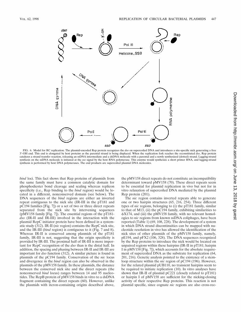
bind loci. This fact shows that Rep proteins of plasmids fromthe same family must have a common catalytic domain forphosphodiester bond cleavage and sealing whereas repliconspecificity (i.e., Rep binding to the bind region) would be lo-cated in a different, nonconserved domain (see below). TheDNA sequences of the bind regions are either an invertedrepeat contiguous to the nick site (IR-III in the pT181 andpC194 families [Fig. 7]) or a set of two or three direct repeatsseparated from the nick site by intervening sequences(pMV158 family [Fig. 7]). The essential regions of the pT181-dso (IR-II and IR-III) involved in the interaction with theplasmid RepC initiator protein have been defined in a system-atic study (312). IR-II (nic region) contains the RepC nick site,and the IR-III (bind region) is contiguous to it (Fig. 7 and 8).Whereas IR-II is conserved among plasmids of the pT181family, IR-III is not, suggesting that the origin specificity isprovided by IR-III. The proximal half of IR-III is more impor-tant for RepC recognition of the dso than is the distal half. Inaddition, the spacing and phasing between IR-II and IR-III areimportant for dso function (312). A similar picture is found inplasmids of the pC194 family. Conservation of the nic locusand divergence in the bind region can also be observed in theplasmids of the pMV158 family. In these plasmids, the distancebetween the conserved nick site and the direct repeats (thenonconserved bind locus) ranges between 14 and 95 nucleo-tides. The RepB protein of pMV158 binds in vitro to a dsDNAfragment containing the direct repeats (60). However, unlikethe plasmids with iteron-containing origins described above,
the pMV158 direct repeats do not constitute an incompatibilitydeterminant toward pMV158 (70). These direct repeats seemto be essential for plasmid replication in vivo but not for invitro relaxation of supercoiled DNA mediated by the plasmidRep protein (201).
The nic region contains inverted repeats able to generateone or two hairpin structures (65, 216, 254). Three differenttypes of nic regions, belonging to (i) the pT181 family, similarto that of M13, (ii) the pC194 family, exhibiting similarities tofX174, and (iii) the pMV158 family, with no relevant homol-ogies to nic regions from known ssDNA coliphages, have beenreported (Table 1) (69, 108, 228). The development of a systemin which DNA strand discontinuities can be mapped with nu-cleotide resolution in vivo has allowed the identification of thenick sites of other plasmids of the pMV158 family, namely,pE194, and pFX2 (106, 328). The DNA sequences recognizedby the Rep proteins to introduce the nick would be located onunpaired regions within these hairpins (IR-II in pT181; hairpinI in pMV158 [Fig. 7]), which accounts for the absolute require-ment of supercoiled DNA as the substrate for replication (64,201, 216). Genetic analysis pointed to the existence of a stem-loop structure within the nic region of pC194 (196). However,for the related plasmid pUB110, no transient hairpins seem tobe required to initiate replication (10). In vitro analyses haveshown that IR-II of plasmid pC221 (closely related to pT181)or hairpin I of pMV158 are sufficient for the nicking-closingactivity of their respective Rep proteins. This reaction is notplasmid specific, since cognate nic regions are also cross-rec-
FIG. 6. Model for RC replication. The plasmid-encoded Rep protein recognizes the dso on supercoiled DNA and introduces a site-specific nick generating a free39-OH end. This end is elongated by host proteins as the parental strand is being displaced. When the replication fork reaches the reconstituted dso, Rep proteincatalyzes a strand transfer reaction, releasing an ssDNA intermediate and a dsDNA molecule with a parental and a newly synthesized (dotted) strand. Lagging-strandsynthesis on the ssDNA molecule is initiated at the sso signal by the host RNA polymerase. This enzyme would synthesize a short primer RNA, and lagging-strandsynthesis is performed by host DNA polymerases. The end products are supercoiled plasmid DNA molecules.
VOL. 62, 1998 REPLICATION OF CIRCULAR BACTERIAL PLASMIDS 447
on June 13, 2018 by guesthttp://m
mbr.asm
.org/D
ownloaded from

ognized by these Rep proteins (201, 292, 332). In addition, invitro replication of plasmid pC221 is reduced if a competingpT181-dso is present (332). In spite of such an in vitro recog-nition and extensive homologies of the Rep proteins and thedso of pT181 and pC221, there is no cross-reactivity betweenthe Rep proteins and the dso of these plasmids in vivo, unlessthe Rep proteins are overexpressed (135).
Rep proteins. Rep proteins of the RC-replicating plasmidshave DNA strand transferase enzymatic activity, so that theyare able to cleave and to join plasmid DNA in a type I topo-isomerase-like fashion. RepC protein of plasmid pT181 waspurified and shown to have nicking activity on the plasmid dso
(156b). This finding, together with the presence of intracellularplasmid ssDNA and with the lack of requirement of a primerRNA for leading-strand synthesis, provided the first indica-tions of the RC mechanism for plasmid replication (156a, 291).Rep-mediated nicking participates in the initiation of replica-tion because the Rep proteins cleave supercoiled DNA withinan unpaired sequence of the nic region. Cleavage and joiningare essential for termination of replication (see below). Repproteins encoded by RC-replicating plasmids have several con-served motifs which are shared with the Tra and Mob proteins(involved in plasmid transfer or conjugative mobilization) andwith the Rep proteins of ssDNA coliphages and geminiviruses
FIG. 7. Origins of replication and related regions of plasmids replicating by the RC mode, as exemplified by plasmids pMV158 (A) and pT181 (B). The bind andnic regions of the dso are indicated. Other symbols are as in Fig. 1.
448 DEL SOLAR ET AL. MICROBIOL. MOL. BIOL. REV.
on June 13, 2018 by guesthttp://m
mbr.asm
.org/D
ownloaded from
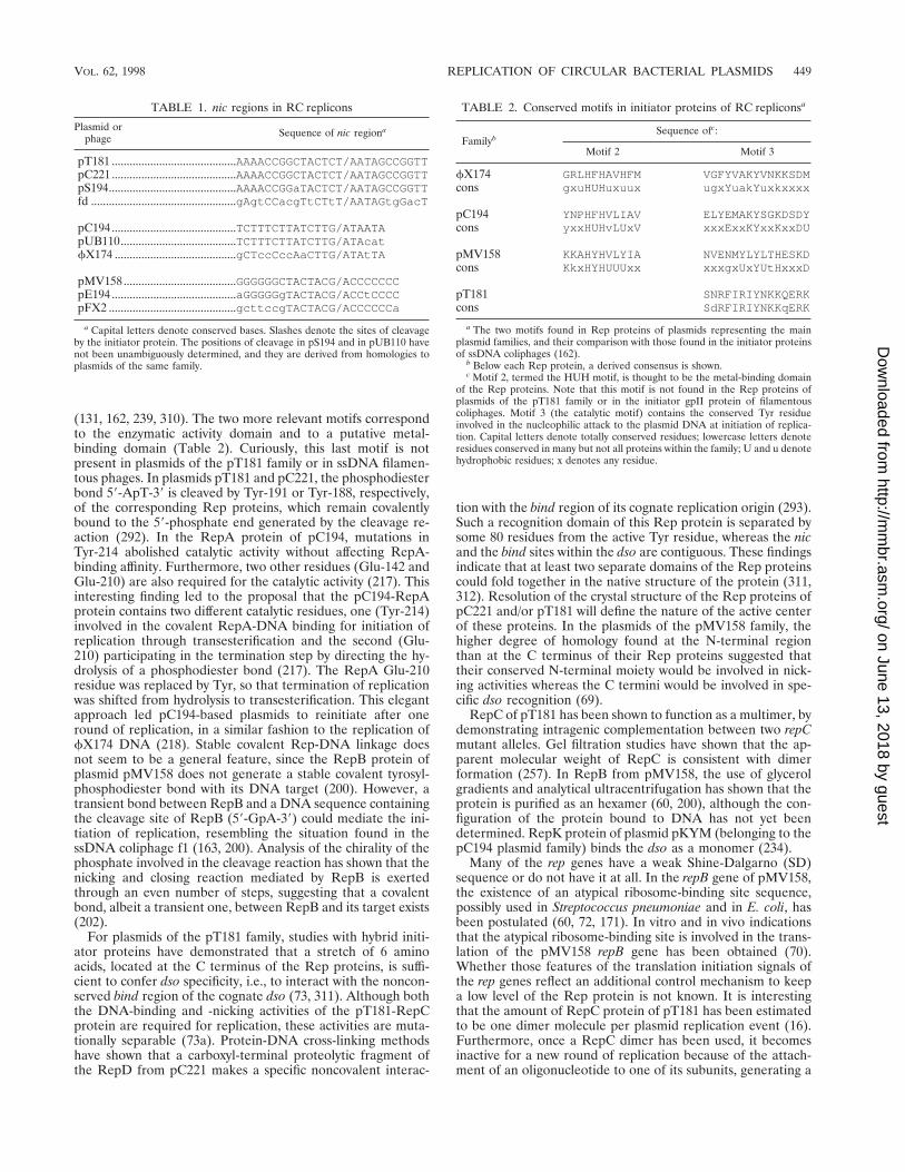
(131, 162, 239, 310). The two more relevant motifs correspondto the enzymatic activity domain and to a putative metal-binding domain (Table 2). Curiously, this last motif is notpresent in plasmids of the pT181 family or in ssDNA filamen-tous phages. In plasmids pT181 and pC221, the phosphodiesterbond 59-ApT-39 is cleaved by Tyr-191 or Tyr-188, respectively,of the corresponding Rep proteins, which remain covalentlybound to the 59-phosphate end generated by the cleavage re-action (292). In the RepA protein of pC194, mutations inTyr-214 abolished catalytic activity without affecting RepA-binding affinity. Furthermore, two other residues (Glu-142 andGlu-210) are also required for the catalytic activity (217). Thisinteresting finding led to the proposal that the pC194-RepAprotein contains two different catalytic residues, one (Tyr-214)involved in the covalent RepA-DNA binding for initiation ofreplication through transesterification and the second (Glu-210) participating in the termination step by directing the hy-drolysis of a phosphodiester bond (217). The RepA Glu-210residue was replaced by Tyr, so that termination of replicationwas shifted from hydrolysis to transesterification. This elegantapproach led pC194-based plasmids to reinitiate after oneround of replication, in a similar fashion to the replication offX174 DNA (218). Stable covalent Rep-DNA linkage doesnot seem to be a general feature, since the RepB protein ofplasmid pMV158 does not generate a stable covalent tyrosyl-phosphodiester bond with its DNA target (200). However, atransient bond between RepB and a DNA sequence containingthe cleavage site of RepB (59-GpA-39) could mediate the ini-tiation of replication, resembling the situation found in thessDNA coliphage f1 (163, 200). Analysis of the chirality of thephosphate involved in the cleavage reaction has shown that thenicking and closing reaction mediated by RepB is exertedthrough an even number of steps, suggesting that a covalentbond, albeit a transient one, between RepB and its target exists(202).
For plasmids of the pT181 family, studies with hybrid initi-ator proteins have demonstrated that a stretch of 6 aminoacids, located at the C terminus of the Rep proteins, is suffi-cient to confer dso specificity, i.e., to interact with the noncon-served bind region of the cognate dso (73, 311). Although boththe DNA-binding and -nicking activities of the pT181-RepCprotein are required for replication, these activities are muta-tionally separable (73a). Protein-DNA cross-linking methodshave shown that a carboxyl-terminal proteolytic fragment ofthe RepD from pC221 makes a specific noncovalent interac-
tion with the bind region of its cognate replication origin (293).Such a recognition domain of this Rep protein is separated bysome 80 residues from the active Tyr residue, whereas the nicand the bind sites within the dso are contiguous. These findingsindicate that at least two separate domains of the Rep proteinscould fold together in the native structure of the protein (311,312). Resolution of the crystal structure of the Rep proteins ofpC221 and/or pT181 will define the nature of the active centerof these proteins. In the plasmids of the pMV158 family, thehigher degree of homology found at the N-terminal regionthan at the C terminus of their Rep proteins suggested thattheir conserved N-terminal moiety would be involved in nick-ing activities whereas the C termini would be involved in spe-cific dso recognition (69).
RepC of pT181 has been shown to function as a multimer, bydemonstrating intragenic complementation between two repCmutant alleles. Gel filtration studies have shown that the ap-parent molecular weight of RepC is consistent with dimerformation (257). In RepB from pMV158, the use of glycerolgradients and analytical ultracentrifugation has shown that theprotein is purified as an hexamer (60, 200), although the con-figuration of the protein bound to DNA has not yet beendetermined. RepK protein of plasmid pKYM (belonging to thepC194 plasmid family) binds the dso as a monomer (234).
Many of the rep genes have a weak Shine-Dalgarno (SD)sequence or do not have it at all. In the repB gene of pMV158,the existence of an atypical ribosome-binding site sequence,possibly used in Streptococcus pneumoniae and in E. coli, hasbeen postulated (60, 72, 171). In vitro and in vivo indicationsthat the atypical ribosome-binding site is involved in the trans-lation of the pMV158 repB gene has been obtained (70).Whether those features of the translation initiation signals ofthe rep genes reflect an additional control mechanism to keepa low level of the Rep protein is not known. It is interestingthat the amount of RepC protein of pT181 has been estimatedto be one dimer molecule per plasmid replication event (16).Furthermore, once a RepC dimer has been used, it becomesinactive for a new round of replication because of the attach-ment of an oligonucleotide to one of its subunits, generating a
TABLE 1. nic regions in RC replicons
Plasmid orphage Sequence of nic regiona
pT181 ..........................................AAAACCGGCTACTCT/AATAGCCGGTTpC221..........................................AAAACCGGCTACTCT/AATAGCCGGTTpS194...........................................AAAACCGGaTACTCT/AATAGCCGGTTfd .................................................gAgtCCacgTtCTtT/AATAGtgGacT
pC194..........................................TCTTTCTTATCTTG/ATAATApUB110.......................................TCTTTCTTATCTTG/ATAcatfX174 .........................................gCTccCccAaCTTG/ATAtTA
pMV158......................................GGGGGGCTACTACG/ACCCCCCCpE194 ..........................................aGGGGGgTACTACG/ACCtCCCCpFX2 ...........................................gcttccgTACTACG/ACCCCCCa
a Capital letters denote conserved bases. Slashes denote the sites of cleavageby the initiator protein. The positions of cleavage in pS194 and in pUB110 havenot been unambiguously determined, and they are derived from homologies toplasmids of the same family.
TABLE 2. Conserved motifs in initiator proteins of RC repliconsa
FamilybSequence ofc:
Motif 2 Motif 3
fX174 GRLHFHAVHFM VGFYVAKYVNKKSDMcons gxuHUHuxuux ugxYuakYuxkxxxx
pC194 YNPHFHVLIAV ELYEMAKYSGKDSDYcons yxxHUHvLUxV xxxExxKYxxKxxDU
pMV158 KKAHYHVLYIA NVENMYLYLTHESKDcons KkxHYHUUUxx xxxgxUxYUtHxxxD
pT181 SNRFIRIYNKKQERKcons SdRFIRIYNKKqERK
a The two motifs found in Rep proteins of plasmids representing the mainplasmid families, and their comparison with those found in the initiator proteinsof ssDNA coliphages (162).
b Below each Rep protein, a derived consensus is shown.c Motif 2, termed the HUH motif, is thought to be the metal-binding domain
of the Rep proteins. Note that this motif is not found in the Rep proteins ofplasmids of the pT181 family or in the initiator gpII protein of filamentouscoliphages. Motif 3 (the catalytic motif) contains the conserved Tyr residueinvolved in the nucleophilic attack to the plasmid DNA at initiation of replica-tion. Capital letters denote totally conserved residues; lowercase letters denoteresidues conserved in many but not all proteins within the family; U and u denotehydrophobic residues; x denotes any residue.
VOL. 62, 1998 REPLICATION OF CIRCULAR BACTERIAL PLASMIDS 449
on June 13, 2018 by guesthttp://m
mbr.asm
.org/D
ownloaded from

heterodimer, RepC/C* (255–257). Inactivation through gener-ation of heterodimers has also been shown for pT181-relatedplasmids (256) and has been suggested to exist for the RepUprotein of pUB110 (207). The footprints generated on super-coiled DNA by the RepC/C homodimer are different fromthose formed by the RepC/C* heterodimer, and whereasRepC/C was able to enhance cruciform extrusion at the dso,RepC/C* was not (140). Nevertheless, biochemical analysis hasshown that RepC and RepC* appeared to be identical, exceptfor the absence of the active Tyr residue in the modified pro-tein (258).
None of the Rep proteins seems to have the HTH motiftypical of many DNA-binding proteins, although a putative LZmotif exists in the RepB protein of plasmid pMV158 (60).Inspection of the DNA sequence of various rep genes showstwo possible translation initiation signals, which would give riseto Rep and Rep9 proteins (similar to the A and A* initiatorproteins, respectively, of the ssDNA coliphage fX174) (13).However, genetic evidence has shown that, at least for RepB ofpMV158, the putative shorter RepB9 protein is not sufficient toconduct replication in vivo (70). Plasmid pUB110 and its rel-ative pRBH1 were reported to contain two putative SD se-quences followed by a Met residue (178, 208). Overproductionof the pUB110-Rep protein showed only one major product,the one translated from the first Met residue (178). Thus, thesignificance of these putative Rep9 proteins (if any) remains tobe clarified.
Initiation and elongation of leading-strand synthesis. Littleinformation is available on the initiation complex in RC-rep-licating plasmids. As stated above, the Rep proteins of pT181and pC221 nick the dso and become covalently attached, by aphosphotyrosine bond, to the thymidilate residue at 39 of thenick. A strict requirement for supercoiled DNA as the sub-strate to initiate replication, most probably for cruciform ex-trusion, has been proposed (10, 64, 65, 216). In pMV158, invitro RepB-dependent cleavage of the nick sequence abso-lutely requires ssDNA as the substrate (200, 201) and thehairpin corresponding to the nic region has been shown toextrude on supercoiled plasmid DNA (65, 254). For pT181, atlow to moderate RepC/pT181 origin ratios, only supercoiledmolecules can be used as the substrate for replication initiation(149), although specific RepC nicking activity has been dem-onstrated in vitro on linearized plasmid DNA (157). Once theRep-DNA complex has formed, several host proteins would berequired for leading-strand synthesis. To generate the replica-tion initiation complex of pT181, the product of the Staphylo-coccus aureus chromosomal pcrA gene is required. PcrA hashomology to helicase II and Rep helicase of E. coli (136). ThePcrA protein is essential for cell viability and for replication ofplasmid pT181. A mutation (pcrA3) found in the pcrA gene ledto an increase in the proportion of nicked DNA plasmid forms,which were identified as replication initiation complexes (138).The copy number of pT181 was reduced in an S. aureus pcrA3background, and suppressor mutations were shown to map inthe plasmid repC gene, indicating a direct interaction betweenRepC and PcrA (137). In addition, it is assumed that DNA PolIII-HE extends the leading strand and that due to the gener-ation of ssDNA intermediates, SSB also participates in thereplication complex.
Termination of leading-strand synthesis. Specific recogni-tion between the Rep protein and the dso is required not onlyfor initiation of RC replication but also for the terminationstep. Rep-dso recognition seems to be more stringent for ini-tiation than for termination, because (i) abortive terminationcan occur at sequences similar but not identical to the origin(15, 196); (ii) part of the dso seems to be sufficient for termi-
nation of replication of pC194 (107); and (iii) an 18-bp se-quence is long enough for termination of pT181 replication,but initiation requires a larger region which includes the RepCbinding site (329). Mutational analyses have shown that thenucleotides of the right arm of the inverted repeat IR-II mustbe conserved for termination to occur whereas changes areallowed in some of the nucleotides of the left arm of the IR-II(330). Genetic approaches suggest that a DNA sequence,probably located 39 of the nic region of pUB110, is required forefficient termination of replication (10). Thus, two partiallyoverlapping sequences, one required for initiation and theother required for termination, should be present in pUB110.For this plasmid, a single amino acid change in its Rep proteinappears to reduce the ability of the Rep protein to terminatereplication specifically (22).
The mechanistic information on termination of RC replica-tion has arisen from the elegant set of experiments performedin Novick’s laboratory with pT181 (141, 255, 257, 311, 312).This information has been extended for other plasmids of thepT181 family (256), as well as for pUB110 (207). The currentmodel for RepC-catalyzed initiation and termination of pT181replication (schematized in Fig. 8; see also Fig. 6 and Table 2)postulates that Tyr-191 of one of the subunits of a RepChomodimer exerts a nucleophilic attack at the dso, generatinga RepC::RepC-DNA covalent complex (141, 255). Replicationof the leading strand proceeds more than one full round andbeyond the nick site (Fig. 8, stage 1). This would permit theIR-II hairpin at the dso to be exposed in both the displacedstrand and the newly synthesized strand. Then the RepC sub-unit which was not used at the initiation step would cleave theregenerated dso, being covalently bound to the 59 end of thenewly synthesized DNA strand (stage 2). The 39-OH end re-leased in this reaction (belonging to the parental strand) wouldexert a nucleophilic attack on the tyrosyl-phosphodiester bondgenerated at the initiation step (stage 3). This last reaction willrelease the ssDNA intermediate. The free OH group of theTyr-191 residue of the RepC subunit involved in the initiationstep attacks the nick site generated on the newly synthesizedstrand by the further extension of DNA synthesis (stage 4).Finally, the 39-OH end generated would attack the tyrosyl-phosphodiester bond generated between RepC and DNA instage 2. The end products of this reaction would be a dsDNAplasmid molecule (containing the newly synthesized plusstrand) and a RepC/C* heterodimer composed of one intactRepC molecule and one modified subunit which has a shortoligonucleotide covalently bound. This oligonucleotide is 12-mer on average, and it contains the 39 half of the IR-II (255).Conversion of RepC/C homodimer to RepC/C* heterodimerhas been shown to be replication dependent (257). TheRepC/C* heterodimer is not able to relax supercoiled pT181DNA or to reinitiate replication, indicating that inactivation ofone subunit (RepC*) may inactivate the whole molecule andthat a RepC dimer is used only once in a replication cycle(255, 257). A RepU/U* heterodimer has also been found inpUB110, which would point to a general inactivation mecha-nism for plasmids replicating by the RC mode (208). Lack ofrecycling of the Rep initiator is a critical feature for a plasmid,since it would prevent overreplication (255). In pMV158, thepT181 model for termination of replication does not seem tobe applicable, since RepB does not remain covalently bound toDNA (200). However, no alternative model for leading-strandtermination has been proposed, and inactivation of the RepBprotein has not been analyzed. A model, similar to the oneproposed by Novick for pT181 RC replication, has been re-cently proposed for termination of ssDNA conjugal transfer
450 DEL SOLAR ET AL. MICROBIOL. MOL. BIOL. REV.
on June 13, 2018 by guesthttp://m
mbr.asm
.org/D
ownloaded from

mediated by a dimer of the TraI protein of the IncPa plasmidRP4 (240).
Replication of the lagging strand. The last stage of RCreplication is the conversion of the ssDNA intermediate into aduplex plasmid DNA molecule. Lagging-strand synthesis ini-tiates and terminates at the sso, which is a specific noncod-ing region with the potential to generate imperfect stem-loop structures. These secondary structures function inan orientation-dependent manner (109). This suggests thatthe ssDNA3dsDNA conversion requires unpaired se-quences within the secondary structures that constitute thesso (65, 68, 109). Based on sequence homologies, varioustypes of sso have been reported (228). The best character-ized are ssoA (pT181 and pC194), ssoU (pUB110), ssoT(pTA1060 and pBAA1) (75, 193), and ssoW (pWV01 andclosely related replicons) (268). The streptococcal plasmidpMV158 has the interesting feature of bearing both ssoA
and ssoU (68, 171, 250, 306). Deletion of plasmid sso regionsleads to reduction in the plasmid copy number (measured asdsDNA), accumulation of ssDNA molecules, and rapid lossof the plasmid in the absence of selective pressure (65, 109).Such an instability does not depend on the average numberof copies of double-stranded plasmid DNA, since it cannotbe overcome by mutations increasing the plasmid copy num-ber of these deleted derivatives (68). Analysis of the DNAsequence and structure of the ssoA of various plasmids (228)showed the presence of two conserved unpaired regions.One of them includes part of recombination site B (RSB),which is highly conserved in almost all of the reported ssoAsequences. In the center of the palindromic sequence of thessoA regions, there is another unpaired region, which isconserved only among the members of each one of twodifferent groups of ssoA sequences, represented by pT181-ssoA and pMV158-ssoA (65, 228). In the ssoA of the plasmid
FIG. 8. Model for termination of RC replication based on results from Novick’s laboratory (140, 141, 255–257). Nucleophilic attacks exerted by the OH groups ofthe Tyr residue of Rep (Y) or by 39-OH groups of the DNA (OH) are indicated by arrows. Solid lines, parental DNA; broken lines, newly synthesized DNA. The thicksolid line indicates the nucleotides that are newly synthesized past the reconstituted dso and that will remain covalently bound to the Rep protein to generate aRep/Rep* inactive dimer. The two subunits of the RepC dimer are differently depicted.
VOL. 62, 1998 REPLICATION OF CIRCULAR BACTERIAL PLASMIDS 451
on June 13, 2018 by guesthttp://m
mbr.asm
.org/D
ownloaded from

group represented by pMV158, the central conserved se-quence, termed CS-6, is 59-TAGCGt/a-39 (65). The CS-6 hasbeen shown to function as a transcriptional terminator forthe synthesis of an RNA primer (see below). Mutationsaffecting the CS-6 sequence of the pMV158-ssoA moder-ately increase the intracellular amount of plasmid ssDNA,although the effect is not as dramatic as that of the deletionof the entire ssoA (165, 167).
The mechanisms of ssDNA3dsDNA plasmid conversionare poorly understood, and no known plasmid-encoded func-tions seem to be involved. In analogy to ssDNA coliphages, apRNA is assumed to be needed to initiate lagging-strand syn-thesis. Experimental evidence suggests that in most cases,ssDNA3dsDNA conversion is mediated by RNAP, sincessDNA accumulates in vivo in the presence of rifampin (27, 74,165). In vitro studies have shown that an ssDNA fragmentcontaining the pMV158-ssoA was able to promote RNAP-directed synthesis of a 20-nucleotide pRNA. This ssDNA frag-ment specifically bound RNAP, with this binding requiring anintact RSB sequence (166).
The ssoW of plasmid pWV01 is made up of two invertedrepeats (IR1 and IR2). Conversion of ssDNA3dsDNA froman intact ssoW seems to require the host RNAP (173). How-ever, deletion of the IR2 resulted in a reduced and RNAP-independent conversion. These findings, together with the ho-mologies between the ssoW-IR1 and the lagging-strand originof fX174, led to the proposal of an alternative mechanism ofpWV01 lagging-strand priming mediated by the host primo-some (173, 268).
In addition to the host RNAP, DNA Pol I is involved inlagging-strand replication both in initiation and in termination(76, 166). Detection of ssDNA intermediates may be maskedbecause the amount of ssDNA accumulated depends upon thespecific host-plasmid pair and, in their natural hosts, single-stranded3double-stranded plasmid DNA conversion is so ef-ficient that the half-lives of ssDNA intermediates are too shortto be detectable (68, 108, 306). Nevertheless, implementationof an in vitro system for replication of ssDNA (23, 210) facil-itated the determination of the RNA-DNA transition points,which corresponded to regions including the above-mentionedconserved central sequences (CS-6) of the secondary structureof the sso regions (74). The use of pMV158-based vectors withno sso indicated the existence of an alternative pathway ofconversion in which plasmid-encoded antisense RNAs, an-nealed to the complementary region on the ssDNA interme-diates, may act as primers in a process mediated by the hostRecA protein (194).
Synopsis. Replication by the RC mechanism is widespreadamong multicopy plasmids from the Archaea and Bacteria. The39-OH end required for initiation of replication is provided bythe site-specific nicking activity of the plasmid-encoded Repprotein on one of the parental plasmid strands. The DNAsubstrate for the Rep-mediated nicking has to be in a single-stranded configuration. This can be achieved by Rep-facili-tated extrusion of a cruciform structure encompassing the nicregion of the origin, where the nick site is unpaired. Leading-strand synthesis is terminated by various specific strand trans-fer reactions, also mediated by the Rep protein. After comple-tion of leading-strand synthesis, the Rep protein is inactivatedand plasmid ssDNA intermediates are generated. RNAP-di-rected synthesis of a short RNA primer initiates lagging-strandreplication. In this step, only host-encoded proteins are in-volved.
CONTROL OF PLASMID REPLICATION
Although plasmid copy number may vary in different bacte-ria, within a given host, and under fixed growth conditions, anyparticular plasmid has a characteristic copy number. This isachieved by plasmid-encoded control elements that regulatethe initiation of the replication step. Control systems maintainthe rate of replication in the steady state at an average of onereplicative event per plasmid copy and cell cycle by correctingdeviations from the average copy number in individual cells.To define and to maintain the copy number, plasmids usenegative regulatory circuits (251). The genetic analysis of rep-lication control mechanisms was first performed for plasmidR1 through the isolation of mutants which exhibited an in-creased copy number (225). Characterization of these mutantsindicated that the determinants of copy number control re-sided in the plasmid itself and that negative regulators (inhib-itors), acting at the initiation step, were involved in this control.A model of control of replication modulated by negative ef-fectors was first proposed and described in quantitative termsby Pritchard et al. (252). When a plasmid colonizes a new host,the concentration of these negative regulators should be neg-ligible. This seems desirable for successful establishment, sinceunhindered plasmid replication would permit the normal copynumber to be reached in a short time. However, once thecharacteristic copy number is reached, keeping the averagecopy number in the population requires adjustments to fluctu-ations in this value in individual cells. The control systems dojust that by either increasing or decreasing the rate of replica-tion per plasmid copy and cell cycle. Individual plasmid copiesare selected for replication at random from a pool that in-cludes replicated and nonreplicated copies. However, mecha-nisms that counterselect newly replicated molecules exist, e.g.,hemimethylation and supercoiling (3, 224).
Control of replication by negative regulators and randomselection of plasmids for replication have an additional conse-quence: two plasmids with identical replicons are unable tostably coexist within a given cell in the absence of selectivepressure. This leads to segregation of plasmids within the hostpopulation, a phenomenon known as plasmid incompatibility.The inhibition of plasmid replication, associated with an in-crease in the gene dosage of copy number control genes, hasbeen used to identify these genes (reviewed in references 12,51, 155, 222, 227, and 251).
Control of replication by inhibitors would require that theycould “measure” the concentration of plasmid copies withinthe cell. This could be achieved by an unstable inhibitor ex-pressed constitutively or by a stable inhibitor synthesizedshortly after each initiation event (252). Any of these alterna-tives would reduce the initiation frequency after each initiationevent and would increase or decrease the rate of initiation ofreplication when the average copy number is, respectively,lower or higher than required. When the frequency of initia-tion is determined by the level of an initiator protein, onemechanism for controlled plasmid replication (unlike phagereplication) would be to inactivate the initiator protein aftereach replication event (255, 319).
Mechanisms controlling replication have been studied invarious systems (reviewed in reference 293a), and several typesof inhibitors have been detected: (i) antisense RNA (ColE1,R1, and pT181); (ii) both an antisense RNA and a protein(pMV158 and pIP501), and (iii) DNA sites for binding initiatorproteins (F, P1, RK2, and R6K). A schematic representation ofthese regulatory loops is depicted in Fig. 9. Control of repli-cation by a protein (lambda-dv) is well documented and quan-tified (222), but it has not been described in natural plasmids
452 DEL SOLAR ET AL. MICROBIOL. MOL. BIOL. REV.
on June 13, 2018 by guesthttp://m
mbr.asm
.org/D
ownloaded from

and is not discussed here. Accessory elements that can alsomodulate replication but that are not directly involved in con-trolling the initiation frequency are not discussed either.
Control by Antisense RNA
Interactions between RNAs can modulate the availability ofeither a pRNA or the level of a rate-limiting Rep protein,which are essential for replication. Antisense RNAs control-ling plasmid copy number are complementary to a region inthe 59 end of the target transcript (preprimer RNA for repli-cation or rep mRNA) and are termed “countertranscribedRNAs” (ctRNAs) (227, 228, 309). In some cases, inhibition byctRNAs is achieved in the region of pairing with the targetRNA. In other cases, formation of the inhibitor-target duplexleads to a conformational change in the target transcript, whichis rendered inactive. Examples of these mechanisms of controlare described below.
Control of primer RNA processing: plasmid ColE1. ColE1was the first example where control of replication was found tobe exerted by RNA (170, 296). In fact, the analyses of copynumber control circuits of ColE1 led to the identification ofantisense RNAs, a milestone discovery in molecular biology.trans-acting antisense RNAs permit the silencing of specificgenes by pairing to mRNA molecules. The bases of the controlof ColE1 replication are well known (reviewed in references 47and 82). Initiation of replication is primed by a 550-nucleotidetranscript, RNA II, which is made by RNAP and is specificallyprocessed by RNase H. This processing is required for thetransition of RNA to DNA synthesis, since it generates a39-OH end on which Pol I initiates the synthesis of the leadingstrand. The availability of this 39-OH end is rate limiting forinitiation and is modulated by an RNA of 108 nucleotides, theantisense RNA I, which is entirely complementary to a regionin the 59 end of the pRNA II. Hybrid formation between theantisense and the preprimer RNA alters the overall secondarystructure of the preprimer. As a consequence of this confor-mational change, the 39 end of the preprimer cannot hybridizeto the DNA. This, in turn, inhibits the initiation of replicationbecause, in the absence of DNA-RNA hybrid formation, thepreprimer is not processed by RNase H. Genetic and biochem-ical analyses showed that interactions of the antisense RNAwith the complementary sequences of the preprimer occurinitially at regions exposed as single-stranded loops on bothfolded RNA molecules. Formation of this initial contact (or“kissing” complex) is the rate-limiting step of the interactionand leads to the full annealing of the antisense RNA I with thepreprimer RNA II, in a process that starts at the 59 end of theRNA I. The RNA I is synthesized from a constitutive promoter(therefore its level is proportional to the plasmid copy number)and is unstable. An auxiliary role is provided by a small basicprotein, termed Rop or Rom (276, 304). Rop interacts with theantisense RNA and with the preprimer, increasing the effi-ciency of formation of the “kissing” complex and thus decreas-ing the efficiency of replication. The gene encoding this proteinis not an incompatibility determinant, but it can reinforce theantisense RNA I-mediated incompatibility.
Copy number control of plasmid R1. Copy number controlin R1 is exerted at the level of RepA synthesis, which is mod-ulated by the products of the copy number control genes, copBand copA (reviewed in references 224 and 309). The productsof copB and of copA inhibit repA expression at the transcrip-tional and posttranscriptional levels, respectively. RepA is ratelimiting for replication, acts preferentially in cis, and cannot bereused. The main copy number control gene is copA, whilecopB plays an accessory role. copA is transcribed from a con-
stitutive promoter, and its product is an unstable (half-life, 2min) RNA, CopA, which is complementary to a leader region(termed CopT) of the repA mRNA. Hybrid formation betweenCopA and its CopT target is also achieved through formationof a “kissing” complex. The CopA-CopT hybrid inhibits RepAsynthesis by hindering the translation of a leader peptide-encoding gene, tap, which is translationally coupled to repA(26). The efficiency of inhibition depends on the affinity ofkissing-complex formation (reviewed in reference 226). Muta-tions in copA that decrease the efficiency of the complex for-mation increase the copy number, but replication still is con-trolled. However, complete inactivation of CopA leads touncontrolled (runaway) replication (reviewed in references224 and 226). The duplex CopA-CopT is specifically cleaved byRNase III, and this cleavage seems to be an important step inthe control of RepA synthesis (25). Mutational analysis per-formed at copA showed that the structure of the major stem-loop in CopA is important for its function. The presence ofbulges in the stem region of CopA is required for rapid duplexformation with its CopT target in vitro, as well as for effectiveinhibition in vivo (126). In addition, the presence of bulgednucleotides in the CopA stem seems to result in a structuraldestabilization that plays a protective role against degradationby RNase III (125). Structural and functional studies on CopAhave permitted us to draw a direct correlation between genedosage (plasmid copy number) and CopA levels. This deter-mines a constant rate of replication, independent of the copynumber of the plasmid or the growth rate of the host. Twoconsequences derive from these results: (i) the frequency ofplasmid replication per copy is .1 when the number of plas-mid copies per cell is below the average and ,1 when thenumber of copies is above the average; and (ii) the copy num-ber of R1 increases with decreasing host growth rate. A con-stant rate of replication per cell, independent of copy number,will slowly correct deviations from the average frequency ofplasmid replication (one per plasmid copy per cell cycle), andtherefore it will actively maintain the average copy numberconstant (222). The second regulatory element in R1, theCopB protein, plays an auxiliary role under normal circum-stances. This small dimeric and basic protein represses tran-scription of repA from a promoter, P2, which is located down-stream of copB (Fig. 9). Under steady-state conditions, CopBis present at saturating concentrations, fully repressing tran-scription from P2 to a basal level. Under these conditions, therepA mRNA is synthesized mainly as a polycistronic copB-tap-repA RNA. However, under conditions in which the copy num-ber drops or is low because of the early stages of plasmidestablishment, the CopB-mediated repression is not efficient.In these circumstances, the P2 promoter is derepressed andrepA is also transcribed as a tap-repA mRNA. This leads to atransient increase in the transcription rate of repA and, as aconsequence, to a temporal increase in the frequency of plas-mid replication. Thus, derepression of the P2 promoter wouldplay an important role during the early phase of plasmid es-tablishment or in situations in which the copy number fallsbelow the wild-type level (224, 320). However, a computer-based simulation suggested that the auxiliary control loop me-diated by CopB should have only a marginal effect not onlyunder steady-state conditions but also during plasmid estab-lishment (259).
Other instances of control by antisense RNAs. There areother examples in which replication is controlled by antisenseRNA. For instance, in plasmids of the IncIa and IncIb groups,control of replication is modulated by a short ctRNA thatinhibits, as in R1, synthesis of a rate-limiting Rep protein,which, in turn, is translationally coupled to the synthesis of a
VOL. 62, 1998 REPLICATION OF CIRCULAR BACTERIAL PLASMIDS 453
on June 13, 2018 by guesthttp://m
mbr.asm
.org/D
ownloaded from
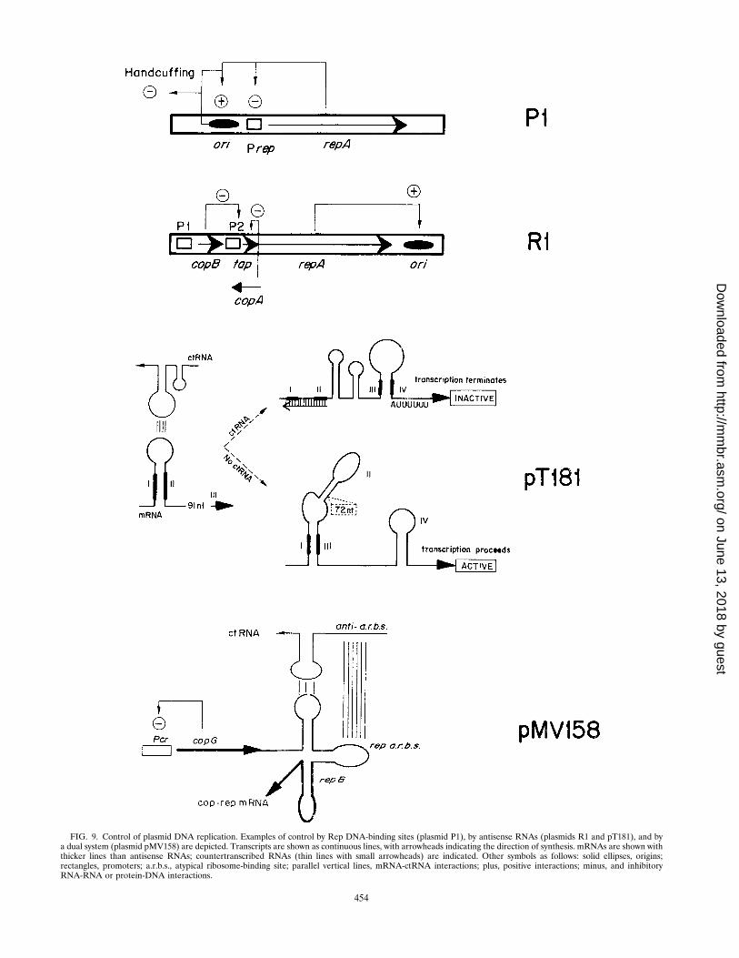
FIG. 9. Control of plasmid DNA replication. Examples of control by Rep DNA-binding sites (plasmid P1), by antisense RNAs (plasmids R1 and pT181), and bya dual system (plasmid pMV158) are depicted. Transcripts are shown as continuous lines, with arrowheads indicating the direction of synthesis. mRNAs are shown withthicker lines than antisense RNAs; countertranscribed RNAs (thin lines with small arrowheads) are indicated. Other symbols as follows: solid ellipses, origins;rectangles, promoters; a.r.b.s., atypical ribosome-binding site; parallel vertical lines, mRNA-ctRNA interactions; plus, positive interactions; minus, and inhibitoryRNA-RNA or protein-DNA interactions.
454
on June 13, 2018 by guesthttp://m
mbr.asm
.org/D
ownloaded from

leader peptide. Inhibition by ctRNA is exerted at the posttran-scriptional level by hindering the synthesis of the leader pep-tide. In addition, ctRNA prevents the formation of an activatorRNA pseudoknot that is required for efficient synthesis of theRep protein (reviewed in reference 309).
Antisense RNA has been involved in the silencing of theori-g of plasmid R6K (243, 244). Replication from this originrequires the synthesis of an activator RNA that starts withinthe seven iterons of the origin and is silenced by the antisenseRNA. The replication initiator protein, p, seems to favor in-teractions between these transcripts. This regulatory system isindependent of the one modulated by iterons. R6K is not theonly theta-replicating iteron-containing plasmid in which anti-sense RNA plays a role in regulating replication. In plasmidColE2, a ctRNA complementary to the leader rep mRNA hasbeen involved in the control of replication at the posttranscrip-tional level through generation of a ctRNA-mRNA hybrid thatsequesters a sequence(s) essential for Rep synthesis other thanthe SD region (287, 323).
Direct inhibition of Rep synthesis: blocking rep translation.A direct way to control plasmid copy number by a ctRNA is theinhibition of Rep synthesis by blocking the accessibility of theribosomes to the rep mRNA translation initiation sequences.This type of control has been proposed for some plasmidsreplicating by the mechanisms of strand displacement (R1162)(154) and RC replication (pMV158) (67). In both instances,the main inc determinant is located in a region encoding actRNA, and the putative signals for initiation of rep translationare placed on a predicted loop of the rep mRNA. In the caseof R1162/RSF1010, the frequency of initiation is determinedby the level of the RepC DNA-binding protein (112, 153).Expression of repC is regulated at the transcriptional level bythe first gene of the repC operon (179) and at the translationallevel by a ctRNA that is complementary to the translationinitiation signals of repC (154). Which of these two regulatorymechanisms makes the RepC synthesis rate limiting is not yetwell understood. In the RC-replicating plasmid pMV158, actRNA has been proposed to inhibit the translation of the repBgene by direct interaction with the initiation of translationsequences present in the repB mRNA (67, 71, 72). Secondarystructures in the ctRNA, other than that corresponding to thetranscriptional terminator, have not been found. Plasmidslacking the DNA region encoding this terminator and thecomplementary structure on the cop-rep mRNA are still sen-sitive to the wild-type ctRNA supplied in trans, suggesting thatformation of a kissing complex may be important but is notessential for the generation of the hybrid ctRNA-mRNA. Amore complex regulatory loop in pMV158, which involves thejoint participation of the ctRNA and of a repressor Cop pro-tein, was described later (see below). Analogous genetic struc-tures in the control-of-replication region have been found inplasmids of the pMV158 family (69). A two-component system(Cop protein and antisense RNA) controlling the replicationof plasmid pIP501 has also been proposed (35; see below). Forplasmid pUB110, synthesis of the initiator RepU proteinseems to be controlled by two antisense ctRNAs that interferewith repU translation, since the putative ctRNAs are comple-mentary to repU initiation of translation sequences (178), al-though a mechanism of control similar to that of pT181 hasalso been suggested (11, 249). However, since the synthesis ofRepU is autoregulated (207), a more complex control levelmay have to be considered.
Transcriptional attenuation: the pT181 paradigm. A singu-lar copy number control mechanism, involving ctRNAs, hasbeen well established for pT181 (119, 169, 227, 228). Thecurrent model for the control of replication of pT181 (refer-
ences 227 and 228 and references therein) proposes that syn-thesis of the Rep initiator is limited by two small ctRNAs(having the same 59 end but different 39 ends), which arecomplementary to the untranslated 59 end of the rep mRNA(44, 119, 227, 229). The initial kissing-complex formation be-tween ctRNAs and rep mRNA would take place through nu-cleotides exposed in complementary loops of hairpin structuresthat can be formed on both RNA species. Formation of thectRNA-mRNA hybrid would lead to a conformational changein a region of the mRNA far apart from the region of comple-mentarity. The new mRNA configuration would be such that anew hairpin ending in an A(U)6 sequence could form. Thissecondary structure resembles a Rho-independent transcrip-tion terminator and therefore can truncate the rep mRNA(230). Thus, transcriptional attenuation (as found in the con-trol of many biosynthetic operons) (161), rather than blockingtranslation initiation, is the mechanism for controlling the copynumber of pT181. A similar control, through premature ter-mination of the rep mRNA promoted by the indirect action ofa ctRNA, has been proposed for plasmids of the pIP501/pAMb1 family (33). For pIP501, it has been shown that stepsoccurring before stable ctRNA-mRNA duplex formation aresufficient to attenuate mRNA synthesis (34).
Control by both Transcriptional Repressorand Antisense RNA
Control by the joint action of a transcriptional repressor andan antisense RNA has been characterized for plasmids R1,pMV158, and, more recently, pIP501, although differences inthe role of the components have been found (35, 69, 71, 72,224). As described above, the main control in R1 is exerted bythe ctRNA CopA whereas CopB plays an auxiliary role. In fact,deletion of copB leads only to a moderate increase in plasmidcopy number, and the copB gene cloned into a compatiblereplicon does not exhibit incompatibility toward R1.
In the case of pMV158, a transcriptional repressor, CopG,binds to and represses transcription from a single promoter forboth the copG and repB genes (66). Mutations or deletions incopG lead to an increase in the plasmid copy number (7), andcopG exerts only a weak incompatibility toward plasmids withthe pMV158 replicon when cloned, under its own transcriptionand translation signals, into a high-copy-number compatiblevector (67). A second element involved in copy number controlis a small countertranscribed RNA, RNA II, which is comple-mentary to a region of the cop-rep mRNA between genes copGand repB. rnaII is a strong inc determinant when supplied intrans at high gene dosage. Mutations that abolish the synthesisof the ctRNA also lead to an increase in copy number, butreplication is still controlled by CopG. However, in this case,great fluctuations in copy number within individual cells wereobserved (71). No significant incompatibility toward plasmidswith the pMV158 replicon was found when the rnaII gene wascloned in a low-copy-number plasmid (71). The control circuitelements (copG and/or rnaII) have been cloned at a physio-logical gene dosage (i.e., at a copy number similar to that of thewild-type plasmid). In this case, a strong incompatibility to-ward pMV158 was found when the entire circuit was providedin trans whereas either component cloned separately exhibitedweak (ctrnaII) or no (copG) incompatibility. The hypothesisderived from these experiments was that the control of repli-cation of pMV158 is exerted by the entire regulatory circuitrather than by a hierarchy of any of the components (71). Dueto the genetic organization of the plasmids of the pMV158family, a similar regulatory circuit seems to exist in all of them(61).
VOL. 62, 1998 REPLICATION OF CIRCULAR BACTERIAL PLASMIDS 455
on June 13, 2018 by guesthttp://m
mbr.asm
.org/D
ownloaded from
The theta-replicating plasmid pIP501 seems to share fea-tures with R1 and pMV158 in their replication control circuit.Similarly to R1, the CopR protein of pIP501 is not a regulatorof its own synthesis but represses transcription from the essen-tial downstream rep promoter (31). However, and unlike R1,the CopR-inhibited rep promoter is not fully repressed, a sit-uation resembling pMV158. The second element regulatingpIP501 copy number is the unusually long-lived antisense RNAIII (35). The stability of this antisense RNA III poses theproblem of corrections of down-fluctuations in copy number,since a decrease in the number of plasmid copies will not berapidly followed by a concomitant reduction in the levels of theinhibitor. This would lead to plasmid loss, which has not beenobserved (139a). To solve this apparent paradox, a modelsimilar to the one proposed for pMV158 (71), in which areduction in the pIP501 copy number would lead to derepres-sion of the rep promoter due to reduction in the CopR levels,has been postulated (35).
The recent finding that the RepU protein of plasmidpUB110 regulates its own synthesis, thus introducing an addi-tional control for plasmid copy number (207), would representanother example of dual control (transcriptional and transla-tional regulation). Mechanistically, the situations for pUB110and the pMV158 may be indistinguishable, which may point toa more general control of replication mechanism in these plas-mids replicating by the RC mode.
Control by Iterons
The Rep proteins of the theta-replicating iteron-containingplasmids are either autoregulated or under transcriptional con-trol, and the iterons play a role in the control of plasmid copynumber (reviewed in references 51, 155, and 223). The role ofthe iterons was initially described for plasmid F, based on theobservation that oriS-dependent replication was inhibitedwhen the iterons of oriS were cloned on a compatible plasmid(295). Similar situations were also found for other iteron-con-taining plasmids, such as P1 or R1162/RSF1010. The extent ofinhibition in P1 was found to be proportional to the number ofcloned iterons (238) whereas in R1162 the inhibition was abol-ished by overproduction of the rate-limiting Rep protein (153).These observations led to the formulation of the titrationmodel (303). The model assumed that the Rep protein is ratelimiting for initiation and that iterons titrate the Rep protein,thus limiting the frequency of initiation. A paradox developedwhen the Rep proteins of most iteron-containing plasmidswere found to be autoregulated, which implies that titrationcould be overcome by derepression (49). Furthermore, inother systems (R6K and RK2), the amount of Rep proteinwithin the cell was apparently too large to be limiting forsaturation of iteron binding (88, 190). This led to the modifi-cation of the initial model and to the proposal that differentforms of the Rep protein were involved in autoregulation andin initiation, so that initiator titration does not cause derepres-sion (302). A computer model that adjust to this hypothesis hasbeen developed (321).
A second solution to the titration/autoregulation paradoxwas provided by the model that Rep molecules bound to the P1iterons were still able to repress the repA promoter (49). Themodel was based on the ability of Rep proteins to bind to twoiterons simultaneously (in both P1 and R6K) (204). This prop-erty of the Rep proteins also provided the basis for a newmodel of negative control of replication called the “steric hin-drance” (238) or “handcuffing” (155, 191) model. According tothis model, when Rep proteins bind to and saturate the iteronsof the origin, initiation occurs if the plasmid copy number is
low. As the number of copies increases, Rep molecules boundto the iterons of one origin begin to interact with similarcomplexes generated on other origins. As a consequence, plas-mid molecules pair through Rep-Rep interactions, causing asteric hindrance to the function of both origins (“handcuff-ing”). The plasmid pairs are apparently separated during sub-sequent cellular growth, and the initiation potential of individ-ual molecules is restored. According to this model, it is theiteron concentration, rather than the level of Rep expression,that determines the rate of replication. The question of therole of autoregulation remains open, and no simple answer hasbeen provided. As mentioned above, in RK2 and R6K, a sub-stantial decrease in the levels of Rep protein has no effect onthe rate of plasmid replication. However, in P1, a twofoldreduction in the level of the Rep protein abolishes replicationand a fourfold increase can lead to inhibition of replication(238). Similarly, in R6K, a twofold increase in initiator con-centration is inhibitory to initiation. These data indicate thatautoregulation is important, at least in some cases, to keep anoptimal concentration of the initiator protein. It has recentlybeen suggested that in particular plasmids in which initiatorsare limiting, initiator titration and initiator pairing (handcuff-ing) could be working together (51).
Some of the replicons containing iterons in the origin alsocontain iterons outside this region, which express incompati-bility and further reduce the rate of replication (237). In prin-ciple, the probability of Rep-mediated iteron pairing greatlyincreases in these replicons, since it can occur in cis by DNAlooping and in various combinations in trans. Since controlsystems should measure the copy number, and since intramo-lecular pairing is independent of this parameter, this pairingcannot correct deviations from the average copy number. How-ever, intramolecular pairing can regulate the level at which thecopy control operates (discussed in reference 223). A differentquestion is whether the iterons within and outside the originregion are equally effective as replication control elements.Recent data on RK2 and P1 plasmids indicate that iteronsoutside of ori are more effective as negative regulators ofreplication than are iterons present in the origin region, prob-ably due to differences in the quantitative and structural fea-tures of the two sets (5, 273). It has been proposed that in RK2these auxiliary iterons stimulate initial pairing between origins.The data for P1 indicate that the concentration of the auxiliaryiterons rather than their relative arrangements is the determin-istic factor for replication control (51).
The role played by Rep proteins of iteron-containing plas-mids in the control of replication and in autoregulation impliesthat specific rep mutations affecting protein-protein or protein-DNA interactions could increase the plasmid copy number. InR6K, copy-up mutations affecting the plasmid initiator proteinhave been obtained (87). Most of the copy-up mutations af-fected a 32-amino-acid region placed between the LZ motifand the putative DNA-binding domain of the p protein, aregion that seems to be involved in high-order oligomerizationof the initiator protein (199). Mutations that affect the LZmotif of the Rep protein and lead to copy-up phenotypes inpSC101 have been described (133). Copy-up mutants of plas-mid RK2 containing mutations encoding TrfA variants defec-tive in binding to DNA have been isolated, indicating that copynumber control is modulated by TrfA-DNA interactions (46).Copy-up mutations in the trfA gene have also been selected asintragenic suppressors of thermosensitive trfA mutations.Some of these copy-up mutations were proposed to reduce astrong protein-protein inhibitory interaction induced, at therestrictive temperature, by the thermosensitive mutation (114).
456 DEL SOLAR ET AL. MICROBIOL. MOL. BIOL. REV.
on June 13, 2018 by guesthttp://m
mbr.asm
.org/D
ownloaded from

Hemimethylation and Regulation ofPlasmid Replication
The heptamer repeats containing the methylation sites in theorigin of replication of plasmid P1 constitute the target of thehost protein SeqA, involved in sequestering hemimethylatedoriC into the bacterial membrane (37). Methylation seems toregulate replication at two different levels. First, hemimethy-lation would exert a negative regulation through ori-membraneinteractions. The newly replicated, hemimethylated DNA issequestered in the cell membrane, thus preventing reinitiation.The sequestration is relieved as the hemimethylated DNAundergoes new methylation (51). At a second level, methyl-ation increases the initiation efficiency both in vivo and in vitro.This stimulation could be due to enhanced bending or unwind-ing of the DNA or to recycling of initiators (3, 51).
Synopsis
Plasmid DNA replication occurs coupled to the cell cycle ofthe bacterial host in such a way that a fixed concentration ofplasmids is maintained in the bacterial population. This isachieved by mechanisms that monitor the initiation frequencyand adjust this frequency to an average of one replication perplasmid copy and cell cycle. The control of plasmid replicationis plasmid encoded and is performed by molecules (antisenseRNA, proteins, or DNA sequences) that inhibit the initiationof replication in a dose-dependent way. This inhibition canlimit the level of active Rep protein, the availability of theprimer for the leading strand, or the number of active origins.Control of plasmid replication may be exerted (i) by a singleinhibitor, (ii) by a main controller and an auxiliary element, or(iii) by the concerted action of two inhibitors (a protein and anantisense RNA or two sets of iterons). In general, any modelon control of plasmid replication should explain how the av-erage plasmid copy number is maintained in a growing popu-lation and how fluctuations on this average are corrected.
SUMMING UP: DIFFERENCES AND SIMILARITIES INPLASMID REPLICATION MECHANISMS
In spite of the progress that has been made in the study ofplasmid replication and its control, there are major gaps in ourknowledge of the various replication systems. Initiation of rep-lication demands melting of the origin and interactions be-tween plasmid-encoded and host-encoded factors, which are ingeneral only poorly understood. Melting of the origin can bestabilized by either manifesting the cryptic ssDNA-binding ac-tivity of the initiation proteins (DnaA and R6K-p) or by theformation of intramolecular secondary structures (e.g., cruci-form extrusion). The configuration of the plasmid origins in-dicates that DNA curvature and Rep-enhanced origin bendingseem to be a common but yet not fully explored feature (81,156c, 205, 206, 283). DNA deformations at the origin mayprovide an appropriate configuration for the initiation event:cruciform extrusion and nicking by the initiator for RC-repli-cating plasmids versus assembly of the initiation complex andsynthesis of an RNA primer for the other replicons. The roleof host-encoded proteins in the generation of the initiationcomplex is relatively well defined for several plasmids replicat-ing by the theta mechanism, but little is known for plasmidsusing the RC mode (apart from the PcrA helicase from S.aureus in pT181 replication) (135–138). Definition of the spec-ificity of the interactions between Rep proteins and host rep-lication factors is relevant to our understanding of the ability ofplasmids to colonize different hosts. The convergence of struc-tural studies with the functional analysis of initiators and in-
hibitors of DNA replication is an important area of futuredevelopment. Host replication factors and plasmid initiatorsform part of the nucleoprotein complex that initiates plasmidreplication. The structural definition of these macromolecularassemblies will provide mechanistic information relevant toour understanding of the initiation of plasmid replication andthe interrelations between plasmids and their hosts. Since thecontinuity of the parental DNA strands is kept in plasmidsreplicating by the theta and strand displacement mechanisms,superhelical tensions accumulate during the elongation stage.A role for topoisomerases in the elongation of DNA synthesisand in the separation of both catenated daughter moleculeshas been documented (8, 9, 117, 245, 246, 327). Such a rolemay not be required for plasmids replicating by the RC mech-anism, although the relaxed DNA molecules (which are theproducts of RC replication) must be supercoiled by DNA gy-rase before they become a substrate for a new round of repli-cation. Finally, information is scarce on three important plas-mid replication events: (i) the role of transcription throughorigins to create waves of supercoiling, (ii) the structuralchanges introduced by chaperones that result in activation ofRep proteins, and (iii) the mechanisms of possible Rep inac-tivation, except for several RC-replicating plasmids (255, 256).
The study of specific signals involved in the termination ofreplication of theta-type plasmid replicons is a matter of grow-ing interest. Elucidation of the structure of termination pro-teins (RTP and Tus) acting at specific termination sites hasopened new avenues to perform detailed mechanistic analysisof this stage of replication (19).
Concerning lagging-strand synthesis, only host factors arethought to be required in plasmids replicating by the RC andtheta modes. This contrasts with the situation for plasmidsreplicating by the strand displacement mechanism, in whichthe same plasmid-encoded primase and helicase proteins areinvolved in replication of both strands, in a continuous processthat starts in two symmetrical origins and proceeds in oppositedirections (263, 266). In this latter plasmid category, uncou-pling synthesis of both DNA strands may occur, leading to thegeneration of ssDNA plasmid intermediates. However, unlikeplasmids replicating by the RC mechanism, there is no strandspecificity in the ssDNA intermediates generated during thestrand displacement replication.
Control of plasmid replication is one of the key features ofthese extrachromosomal elements. Such control is always ex-erted at the initiation stage, perhaps because the initiationevents, as opposed to the elongation and termination steps, areinvariably replicon specific. The role of RNA inhibitors in thecontrol of replication is relatively well understood (226, 309).However, replication control modulated by iterons is less wellknown and is an object of intense research (51, 155). Many ofthe initiators that bind to iterons autoregulate their own syn-thesis. How this autoregulation influences the frequency ofinitiation is important to a full understanding of control ofreplication in these plasmids. Interestingly, the strategies usedto control plasmid replication do not correlate with the mech-anism by which the plasmid replication initiates: replicationinitiation by different mechanisms may have similarities in con-trol circuits (pIP501 and pMV158; pIP501 and pT181; andR1162 and pMV158). Finally, although the elements control-ling replication have been identified in many instances andtheir modes of action have been described, there has not beenmuch efforts to understand the control in terms of kinetics,except for a few cases (12, 223).
VOL. 62, 1998 REPLICATION OF CIRCULAR BACTERIAL PLASMIDS 457
on June 13, 2018 by guesthttp://m
mbr.asm
.org/D
ownloaded from
CONCLUDING REMARKS AND PROSPECTS
The main emphasis of this review has been to show thatplasmids are important models with which the combination ofgenetic, biochemical, and molecular approaches have revealedmany biological phenomena. An important focus for molecularbiologists studying plasmids has been on the mechanisms in-volved in their replication and the control of this process.Replication studies have revealed a variety of mechanisms thatare used to replicate circular or linear plasmids. Plasmids con-tribute with specialized sequences and functions to their ownreplication but, in general, use extensively the replication ma-chinery of the host. Replication of these genetic elementstherefore provides a window to gain information on the com-munication between plasmid- and host-specific replication ma-chinery. Buried in these interrelationships is the ability of plas-mids to colonize different hosts (i.e., their host range), whichhas important ecological and biotechnological implications.
Plasmid initiator proteins have drawn much interest, and thestudies on these activators have been focused on the specificprotein-protein and protein-DNA interactions involved in theformation of an active initiation complex. Although geneticand biochemical analyses have allowed the isolation and char-acterization of Rep variants affected in their interaction withDNA, the relevant protein domains involved in contactingDNA are not clearly known in most cases. Concerning protein-protein interaction, the available information indicates that theinitiation complex is formed by multiple copies of the initiatorprotein interacting not only with the DNA in the origin regionbut also with each other and with host initiation factors, not tomention the role of membranes in DNA replication. The anal-ysis of the Rep protein of plasmid pPS10 has given importantclues on protein motifs involved in these interactions, althoughthe picture is far from complete. Atomic resolution of thestructure of an initiator and of a multiprotein-DNA initiationcomplex will be an important step toward a clear understand-ing of these interactions and the changes introduced in thetarget DNA. This could be done not only through the resolu-tion of crystal structures of protein-DNA complexes but alsothrough the use of many other available physicochemical ap-proaches (such as nuclear magnetic resonance, light and neu-tron scattering, circular dichroism, and analytical ultracentrif-ugation, to name a few). Some steps in these directions haverecently been taken for the RepA protein of plasmid P1 (50).
Studies on the control of plasmid replication have revealedthe role played by small antisense RNAs in the processing ofRNAs or in the specific inhibition of Rep-protein synthesis atthe transcriptional or translational level. The biotechnologicaland clinical implication of this important discovery is wellknown and reveals, once more, the important practical impli-cation of basic studies. The different ways in which antisenseRNAs modulate the control of replication are well established.A less well known mechanism is the control of replicationmodulated by iterons, and this is an active field of research.
In contrast to molecular biologists, microbial ecologists havefocused their attention mostly on lateral spread and persis-tence of plasmids in natural environments. However, in thisarea there is a lack of application of molecular biology tech-niques, which may result in a biased identification and charac-terization of plasmids isolated from new environments or fromenvironments which have not been subjected to extreme selec-tive pressures. Molecular analysis of plasmid replication and itscontrol occurring in bacterial populations growing under nat-ural conditions should be considered. Growth phases otherthan the exponential phase should also be considered andanalyzed, and new methods to study plasmid replication in
mixed bacterial populations and in bacteria growing on micro-films should be developed. Analysis of the so-called crypticfunctions harbored by plasmids may reveal new areas of po-tential interest. A study on the presence of plasmids in thenonculturable bacteria, the vast majority of the microorgan-isms contributing to the biodiversity, remains to be done. Theinterests of biotechnologists in plasmids harboring “food-grade” selectable markers, in the mechanisms of stable inher-itance, and in microbially aided improved crops are very dif-ferent from those of people working in hospitals, who are moreconcerned with clinical outbreaks of pathogenic strains harbor-ing antibiotic resistance plasmids and in the mechanisms oftheir elimination. The recent report on gene transfer fromplasmid-harboring bacteria to mammalian cells opens the pos-sibility of targeted gene therapy of human diseases (55).
Be that as it may, a wealth of information has been accu-mulated in recent years on the genetic and functional organi-zation of plasmids, which can now be applied to study themechanisms involved in the persistence and spread of plasmidsin the environment. The incorporation of plasmid-encodedhost-killing systems is an interesting feature in the design ofmicrobes to be released into the environment. Knowledge ofmechanisms which can specifically hinder plasmid replication,as well as of phenomena underlying plasmid population kinet-ics, is an important tool for microbial pathologists. In general,we may conclude that although a great deal has been achieved,the new stages in the research of plasmid biology should bebased on the confluence of the areas of research outlined hereand, ultimately, on the study of plasmids within their naturalhosts and in more natural environments.
ACKNOWLEDGMENTS
We are indebted to Deepak Bastia, Dhruba Chattoraj, Don Helin-ski, and Martine Couturier for critical reading of the manuscript andmany useful comments and to Saleem Khan for many interestingdiscussions. We thank all the colleagues who provided us with unpub-lished information, and we apologize to those colleagues who feel thattheir work is insufficiently cited. Thanks are due also to our past andpresent collaborators.
During the writing of this review, our labs were financed by Comi-sion Interministerial de Ciencia y Tecnologıa, grants PB94-0127 andPB96-0917 (to R.D.) and grant BIO97-0347 (to M.E.).
REFERENCES
1. Abeles, A. L., and S. J. Austin. 1987. P1 plasmid replication requires meth-ylated DNA. EMBO J. 10:3185–3189.
2. Abeles, A. L., and S. J. Austin. 1988. P1 plasmid replication requires Esch-erichia coli Dam-methylated DNA. Gene 74:185–186.
3. Abeles, A. L., T. Brendler, and S. J. Austin. 1993. Evidence of two levels ofcontrol of P1 oriR and host oriC replication origins by DNA adeninemethylation. J. Bacteriol. 175:7801–7807.
4. Abeles, A. L., L. D. Reaves, and S. J. Austin. 1990. A single DnaA box issufficient for initiation from the P1 plasmid origin. J. Bacteriol. 172:4386–4391.
5. Abeles, A. L., L. D. Reaves, B. Youngre-Grimes, and S. J. Austin. 1995.Control of P1 plasmid replication by iterons. Mol. Microbiol. 18:903–912.
6. Abeles, A. L., K. M. Snyder, and D. K. Chattoraj. 1984. P1 plasmid repli-cation: replicon structure. J. Mol. Biol. 173:307–324.
7. Acebo, P., M. T. Alda, M. Espinosa, and G. del Solar. 1996. Isolation andcharacterization of pLS1 plasmid mutants with increased copy numbers.FEMS Microbiol. Lett. 140:85–91.
8. Adams, D. E., E. M. Shektman, E. L. Zechiedrich, M. B. Schmid, and N. R.Cozzarelli. 1992. The role of topoisomerase IV in partitioning bacterialreplicons and the structure of catenated intermediates in DNA replication.Cell 71:277–288.
9. Adams, D. E., J. B. Bliska, and N. R. Cozzarelli. 1992. Cre-lox recombina-tion in Escherichia coli cells. Mechanistic differences from the in vitroreaction. J. Mol. Biol. 226:661–673.
10. Alonso, J. C., H. Leonhardt, and C. Stiege. 1988. Functional analysis of theleading strand replication origin of plasmid pUB110 in Bacillus subtilis.Nucleic Acids Res. 16:9127–9145.
11. Alonso, J. C., and R. H. Taylor. 1987. Initiation of plasmid pC194 replica-
458 DEL SOLAR ET AL. MICROBIOL. MOL. BIOL. REV.
on June 13, 2018 by guesthttp://m
mbr.asm
.org/D
ownloaded from
tion and its control in Bacillus subtilis. Mol. Gen. Genet. 210:476–484.12. Austin, S. J., and K. Nordstrom. 1990. Partition-mediated incompatibility
of bacterial plasmids. Cell 60:351–354.13. Baas, P. D., and H. S. Jansz. 1988. Single-stranded DNA phage origins.
Curr. Top. Microbiol. Immunol. 136:31–70.14. Baker, T. A. 1995. Replication arrest. Cell 80:521–524.15. Ballester, S., P. Lopez, M. Espinosa, J. C. Alonso, and S. A. Lacks. 1989.
Plasmid structural instability associated with pC194 replication functions. J.Bacteriol. 171:2271–2277.
16. Bargonetti, J., P. Z. Wang, and R. P. Novick. 1993. Measurement of geneexpression by translational coupling: effect of copy mutations on pT181initiator synthesis. EMBO J. 12:3659–3667.
16a.Bastia, D. Personal communication.17. Bastia, D., J. Germino, J. Crossa, and J. Ram. 1981. The nucleotide se-
quence surrounding the replication terminus of R6K. Proc. Natl. Acad. Sci.USA 78:2095–2099.
18. Bastia, D., J. Germino, J. Crossa, and P. Hale. 1981. Molecular cloning ofthe replication terminus of the plasmid R6K. Gene 14:81–89.
19. Bastia, D., and B. K. Mohanthy. 1996. Mechanisms for completing DNAreplication, p. 177–215. In M. L. DePanphilis (ed.), DNA replication ineukaryotic cells. Cold Spring Harbor Laboratory Press, Cold Spring Har-bor, N.Y.
20. Bernander, R., S. DasGupta, and K. Nordstrom. 1991. The Escherichia colicell cycle and the plasmid R1 replication cycle in the absence of DnaAprotein. Cell 64:1145–1153.
21. Bernander, R., M. Krabbe, and K. Nordstrom. 1992. Mapping of the in vivostart site for leading strand DNA synthesis in plasmid R1. EMBO J. 11:4481–4487.
22. Bidnenko, V. E., A. Gruss, and S. D. Ehrlich. 1993. Mutation in the plasmidpUB110 Rep protein affects termination of rolling circle replication. J.Bacteriol. 175:5611–5616.
23. Birch, P., and S. Khan. 1992. Replication of single-stranded plasmid pT181DNA in vitro. Proc. Natl. Acad. Sci. USA 89:290–294.
24. Bird, R. E., and J. Tomizawa. 1978. Ribonucleotide-deoxyribonucleotidelinkages at the origin of DNA replication of colicin E1 plasmid. J. Mol. Biol.120:137–143.
25. Blomberg, P., E. G. H. Wagner, and K. Nordstrom. 1990. Control of rep-lication of plasmid R1: the duplex between the antisense RNA, CopA, andits target, CopT, is processed specifically in vivo and in vitro by RNase III.EMBO J. 9:2331–2340.
26. Blomberg, P., K. Nordstrom, and E. G. H. Wagner. 1992. Replicationcontrol of plasmid R1: RepA synthesis is regulated by CopA RNA throughinhibition of leader peptide translation. EMBO J. 11:2675–2683.
27. Boe, L., M.-F. Gros, H. te Riele, S. D. Ehrlich, and A. Gruss. 1989. Repli-cation origins of single-stranded-DNA plasmid pUB110. J. Bacteriol. 171:3366–3372.
28. Boldicke, T. W., E. Lanka, and W. L. Staudenbauer. 1981. Rifampicin-resistant initiation of DNA synthesis on the isolated strands of ColE1plasmid DNA. Nucleic Acids Res. 20:5215–5231.
29. Bolivar, F., R. I. Rodriguez, M. C. Betlach, and H. W. Boyer. 1977. Ampi-cillin-resistant derivatives of the plasmid pMB9: construction and charac-terization of new cloning vehicles. I. Proc. Natl. Acad. Sci. USA 74:5265–5269.
30. Bramhill, D., and A. Kornberg. 1988. A model for initiation at origins ofDNA replication. Cell 54:915–918.
31. Brantl, S. 1994. The copR gene product of plasmid pIP501 acts as a tran-scriptional repressor at the essential repR promoter. Mol. Microbiol. 14:473–483.
32. Brantl, S., D. Behnke, and J. C. Alonso. 1990. Molecular analysis of thereplication region of the conjugative Streptococcus agalactiae plasmidpIP501 in Bacillus subtilis. Comparison with plasmids pAMb1 andpSM19035. Nucleic Acids Res. 18:4783–4789.
33. Brantl, S., E. Birch-Hirschfeld, and D. Behnke. 1993. Rep protein expres-sion on plasmid pIP501 is controlled by an antisense RNA-mediated tran-scription attenuation mechanism. J. Bacteriol. 175:4052–4061.
34. Brantl, S., and E. G. H. Wagner. 1994. Antisense RNA-mediated transcrip-tional attenuation occurs faster than stable antisense/target RNA pairing:an in vitro study of plasmid pIP501. EMBO J. 13:3599–3607.
35. Brantl, S., and E. G. H. Wagner. 1996. An unusually long-lived antisenseRNA in plasmid copy number control: in vivo RNAs encoded by thestreptococcal plasmid pIP501. J. Mol. Biol. 255:275–288.
36. Brendler, T., A. Abeles, and S. J. Austin. 1991. Critical sequences in thecore of the P1 plasmid replication origin. J. Bacteriol. 173:3935–3942.
37. Brendler, T., A. Abeles, and S. J. Austin. 1995. A protein that binds to theP1 origin core and the oriC 13mer region in a methylation-specific fashionis the product of the host seqA gene. EMBO J. 14:4083–4089.
38. Brendler, T. G., A. L. Abeles, L. D. Reaves, and S. J. Austin. 1991. Uniquesequence requirements for the P1 plasmid replication origin. Res. Micro-biol. 142:209–216.
39. Brennan, R. G., and B. W. Matthews. 1989. The helix-turn-helix DNA-binding motif. J. Biol. Chem. 264:1903–1906.
40. Bruand, C., S. D. Ehrlich, and L. Janniere. 1991. Unidirectional replication
of the structurally stable Enterococcus faecalis plasmid pAMb1. EMBO J.10:2171–2177.
41. Bruand, C., E. Le Chatelier, S. D. Ehrlich, and L. Janniere. 1993. A fourthclass of theta-replicating plasmids: the pAMb1 family from Gram-positivebacteria. Proc. Natl. Acad. Sci. USA 90:11668–11672.
42. Bruand, C., S. D. Ehrlich, and L. Janniere. 1995. Primosome assembly sitein Bacillus subtilis. EMBO J. 14:2642–2650.
43. Bussiere, D. E., D. Bastia, and S. White. 1995. Crystal structure of thereplication terminator protein from Bacillus subtilis at 2.6 A. Cell 80:651–660.
44. Carleton, S. M., S. J. Projan, S. K. Highlander, S. M. Moghazeh, and R. P.Novick. 1984. Control of pT181 replication. II. Mutational analysis. EMBOJ. 3:2407–2414.
45. Cereghino, J. L., and D. R. Helinski. 1993. Essentiality of the three car-boxyl-terminal amino acids of the plasmid RK2 replication initiation pro-tein TrfA for DNA binding and replication activity in gram-negative bac-teria. J. Biol. Chem. 33:24926–24933.
46. Cereghino, J. L., D. R. Helinski, and A. E. Toukdarian. 1994. Isolation andcharacterization of DNA-binding mutants of a plasmid replication initiationprotein utilizing an in vivo binding assay. Plasmid 31:89–99.
47. Cesareni, G., M. Helmer-Citterich, and L. Castagnoli. 1991. Control ofColE1 plasmid replication by antisense RNA. Trends Genet. 7:230–235.
48. Chang, P. C., and S. N. Cohen. 1994. Bidirectional replication from aninternal origin in a linear streptomyces plasmid. Science 265:952–954.
49. Chattoraj, D. K., R. J. Mason, and S. H. Wickner. 1988. Mini-P1 plasmidreplication: the autoregulation-sequestration paradox. Cell 52:551–557.
50. Chattoraj, D. K., R. Ghirlando, K. Park, J. A. Dibbens, and M. S. Lewis.1996. Dissociation kinetics of RepA dimers: implications for mechanisms ofactivation of DNA binding by chaperones. Genes Cells 1:189–199.
51. Chattoraj, D. K., and T. D. Schneider. 1997. Replication control of plasmidP1 and its host chromosome: the common ground. Prog. Nucleic Acid Res.Mol. Biol. 57:145–186.
52. Churchward, G., P. Linder, and L. Caro. 1983. The nucleotide sequence ofreplication and maintenance functions encoded by plasmid pSC101. Nu-cleic Acids Res. 11:5645–5659.
53. Conley, D. L., and S. N. Cohen. 1995. Effects of the pSC101 partition (par)locus on in vivo DNA supercoiling near the plasmid replication origin.Nucleic Acids Res. 23:701–707.
54. Conley, D. L., and S. N. Cohen. 1995. Isolation and characterization ofplasmid mutations that enable partitioning of pSC101 replicons lacking thepartition (par) locus. J. Bacteriol. 177:1086–1089.
55. Courvalin, P., S. Goussard, and C. Grillot-Courvalin. 1995. Gene transferfrom bacteria to mammalian cells. C. R. Acad. Sci. Paris 318:1207–1212.
56. Cross, M. A., S. R. Warne, and C. M. Thomas. 1986. Analysis of thevegetative replication origin of broad-host-range plasmid RK2 by transpo-son mutagenesis. Plasmid 15:136–146.
57. DasGupta, S., H. Masukata, and J. I. Tomizawa. 1987. Multiple mecha-nisms for initiation of ColE1 DNA replication: DNA synthesis in the pres-ence and absence of ribonuclease H. Cell 51:1113–1122.
58. DasGupta, S., G. Mukhopadhyay, P. P. Papp, M. S. Lewis, and D. K.Chattoraj. 1993. Activation of DNA binding by the monomeric form of theP1 replication initiator RepA by heat shock proteins DnaJ and DnaK. J.Mol. Biol. 232:23–34.
59. de Graaff, J., J. H. Crosa, F. Heffron, and S. Falkow. 1978. Replication ofthe nonconjugative plasmid RSF1010 in Escherichia coli K-12. J. Bacteriol.134:1117–1122.
60. de la Campa, A. G., G. del Solar, and M. Espinosa. 1990. Initiation ofreplication of plasmid pLS1. The initiator protein RepB acts on two distantDNA regions. J. Mol. Biol. 213:247–262.
61. Dellis, S., and M. Filutowicz. 1991. Integration host factor of Escherichiacoli reverses the inhibition of R6K plasmid replication by pi initiator pro-tein. J. Bacteriol. 173:1279–1286.
62. Dellis, S., T. Schatz, K. Rutlin, R. B. Inman, and M. Filutowicz. 1992. Twoalternative structures can be formed by IHF protein binding to the plasmidR6K gamma origin. J. Biol. Chem. 267:24426–24432.
63. del Solar, G. Unpublished data.64. del Solar, G., R. Dıaz, and M. Espinosa. 1987. Replication of the strepto-
coccal plasmid pMV158 and derivatives in cell-free extracts of Escherichiacoli. Mol. Gen. Genet. 206:428–435.
65. del Solar, G., A. Puyet, and M. Espinosa. 1987. Initiation signals for theconversion of single stranded to double stranded DNA forms in the strep-tococcal plasmid pLS1. Nucleic Acids Res. 15:5561–5580.
66. del Solar, G., A. G. de la Campa, J. Perez-Martın, T. Choli, and M.Espinosa. 1989. Purification and characterization of RepA, a protein in-volved in the copy number control of plasmid pLS1. Nucleic Acids Res.17:2405–2420.
67. del Solar, G., and M. Espinosa. 1992. The copy number of plasmid pLS1 isregulated by two trans-acting plasmid products: the antisense RNA II andthe repressor protein RepA. Mol. Microbiol. 6:83–94.
68. del Solar, G., G. Kramer, S. Ballester, and M. Espinosa. 1993. Replicationof the promiscuous plasmid pLS1: a region encompassing the minus originof replication is associated with stable plasmid inheritance. Mol. Gen.Genet. 241:97–105.
VOL. 62, 1998 REPLICATION OF CIRCULAR BACTERIAL PLASMIDS 459
on June 13, 2018 by guesthttp://m
mbr.asm
.org/D
ownloaded from

69. del Solar, G., M. Moscoso, and M. Espinosa. 1993. Rolling circle-replicat-ing plasmids from gram-positive and -negative bacteria: a wall falls. Mol.Microbiol. 8:789–796.
70. del Solar, G., M. Moscoso, and M. Espinosa. 1993. In vivo definition of thefunctional origin of replication (ori(1)) of the promiscuous plasmid pLS1.Mol. Gen. Genet. 237:65–72.
71. del Solar, G., P. Acebo, and M. Espinosa. 1995. Replication control ofplasmid pLS1: efficient regulation of plasmid copy number is exerted by thecombined action of two plasmid components, CopG and RNA II. Mol.Microbiol. 18:913–924.
72. del Solar, G., P. Acebo, and M. Espinosa. 1997. Replication control ofplasmid pLS1: the antisense RNA II and the compact rnaII region areinvolved in translational regulation of the initiator RepB synthesis. Mol.Microbiol. 23:95–108.
73. Dempsey, L. A., P. Birch, and S. A. Khan. 1992. Six amino acids determinethe sequence-specific DNA binding and replication specificity of the initi-ator proteins of the pT181 family. J. Biol. Chem. 267:24538–24543.
73a.Dempsey, L. A., P. Birch, and S. A. Khan. 1992. Uncoupling of the DNAtopoisomerase and replication activities of an initiator protein. Proc. Natl.Acad. Sci. USA 89:3083–3087.
74. Dempsey, L. A., A. C. Zhao, and S. A. Khan. 1995. Localization of the startsites of lagging-strand replication of rolling-circle plasmids from gram-positive bacteria. Mol. Microbiol. 15:679–687.
75. Devine, K. M., S. T. Hogan, D. G. Higgins, and D. J. McConnell. 1989.Replication and segregational stability of the Bacillus plasmid pBAA1. J.Bacteriol. 171:1166–1172.
76. Dıaz, A., S. A. Lacks, and P. Lopez. 1994. Multiple roles for DNA poly-merase I in establishment and replication of the promiscuous plasmid pLS1.Mol. Microbiol. 14:773–783.
77. Dıaz, R., K. Nordstrom, and W. L. Staudenbauer. 1981. Plasmid R1 DNAreplication dependent on protein synthesis in cell-free extracts of E. coli.Nature 289:326–328.
78. Dıaz, R., and W. L. Staudenbauer. 1982. Origin and direction of mini-R1plasmid DNA replication in cell extracts of Escherichia coli. J. Bacteriol.150:1077–1084.
78a.Dibbens, J. A., K. Muraiso, and D. K. Chattoraj. 1997. Chaperone-medi-ated reduction of RepA dimerization is associated with RepA conforma-tional change. Mol. Microbiol. 26:185–195.
79. Dong, X., K. P. Rovillard, D. D. Womble, and R. H. Rownd. 1989. DNAbending near the replication origin of Inc FII plasmid NR1. J. Bacteriol.171:703–707.
79a.Doran, K. S., D. R. Helinski, and I. Konieczny. Unpublished data.79b.Doran, K. S., I. Konieczny, and D. R. Helinski. Replication origin of the
broad host range plasmid RK2: positioning of various motifs is critical forinitiation of replication. J. Biol. Chem., in press.
80. Durland, R. H., and D. R. Helinski. 1987. The sequence encoding the43-kilodalton trfA protein is required for efficient replication or mainte-nance of minimal RK2 replicons in Pseudomonas aeruginosa. Plasmid 18:164–169.
81. Eckdahl, T. T., and J. N. Anderson. 1990. Conserved DNA structures inorigins of replication. Nucleic Acids Res. 18:1609–1612.
82. Eguchi, Y., T. Itoh, and J. Tomizawa. 1991. Antisense RNA. Annu. Rev.Biochem. 60:631–652.
83. Erauso, G., S. Marsin, N. Benbouzid-Rollet, M. F. Baucher, T. Barbeyron,Y. Zivanovic, D. Prieur, and P. Fonterre. 1996. Sequence of plasmid pGT5from the archaeon Pyrococcus abyssi: evidence for rolling-circle replicationin a hyperthermophile. J. Bacteriol. 178:3232–3237.
84. Espinosa, M., G. del Solar, F. Rojo, and J. C. Alonso. 1995. Plasmid rollingcircle replication and its control. FEMS Microbiol. Lett. 130:111–120.
85. Fernandez-Tresguerres, M. E., M. Martın, D. Garcıa de Viedma, R.Giraldo, and R. Dıaz-Orejas. 1995. Host growth temperature and a con-servative amino acid substitution in the replication protein of pPS10 influ-ence plasmid host range. J. Bacteriol. 177:4377–4384.
86. Filutowicz, M., E. Uhlenhopp, and D. R. Helinski. 1985. Binding of purifiedwild-type and mutant Pi initiation proteins to a replication origin region ofplasmid R6K. J. Mol. Biol. 187:225–239.
87. Filutowicz, M., S. Dellis, I. Levchenko, M. Urh, F. Wu, and D. York. 1994.Regulation of replication in an iteron-containing DNA molecule. Prog.Nucleic Acid Res. Mol. Biol. 68:239–273.
88. Filutowicz, M., M. J. MacEachern, and D. R. Helinski. 1986. Positive andnegative roles of an initiator protein at an origin of replication. Proc. Natl.Acad. Sci. USA 83:9645–9649.
89. Flashner, Y., J. Shlomai, and A. Shafferman. 1996. Three novel plasmidR6K proteins act in concert to distort DNA within the alpha and betaorigins of DNA replication. Mol. Microbiol. 19:985–996.
90. Frey, J., and M. Bagdasarian. 1989. The molecular biology of IncQ plas-mids, p. 79–94. In C. M. Thomas (ed.), Promiscuous plasmids of Gram-negative bacteria. Academic Press, Ltd., London, United Kingdom.
91. Gamas, P., A. C. Burger, G. Churchward, L. Caro, D. Galas, and M.Chandler. 1986. Replication of pSC101: effects of mutations in the E. coliDNA binding protein IHF. Mol. Gen. Genet. 204:85–89.
92. Garcıa de Viedma, D., R. Giraldo, M. J. Ruiz-Echevarria, R. Lurz, and R.
Diaz-Orejas. 1995. Transcription of repA, the gene of the initiation proteinof the Pseudomonas plasmid pPS10, is autoregulated by interactions of theRepA protein at a symmetrical operator. J. Mol. Biol. 247:211–223.
93. Garcıa de Viedma, D., A. Serrano-Lopez, and R. Diaz-Orejas. 1995. Specificbinding of the replication protein of plasmid pPS10 to direct and invertedrepeats is mediated by an HTH motif. Nucleic Acids Res. 23:5048–5054.
94. Garcıa de Viedma, D., R. Giraldo, G. Rivas, E. Fernandez-Tresguerres, andR. Diaz-Orejas. 1996. A leucine zipper motif determines different functionsin a DNA replication protein. EMBO J. 15:925–934.
95. Genetics Computer Group. 1995. Wisconsin sequence analysis package,version 8.1. Genetics Computer Group, Madison, Wis.
96. Germino, J., and D. Bastia. 1982. Primary structure of the replicationinitiation protein of plasmid R6K. Proc. Natl. Acad. Sci. USA 79:5475–5479.
97. Germino, J., and D. Bastia. 1983. The replication initiator protein of plas-mid R6K tagged with beta-galactosidase shows sequence-specific DNA-binding. Cell 32:131–140.
98. Germino, J., and D. Bastia. 1983. Interaction of the plasmid R6K-encodedreplication initiator protein with its binding sites on DNA. Cell 34:125–134.
99. Gielow, A., L. Diederich, and W. Messer. 1991. Characterization of a phage-plasmid hybrid (Phasyl) with two independent origins of replication isolatedfrom Escherichia coli. J. Bacteriol. 173:73–79.
100. Giraldo, R. Unpublished data.101. Giraldo, R., and R. Dıaz. 1992. Differential binding of wild-type and mutant
RepA protein to oriR sequence suggest a model for the initiation of plas-mid R1 replication. J. Mol. Biol. 228:787–802.
102. Giraldo, R., J. M. Andreu, and R. Dıaz. Unpublished data.103. Giraldo, R., C. Nieto, M. E. Fernandez-Tresguerres, and R. Dıaz. 1989.
Bacterial zipper. Nature 342:866.104. Giraldo, R., M. Martın, M. E. Fernandez-Tresguerres, C. Nieto, and R.
Dıaz. 1992. Mutations within the minimal replicon of plasmid pPS10 in-crease its host range, p. 225–237. In P. Hughes, E. Fanning, and M. Ko-hiyama (ed.), DNA replication: the regulatory mechanisms. Springer-Ver-lag KG, Berlin, Germany.
105. Giraldo-Suarez, R., M. E. Fernandez-Tresguerres, R. Dıaz-Orejas, A.Malki, and M. Kohiyama. 1993. The heat-shock DnaK protein is requiredfor plasmid R1 replication and it is dispensable for plasmid ColE1 replica-tion. Nucleic Acids Res. 21:5495–5499.
106. Grohmann, E., E. L. Zechner, and M. Espinosa. 1997. Determination ofspecific DNA strand discontinuities with nucleotide resolution in exponen-tionally growing bacteria harbouring rolling circle-replicating plasmids.FEMS Microbiol. Lett. 152:363–369.
107. Gros, M.-F., H. te Riele, and S. D. Ehrlich. 1987. Rolling circle replicationof single-stranded DNA plasmid pC194. EMBO J. 6:3863–3869.
108. Gruss, A. D., and S. D. Ehrlich. 1989. The family of highly interrelatedsingle-stranded deoxyribonucleic acid plasmids. Microbiol. Rev. 53:231–241.
109. Gruss, A. D., H. Ross, and R. P. Novick. 1987. Functional analysis of apalindromic sequence required for normal replication of several staphylo-coccal plasmids. Proc. Natl. Acad. Sci. USA 84:2165–2169.
110. Hansen, E. B., and M. B. Yarmolinsky. 1986. Host participation in plasmidmaintenance: dependence upon dnaA of replicons derived from P1 and F.Proc. Natl. Acad. Sci. USA 83:4423–4427.
111. Haring, V., and E. Scherzinger. 1989. Replication proteins of the IncQplasmids, p. 95–124. In C. M. Thomas (ed.), Promiscuous plasmids ofGram-negative bacteria. Academic Press, Ltd., London, United Kingdom.
112. Haring, V., P. Scholz, E. Scherzinger, J. Frey, K. Derbyshire, G. Hatfull,N. S. Willetts, and M. Bagdasarian. 1985. Protein RepC is involved in copynumber control of the broad host range plasmid RSF1010. Proc. Natl.Acad. Sci. USA 82:6090–6094.
113. Hasunuma, K., and M. Sekiguchi. 1977. Replication of plasmid pSC101 inEscherichia coli K12: requirement for dnaA function. Mol. Gen. Genet.154:225–230.
114. Haugan, K., P. Karunakan, J. M. Blatny, and S. Valla. 1992. The pheno-types of temperature-sensitive mini-RK2 replicons carrying mutations inthe replication control gene trfA are suppressed nonspecifically by intra-genic cop mutations. J. Bacteriol. 174:7026–7032.
115. Hefford, M. A., R. M. Teather, and R. J. Forster. 1993. The completenucleotide sequence of a small cryptic plasmid from a rumen bacterium ofthe genus Butyrivibrio. Plasmid 29:63–69.
116. Hiasa, H., and K. J. Marians. 1992. Differential inhibition of the DNAtranslocation and DNA unwinding activities of DNA helicases by the Esch-erichia coli Tus protein. J. Biol. Chem. 267:11379–11385.
117. Hiasa, H., and K. J. Marians. 1994. Topoisomerase IV can support oriCDNA replication in vitro. J. Biol. Chem. 269:16371–16375.
118. Higashi, A., H. Sakai, Y. Honda, T. Tanaka, D. M. Miao, T. Nakamura, Y.Taguchi, T. Komano, and M. Bagdasarian. 1994. Functional features oforiV of the broad host range plasmid RSF1010 in Pseudomonas aeruginosa.Plasmid 31:196–200.
119. Highlander, S. K., and R. P. Novick. 1990. Mutational and physiologicalanalyses of plasmid pT181 functions expressing incompatibility. Plasmid23:1–15.
120. Hill, T. M. 1992. Arrest of bacterial DNA replication. Annu. Rev. Micro-biol. 46:603–633.
460 DEL SOLAR ET AL. MICROBIOL. MOL. BIOL. REV.
on June 13, 2018 by guesthttp://m
mbr.asm
.org/D
ownloaded from

121. Hill, T. M., M. Tecklenburg, A. Pelletier, and P. L. Kuempel. 1989. tus, thetrans-acting gene required for termination of DNA replication in Esche-richia coli, encodes a DNA-binding protein. Proc. Natl. Acad. Sci. USA86:1593–1597.
122. Hillenbrand, G., and W. L. Staudenbauer. 1982. Discriminatory function ofribonuclease H in the selective initiation of plasmid DNA replication.Nucleic Acids Res. 10:834–853.
123. Hinnebusch, J., and K. Tilly. 1993. Linear plasmids and chromosomes inbacteria. Mol. Microbiol. 10:917–922.
124. Hiraga, S. I., T. Sugiyama, and T. Itoh. 1994. Comparative analysis of thereplicon regions of eleven ColE2-related plasmids. J. Bacteriol. 176:7233–7234.
125. Hjalt, T. A. H., and E. G. H. Wagner. 1995. Bulged-out nucleotides protectan antisense RNA from RNase III cleavage. Nucleic Acids Res. 23:571–579.
126. Hjalt, T. A. H., and E. G. H. Wagner. 1995. Bulged-out nucleotides in anantisense RNA are required for rapid target RNA binding in vitro andinhibition in vivo. Nucleic Acids Res. 23:580–587.
127. Holmes, M. L., F. Pfeifer, and M. L. Dyall-Smith. 1995. Analysis of thehalobacterial plasmid pHK2 minimal replicon. Gene 153:117–121.
128. Honda, Y., H. Sakai, T. Komano, and M. Bagdasarian. 1989. RepB9 isrequired in trans for the two single-strand DNA initiation signals in oriV ofplasmid RSF1010. Gene 80:155–159.
129. Honda, Y., H. Sakai, H. Hiasa, K. Tanaka, T. Komano, and M. Bagdasar-ian. 1991. Functional division and reconstruction of a plasmid replicationorigin: molecular dissection of the oriV of the broad host-range plasmidRSF1010. Proc. Natl. Acad. Sci. USA 88:179–183.
130. Horiuchi, T., and M. Hidaka. 1988. Core sequence of two separable ter-minus sites is a 20 bp inverted repeat. Cell 54:515–523.
131. Ilyina, T. V., and E. V. Koonin. 1992. Conserved sequence motifs in theinitiator proteins for rolling circle DNA replication encoded by diversereplicons from eubacteria, eucaryotes, and archaebacteria. Nucleic AcidsRes. 20:3279–3285.
132. Ingmer, H., and S. N. Cohen. 1993. The pSC101 par locus alters protein-DNA interactions in vivo at the plasmid replication origin. J. Bacteriol.175:6046–6048.
133. Ingmer, H., and S. N. Cohen. 1993. Excess intracellular concentration ofpSC101 RepA protein interferes with both plasmid DNA replication andpartitioning. J. Bacteriol. 175:7834–7841.
134. Ingmer, H., E. L. Fong, and S. N. Cohen. 1995. Monomer-dimer equilib-rium of the pSC101 RepA protein. J. Mol. Biol. 250:309–314.
135. Iordanescu, S. 1989. Specificity of the interactions between the Rep pro-teins and the origins of replication of Staphylococcus aureus plasmids pT181and pC221. Mol. Gen. Genet. 217:481–487.
136. Iordanescu, S. 1993. Characterization of the Staphylococcus aureus chro-mosomal gene pcrA, identified by mutations affecting plasmid pT181 rep-lication. Mol. Gen. Genet. 241:185–192.
137. Iordanescu, S. 1993. Plasmid pT181-linked suppressors of the Staphylococ-cus aureus pcrA3 chromosomal mutation. J. Bacteriol. 175:3916–3917.
138. Iordanescu, S., and R. Basheer. 1991. The Staphylococcus aureus mutationpcrA3 leads to the accumulation of pT181 replication initiation complexes.J. Mol. Biol. 221:1183–1189.
139. Itoh, T., and J. Tomizawa. 1980. Formation of an RNA primer for initiationof replication of ColE1 DNA by ribonuclease H. Proc. Natl. Acad. Sci. USA77:2450–2454.
139a.Janniere, L., C. Bruand, and S. D. Ehrlich. 1990. Structurally stable DNAcloning vectors. Gene 87:53–61.
140. Jin, R., X. Zhou, and R. P. Novick. 1996. The inactive pT181 initiatorheterodimer, RepC/C*, binds but fails to induce melting of the plasmidreplication origin. J. Biol. Chem. 271:31086–31091.
141. Jin, R., A. Rasooly, and R. P. Novick. 1997. In vitro inhibitory activity ofRepC/C*, the inactivated form of the pT181 plasmid initiation protein,RepC. J. Bacteriol. 179:141–147.
142. Jovanovic, O. S., E. K. Ayres, and D. H. Figurski. 1994. Host-inhibitoryfunctions encoded by promiscuous plasmids. Transient arrest of Escherichiacoli segregants that fail to inherit plasmid RK2. J. Mol. Biol. 237:52–64.
143. Kamada, K., T. Horiuchi, K. Ohsumi, N. Shimamoto, and K. Morikawa.1996. Structure of a replication-terminator protein complexed with DNA.Nature 383:598–603.
144. Kamio, Y., and Y. Terawaki. 1983. Nucleotide sequence of an incompati-bility region of mini-Rts1 that contains five direct repeats. J. Bacteriol.155:1185–1191.
145. Kato, J., Y. Nishimura, R. Imamura, H. Niki, S. Hiraga, and H. Suzuki.1988. Gene organization in a region containing a new gene involved inchromosome partitioning in Escherichia coli. J. Bacteriol. 170:3967–3977.
145a.Kelley, W. L., and D. Bastia. 1991. Conformational changes induced byintegration host factor at origin gamma of R6K and copy control. J. Biol.Chem. 266:15924–15937.
146. Kelley, W. L., and D. Bastia. 1992. Activation in vivo of the minimalreplication origin beta of plasmid R6K requires a small target sequenceessential for DNA looping. New Biol. 4:569–580.
147. Kelley, W. L., I. Patel, and D. Bastia. 1992. Structural and functionalanalysis of a replication enhancer: separation of the enhancer activity from
origin function by mutational dissection of the replication origin gamma ofplasmid R6K. Proc. Natl. Acad. Sci. USA 89:5078–5082.
148. Kelman, Z., and M. O’Donnell. 1995. DNA polymerase III holoenzyme:structure and function of a chromosomal replicating machine. Annu. Rev.Biochem. 64:171–200.
149. Khan, S. A., S. M. Carleton, and R. P. Novick. 1981. Replication of plasmidpT181 in vitro: requirement for a plasmid-encoded product. Proc. Natl.Acad. Sci. USA 78:4902–4906.
150. Khan, S. A. 1996. Mechanism of replication and copy number control ofplasmids in Gram-positive bacteria. Genet. Eng. 18:183–201.
150a.Khan, S. A. 1997. Rolling-circle replication of bacterial plasmids. Microbiol.Mol. Biol. Rev. 61:442–455.
151. Khatri, G. S., T. MacAllister, P. R. Sista, and D. Bastia. 1989. The repli-cation terminator protein of Escherichia coli is a DNA sequence-specificcontra-helicase. Cell 59:667–674.
152. Kiewiet, R., S. Bron, K. de Jonge, G. Venema, and J. F. M. L. Seegers. 1993.Theta replication of the lactococcal plasmid pWVO2. Mol. Microbiol. 10:319–327.
153. Kim, K., and R. J. Meyer. 1985. Copy number of the broad host-rangeplasmid R1162 is determined by the amount of essential plasmid-encodedproteins. J. Mol. Biol. 185:755–767.
154. Kim, K., and R. J. Meyer. 1986. Copy number of broad host-range plasmidR1162 is regulated by a small RNA. Nucleic Acids Res. 14:8027–8046.
155. Kittell, B. L., and D. R. Helinski. 1992. Plasmid incompatibility and repli-cation control, p. 223–242. In D. B. Clewell (ed.), Bacterial conjugation.Plenum Press, New York, N.Y.
156. Kleanthous, H., C. L. Clayton, and S. Tabaqchali. 1991. Characterization ofa plasmid from Helicobacter pylori encoding a replication protein commonto plasmids in Gram-positive bacteria. Mol. Microbiol. 5:2377–2389.
156a.Koepsel, R. R., R. W. Murray, and S. A. Khan. 1986. Sequence-specificinteraction between the replication initiator protein of plasmid pT181 andits origin of replication. Proc. Natl. Acad. Sci. USA 83:5484–5488.
156b.Koepsel, R. R., R. W. Murray, W. D. Rosenblum, and S. A. Khan. 1985.Purification of pT181-encoded RepC protein required for the initiation ofplasmid replication. J. Biol. Chem. 260:8571–8577.
156c.Koepsel, R. R., and S. A. Khan. 1986. Static and initiator protein-enhancedbending of DNA at a replication origin. Science 233:1316–1318.
157. Koepsel, R. R., and S. A. Khan. 1987. Cleavage of single-stranded DNA byplasmid pT181-encoded RepC protein. Nucleic Acids Res. 15:4085–4097.
158. Kogoma, T. 1984. Absence of RNase H allows replication of pBR322 inEscherichia coli. Proc. Natl. Acad. Sci. USA 81:7845–7849.
159. Kollek, R., W. Oertel, and W. Goebel. 1978. Isolation and characterizationof the minimal fragment required for autonomous replication (“basic rep-licon”) of a copy mutant (pKN102) of the antibiotic resistance factor R1.Mol. Gen. Genet. 162:51–57.
160. Kolter, R., and D. Helinski. 1978. The activity of the replication terminus ofplasmid R6K in hybrid replicons in Escherichia coli. J. Mol. Biol. 124:425–441.
161. Kolter, R., and C. Yanowsky. 1982. Attenuation in amino acid biosyntheticoperons. Annu. Rev. Genet. 16:113–134.
161a.Konieczny, I., K. S. Doran, D. R. Helinski, and A. Blasina. 1997. Role ofTrfA and DnaA proteins in origin opening during initiation of DNA rep-lication of the broad host range plasmid RK2. J. Biol. Chem. 272:20173–20178.
161b.Konieczny, I., and D. R. Helinski. Personal communication.162. Koonin, E. V., and T. V. Ilyina. 1993. Computer-assisted dissection of
rolling circle DNA replication. BioSystems 30:241–268.163. Kornberg, A., and T. Baker. 1992. DNA replication. W. H. Freeman & Co.
New York, N.Y.164. Kozlowski, M., V. Thatte, P. C. K. Lau, L. P. Visentin, and V. N. Iyer. 1987.
Isolation and structure of the replicon of the promiscuous plasmid pCU1.Gene 58:217–228.
165. Kramer, M. G., G. del Solar, and M. Espinosa. 1995. Lagging-strand originsof the promiscuous plasmid pMV158: physical and functional characteriza-tion. Microbiology 141:655–662.
166. Kramer, M. G., S. A. Khan, and M. Espinosa. 1997. Plasmid rolling circlereplication: identification of the RNA polymerase-directed primer RNAand requirement of DNA polymerase I for lagging strand initiation. EMBOJ. 16:5784–5795.
167. Kramer, M. G., S. A. Khan, and M. Espinosa. 1998. Lagging-strand repli-cation from the ssoA origin of plasmid pMV158 in Streptococcus pneu-moniae: in vivo and in vitro influences of mutations in two conserved ssoAregions. J. Bacteriol. 180:83–89.
168. Kubota, Y.-H., K.-I. Arai, and H. Masai. 1993. Roles of the G-site andfX174-type primosome assembly site in priming of leading-strand synthe-sis: initiation by a mobile primosome and replication-fork arrest by RepAprotein bound to oriR. Gene 126:9–16.
168a.Kues, U., and U. Stahl. 1989. Replication of plasmids in gram-negativebacteria. Microbiol. Rev. 53:491–516.
169. Kumar, C., and R. P. Novick. 1985. Plasmid pT181 replication is regulatedby two countertranscripts. Proc. Natl. Acad. Sci. USA 82:638–642.
170. Lacatena, R. M., and G. Cesareni. 1981. Base pairing of RNA I with its
VOL. 62, 1998 REPLICATION OF CIRCULAR BACTERIAL PLASMIDS 461
on June 13, 2018 by guesthttp://m
mbr.asm
.org/D
ownloaded from

complementary sequence in the primer precursor inhibits ColE1 replica-tion. Nature 294:623–626.
171. Lacks, S. A., P. Lopez, B. Greenberg, and M. Espinosa. 1986. Identificationand analysis of genes for tetracycline resistance and replication functions inthe broad-host-range plasmid pLS1. J. Mol. Biol. 192:753–765.
172. Landschultz, W. H., P. F. Johnson, and S. L. Mcknight. 1988. The leucinezipper: a hypothetical structure common to a new class of DNA bindingproteins. Science 240:1759–1764.
172a.Lee, E. H., A. Kornberg, M. Hidaka, T. Kobayashi, and T. Horiuchi. 1989.Escherichia coli replication termination protein impedes the action of he-licases. Proc. Natl. Acad. Sci. USA 86:9104–9108.
173. Leenhouts, K. J., B. Tolner, S. Bron, J. Kok, G. Venema, and J. F. M. L.Seegers. 1991. Nucleotide sequence and characterization of the broad-host-range lactococcal plasmid pWVO1. Plasmid 26:55–66.
174. Levchenco, I., and M. Filutowicz. 1996. Initiator protein Pi can bind inde-pendently to two domains of the gamma origin core of plasmid R6K: thedirect repeats and the A1T-rich segment. Nucleic Acids Res. 24:1936–1942.
175. Lin, J., and D. R. Helinski. 1992. Analysis of mutations in trfA, the repli-cation initiation gene of the broad-host-range plasmid RK2. J. Bacteriol.174:4110–4119.
176. Lin, L. S., and R. J. Meyer. 1986. Directly repeated, 20 bp sequence ofplasmid R1162 is required for replication, expression of incompatibility,and copy-number control. Plasmid 15:35–47.
177. Lovett, M. A., R. B. Sparks, and D. R. Helinski. 1975. Bidirectional repli-cation of plasmid R6K DNA in Escherichia coli: correspondence betweenorigin of replication and position of single-stranded break in relaxed com-plex. Proc. Natl. Acad. Sci. USA 72:2905–2909.
178. Maciag, I. E., J.-F. Viret, and J. C. Alonso. 1988. Replication and incom-patibility properties of plasmid pUB110 in Bacillus subtilis. Mol. Gen.Genet. 212:232–240.
179. Maeser, S., P. Scholz, S. Otto, and E. Scherzinger. 1990. Gene F of plasmidRSF1010 codes for a low-molecular-weight repressor protein that autoregu-lates expression of the repAC operon. Nucleic Acids Res. 18:6215–6222.
180. Maestro, B. Personal communication.181. Manen, D., L. C. Upegui-Gonzalez, and L. Caro. 1992. Monomers and
dimers of the RepA protein in plasmid pSC101 replication: domains inRepA. Proc. Natl. Acad. Sci. USA 89:8923–8927.
182. Marians, K. J. 1992. Prokaryotic DNA replication. Annu. Rev. Biochem.61:673–719.
183. Masai, H., Y. Kaziro, and K.-I. Arai. 1983. Definition of oriR, the minimumDNA segment essential for initiation of R1 plasmid replication in vivo.Proc. Natl. Acad. Sci. USA 80:6814–6818.
184. Masai, M., and K.-I. Arai. 1987. RepA and DnaA proteins are required forinitiation of R1 plasmid replication in vitro and interact with the oriRsequence. Proc. Natl. Acad. Sci. USA 84:4781–4785.
185. Masai, M., and K. I. Arai. 1988. R1 plasmid replication in vitro: RepA- andDnaA-dependent initiation at oriR, p. 113–121. In R. E. Moses and W. C.Sammers (ed.), DNA replication and mutagenesis. American Society forMicrobiology, Washington, D.C.
186. Masai, H., and K. I. Arai. 1989. Leading strand synthesis of R1 plasmidreplication in vitro is primed by primase alone at a specific site downstreamof oriR. J. Biol. Chem. 264:8082–8090.
187. Masai, H., N. Nomura, Y. Kubota, and K. I. Arai. 1990. Roles of fX174type primase-dependent priming in initiation of lagging and leading strandsynthesis of DNA replication. J. Biol. Chem. 265:15124–15133.
188. Masukata, H., and J. Tomizawa. 1984. Effects of point mutations on for-mation and structure of the RNA primer for ColE1 DNA replication. Cell36:513–522.
189. Masukata, H., and J. Tomizawa. 1990. A mechanism of formation of apersistent hybrid between elongating RNA and template DNA. Cell 62:331–338.
190. McEachern, M. J., M. Filutowicz, S. Yang, A. Greener, P. Mukhopadyay,and D. R. Helinski. 1986. Elements involved in the copy number regulationof the antibiotic resistance plasmid R6K. Banbury Rep. 24:195–204.
191. McEachern, M. J., M. A. Bott, P. A. Tooker, and D. R. Helinski. 1989.Negative control of plasmid R6K replication: possible role of intermolec-ular coupling of replication origins. Proc. Natl. Acad. Sci. USA 86:7942–7946.
192. Meijer, W. J. J., A. J. de Boer, S. van Tongeren, G. Venema, and S. Bron.1995. Characterization of the replication region of the Bacillus subtilisplasmid pLS20: a novel type of replicon. Nucleic Acids Res. 23:3214–3223.
193. Meijer, W. J. J., G. Venema, and S. Bron. 1995. Characterization of singlestrand origins of cryptic rolling-circle plasmids from Bacillus subtilis. Nu-cleic Acids Res. 23:612–619.
194. Meijer, W. J. J. 1995. Alternative, RecA-mediated pathway for the initia-tion of lagging strand synthesis of the broad-host-range rolling circle plas-mid pMV158 in Bacillus subtilis. Ph.D. thesis. R.U. University Groningen,Groningen, The Netherlands.
195. Miao, D.-M., Y. Honda, K. Tanaka, A. Higashi, T. Nakamura, Y. Taguchi,H. Sakai, T. Komano, and M. Bagdasarian. 1993. A base-paired hairpinstructure essential for the functional priming signal for DNA replication ofthe broad host-range plasmid RSF1010. Nucleic Acids Res. 21:4900–4903.
196. Michel, B., and S. D. Ehrlich. 1986. Illegitimate recombination at the
replication origin of the plasmid pC194. EMBO J. 5:3691–3696.197. Miller, C. A., H. Ingmer, and S. N. Cohen. 1995. Boundaries of the pSC101
minimal replicon are conditional. J. Bacteriol. 177:4865–4871.198. Minden, J. S., and K. J. Marians. 1985. Replication of pBR322 DNA in
vitro with purified proteins. Requirement for topoisomerase I in the main-tenance of template specificity. J. Biol. Chem. 261:11906–11917.
199. Miron, A., S. Mukherjee, and D. Bastia. 1992. Activation of distant repli-cation origins in vivo by DNA looping as revealed by a novel mutant formof an initiator protein defective in cooperativity at a distance. EMBO J.11:1205–1206.
200. Moscoso, M., G. del Solar, and M. Espinosa. 1995. Specific nicking-closingactivity of the initiator of replication protein RepB of plasmid pMV158 onsupercoiled or single-stranded DNA. J. Biol. Chem. 270:3772–3779.
201. Moscoso, M., G. del Solar, and M. Espinosa. 1995. In vitro recognition ofthe replication origin of pLS1 and of plasmids of the pLS1 family by theRepB initiator protein. J. Bacteriol. 177:7041–7049.
202. Moscoso, M., R. Eritja, and M. Espinosa. 1997. Initiation of replication ofplasmid pMV158: mechanisms of DNA strand transfer reactions mediatedby the initiator RepB protein. J. Mol. Biol. 268:840–856.
203. Mukherjee, S., H. Erickson, and D. Bastia. 1988. Detection of DNA loop-ing due to simultaneous interaction of a DNA-binding protein with twospatially separated binding sites on DNA. Proc. Natl. Acad. Sci. USA85:6287–6291.
204. Mukherjee, S., H. Erickson, and D. Bastia. 1988. Enhancer-origin interac-tion in plasmid R6K involves a DNA loop mediated by initiator protein.Cell 52:375–383.
205. Mukherjee, S., I. Patel, and D. Bastia. 1985. Conformational changes in areplication origin induced by an initiator protein. Cell 43:189–197.
206. Mukhopadhyay, G., K. M. Carr, J. M. Kaguni, and D. K. Chattoraj. 1993.Open-complex formation by the host initiator, DnaA, at the origin of P1plasmid replication. EMBO J. 12:4547–4554.
207. Muller, A. K., F. Rojo, and J. C. Alonso. 1995. The level of the pUB110replication initiator protein is autoregulated, which provides an additionalcontrol for plasmid copy number. Nucleic Acids Res. 23:1894–1900.
208. Muller, R. E., T. Ano, T. Imanaka, and S. Aiba. 1986. Complete nucleotidesequences of Bacillus plasmids pUB110dB, pRBH1, and its copy mutants.Mol. Gen. Genet. 202:169–171.
209. Murotsu, T., K. Matsubara, H. Sugisaki, and M. Takanami. 1981. Nineunique repeating sequences in a region essential for replication and incom-patibility of the mini-F plasmid. Gene 15:257–271.
210. Murray, R. W., R. R. Koepsel, and S. A. Khan. 1989. Synthesis of single-stranded plasmid pT181 DNA in vitro. J. Biol. Chem. 264:1051–1057.
211. Naito, S., and H. Uchida. 1986. RNase H and replication of ColE1 DNA inEscherichia coli. J. Bacteriol. 166:143–147.
212. Nakasu, S., and J. Tomizawa. 1992. Structure of the ColE1 DNA moleculebefore segregation to daughter molecules. Proc. Natl. Acad. Sci. USA89:10139–10143.
213. Nash, J., and V. Krishnapillai. 1987. DNA sequence analysis of host rangemutants of the promiscuous incP-1 plasmid R18 and R86 with Tn7. Plasmid18:35–45.
214. Natarajan, S., S. Kaul, A. Miron, and D. Bastia. 1993. A 27 kd protein ofEscherichia coli promotes antitermination of replication in vitro at a se-quence-specific replication terminus. Cell 72:113–120.
215. Nieto, C., R. Giraldo, E. Fernandez-Tresguerres, and R. Dıaz. 1992. Ge-netic and functional analysis of the basic replicon of pPS10, a plasmidspecific for Pseudomonas isolated from Pseudomonas syringae pathovarsavastanoi. J. Mol. Biol. 223:415–426.
216. Noirot, P., J. Bargonetti, and R. P. Novick. 1990. Initiation of rolling-circlereplication in pT181 plasmid: initiator protein enhances cruciform extru-sion at the origin. Proc. Natl. Acad. Sci. USA 87:8560–8564.
217. Noirot-Gros, M. F., V. Bidnenko, and S. D. Ehrlich. 1994. Active site of thereplication protein of the rolling circle plasmid pC194. EMBO J. 13:4412–4420.
218. Noirot-Gros, M. F., and S. D. Ehrlich. 1996. Change of a catalytic reactioncarried out by a DNA replication protein. Science 274:777–780.
219. Nomura, N., H. Masai, M. Inuzuka, C. Miyazaki, E. Ohtsubo, T. Itoh, S.Sasamoto, M. Matsui, R. Ishizaki, and K. I. Arai. 1991. Identification ofeleven single-strand initiation sequences (ssi) for priming of DNA replica-tion in the F, R6K, R100, and ColE2 plasmids. Gene 108:15–22.
220. Nomura, N., and D. S. Ray. 1980. Expression of DNA strand initiationsequence of ColE1 plasmid in a single-stranded DNA phage. Proc. Natl.Acad. Sci. USA 77:6566–6570.
221. Nordstrom, K. 1983. Control of plasmid replication. Plasmid 9:1–7.222. Nordstrom, K. 1985. Control of plasmid replication: theoretical consider-
ations and practical solutions, p. 189–214. In D. R. Helinski, S. N. Cohen,D. B. Clewell, D. A. Jackson, and A. Hollaender (ed.), Plasmids in bacteria.Plenum Press, New York, N.Y.
223. Nordstrom, K. 1990. Control of plasmid replication. How do DNA iteronsset the replication frequency? Cell 63:1121–1124.
224. Nordstrom, K., S. Molin, and J. Light. 1984. Control of replication ofbacterial plasmids: genetics, molecular biology, and physiology of the plas-mid R1 system. Plasmid 12:71–90.
462 DEL SOLAR ET AL. MICROBIOL. MOL. BIOL. REV.
on June 13, 2018 by guesthttp://m
mbr.asm
.org/D
ownloaded from

225. Nordstrom, K., L. C. Ingram, and A. Lundback. 1972. Mutations in Rfactors of Escherichia coli causing an increased number of R-factor copiesper chromosome. J. Bacteriol. 110:562–569.
226. Nordstrom, K., and E. G. H. Wagner. 1994. Kinetic aspect of control ofplasmid replication by antisense RNA. Trends Biochem. Sci. 19:294–300.
227. Novick, R. P. 1987. Plasmid incompatibility. Microbiol. Rev. 51:381–395.228. Novick, R. P. 1989. Staphylococcal plasmids and their replication. Annu.
Rev. Microbiol. 43:537–565.229. Novick, R. P., G. K. Adler, S. J. Projan, S. M. Carleton, S. K. Highlander,
S. A. Gruss, S. A. Khan, and S. Iordanescu. 1984. Control of pT181 repli-cation. I. The pT181 copy control function acts by inhibiting the synthesis ofa replication protein. EMBO J. 3:2399–2405.
230. Novick, R. P., S. Iordanescu, S. J. Projan, J. Kornblum, and I. Edelman.1989. pT181 plasmid replication is regulated by a countertranscript-driventranscriptional attenuator. Cell 59:395–404.
230a.Nunez, B., P. Avila, and F. de la Cruz. 1997. Genes involved in conjugativeDNA processing of plasmid R6K. Mol. Microbiol. 24:1157–1168.
231. Ortega, S., E. Lanka, and R. Dıaz. 1986. The involvement of host replica-tion proteins and of specific origin sequences in the in vitro replication ofminiplasmid R1 DNA. Nucleic Acids Res. 14:4865–4879.
232. Ortega, S., G. de Torrontegui, and R. Dıaz. 1989. Isolation and character-ization of a conditional replication mutant of the antibiotic resistance factorR1 affected in the gene of the replication protein repA. Mol. Gen. Genet.217:111–117.
233. Ortega-Jimenez, S., R. Giraldo-Suarez, M. E. Fernandez-Tresguerres, A.Berzal-Herranz, and R. Dıaz-Orejas. 1992. DnaA dependent replication ofplasmid R1 occurs in the presence of point mutations that disrupt the dnaAbox of oriR. Nucleic Acids Res. 20:2547–2551.
234. Ozaki, E., H. Yashubara, and Y. Masamune. 1994. Purification of pKYM-encoded RepK, a protein required for the initiation of plasmid replication.J. Gen. Appl. Microbiol. 40:365–375.
235. Pabo, C. O., and R. T. Sauer. 1984. Protein-DNA recognition. Annu. Rev.Biochem. 53:293–321.
236. Pak, M., and S. Wickner. 1997. Mechanism of protein remodeling by ClpAchaperone. Proc. Natl. Acad. Sci. USA 94:4901–4906.
237. Pal, S., R. J. Mason, and D. K. Chattoraj. 1986. Role of initiator titrationin copy number control. J. Mol. Biol. 192:275–285.
238. Pal, S. K., and D. K. Chattoraj. 1988. P1 plasmid replication: initiatorsequestration is inadequate to explain control by initiator-binding sites. J.Bacteriol. 170:3554–3560.
239. Pansegrau, W., and E. Lanka. 1991. Common sequence motifs in DNArelaxases and nick regions from a variety of DNA transfer systems. NucleicAcids Res. 19:3455.
240. Pansegrau, W., and E. Lanka. 1996. Mechanisms of initiation and termi-nation reactions in conjugative DNA processing. Independence of tightsubstrate binding and catalytic activity of relaxase (TraI) of IncPa plasmidRP4. J. Biol. Chem. 271:13068–13076.
241. Pansegrau, W., E. Lanka, P. T. Barth, D. H. Figurski, D. G. Guiney, D.Haas, D. R. Helinski, H. Schwab, V. A. Stanisich, and C. M. Thomas. 1995.Complete nucleotide sequence of Birmingham IncP alpha plasmids. Com-pilation and comparative analysis. J. Mol. Biol. 239:623–663.
242. Papp, P. P., D. K. Chattoraj, and T. D. Schneider. 1993. Informationanalysis of sequences that bind the replication initiator RepA. J. Mol. Biol.233:219–230.
243. Patel, I., and D. Bastia. 1986. A replication origin is turned off by an origin“silencer” sequence. Cell 47:785–792.
244. Patel, I., and D. Bastia. 1987. A replication initiator protein enhances therate of hybrid formation between a silencer RNA and an activator RNA.Cell 51:455–462.
245. Peng, H., and K. J. Marians. 1993. Decatenation activity of topoisomeraseIV during oriC and pBR322 DNA replication in vitro. Proc. Natl. Acad. Sci.USA 90:8571–8575.
246. Peng, H., and K. J. Marians. 1995. The interaction of Escherichia colitopoisomerase IV with DNA. J. Biol. Chem. 270:25286–25290.
247. Perez-Casal, J., A. E. Gammie, and J. H. Crosa. 1989. Nucleotide sequenceanalysis and expression of the minimum RepI replication region and in-compatibility determinants of pCo9lV-K30. J. Bacteriol. 171:2195–2201.
248. Polisky, B. 1988. ColE1 replication control circuitry: sense from antisense.Cell 55:929–932.
249. Pouwels, P. H., N. van Luijk, R. J. Leer, and M. Posno. 1994. Control ofreplication of the Lactobacillus pentosus plasmid p353-2: coding for thereplication protein. Mol. Gen. Genet. 242:614–622.
250. Priebe, S. D., and S. A. Lacks. 1989. Region of the streptococcal plasmidpMV158 required for conjugative mobilization. J. Bacteriol. 171:4778–4784.
251. Pritchard, R. H. 1978. Control of DNA replication in bacteria, p. 1–22. InI. Molineux and M. Kohiyama (ed.), DNA synthesis: present and future.Plenum Press, New York, N.Y.
252. Pritchard, R. H., P. T. Barth, and J. Collins. 1969. Control of DNAsynthesis in bacteria. Symp. Soc. Gen. Microbiol. 19:263–297.
253. Projan, S. J., and R. P. Novick. 1988. Comparative analysis of five relatedstaphylococcal plasmids. Plasmid 19:203–221.
254. Puyet, A., G. del Solar, and M. Espinosa. 1988. Identification of the originand direction of replication of the broad-host-range plasmid pLS1. NucleicAcids Res. 16:115–133.
255. Rasooly, A., and R. P. Novick. 1993. Replication-specific inactivation of thepT181 plasmid initiator protein. Science 262:1048–1050.
256. Rasooly, A., S. J. Projan, and R. P. Novick. 1994. Plasmids of the pT181family show replication-specific initiator protein modification. J. Bacteriol.176:2450–2453.
257. Rasooly, A., P.-Z. Wang, and R. P. Novick. 1994. Replication-specific con-version of the Staphylococcus aureus pT181 initiator protein from an activehomodimer to an inactive heterodimer. EMBO J. 13:5245–5251.
258. Rasooly, A., and R. S. Rasooly. 1996. Modification of the plasmid initiatorprotein RepC active site during replication. FEMS Microbiol. Lett. 145:245–253.
258a.Ratnakar, P. V., B. K. Mohanty, M. Lobert, and D. Bastia. 1996. Thereplication initiator protein Pi of plasmid R6K specifically interacts with thehost-encoded helicase DnaB. Proc. Natl. Acad. Sci. USA 86:3026–3030.
259. Rosenfeld, R., and N. B. Grover. 1993. Control of mini-R1 plasmid repli-cation: a computer simulation. Plasmid 29:94–116.
260. Rost, B., and C. Sander. 1993. Prediction of protein structure at better than70% accuracy. J. Mol. Biol. 232:584–599.
261. Sahoo, T., B. K. Mohanty, A. Manna, M. Lobert, and D. Bastia. 1995. Thecontrahelicase activities of the replication terminator proteins of Esche-richia coli and Bacillus subtilis are helicase-specific and impede both heli-case translocation and authentic DNA unwinding. J. Biol. Chem. 270:29138–29144.
262. Sahoo, T., B. K. Mohanty, I. Patel, and D. Bastia. 1995. Termination ofDNA replication in vitro. Requirement for stereospecific interactions be-tween two dimers of the replication termination protein of Bacillus subtilisand with the terminator site to elicit polar contrahelicase and fork impe-dence. EMBO J. 14:619–628.
263. Sakai, H., and T. Komano. 1996. DNA replication of the IncQ broad-host-range plasmids in gram-negative bacteria. Biosci. Biotechnol. Biochem.60:377–382.
264. Salas, M. 1991. Protein-priming of DNA replication. Annu. Rev. Biochem.60:39–71.
265. Scherzinger, E., M. M. Bagdasarian, P. Scholz, R. Lurz, B. Ruckert, and M.Bagdasarian. 1984. Replication of the broad host-range plasmid RSF1010:requirement for three plasmid encoded proteins. Proc. Natl. Acad. Sci.USA 81:654–658.
266. Scherzinger, E., V. Haring, R. Lurz, and S. Otto. 1991. Plasmid RSF1010DNA replication in vitro promoted by purified RSF1010 RepA, RepB andRepC proteins. Nucleic Acids Res. 19:1203–1211.
267. Scholz, P., V. Haring, B. Wittmann-Liebold, K. Ashman, M. Bagdasarian,and E. Scherzinger. 1989. Complete nucleotide sequence and gene orga-nization of the broad-host-range plasmid RSF1010. Gene 75:271–288.
268. Seegers, J. F. M. L., A. C. Zhao, W. J. J. Meijer, S. A. Khan, G. Venema, andS. Bron. 1995. Structural and functional analysis of the single-strand originof replication from the lactococcal plasmid pWV01. Mol. Gen. Genet.249:43–50.
269. Seufert, W., B. Dobrinski, R. Lurz, and W. Messer. 1988. Functionality ofDnaA protein binding site in DNA replication is orientation-dependent.J. Biol. Chem. 263:2719–2723.
270. Seufert, W., and W. Messer. 1987. DnaA protein binding to the plasmidorigin region can substitute for primosome assembly during replication ofpBR322 in vitro. Cell 48:73–78.
271. Seufert, W., R. Lurz, and W. Messer. 1988. A novel replicon occurringnaturally in Escherichia coli is a phage-plasmid hybrid. EMBO J. 7:4005–4010.
272. Shafferman, A., and D. R. Helinski. 1983. Structural properties of the betaorigin of replication of plasmid R6K. J. Biol. Chem. 258:4083–4090.
273. Shah, D. S., M. A. Cross, D. Porter, and C. M. Thomas. 1995. Dissection ofthe core and auxiliary sequences in the vegetative replication origin ofpromiscuous plasmid RK2. J. Mol. Biol. 254:608–622.
274. Shingler, V., and C. M. Thomas. 1984. Analysis of the trfA region of broadhost-range plasmid RK2 by transposon mutagenesis and identification ofpolypeptide products. J. Mol. Biol. 175:229–249.
275. Sista, P. R., S. Mukherjee, P. Patel, G. S. Khatri, and D. Bastia. 1989. Ahost-encoded DNA-binding protein promotes termination of plasmid rep-lication at a sequence-specific replication terminus. Proc. Natl. Acad. Sci.USA 86:3026–3030.
276. Som, T., and J. Tomizawa. 1983. Regulatory regions of ColE1 that areinvolved in determination of plasmid copy number. Proc. Natl. Acad. Sci.USA 80:2257–2261.
277. Stalker, D. M., R. Kolter, and D. R. Helinski. 1979. Nucleotide sequence ofthe region of an origin of the antibiotic resistance plasmid R6K. Proc. Natl.Acad. Sci. USA 76:1150–1154.
278. Stalker, D. M., R. Kolter, and D. R. Helinski. 1982. Plasmid R6K DNAreplication. I. Complete nucleotide sequence of an autonomously replicat-ing segment. J. Mol. Biol. 161:33–43.
279. Stalker, D. M., C. M. Thomas, and D. R. Helinski. 1981. Nucleotide se-quence of the region of the origin of replication of the broad host range
VOL. 62, 1998 REPLICATION OF CIRCULAR BACTERIAL PLASMIDS 463
on June 13, 2018 by guesthttp://m
mbr.asm
.org/D
ownloaded from
plasmid RK2. Mol. Gen. Genet. 181:8–12.280. Staudenbauer, W. L. 1978. Structure and replication of the colicin E1
plasmid. Curr. Top. Microbiol. Immunol. 83:93–156.281. Staudenbauer, W. L., H. Scherzinger, and E. Lanka. 1979. Replication of
the colicin E1 plasmid in extracts of Escherichia coli: uncoupling of leadingstrand from lagging strand synthesis. Mol. Gen. Genet. 177:113–120.
282. Stenzel, T. T., T. MacAllister, and D. Bastia. 1991. Cooperativity at adistance promoted by the combined action of two replication initiatorproteins and a DNA bending protein at the replication origin of pSC101.Genes Dev. 5:1453–1463.
283. Stenzel, T. T., P. Patel, and D. Bastia. 1987. The integration host factor ofEscherichia coli binds to bent DNA at the origin of replication of theplasmid pSC101. Cell 49:709–717.
284. Sugiura, S., S. Ohkubo, and K. Yamaguchi. 1993. Minimal essential originof plasmid pSC101 replication: requirement of a region downstream ofiterons. J. Bacteriol. 175:5993–6001.
285. Swindells, M. B. 1995. Identification of a common fold in the replicationterminator protein suggests a possible model for DNA binding. TrendsBiochem. Sci. 20:300–302.
286. Tait, R. C., C. I. Kado, and R. L. Rodriguez. 1983. A comparison of theorigin of replication of pSa with R6K. Mol. Gen. Genet. 192:32–38.
287. Takechi, S., H. Yasueda, and T. Itoh. 1994. Control of ColE2 plasmidreplication: regulation of Rep expression by a plasmid-coded antisenseRNA. Mol. Gen. Genet. 244:49–56.
288. Takechi, S., H. Matsui, and T. Itoh. 1995. Primer RNA synthesis by plas-mid-specific Rep protein for initiation of ColE2 DNA replication. EMBOJ. 14:5141–5147.
289. Tanaka, K., K. Kino, Y. Taguchi, D. M. Miao, Y. Honda, H. Sakai, T.Komano, and M. Bagdasarian. 1994. Functional difference between the twooppositely oriented priming signals essential for the initiation of the broadhost-range plasmid RSF1010 DNA replication. Nucleic Acids Res. 22:767–772.
290. Tang, X. B., D. D. Womble, and R. H. Rownd. 1989. DnaA protein is notessential for replication of incF II plasmid NR1. J. Bacteriol. 171:5290–5295.
291. te Riele, H., B. Michel, and S. D. Ehrlich. 1986. Single-stranded plasmidDNA in Bacillus subtilis and Staphylococcus aureus. Proc. Natl. Acad. Sci.USA 83:2541–2545.
292. Thomas, C. D., D. F. Balson, and W. V. Shaw. 1990. In vitro studies of theinitiation of staphylococcal plasmid replication. J. Biol. Chem. 265:5519–5530.
293. Thomas, C. D., T. T. Nikiforov, B. A. Connolly, and W. V. Shaw. 1995.Determination of sequence specificity between a plasmid replication initi-ator protein and the origin of replication. J. Mol. Biol. 254:381–391.
293a.Thomas, C. M. 1988. Recent studies on the control of plasmid replication.Biochim. Biophys. Acta 949:253–263.
294. Thompson, J. D., D. G. Higgins, and T. J. Gibson. 1994. CLUSTAL W:improving the sensitivity of progressive multiple sequence alignmentthrough sequence weighting, position-specific gap penalties and weightmatrix choice. Nucleic Acids Res. 22:4673–4680.
295. Tolun, A., and D. R. Helinski. 1981. Direct repeats of the plasmid incCregion express F incompatibility. Cell 24:687–694.
296. Tomizawa, J., and T. Itoh. 1981. Plasmid ColE1 incompatibility determinedby interaction of RNA1 with primer transcript. Proc. Natl. Acad. Sci. USA78:6096–6100.
297. Tomizawa, J., Y. Sakakibara, and T. Kakefuda. 1974. Replication of colicinE1 plasmid DNA in cell extracts. Origin and direction of replication. Proc.Natl. Acad. Sci. USA 71:2260–2264.
298. Tomizawa, J., Y. Sakakibara, and T. Kakefuda. 1975. Replication of colicinE1 plasmid DNA added to cell extracts. Proc. Natl. Acad. Sci. USA 72:1050–1054.
299. Tomizawa, J. 1975. Two distinct mechanisms of synthesis of DNA frag-ments on colicin E1 plasmid DNA. Nature 257:253–254.
300. Tomizawa, J., H. Ohmori, and R. E. Bird. 1977. Origin of replication ofcolicin E1 plasmid DNA. Proc. Natl. Acad. Sci. USA 74:1865–1869.
301. Tomizawa, J. 1978. Replication of colicin E1 plasmid DNA in vitro, p.797–826. In I. Molineux and M. Kohiyama (ed.), DNA synthesis: presentand future. Plenum Publishing Co., New York, N.Y.
302. Trawick, J. D., and C. Kline. 1985. A two-stage molecular model for controlof mini-F replication. Plasmid 13:59–69.
303. Tsutsui, H., A. Fujiyama, T. Murotsu, and K. Matsubara. 1983. Role ofnine repeating sequences of the mini-F genome for expression of F-specificincompatibility phenotype and copy number control. J. Bacteriol. 155:337–344.
304. Twigg, A. J., and D. Sherrat. 1980. Trans-complementable copy-numbermutants of plasmid ColE1. Nature 283:216–218.
305. Uhlin, B. E., and K. Nordstrom. 1978. A runaway-replication mutant ofplasmid R1drd-19: temperature-dependent loss of copy number control.Mol. Gen. Genet. 165:167–179.
306. van der Lelie, D., S. Bron, G. Venema, and L. Oskam. 1989. Similarity ofminus origins of replication and flanking open reading frames of plasmidpUB110, pTB913 and pMV158. Nucleic Acids Res. 17:7283–7294.
307. Viguera, E., P. Hernandez, D. B. Krimer, A. S. Boistov, R. Lurz, J. C.Alonso, and J. B. Schvartzman. 1996. The ColE1 unidirectional origin acts
as polar replication fork pausing site. J. Biol. Chem. 271:22414–22421.308. Vocke, C., and D. Bastia. 1983. DNA-protein interaction at the origin of
DNA replication of the plasmid pSC101. Cell 35:495–502.309. Wagner, E. G. H., and R. W. Simons. 1994. Antisense RNA control in
bacteria, phages, and plasmids. Annu. Rev. Microbiol. 48:713–742.310. Waters, V. L., and D. G. Guiney. 1993. Processes at the nick region link
conjugation, T-DNA transfer and rolling circle replication. Mol. Microbiol.9:1123–1130.
311. Wang, P.-Z., S. J. Projan, V. Henriquez, and R. P. Novick. 1992. Specificityof origin recognition by replication initiator protein in plasmids of thepT181 family is determined by a six amino acid residue element. J. Mol.Biol. 223:145–158.
312. Wang, P.-Z., S. J. Projan, V. Henriquez, and R. P. Novick. 1993. Originrecognition specificity in pT181 plasmid is determined by a functionallyasymmetric palindromic DNA element. EMBO J. 12:45–52.
312a.Wickner, S. 1990. Three Escherichia coli heat shock proteins are requiredfor P1 plasmid DNA replication: formation of an active complex between E.coli DnaJ protein and the P1 initiator protein. Proc. Natl. Acad. Sci. USA87:3690–3694.
313. Wickner, S., and D. Chattoraj. 1987. Replication of mini-P1 plasmid DNAin vitro requires two initiation proteins, encoded by the repA gene of phageP1 and the dnaA gene of Escherichia coli. Proc. Natl. Acad. Sci. USA84:3668–3672.
314. Wickner, S., J. Hoskins, D. Chattoraj, and K. McKenney. 1990. Deletionanalysis of the mini-P1 plasmid origin of replication and the role of Esch-erichia coli DnaA protein. J. Biol. Chem. 165:11622–11627.
315. Wickner, S., J. Hoskins, and K. McKenney. 1991. Function of DnaJ andDnaK as chaperones in origin-specific DNA binding by RepA. Nature350:165–167.
316. Wickner, S., S. Gottesman, D. Skowyra, J. Hoskins, K. McKenney, andM. R. Maurizi. 1994. A molecular chaperone, ClpA, functions like DnaKand DnaJ. Proc. Natl. Acad. Sci. USA 91:12218–12222.
317. Woelker, B., and W. Messer. 1993. The structure of the initiation complexat the replication origin, oriC, of Escherichia coli. Nucleic Acids Res. 21:5025–5033.
318. Woese, C. R., O. Kandler, and M. L. Wheelis. 1990. Towards a naturalsystem of organisms. Proposal for the domains Archaea, Bacteria andEucarya. Proc. Natl. Acad. Sci. USA 87:4576–4579.
319. Wojtkowiak, D., C. Georgopoulos, and M. Zylicz. 1993. Isolation and char-acterization of ClpX, a new ATP-dependent specificity component of theClp protease of Escherichia coli. J. Biol. Chem. 268:22609–22617.
320. Womble, D. D., and R. H. Rownd. 1986. Regulation of IncFII plasmid DNAreplication. A quantitative model for control of plasmid NR1 replication inthe bacterial cell division cycle. J. Mol. Biol. 192:529–548.
321. Womble, D. D., and R. H. Rownd. 1987. Regulation of mini-F plasmid DNAreplication. A quantitative model for control of plasmid mini-F replicationin the bacterial cell division cycle. J. Mol. Biol. 195:99–113.
322. Yasueda, H., T. Horii, and T. Itoh. 1989. Structural and functional organi-zation of ColE2 and ColE3 replicons. Mol. Gen. Genet. 215:209–216.
323. Yasueda, H., S. Takechi, T. Sugiyama, and T. Itoh. 1994. Control of ColE2plasmid replication: negative regulation of the expression of the plasmid-specified initiator protein, Rep, at a posttranscriptional step. Mol. Gen.Genet. 244:41–48.
324. Yasukawa, H., T. Hase, A. Sakai, and Y. Masamune. 1991. Rolling-circlereplication of the plasmid pKYM isolated from a Gram-negative bacterium.Proc. Natl. Acad. Sci. USA 88:10282–10286.
325. York, D., and M. Filutowicz. 1993. Autoregulation-deficient mutant of theplasmid R6K-encoded pi protein distinguishes between palindromic andnonpalindromic binding sites. J. Biol. Chem. 29:21854–21861.
326. Zavitz, K. H., and K. J. Marians. 1991. Dissecting the functional role ofPriA protein-catalyzed primosome assembly in Escherichia coli DNA rep-lication. Mol. Microbiol. 5:2869–2873.
327. Zechiedrich, E. L., and N. R. Cozzarelli. 1995. Roles of topoisomerase IVand DNA gyrase in DNA unlinking during replication in Escherichia coli.Genes Dev. 9:2859–2869.
328. Zechner, E. L., H. Pruger, E. Grohmann, M. Espinosa, and G. Hogenauer.1997. Specific cleavage of chromosomal and plasmid DNA strands in Gram-positive and Gram-negative bacteria can be detected with nucleotide res-olution. Proc. Natl. Acad. Sci. USA 94:7435–7440.
329. Zhao, A. C., and S. A. Khan. 1996. An 18-base pair sequence is sufficient fortermination of rolling-circle replication of plasmid pT181. J. Bacteriol.178:5222–5228.
330. Zhao, A. C., and S. A. Khan. 1997. Sequence requirements for the termi-nation of rolling-circle replication of plasmid pT181. Mol. Microbiol. 24:535–544.
331. Zillig, W., A. Kletzin, C. Schleper, I. Holz, D. Janekovick, J. Hain, M.Lanzendorfer, and J. K. Kristjansson. 1994. Screening for Sulfolobales,their plasmids and their viruses in Icelandic solfataras. Syst. Appl. Micro-biol. 16:609–628.
332. Zock, J. M., P. Birch, and S. A. Khan. 1990. Specificity of RepC protein inplasmid pT181 DNA replication. J. Biol. Chem. 265:3484–3488.
464 DEL SOLAR ET AL. MICROBIOL. MOL. BIOL. REV.
on June 13, 2018 by guesthttp://m
mbr.asm
.org/D
ownloaded from