relevance of light, fluorescence and transmission electron microscopy ... · relevance of light,...
TRANSCRIPT

Relevance of light, fluorescence and transmission electron microscopy
techniques to differential diagnoses of kidney allograft rejection
Faleiros ACG1, Aguiar CF
1, Breda LCD
1, Machado JR
3, Llaguno MM
1, Abate DTRS
3, Rodrigues MLP
2
and Reis MA3.
1Federal University of Triângulo Mineiro, Biological Sciences - Cell Biology, Uberaba, Minas Gerais, Brasil. Frei Paulino
Street, # 30, ZIP CODE: 38025-180. TEL 0055 (xx34) 3318.5207. 2Federal University of Triângulo Mineiro, Biological Sciences - Embriology, Uberaba, Minas Gerais, Brasil. Frei Paulino
Street, # 30, ZIP CODE: 38025-180. TEL 0055 (xx34) 3318.5449. 3Federal University of Triângulo Mineiro, Biological Sciences – Pathology - Nephropatology Center, Uberaba, Minas
Gerais, Brasil. Frei Paulino Street, # 30, ZIP CODE: 38025-180. TEL 0055 (xx34) 3318.5428.
The correct diagnosis of kidney graft disease is crucial to an effective treatment and to prognosis. This technical note
report the practical aspects of different histological techniques routinely used on a nephropathology diagnosis center.
Anatomopathologic exam combine resources of light microscopy (histochemistry and immunohistochemistry),
fluorescence and transmission electron microscopy (TEM) to determine the subsets of renal allograft rejection. Biopsies
are scored individually for the intensity of interstitial inflammation, glomerulitis, tubulitis, and intimal arteritis.Three
fragments of each biopsy are properly fixed, processed and stained according to the protocol established to the specific
microscopy technique used. Histochemical stains use hematoxylin-eosin, picro-sírius, silver metenamine and blue
masson’s trichrome. Indirect immunohistochemical reactions to C4d are evaluated to determine the percentage of positive
stain in peritubular capillaries helping in diagnose cellular or humoral reaction. Direct immunofluorescence reactions to
IgA, IgG, IgM, C3, C1q, kappa, Lambda and fibrinogen are evaluated regarding their presence, labeled area and pattern of
distribution. TEM analysis is used to evaluate ultrastructural alterations. All informations provided by several stains are
used to establish the diagnosis hypothesis. Graft histology is recorded according to strict Banff criteria. Therefore it is
possible recognize acute or cronic humoral rejection and their subsets using information related to the specific cell type
involved. On the same way cell mediated rejection can be determined as acute or cronic in activity based on possible
associations between morphological aspects of histochemical stains and specific immunolabel for C4d. TEM is as a
complementary research tool to ultrastructural identification of subcellular structures that helping to find disease cause as
virus or exclude a specific diagnosis hypothesis. During thirteen years (1996-2009) the methodology described evaluated
258 biopsies and found 36.7% of antibody mediated rejection, 16.7% cell mediated rejection, 25% borderline rejection and
18,3% interstitial fibrosis and tubular atrophy without specific etiology. Using these complementary histological
techniques evaluated according to Banff 07 criteria is possible a precise differential diagnose to allograft rejection which is
a crucial end point for therapeutic success of rejection therapy.
Key words: renal allograft, histological techniques, TEM, diagnosis
1. Introduction
Chronic kidney disease is a worldwide threat to public health, but the scale of the problem is probably not fully
appreciated. Estimates of the global burden of diseases report that diseases of the kidney and urinary tract contribute to
approximately 830,000 deaths annually [1,2]. Kidney transplant improves survival in recipients compared with patients
who have end-stage renal disease and remain on the waiting list [3], because the therapeutic increase the survive and the
life quality [4].
However, the loss of graft rejection, whether acute or chronic, constituted a major obstacle in the survival of the
transplanted organ. Another point to consider is the almost complete absence of signs and symptoms in the current
processes of rejection, due to a potent immunosuppression, which makes it necessary to use invasive techniques for the
diagnosis of acute rejection.
Currently one of the ways to monitor the renal graft is by renal biopsy [5]. The purpose of a renal biopsy varies with
each case, but is likely to include one or all of the following, with varying priority with to establish a tissue diagnosis, or
at least to exclude other diagnostic possibilities that could have a similar clinical presentation, to assess the severity and
activity of the lesion (“grade”), and to assess the amount of irreversible scarring (“stage”). Therefore, the renal
pathology laboratory must have an on call service. In biopsies taken for the diagnosis or exclusion of acute rejection, it
is often helpful if the pathologist also integrates the morphological appearances with the clinical information provided,
and suggests whether treatment for an episode of acute rejection is likely to be justified or not [6].
The correct diagnosis of kidney graft disease is crucial to an effective treatment and to prognosis. This technical note
report the practical aspects of different histological techniques routinely used on a nephropathology diagnosis center.
Anatomopathologic exam combine resources of light microscopy – histochemistry (HC) and immunohistochemistry
(IHC), fluorescence microscopy (FM) and transmission electron microscopy (TEM) to determine the subsets of renal
allograft rejection.
Microscopy: Science, Technology, Applications and Education A. Méndez-Vilas and J. Díaz (Eds.)
©FORMATEX 2010 1155
______________________________________________

2. Material and Methods
2.1 Patients
Clinical and laboratory records of renal transplanted patients underwent biopsy were reviewed. The study was approved
by the Ethics of our institution. Renal tissue specimens were fixed: one fragment in the bouin or paraformaldehyde for
light microscopic examination, one fragment frozen in liquid nitrogen for immunofluorescence procedure and one
fragment in glutaraldehyde for ultrastructural analysis in TEM. All the specimens were send to the Nephropathology
Departament of Clinic`s Hospital, Federal University of Triangulo Mineiro, Uberaba, Minas Gerais, Brazil.
2.2 Diagnostic
All histologic lesions were classified and scored according to the Banff 2007 classification. The 9th Banff Conference
on Allograft Pathology met pathologists, clinicians and scientists to address unsolved issues in transplantation and adapt
the Banff schema for renal allograft rejection in response to emerging data and technologies [7]. The
anatomopathological diagnosis was based in light, fluorescence and transmission electron microscopic analysis.
2.3 Light microscopy
2.3.1 Histochemical (HC)
All biopsies were routinely fixed in 10% buffered formalin and blocked in paraffin. Briefly, paraffin sections were cut
at 5-µm thickness and were placed on the positively charged slides. Sections were deparaffinized and rehydrated
through a series of xylene and graded alcohols. Thin tissue sections were stained haematoxylin and eosin, Masson`s
trichrome periodic acid Schiff, periodic acid methenamine and picro-sirius (Figure 1).
a) b)
c) d)
e) f)
Figure 1: Renal tissue sections stained a) haematoxylin and eosin (40x), b) blue masson`s trichrome (40x), c) periodic acid Schiff,
(40x), d) periodic acid methenamine, (40x) e) picro-sirius, without polarization, (20x) and f) picro-sirius, with polarization (20x).
Microscopy: Science, Technology, Applications and Education A. Méndez-Vilas and J. Díaz (Eds.)
1156 ©FORMATEX 2010
______________________________________________

2.3.2 Immunohistochemistry (IHC)
For IHC, all biopsies were routinely fixed in 10% buffered formalin and embedded in paraffin too. Briefly, paraffin
sections were cut at 2-µm thickness and were placed on the positively charged slides. Sections were deparaffinized and
rehydrated through a series of xylene and graded alcohols. Endogenous peroxidase was blocked in 3% H2O2 for 20
minutes. Antigen retrieval was performed by placing the slides in pressure cook PASCAL with Target Retrieval
Solution of pH 6.1 (DAKO Cytomation). After was blocked endogenous protein with Protein block (DAKO). The
primary polyclonal rabbit antihuman C4d antibody (marketed in the United States by ALPCO Diagnostic, cat no. 004-
BI-RC4D) was applied in a dilution of 1:600 for overnight at 4ºC. In the other day was used the kit Advanced (DAKO)
and after the slides were used with 3,3V-diaminobenzidine as chromogen. Slides counterstained with Harrys
hematoxylin for 30 seconds and dehydrated through graded alcohols, cleared in xylene, and coverslipped with Cytoseal
60 (Richard-Allen Scientific).
C4d staining was scored as positive (or diffuse positive) when there was uniform finely granular decoration involving
>50% of the peritubular capillaries. Focal C4d positivity was defined when 10 to 50% of the sampled capillaries were
involved accompanied by lesions humoral response mediated. C4d negative when involving 1 to 10% or 0% of the
peritubular capillaries stained (Figure 2).
a) b)
c) d)
Figure 2: Score IHC for C4d a) 0% negative (40x), b) negative 1-10% (20x), c) focal, (20x), d) diffuse positive (20x).
2.4 Fluorescence microscopy (FM)
Serial sections of fresh tissue were cut at 3-µm thickness and stained by direct immunostaining, they were fixed in cold
acetone for 10 min and washed with PBS (Phosphate Buffer Solution) and subsequently incubated with fluorescein
isothiocynate-conjugated anti-IgA (Rabbit anti-human IgA specific for alpha-chains, 1:5), anti-IgG (Rabbit anti-human
IgG specific for gamma-chains, 1:20), anti-IgM (Rabbit anti-human IgM specific for Mu-chains, 1:5), anti-C3 (Rabbit
anti-human C3c complement, Code F201, 1:10), anti-C1q (Rabbit anti-human C1q complement, 1:5), anti-Kappa
(Rabbit anti-human Kappa light chains, 1:5), anti-Lambda (Rabbit anti-human Lambda light chains, 1:5), anti-
fibrinogen (Rabbit anti-human fibrinogen, 1:5) for 30 minutes at room temperature. Slices were analyzed by
fluorescence microscope according to the location, shape and intensity of staining.
2.5 Transmission electron microscopy (TEM)
TEM analysis is used to evaluate ultrastructural alterations. One fragment was rapidly excised and sliced in a pool of
glutaraldehyde fixative (1% tannic acid and 2.5% glutaraldehyde in 0.1 M cacodylate buffer, pH 7.3). After a minimum
Microscopy: Science, Technology, Applications and Education A. Méndez-Vilas and J. Díaz (Eds.)
©FORMATEX 2010 1157
______________________________________________
of 3 h of fixation, the tissues were left in fresh fixative overnight, then washed in 3-10 min changes of 0.1 M cacodylate
buffer (pH 7.3, 4°C). The tissues were postfixed for 1 h in either 1% osmium tetroxide in 0.1M cacodylate buffer (pH
7.3, 4°C) or 3% methylamine tungstate (pH 7.3, 25°C) (12). Tissues were washed again in either cacodylate buffer and
then distilled water (3-15 min changes, osmium fixed) or distilled water (4-15 min changes, tungstate fixed) before
dehydration with ethanol, infiltration and embedding in Araldite resin (Agar Scientific, Stansted, UK) (Figure 3).
Fugure 3: Tissues were infiltration and embedding in Araldite resin.
Survey sections (50 µm thick) were cut from each kidney and stained with Toluidine Blue (1% in 1% [aqueous] borax)
for light microscopy (Figure 4a). Glomeruli were identified before the cut surface was trimmed to include one to three
glomeruli (clustered) in a smaller block face suitable for ultrathin serial sectioning (Figure 4b). Serial section runs of
50-70 nm thickness were cut and laid on consecutive carbon Formvar slot grids and stained with 3% (aqueous) uranyl
acetate and lead citrate. Digital micrographs were taken on a Zeiss EM-900 transmission electron microscope (Figure
4c).
a) b) c)
Figure 4: a) Glomeruli in section stained with toluidine blue analyzed in light microscopy, 40x, b) ultrathin serial sections, c) Digital
micrograph (TEM): glomerular capillary with endothelial cell (left) and basal membrane (right).
3. Results
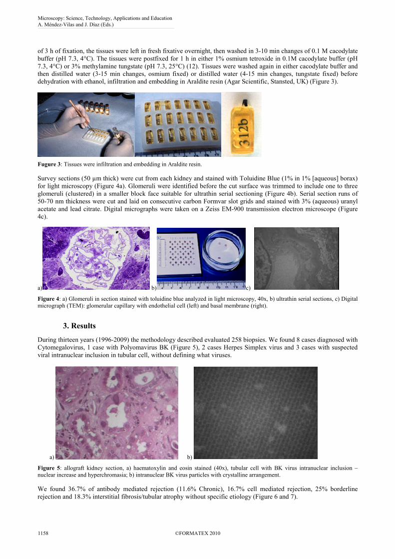
During thirteen years (1996-2009) the methodology described evaluated 258 biopsies. We found 8 cases diagnosed with
Cytomegalovirus, 1 case with Polyomavirus BK (Figure 5), 2 cases Herpes Simplex virus and 3 cases with suspected
viral intranuclear inclusion in tubular cell, without defining what viruses.
a) b)
Figure 5: allograft kidney section, a) haematoxylin and eosin stained (40x), tubular cell with BK virus intranuclear inclusion –
nuclear increase and hyperchromasia; b) intranuclear BK virus particles with crystalline arrangement.
We found 36.7% of antibody mediated rejection (11.6% Chronic), 16.7% cell mediated rejection, 25% borderline
rejection and 18.3% interstitial fibrosis/tubular atrophy without specific etiology (Figure 6 and 7).
Microscopy: Science, Technology, Applications and Education A. Méndez-Vilas and J. Díaz (Eds.)
1158 ©FORMATEX 2010
______________________________________________

a) b)
Figure 6: allograft kidney section, (40x) light microscopy a) C4d IHC diffuse positive; b) haematoxylin and eosin stained, illustrates
acute tubular necrosis. Diagnostic: acute humoral rejection I.
a) b)
c) d)
Figure 7: allograft kidney section, a) green masson`s trichrome stained, light microscopy (5x), illustrates glomerular sclerosis,
mesangial matrix increase and interstitial fibrosis; b) TEM, illustrates peritubular capillary basement membrane thickening, c)
positive IgM in vascular wall d) positive C3 in vascular wall. Diagnostic: chronic humoral rejection.
4. Discussion
A small needle biopsy of a renal allograft is easy to obtain and quite safe. When studied carefully, it can define the
nature of a rejection or a nonrejection process. Thus, it can prove quite valuable to direct therapy. Most determinations
can be made by standard hematoxylin and eosin, periodic acid Schiff, and trichrome stains. Immunofluorescence studies
are used primarily to look for peritubular capillary C4d deposition, which is indicative of a humoral rejection process,
or for evidence of recurrent disease, such as IgA glomerulopathy. Electron microscopy generally is not necessary to
evaluate a renal allograft biopsy, but it can be particularly helpful to confirm recurrent disease in a renal allograft,
especially IgA glomerulopathy and focal segmental glomerulosclerosis [8].
The gold standard for the diagnosis of rejection and for guiding patient management is the histological evaluation of
a renal allograft biopsy [9]. Over the past decades, morphological criteria of acute and chronic rejection have been
Microscopy: Science, Technology, Applications and Education A. Méndez-Vilas and J. Díaz (Eds.)
©FORMATEX 2010 1159
______________________________________________

defined, and classification schemes of rejection have been introduced, such as the CCTT and the Banff schemes [10,11].
They form the backbone for the clinical decision making, outcome studies and multicentre analyses of the efficacy of
new immunosuppressive drugs. However, all current classification schemes of renal allograft rejection have major
shortcomings. In particular, the proper identification of humoral rejection episodes after the immediate
posttransplantation period causes problems. The difficulties with identifying humoral rejection are due mainly to the
lack of typical morphological and immunohistochemical changes characterizing different forms of an antibody
response. Hence, antibody-mediated rejection episodes frequently remained undiagnosed and unclassified.
Consequently, nearly all acute rejection episodes have been classified as ‘cell mediated’. Tubulointerstitial rejection is a
prime example [12,13].
In multiple studies the biopsies changed the clinical diagnosis in 26% to 46% of cases and the therapy in 38% to 83%
of the cases [14]. Since signs of rejection can be focal, an adequate biopsy is critical. One study showed that sensitivity
of one core is 90% and of two cores, 99% [15].
This approach ensures an adequate light (HC and IHC), fluorescence and transmission electron microscopy
correlation at the present time. Using these complementary histological techniques evaluated according to Banff 2007
criteria is possible a precise differential diagnose to humoral or cellular rejection and others allograft dysfunctions,
which is a crucial end point for therapeutic success and better progosis.
Acknowledgements. This study was financial support by Fundação de Ensino e Pesquisa de Uberaba (FUNEPU), Fundação de
Amparo à Pesquisa de Minas Gerais (FAPEMIG), Conselho Nacional de Desenvolvimento Científico e Tecnológico (CNPq),
Coordenação de Aperfeiçoamento de Pessoal de Nível Superior (CAPES).
References
[1] Ravera M, Deferrari L, Vettoretti S, and Deferrari G. Importance of Blood Pressure Control in Chronic Kidney Disease. J Am Soc
Nephrol 17: S98–S103, 2006.
[2] Perico N, Benigni A and Remuzzi G. Present and future drug treatments for chronic kidney diseases: evolving targets in
renoprotection. Nature Reviews drug discovery. Volume 7; november 2008 p. 937.
[3] Goldfarb-Rumyantzev AS, Smith L, Shihab FS, Baird BC, Habib AN, Lin SJ, and Barenbaum LL. Role of Maintenance
Immunosuppressive Regimen in Kidney Transplant Outcome. Clin J Am Soc Nephrol 1: 563–574, 2006. doi:
10.2215/CJN.00640805.
[4] Humar A, Denny R, Matas AJ, Najarian JS. Graft and quality of life outcomes in older recipients of a kidney transplant. Exp Clin
Transplant. 2003 Dec;1(2):69-72.
[5] Dias ECA, Câmara NOS, Silva-Filho AP, Manfro RC. Monitorização Molecular da Rejeição de Transplantes Renais. J Bras
Nefrol Volume XXVII - nº 2 - Junho de 2005.
[6] Furness PN. Renal biopsy specimens. J Clin Pathol 2000 53: 433-438.
[7] Solez, K., Colvin, R. B., Racusen, L. C., Haas, M., Sis, B., Mengel, M., Halloran, P. F., Baldwin, W., Banfi, G., Collins, A. B.,
Cosio, F., David, D. S., Drachenberg, C., Einecke, G., Fogo, A. B., Gibson, I. W., Glotz, D., Iskandar, S. S., Kraus, E., Lerut, E.,
Mannon, R. B., Mihatsch, M., Nankivell, B. J., Nickeleit, V., Papadimitriou, J. C., Randhawa, P., Regele, H., Renaudin, K.,
Roberts, I., Seron, D., Smith, R. N. e Valente, M. Banff 07 classification of renal allograft pathology: updates and future
directions. Am J Transplant, v.8, n.4, Apr, p.753-60. 2008.
[8] Crosson JT. Transplant Rejection Under the Microscope. Transplantation Proceedings, 2007, 39, 662–666.
[9] Colvin RB. Renal transplant pathology. In: Jennette JC, Olson JC, Schwartz ML, Silva FG, eds. Heptinstall’s Pathology of the
Kidney, 5th edn. Lippincott-Raven, Philadelphia, PA, USA; 1998: 1409–1540.
[10] Colvin RB, Cohen AH, Saiontz C et al. Evaluation of pathologic criteria for acute renal allograft rejection: reproducibility,
sensitivity, and clinical correlation. J Am Soc Nephrol 1997; 8: 1930–1941.
[11] Racusen LC, Solez K, Colvin RB et al. The Banff 97 working classification of renal allograft pathology. Kidney Int 1999; 55:
713–723.
[12] Mauiyyedi S, Colvin RB. Pathology of kidney transplantation. In: Morris PJ, ed. Kidney Transplantation, 5th edn. W. B.
Saunders Co., Philadelphia, PA, USA; 2001: 343–391.
[13] Nickeleit V and Mihatsch MJ. Kidney transplants, antibodies and rejection: is C4d a magic marker? Nephrol Dial Transplant,
2003, 18: 2232–2239.
[14] Colvin RB, Nickeleit V: Renal Transplant Pathology in Jennette JC, Olson JJ, Schwartz MM, et al (eds): Heptinstall’s
Pathology of the Kidney, 6th ed. Philadelphia: Lippincott Williams & Wilkins; 2007, p 1347.
[15] Soroff JM, Vartanian RK, Olson JL, et al: Histopathological concordance of paired renal allograft biopsy cores: effect on the
diagnosis and management of acute rejection. Transplantation 60:1215, 1995
Microscopy: Science, Technology, Applications and Education A. Méndez-Vilas and J. Díaz (Eds.)
1160 ©FORMATEX 2010
______________________________________________