relationship between monitored elements and prescribed ... · ventilator setting modifications in...
TRANSCRIPT

Université de Montréal
Relationship between monitored elements and prescribed
ventilator setting modifications in critically ill children
by
Kay Allen Eddington, MD, FAAP
Faculté de Médecine
Mémoire présenté à la Faculté de Médecine
en vue de l’obtention du grade de Maîtrise
en Sciences biomédicales
option Recherche clinique
Mars, 2012
© Kay Allen Eddington, MD, FAAP, 2012

Université de Montréal
Faculté des études supérieures et postdoctorales
Ce mémoire intitulé :
Relationship between monitored elements and prescribed ventilator setting modifications in
critically ill children
Présenté par :
Kay Allen Eddington, MD, FAAP
a été évalué par un jury composé des personnes suivantes :
Yves Tremblay, président-rapporteur
Philippe Jouvet, directeur de recherche
Guillaume Emeriaud, co-directeur
Jean Paul Prauld, membre du jury

i
Résumé
Les pédiatres intensivistes ont plusieurs éléments disponibles pour guider leurs décisions
par rapport à la ventilation mécanique. Par contre, aucune étude prospective ne décrit les
éléments auxquels les intensivistes se réfèrent pour modifier les paramètres du respirateur.
Objectifs : Décrire la pratique actuelle de la modification des paramètres du respirateur aux
soins intensifs du CHU Sainte-Justine, un hôpital pédiatrique tertiaire.
Hypothèse : 80% des modifications des paramètres du respirateur influant sur l’épuration
du CO2 sont liées à l’analyse de la PCO2 ou du pH et 80% des modifications des paramètres
d’oxygénation sont liés à l’analyse de l’oxymétrie de pouls.
Méthodes : En se servant d’un logiciel de recueil de données, les soignants ont enregistré
un critère de décision primaire et tous les critères de décision secondaires menant à chaque
modification de paramètre du respirateur au moment même de la modification.
Résultats : Parmi les 194 modifications des paramètres du respirateur influant sur
l’épuration du CO2, faites chez vingts patients, 42.3% ±7.0% avaient pour critère primaire
la PCO2 ou le pH sanguin. Parmi les 41 modifications de la pression expiratoire positive et
les 813 modifications de la fraction d’oxygène inspirée, 34.1% ±14.5% et 84.5% ±2.5%
avaient pour critère primaire l’oxymétrie de pouls, respectivement.
Conclusion : Les médecins surestiment le rôle de la PCO2 et du pH sanguins et
sousestiment le rôle d’autres critères de décision dans la gestion de la ventilation
mécanique. L’amélioration de notre compréhension de la pratique courante devrait aider à
l’éboration des systèmes d’aide à la décision clinique en assistance respiratoire.

ii
Mots-clés : ventilation mécanique, pratique courante, soins intensifs pédiatriques

iii
Abstract
Pediatric intensivists have a multiplicity of elements available to guide them in mechanical
ventilator decision-making; however, no prospective studies describe which elements
intensivists currently use to make ventilator setting changes.
Objectives: We describe the current practice of ventilator setting modification in the
intensive care unit at Sainte-Justine Hospital, a tertiary care pediatric hospital.
Hypothesis: Eighty percent of ventilator settings affecting carbon dioxide clearance are
based on the PCO2 or pH while eighty percent of settings affecting oxygenation are based
on pulse oximetry.
Methods: Caregivers recorded the primary element and any secondary elements leading to
a ventilator setting change at the time of the change via a custom-designed data gathering
software.
Results: We included twenty patients. Of a combined 194 changes affecting CO2 clearance,
42.3% ±7.0% were in reference to blood PCO2 or pH. Of forty-one changes to positive
end-expiratory pressure, 34.1% ±14.5% were in reference to pulse oximetry, as were 84.5%
±2.5% of the 813 changes to the fraction of inspired oxygen.
Conclusion: Physicians over-estimate the role of blood pH and PCO2 in their ventilator
management, while under-estimating the role of other elements. Improving our
understanding of current practice patterns can help in the development of systems to aid in
clinical decision-making in mechanical ventilation, improving clinical outcomes.
Keywords : mechanical ventilation, practice patterns, pediatric intensive care

iv
Table of Contents
Introduction………………………………………………………. 1
Preliminary research....……………………………………………4
Review of the literature...……………………………………4
Two preliminary surveys....…………………………………5
Study Objectives..………………………………………………….8
Methods……………………………………………..…………….10
Study design……………………………………………...…10
Data collection………………………………………………12
Sample size………………………………………………….15
Results…………………………………………………………….16
Discussion…………………………………………………………21
Conclusions…………………………………………………….…28
References………………………………………………………...30
List of abbreviations……………………………………………...33

v
List of Figures
Figure 1: Patient Enrolment..........……...........................................................................34
List of Tables
Table 1: Patient Characteristics.......................................................................................35
Table 2: Modes of Ventilation Employed at Inclusion....................................................36
Table 3: Proportion of all changes of a given
Ventilator setting due to a given primary element...........................................................37
Table 4: Proportion of ventilator setting changes
in which secondary elements were identified....................................................................38
Table 5: Categorization of elements which could
be incorporated into automatically adjusting
ventilator modes based on current technology.................................................................39
Appendices
Appendix 1: Preliminary survey number 1……………………………………………….i
Appendix 2: Preliminary survey number 2………………………………………………ii
Appendix 3 : Screen shot of the data-gathering software………………………………iii

vi
To Mom and Dad
And to Erin, Marianna, Emily and Amberlee

vii
Special Thanks
To my family, friends; to my co-fellows and residents; to the many nurses and
respiratory therapists who participated in this study; to my patients; to my mentors (who
have become my friends); and especially to Philippe for his guidance throughout this
process, Thank you! Je vous remercie!

Introduction
Invasive mechanical ventilation in children is managed by attending physicians,
physicians in training (fellows or residents), and respiratory therapists. [1] Little evidence
is available to describe how these caregivers manage invasive mechanical ventilation on a
day to day basis, and no published data exists describing the relationship between patient
information available to the caregiver and how that information is processed and ultimately
leads to the prescription of setting changes on the mechanical ventilator.
Caregivers have multiple variables to consider when implementing or adjusting a
ventilation strategy, including the patient’s age, weight, chronic illness, acute illness, level
of sedation, physical exam findings, chest radiography findings, blood gas analysis, and
non-invasive monitoring. Once a ventilation strategy is chosen and a mode of ventilation
and settings are prescribed, changes in any of above factors can potentially lead to changes
in the settings, the mode, or even the overall strategy.
It has been established in a recent point prevalence study that significant variability exists
in ventilator settings currently being used in children with acute lung injury (ALI),
including settings outside of current recommendations. [2] A survey of pediatric
intensivists from Canada and Europe has also revealed wide variability in acceptable
physiologic elements (especially respiratory rate, tidal volume, and PCO2) during the
weaning phase of mechanical ventilation. [3] The presence of this variability in ventilator
settings and in acceptable physiologic elements strongly suggests that variability also exist

2
in which elements caregivers consider most important when prescribing changes to
ventilator settings, not only in the weaning phase, but throughout the patient’s entire course
of mechanical ventilation.
This practice variability may be leading to sub optimal patient outcomes and is a barrier to
the evaluation of different ventilation strategies and new modes of ventilation, because
there is no clearly described “best practice” to use as a standard of comparison. Studying
and describing how basic patient information, physiologic elements, and other information
at the physician’s disposition lead to ventilator setting changes has the potential of bringing
about improved patient outcomes if a “best practice” can be described, accepted and
implemented within the pediatric intensive care community. It may simplify patient care
by focusing physician’s attention on the elements which matter the most in decision-
making regarding mechanical ventilation. Furthermore, it may allow for the development
of computer assisted decision-making protocols (software packages in which caregivers
enter a patient’s pertinent clinical information and which, in turn, propose a management
plan adapted for that patient’s specific situation, based on a pre-defined protocol) and/or
automatically adjusting ventilator modes (modes of ventilation which automatically adjust
settings based on protocols integrated into the ventilator’s computer and relying on
information gathered directly from the patient via patient monitoring devices integrated
onto to the ventilator). Such protocols and automatic ventilator modes have been
developed and could potentially simultaneously improve outcomes and free caregivers to
tend to other aspects of patient care, but they have not enjoyed wide spread acceptance,

3
perhaps in part because data concerning current practice patterns are lacking. [4]
Physicians may be understandably reluctant to turn decision making over to a protocol or
an automatic ventilator in an area where they do not fully understand their own decision
making process.
The following is a pilot study which identifies the key elements among the multiplicity of
invasive and non-invasive monitored data at a caregiver’s disposition and describes the
relationships between those elements and the specific ventilator setting changes to which
they led, as reported by the caregiver at the time the change was prescribed.

Preliminary research
Review of the literature
Prior to beginning this study, I conducted an extensive literature search primarily
using the US National Library of Medicine website Pubmed.com. Broad search terms were
employed, aimed at identifying studies on ventilator settings, monitoring techniques, and
ventilation protocols. Examples include “mechanical ventilator settings”, “ventilation
wean”, “ventilation monitoring”, “ventilation protocol”, “pulse oximetry” along with
multiple permutations of these terms, including adding “pediatric” to each term. Many
studies have been published which establish the validity of specific monitoring techniques
(both invasive and non-invasive). [5-6] And there are some studies which attempt to
establish extubation readiness criteria in children and weaning protocols in the event that a
patient fails extubation readiness tests. [7-8] Other studies aim to provide guidelines for
ventilator settings in specific disease entities, especially guidelines to establish the role of
lung protective strategies (by means of low set tidal volumes) for children with acute
respiratory distress syndrome (ARDS). [9-10] Although there is a great deal of research
activity on the subject of pediatric ventilation in general, there are no published studies
which specifically evaluate which monitored elements caregivers rely on as they make
decisions to change a patient’s ventilator settings. The studies by Santschi et al, as well as
other studies on practice variability in pediatric ventilation, which have been published
since the inception of the present study, demonstrate a growing interest in the pediatric

5
intensive care community in understanding the current state of the art and improving our
management of invasively ventilated children.[2-3,11] The present study provides valuable
information on the clinical practice of the center in which it was carried out and provides a
model upon which large-scale, multi-center studies can be carried out in order to
understand the current clinical decision-making processes and ultimately improve upon
them.
Two preliminary surveys
In the pediatric intensive care community, it is taken for granted that the majority of
changes to the ventilator settings which control oxygenation (the fraction of inspired
oxygen and the positive end-expiratory pressure) are based almost exclusively on pulse
oximetry monitoring, and that the majority of modifications to the remaining principle
ventilator settings (respiratory rate, set tidal volume, positive inspiratory pressure, and
pressure support), though they are more complex, are based primarily on arterial blood pH,
arterial partial pressure of carbon dioxide (PaCO2), or a surrogate measure of the latter. In
order to better understand local perceptions of practice patterns, and to generate necessary
data to prepare for this study, we performed two informal surveys amongst the pediatric
intensivists, neonatologists, pediatric intensive care fellows, and neonatology fellows of
Sainte-Justine Hospital. (See appendices 1 and 2) In the first survey, participants were
asked to identify the elements they considered when increasing or decreasing the imposed
respiratory rate, positive inspiratory pressure or tidal volume (depending on the mode of

6
ventilation), positive end-expiratory pressure, and fraction of inspired oxygen
(respectively). All questions were open-ended. The survey was distributed via e-mail and
in paper format to twenty individuals, and eight surveys were completed (forty percent
response rate). The results of the first survey were reviewed, and similar responses were
combined. The second survey included the elements identified in the first survey, and
asked respondents to estimate the frequency (in terms of a percentage) with which a
particular element was included in the decision-making process amongst all of their
prescriptions to change a particular ventilator setting. (e.g., Among all of your
prescriptions to change the imposed respiratory rate, what percentage are based on the
partial pressure of carbon dioxide from an arterial blood gas?) The format required that
participants write a percentage for each of seventeen questions. Again, the survey was
distributed to twenty individuals and thirteen individuals responded (sixty-five percent
response rate).
The results of these surveys were consistent with the current perceptions of clinical practice
in the pediatric intensive care community. The results of the first survey were instrumental
in the design of a computer program used as the primary data gathering utility for this
project. The results of the second survey—specifically, that seventy-eight percent of
changes to the imposed rate, positive inspiratory pressure, set tidal volume, and pressure
support are made in reference to arterial blood pH or arterial partial pressure or carbon
dioxide and that eighty-one percent of changes to the fraction of inspired oxygen and

7
positive end-expiratory pressure are made in reference to pulse oximetry monitoring—
served as the basis for the hypothesis of this study.

8
Study Objectives
The purpose of this study is to describe the current practice of ventilation
management at Sainte-Justine Hospital, with respect to which elements caregivers rely on
in their decisions to change the principle ventilator settings, namely, set respiratory rate,
tidal volume, positive inspiratory pressure, positive end-expiratory pressure, pressure
support and the fraction of inspired oxygen. This study serves as a pilot for a larger
multicenter study. A novel software product was developed for this study in order to
survey caregivers in a minimally labor intensive fashion at the time a decision was made to
change a ventilator setting. In part, this study, as does any pilot study, serves as learning
experience to improve our processes and tools, including our software product, to refine our
protocol, and to identify pitfalls of the study methods prior to initiating a large-scale study.
As such, the direct interpretation of the results of this study are limited, especially
considering the presumed practice differences that exist from center to center and from
region to region. Nevertheless, as previously mentioned, there are no prior published
studies elucidating this relationship, so this publication will directly contribute to the
knowledge base of the subject, and will directly inform the practitioners at Sainte-Justine
Hospital—granted, in a limited fashion—of their practice.
There are several applications of the data generated from this study, and clearly to a
greater extent the data anticipated from the multicenter study. First, and most simply, this
data will call the pediatric critical care community’s attention to their practice patterns and
possibly inform caregivers of significant differences between their actual practice and
perceived practice. There is a great deal of information gathering and processing in an
ongoing fashion implicated in the care of critically ill children. Obtaining this information
can consume time and resources and may directly negatively impact the patient (e.g. pain,

9
blood loss, and risks associated with indwelling angio catheters for obtaining blood gases;
dead space introduced into the ventilator circuit for continuous end-tidal carbon dioxide
monitoring; radiation from chest radiography, etc). Although the benefits of obtaining this
information are generally perceived to outweigh the injury, the results of this study may
provide a better understanding of how we actually use that information and may change
clinical practice by helping us more appropriately select which information we gather.
Second, an understanding our practice patterns based on evidence will help us educate
young physicians about mechanical ventilation more accurately. Third, this study is a first
and necessary step toward establishing standards and guidelines in pediatric mechanical
ventilation, which could ultimately improve patient outcomes. And lastly, the data
obtained from this study may contribute to establishing mechanical ventilation protocols,
which could be integrated into computer assisted decision-making software and potentially
into automatically adjusting ventilator modes, which could simplify mechanical ventilation,
freeing the physician to focus on other areas of patient care.

Methods
Study design
This was a prospective observational cohort study which enrolled critically ill
children admitted to the pediatric intensive care unit (PICU) of Sainte-Justine Hospital, a
free-standing, tertiary care hospital for woman and children, affiliated with the University
of Montreal, Montreal, Quebec, Canada. Patient enrollment took place from January 2010
to January 2011 with follow up data gathered until June 2011.
The research ethics committee of Sainte- Justine Hospital approved this study, and the need
to obtain informed consent from patients or their guardians was waived due to the strictly
observational nature of the study design.
During the study period, all consecutive critically ill children were considered eligible to
participate in the study, regardless of their indication for mechanical ventilation (whether
pulmonary, neurologic, etc) and regardless of the mode of ventilation, if they met the
following inclusion criteria: 1) presence of invasive mechanical ventilation (via
endotracheal tube or tracheostomy) and 2) expected duration of mechanical ventilation
greater than three days. Expected duration of mechanical ventilation greater than three
days was determined by the presence of one of the following three criteria that were
demonstrated as risk factors for prolonged mechanical ventilation [12]: a) mean airway
pressure greater than or equal to thirteen centimeters of water maintained for at least sixty

11
minutes at any time in the first twenty-four hours of mechanical ventilation, b) Pediatric
Risk of Mortality score (PRISM) greater than or equal to ten on the day of PICU admission,
or c) continuous infusion of a sedating medicine for any amount of time during the first
twenty-four hours of mechanical ventilation. [12-13] Patients were excluded if they met
any of the following criteria: 1) presence of a “Do not resuscitate” or “do not reintubate”
order in the chart, 2) suspected or confirmed brain death, 3) history of mechanical
ventilation (invasive or non invasive) at home, 4) patient ventilated with a machine other
than the Servo-i (Maquet GmbH & Co. KG, Rastatt, Germany), 5) a data gathering
computer required for the study was not available, or 6) permission was not granted by
treating physician. (See figure 1) Of note, only patients ventilated with a Servo-I ventilator
were included in the study, because that ventilator allows simple recording of the internal
ventilator log via a memory card. That particular ventilator may be equipped to ventilate in
NAVA (Neurally Adjusted Venilatory Assist) mode, a mode for spontaneously breathing
patients in which breaths are triggered by and the inspiratory pressure delivered is modified
as a function of the electrical activity of the patient’s diaphragm as detected by a special
nasogastric tube. While we were in possession of two ventilators so equipped, the use of
NAVA mode at Sainte-Justine Hospital at the time of this study was limited to very brief
periods for research purposes, and always under direct observation by a physician.
Therefore, while there were no specific exclusion criteria for patients using NAVA, those
patients were de facto excluded. Furthermore, provisions were made to suspend the study,
should any patients be transitioned to high frequency oscillatory ventilation, and to resume

12
the study when the patients were returned to conventional ventilation; however, those
provisions were never necessary.
Data collection
Patient characteristics including demographic data, diagnosis, severity scores,
clinical data at inclusion and outcomes were collected from the charts. When a patient was
selected for the study, a laptop computer was installed at the bedside. A custom-designed
computer program was used to record each ventilator setting change along with one
primary element which prompted the caregiver to prescribe the setting change as well as an
unlimited number of secondary elements included in the caregiver’s decision-making
process. (See appendix 3.) Caregivers filled out this electronic survey while at the
patient’s bedside at the time of any ventilator setting modification. They had the option of
selecting from a list of pre-determined ventilator setting changes and monitored elements
and/or manually entering changes or monitored elements not found in the lists. The list of
ventilator setting changes included the increase or decrease of the fraction of inspired
oxygen (FiO2), imposed respiratory rate (RR), tidal volume (Vt), positive inspiratory
pressure (PIP), positive end-expiratory pressure (PEEP) and pressure support (PS). The list
of monitored elements included pulse oximetry (SpO2), respiratory therapy or endotracheal
tube suctioning, blood pH, arterial partial pressure of carbon dioxide (PaCO2), arterial
partial pressure of oxygen (PaO2), measured tidal volume, minute ventilation, end-tidal

13
carbon dioxide (EtCO2), transcutaneous carbon dioxide (TcCO2), chest radiography
findings (CXR), and physical exam. If chest radiography findings or physical exam were
selected, addition information could be provided to specify the findings. The role of the
caregiver responsible for the setting change (nurse, respiratory therapist, resident, fellow or
attending physician) was also recorded for each change.
Data collection was initially planned to proceed from study inclusion until
termination of invasive mechanical ventilation; however, the first patient included remained
ventilated for far greater than one month. She remained on minimal, stable settings for
several days, but could not tolerate extubation. After thirty days of inclusion, the decision
was made to modify the protocol to end data collection at the termination of mechanical
ventilation or at twenty-eight days after inclusion. Data capture for all remaining patients
ended at the termination of invasive mechanical ventilation.
Multiple training sessions were held with the various groups of caregivers to inform them
of the study and to instruct them on how to use the data collection software. Sessions with
nurses and respiratory therapists were approximately fifteen minutes long, while the
sessions with physicians were typically thirty minutes long. Physicians tended to foresee
more complicated clinical scenarios and asked more probing questions during their
sessions.

14
The endpoint of this study was the change to the ventilator setting in reference to the
primary and secondary elements included in the decision-making process. As our goal was
to identify which elements were used to guide changes in which ventilator settings,
determining acceptable limits of the various monitored elements and determining the
magnitude of the resultant setting changes were beyond the scope of our study. Therefore,
the relationship between monitored elements and ventilator setting changes are expressed
as the percentage of ventilator setting changes for which a particular monitored element
played a role.
During endotracheal tube suctioning, chest physical therapy, or other patient manipulation,
temporary changes to a variety of ventilator settings may be made as a matter of routine,
most notably to the fraction of inspired oxygen. All such temporary changes were recorded
but were analyzed separately from other ventilator setting changes.
Caregiver compliance with the protocol was estimated by recording the internal ventilator
log of ventilator setting modifications using a compact flash reader connected to the
ventilator (Servo-I, Maquet GmbH & Co. KG, Rastatt, Germany). The ventilator setting
changes recorded by the ventilator log were then matched with the ventilator setting
changes recorded by caregivers. The number of setting changes in the ventilator log with a
corresponding entry in the study software was divided by the total number of setting
changes in the ventilator log to yield a percent compliance. The compliance was measured
in two (ten percent) of the patients.

15
Sample size
Based on an average prevalence of monitored elements leading to ventilator setting
prescription changes equal to fifty percent (P0) and a ninety-five percent confidence
interval, measurement of 384 ventilator changes would be required to reach a level of
precision of plus or minus five percent around the estimate. Also, based on an expected
mean length of mechanical ventilation of three days and an average of seven ventilator
setting changes per day, a sample-size of twenty subjects (approx. 420 measurements) was
targeted. The average of seven ventilator changes per day was obtained by a reviewing the
data of a prior study carried out at Sainte-Justine Hospital amongst a similar patient
population. [12]

16
Results
Twenty patients were included, with both medical and surgical indications for
admission to the intensive care unit, including post operative patients with congenital heart
disease. The patient ages ranged from two days to sixteen years (mean 2.4 years, standard
deviation 2.0). Half were boys. The mean length of PICU stay was twenty-nine days
(standard deviation sixty-nine days). Of note, one patient remained admitted to the PICU
upon completion of the follow-up period; however, he was no longer mechanically
ventilated. The mean duration of invasive ventilation was twenty-eight days, with a
standard deviation of seventy days. The twenty-eight day mortality was fifteen percent. The
mean duration of electronic capture of ventilator setting changes was six days. There were
six patients admitted to the PICU for primary respiratory diagnoses, four for post-operative
care after surgery for congenital cardiopathy, four for sepsis, and one each for liver
transplant, multiple trauma, meningitis, major burns, hemolytic-uremic syndrome, and
acute lymphoblastic leukemia (with sepsis). The initial modes of ventilation at the time of
inclusion were as follows: pressure control (without pressure support), seven patients;
pressure regulated volume control with pressure support, seven patients; pressure control
with pressure support, four patients; and volume control with pressure support, two
patients. The mode of ventilation did change occasionally in some patients. Of note, use of
pressure support ventilation at Sainte-Justine Hospital is routine practice as a test for
extubation readiness and as the final mode of ventilation just prior to extubation; however,

17
even spontaneously breathing patients are not routinely initially ventilated with that mode.
Furthermore, patients are routinely weaned to pressure support ventilation but very rarely
are settings changed when a patient is using that mode (i.e. patients deemed successful are
extubated rather than decreasing their settings, and patients deemed unsuccessful are placed
on a different mode of ventilation). Therefore, very little data from this study was captured
from patients being actively ventilated in pressure support mode. (Table 1)
Eighty caregivers participated in the study; forty-three nurses, fifteen respiratory therapists,
eight residents, six fellows, and eight attending physicians. The median caregiver
compliance with the protocol was 74.5% with a median duration of observation of 16.5
days per patient. No trends in compliance were found between the different shifts nor over
the days of observation for each patient. The median number of setting changes per patient
per day was 19.6 with twenty-fifth and seventy-fifth percentiles of 13.3 and 26.6,
respectively. Excluding changes to the fraction of inspired oxygen, the median number of
setting changes per patient per day was 2.5 with twenty-fifth and seventy-fifth percentiles
of 2.0 and 4.3, respectively.
After exclusion of temporary changes made during endotracheal tube aspiration and other
patient manipulation, pulse oximetry was identified as the primary monitored element for
84.5% ( ±3.0%) of changes to the fraction of inspired oxygen (Table 2). Physical exam
(1.0 ±0.4 %) and arterial partial pressure of oxygen (0.9 ±0.4 %) were also identified as
elements influencing FiO2 changes, but in much smaller proportions. Pulse oximetry was
18
identified as the primary reason for 34.1% ±14.5% of changes to positive end-expiratory
pressure, followed by physical exam (12.2% ±10.0%), arterial partial pressure of oxygen
(9.8% ±9.1%), and chest radiography findings (9.8% ±9.1%). One notion which was not
included in the list of factors leading to ventilator setting changes, but which was expressed
by several participants via free text was the idea that a patient was expected to tolerate the
step-wise weaning of a given setting. Participants seemed to struggle to identify any given
monitored element which they were following; rather, they expected to be able to
successfully wean a given setting over time without significant changes to ANY of the
monitored elements or to the physical exam. These changes tended to be discussed on
morning rounds, mentioned in the daily progress notes, and carried out over a number of
days. A common example was the plan to wean the PEEP by one centimeter of water per
day until a setting of five was achieved. I refer to this notion as planned, step-wise
weaning. Planned, step-wide weaning and measured tidal volume were identified as
primary reasons for 7.3% ±8.0% of PEEP modifications, each.
The blood partial pressure of carbon dioxide (pCO2)—including arterial, capillary and
venous blood—was only identified as the primary factor for 47.9% ±11.5% of changes to
the imposed respiratory rate, with blood pH (9.9% ±6.5%) and increase in patient work of
breathing (7.0% ±5.6% each) as the next most frequently cited factors. The pCO2 was
similarly influential for changes to the tidal volume at 47.4% ±21.9%, followed by the
patients’ spontaneous respiratory rate (21.1% ±17.0%), blood pH (5.3% ±9.2%), and the
measured inspiratory pressure (5.3% ±9.2%). pCO2 was cited much less frequently for

19
changes to the positive inspiratory pressure (23.9% ±8.1), with measured tidal volume
being the most commonly recorded primary monitored factor at 31.8% ±9.0%. Planned,
stepwise weaning (9.1% ±5.3%) and pulse oximetry (8.0% ±5.0%) were also significant
factors. Blood pH, pCO2, spontaneous respiratory rate, and minute ventilation were all
cited as primary factors for 2-5% of tidal volume changes. pCO2 was also the most
commonly recorded factor influencing changes to the level of pressure support at 31.3%
±21.2%, followed by increased patient work of breathing (12.5% ±14.7%).
Though seven patients (thirty-five percent) had EtCO2 monitors at some point during the
study, only two decisions to modify settings were based on that criterion, one for the set
respiratory rate, 1.2% ±2.4% of RR changes, and one for the PIP, 0.9% ±1.7% of those
changes.
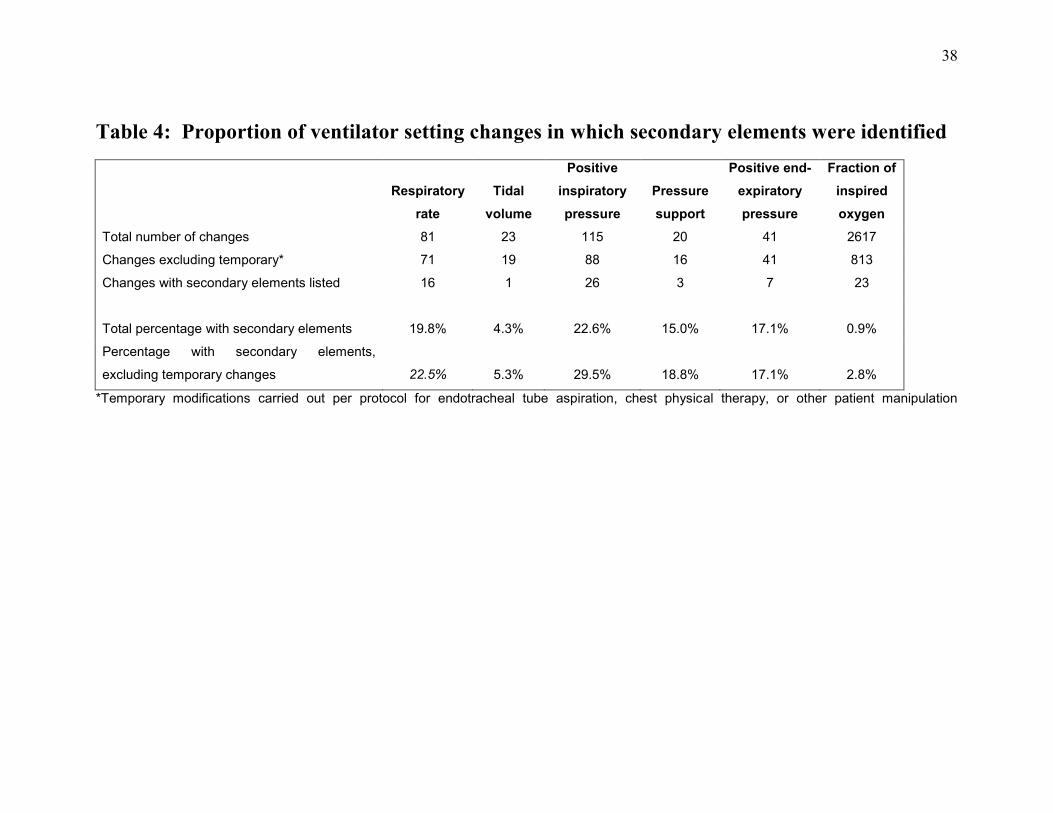
For the different ventilator settings, there were between 3.4% and 16.1% of changes for
which respondents recorded a setting change, but did not record their motivation for
making the change. The frequency with which respondents identified secondary monitored
elements assuming a primary element was identified varied as follows: imposed respiratory
rate 19.8%, tidal volume 4.3%, positive inspiratory pressure 22.6%, pressure support
15.0%, positive end-expiratory pressure 17.1%, and fraction of inspired oxygen 0.9%.
(Table 3)
20
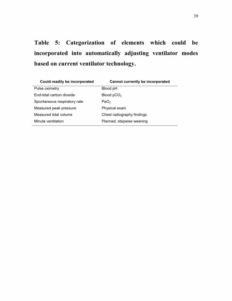
In order to estimate the potential gains of employing either computer assisted ventilation
management protocols or ventilator modes with automatic setting adjustment, we
categorized the reasoning for any given setting change (including the primary and all
secondary elements) as readily incorporatable into an automatic protocol based on current
technology or non-incorporatable. For this analysis, an element is considered incorporatable
into an automatic protocol if data can be gathered from the patient and digitized without
further human intervention for data entry via equipment which is commonplace in most
pediatric intensive care units. The list of elements considered as readily incorporatable into
an automatic protocol is shown in Table 4. When multiple elements were reported for one
ventilator setting change, if any one element was considered non-readily incorporatable into
an automatic protocol, that ventilator setting change was considered to be based on non-
incorporatable elements. Temporary changes for respiratory therapy and suctioning were
again excluded, as were survey entries in which no reason for the setting change was
reported.
Of the sixty changes to the imposed respiratory rate, fourteen (23.3%) were based on
elements which are potentially incorporatable into automatic protocols. Such was also the
case for five (33.3%) of the fifteen tidal volume changes, forty-seven (56%) of the eighty-
four positive inspiratory pressure changes, five (41.7%) of the twelve pressure support
changes, nineteen (51.4%) of thirty-seven positive end-expiratory pressure changes, and
659 (97.1%) of 679 fraction of inspired oxygen changes.

21
Discussion
Contrary to the results of our preliminary surveys in which caregivers estimated that
roughly eighty percent of their prescribed setting changes to the imposed respiratory rate,
positive inspiratory pressure, tidal volume and pressure support were motivated by pCO2 or
blood pH, those elements were only cited as primary elements in 35.1% of setting changes
when caregivers recorded their motivation at the time the prescription was made. The
common perception among pediatric intensivists is that most changes to the settings listed
above are made either because the level of carbon dioxide or pH is outside of an acceptable
target range or because targets are being met, and physicians feel that the patient could be
weaned without those elements going out of target range. But according to this study, such
is only the case in roughly a third of the setting changes we make. Rather, other
considerations are driving us to modify ventilator settings, specifically peak or plateau
pressures, measured tidal volumes, and spontaneous respiratory rates. If we use the
presence or absence of recorded secondary factors as an indirect measure of the complexity
of decision making, the majority of the prescriptions made during this study were lower
complexity, as 80.7 percent of the changes to respiratory rate, tidal volume, positive
inspiratory pressure and pressure support were made in reference to a single primary
monitored element.
These results provide new insight into our daily practice and into our ability to correctly
perceive our daily practice. The imposed respiratory rate, tidal volume, positive inspiratory

22
pressure and pressure support are the settings that primarily determine a patient’s partial
pressure of carbon dioxide and, along with metabolic and renal factors, the patient’s blood
pH. But those settings are also important in determining patient synchrony and comfort on
the ventilator and determining the risk of lung injury secondary to mechanical ventilation.
Perhaps, then, when questioned retrospectively, pediatric intensivists lend greater
importance to the role of those settings in maintaining carbon dioxide level and blood pH,
than to the other outcomes determined by those settings, leading to a very significant recall
bias. The complexity (or simplicity) of our decision-making may also be difficult for
physicians to perceive accurately. With eighty percent of changes to these four ventilator
settings based on a single element, it seems feasible to develop guidelines and ventilation
strategy protocols which could reduce the practice variability both within and across
centers, allowing the pediatric intensive care community to measure pertinent outcomes and
determine a best practice. When considering secondary elements, and categorizing the
monitored elements as incorporatable or non-incorporatable into automatic ventilator
modes as described in Table 4, 41.5% of the setting changes to imposed respiratory rate,
positive inspiratory pressure, tidal volume and pressure support could have been managed
by use of automatic ventilator modes using readily available technology. Of note, end-tidal
carbon dioxide monitoring is not standard practice in our ICU. Seven of the twenty patients
had end-tidal carbon dioxide monitors attached during our observation period, and only two
setting changes were made primarily in response to their readings. This could be
interpreted in two ways: either ventilator setting changes are not often based on EtCO2
monitoring because few patients have the monitor at Sainte-Justine Hospital, or,

23
conversely, physicians seldom request that the monitor be installed, because it does not
influence their clinical decision-making. It remains to be seen if other centers where end-
tidal carbon dioxide monitors are used as a matter of routine for all intubated patients see
higher proportions of prescribed ventilator changes based on that technology.
While our results for imposed respiratory rate, tidal volume, etc, diverged from physician’s
perceptions, the results for the fractions of inspired oxygen were consistent with perceived
clinical experience. Changes to the fraction of inspired oxygen were the most numerous,
were almost uniformly associated with pulse oximetry (84.5% of changes), and were the
least complex. Less than one percent of changes were associated with multiple elements.
Lack of complexity is demonstrated by the fact that the routine management of the fraction
of inspired oxygen has been largely delegated to bedside nurses, who are instructed to
adjust it in order to maintain target pulse oximetry readings and in accordance with the
patient’s skin coloration and general status. Our findings show that 97.1% of fraction of
inspired oxygen setting changes in our center could potentially be managed by
automatically adjusting ventilator modes. Such technology is currently under development,
and is of particular interest in neonatal intensive care units. [14]
Changes to positive end-expiratory pressure were made with reference to various elements;
however, multiple elements were only recorded for 17.1% of modifications. In other
words, several elements were tracked, but usually, only one of these elements became
clinically significant at a time. These findings may be explained by considering the clinical

24
scenarios that might develop during the course of mechanical ventilation and that influence
the preferred level of PEEP (although the “preferred” level of PEEP for some situations
remains controversial). For example, the development of a pneumothorax (diagnosed on
physical exam or by chest radiography) may lead physicians to decrease the PEEP in a
patient who might otherwise benefit from higher levels, or a patient with poor pulmonary
compliance who develops increasing levels of CO2, and who is experiencing very high
inspiratory pressures may require a reduction in PEEP to improve CO2 removal via a
relative increase in PIP without actually increasing the inspiratory pressure. The
development of bronchospasm might lead some physicians to increase PEEP, some to
decrease PEEP, and others may not adjust PEEP in light of bronchospasm. Pulse oximetry
was by far the most frequently cited element at about one third of the changes, while
physical exam, arterial partial pressure of oxygen, chest radiography findings, planned,
step-wise weaning and measured tidal volume were all identified at similar frequencies
(7.3% to 12.2%). Assuming that an adaptable weaning plan can be incorporated into
computer assisted decision-making protocols and/or automatically adjusting ventilator
modes, about half of the changes to positive end-expiratory pressure which we observed
could have been managed by such methods. [4]
Of note, heart-lung interactions were never specifically reported as a reason to modify the
level of positive end-expiratory pressure. This could be explained first by assuming that
when physicians cited the physical exam as a reason for making a change, that heart-lung
interactions were considered as part of that assessment, and second, by recalling that we

25
only observed changes to ventilator settings as opposed to initial settings and/or magnitude
of change. Therefore, patients whose initial assessment included cardiovascular reasons to
limit positive end-expiratory pressure may have had their level modified as a function of
pulse oximetry and the other elements mentioned above, but with smaller increments of
change and lower total values throughout their entire course of ventilation than those
without special heart-lung considerations.
As one would expect, by selecting intubated patients with a projected duration of
mechanical ventilation of greater and three days, the patients included in this study had
longer lengths of stay, higher severity of illness scores (PRISM and PELOD), and a higher
mortality rate than the average for our ICU. [13,15] One may also suppose that the
ventilation management of these patients is more complex than the average pediatric ICU
patient.
By virtue of the survey method we used, we were able to achieve higher response rates than
we would have expected using a survey given periodically, after a call night, for example.
One of the strengths of this technique is that it reduces caregivers’ recall bias and
eliminates the bias created by allowing caregivers to observe the results of the ventilator
changes they prescribe prior to filling out the survey. Special care was also taken in the
design of the software to ensure ease of use and a minimal amount of computer navigation
in an effort to increase compliance.
26
Caution should be used when interpreting our data because it represents the practice of a
single center in an area of medicine with few standards or guidelines. In addition, we
remind the reader that we only identified monitored elements that led to ventilator changes.
If the default position of a ventilation management plan was to change (wean) a setting, and
a monitored parameter prevented that change, we did not capture such information. Also,
as is the case with any study attempting to understand clinical reasoning, our observations
are limited to what caregivers reported, and may therefore be inherently oversimplified.
For standards and guidelines to be established, data must be gathered from multiple centers
representative of pediatric intensive care as a whole and should represent the full range of
mechanical ventilation. To that end, in preparing for a multi-centered study, there are
modifications to this pilot study which would improve the quality of data. First, for ease of
patient inclusion by a single researcher, we allowed data capture after several hours of
mechanical ventilation. In doing so, we failed to capture the acute—and in terms of
ventilator settings, rapidly escalating—phase of ventilation some patients. A future, multi-
center study should attempt to initiate data capture from the time of intubation, or from the
time of transfer to the PICU. Second, controversy surrounding the management of the
fraction of inspired oxygen is much less than the management of the other settings. While
the target of oxygen saturation may be debated in particular situations, most agree that the
adjustment up or down of the fraction of inspired oxygen is almost exclusively based on
pulse oximetry. This study confirmed that assumption, and most PICUs have a policy
delegating that management to nurses with specific guidelines, so further studies of that
particular setting could be omitted from future studies. Finally, we gathered data regarding
27
the role of the caregiver prescribing each setting change; however, the number of settings
changes we gathered were far too small to perform any sort of analysis stratifying by
caregiver role. There may be significant practice differences between physicians in
training, attending physicians, and respiratory therapists, and elucidating those differences
may prove particularly beneficial to ventilator management education.

Conclusions
This pilot study demonstrates that observation and surveying of clinical decision
making in mechanical ventilation at the time decisions are being made is feasible and that
the results may lead to a more accurate and objective understanding of our own practice.
The methods used are easily transportable to other intensive care units and do not require
special equipment, other than a computer. Staff training sessions were brief, and
compliance was high. The results obtained using the bedside computerized survey varied
greatly from results obtained from a preliminary paper/e-mail survey, which demonstrates
the significance of recall bias in studies of practice patterns and the benefit of using a
methodology similar to ours.
The data obtained in this study reveal that physicians in our center do not use end tidal
pCO2 to modify ventilator settings and over-estimate the role of blood pH and carbon
dioxide in their ventilator management, while the role of elements such as the patient’s
spontaneous respiratory rate, measured tidal volume, and peak pressure were under-
estimated. This discrepancy between perceived practice patterns and actual practice
patterns and the relatively low-complexity of the decisions made may be a reflection of
physicians’ priorities with regards to ventilation; specifically, assuring an acceptable blood
pH and carbon dioxide level takes priority over limiting the risk of secondary pulmonary
lesions due to mechanical ventilation, maximizing patient synchrony with the ventilator,
and assuring patient comfort. Caregivers are more concerned about assuring adequate
blood pH and carbon dioxide, so they believe the majority of their prescriptions to change

29
ventilator setting are to achieve that goal, when in fact, even among the sickest patients,
those goals are often met, but “fine tuning” to meet secondary objectives leads to the
majority of setting changes.
Roughly half of the changes to the positive end-expiratory pressure, forty percent of the
changes to the imposed respiratory rate, tidal volume, positive inspiratory pressure, and
pressure support, and ninety-seven percent of changes to the fraction of inspired oxygen
which we observed could have potentially be managed by automatic ventilator modes based
on technology which already exists. This serves as evidence that establishing standards and
practice guidelines (if not protocols and automatic ventilation modes) in pediatric
mechanical ventilation is feasible, and the current level of practice variability as can be
surmised from the available studies suggests that patient outcomes would improve as a
result of such endeavors. Furthermore, caregiver efficiency and the education of medical
students, respiratory therapy students, nurses, residents and fellows would benefit from
clearly established “best practice” guidelines. However a multicenter observational study is
necessary to generalize our results and I am currently collecting data with the same
methodology from two sites in US (University of Virginia and Children Hospital of Los
Angeles) and plan to include more centers.
30
References
[1] Jouvet P, Ben Jaballah N, Hamlaoui M, Newth C, Farias J, Monteverde E, Poterala
R, von Dessauer B, Leclerc F, Erickson S. Organization and management of mechanical
ventilation worldwide. In P Rimensberger. Textbook on Pediatric and Neonatal
Mechanical Ventilation Ed : Springer Verlag 2012
[2] Santschi M, Jouvet P, Leclerc F, Tasker R, Rimensberger P, Randolph A; for the
PALIVE investigators, the European Society of Pediatric and Neonatal Intensive Care
Medicine, and the Pediatric Acute Lung Injury and Sepsis Investigators Network.
Therapeutic practice and feasibility of international clinical trials. Pediatr Crit Care Med
2010;11:681-689
[3] Santschi M, Gauvin F, Hatzakis G, Lacroix J, Jouvet P. Acceptable respiratory
physiological limits for children during the weaning phase of mechanical ventilation.
Intens Care Med 2007 33:319-25
[4] Lellouche F, Brochard L. Advanced closed loops during mechanical ventilation
(PAV, NAVA, ASV, SmartCare) Best Pract Res Clin Anaesthesiol 2009 Mar; 23(1) : 81-
93.
[5] Van de Louw A, Cracco C, Cerf C, Harf A, Duvaldenstin P, Lemaire F, Brochard L.
Accuracy of pulse oximetry in the intensive care unit. Intensive Care Med. 2001 Oct; 27
(10): 1606-13.
[6] Numa AH, Newth CJ. Assessment of lung function in the intensive care unit. Pediatr
pulmonol. 1995 Feb; 19(2): 118-28.
[7] Randoph AG, Wypij D, Venkataraman ST, Hanson JH, Gedeit RG, Meert KL,
Luckett PM, Forbes P, Lilley M, Thompson J, Cheifetz IM, Hibberd P, Wetzel R, Cox

31
PN, Arnold JH; Pediatric Acute Lung Injury and Sepsis Investigators (PALISI) Network.
Effect of mechanical ventilator weaning protocols on respiratory outcomes in infants and
children: a randomized controlled trial. JAMA 2002 Nov 27; 288(20): 2561-8
[8] Schultz TR, Lin RJ, Watzman HM, Durning SM, Hales R, Woodson A, Francis B,
Tyler L, Napoli L, Godinez RI. Weaning children from mechanical ventilation: a
prospective randomized trial of protocol-directed versus physician-directed weaning.
Respir Care. 2001 Aug; 46(8): 772-82
[9] Turner DA, Arnold JH. Insights in pediatric ventilation: timing of intubation,
ventilator strategies, and weaning. Curr Opin Crit Care 2007 Feb; 13(1):57-63
[10] Mehta NM, Arnold JH. Mechanical ventilation in children with acute respiratory
failure. Curr Opin Crit Care. 2004 Feb; 10(1):7-12
[11] Khemani RG, Sward K, Morris A, Dean JM, Newth CJ; NICHD Collaborative
Pediatric Critical Care Reesearch Network (CPCCRN). Variability in usual care
mechanical ventilation for pediatric acute lung injury: the potential benefit of a lung
protective computer protocol. Intensive Care Med. 2011 Nov; 37(11): 1840-8. Epub 2011
Oct 1
[12] Payen V, Jouvet P, Lacroix J, Ducruet T, Gauvin F. Risk factors associated with
increase mechanical ventilation duration in children. Pediatr Crit Care Med 2011 Jul 14
[Epub ahead of print]
[13] Pollack MM, Ruttimann UE, Getson PR. Pediatric Risk of Mortality score. Crit
Care Med 1988 Nov; 16(11):1110-6

32
[14] Claure N, Gerhardt T, Everett R, Musante G, Herrera C, Bancalari E.Closed-loop
controlled inspired oxygen concentration for mechanically ventilated very low birth
weight infants with frequent episodes of hypoxemia. Pediatrics. 2001 May;107(5):
1120-4.
[15] Leteurtre S, Martinot A, Duhamel A, Proulx F, Grandbastien B, Cotting J,
Gottesman R, Joffe A, Pfenninger J, Hubert P, Lacroix J, Leclrec F. Validation of the
paediatric logistic organ dysfunction (PELOD) score: prospective, observational,
multicentre study. Lancet. 2003 Jul 19; 362(9379):192-7
33
List of abbreviations
ALI Acute lung injury
ARDS Acute respiratory distress syndrome
CXR Chest radiograph
EtCO2 End-tidal carbon dioxide
FiO2 Fraction of inspired oxygen
PaCO2 Arterial partial pressure of carbon dioxide
PaO2 Arterial partial pressure of oxygen
PCO2 Partial pressure of carbon dioxide (of blood from any source)
PEEP Positive end-expiratory pressure
PELOD Paediatric logistic organ dysfunction score
PICU Pediatric intensive care unit
PIP Positive inspiratory pressure
PRISM Pediatric risk of mortality score
PS Pressure support
RR Respiratory rate
SpO2 Oxygen saturation measured by pulse oxymetry
TcCO2 Transcutaneous carbon dioxide
Vt Tidal volume

34
Figure 1: Patient enrolment
185 consecutively admitted patients underwent mechanical ventilation
165 Patients excluded
38 anticipated ventilation for three days or less
44 ventilated with machines other than the Servo-i
66 bedside laptop not available, or researcher not available to enroll patient
17 permission not granted by treating physician
20 patients enrolled

35
Table 1: Patient Characteristics
n=20
(Mean ± STD or %) (Range)
Age (years) 2.4 ± 2.0 2 days to 16 years
Male gender 50%
Weight (kg) 12.1 ± 7.1 2.6 to 63
PRISM 10 ± 6 0 to 22
PELOD 8 ± 6 1 to 22
PICU length of stay* 29 ± 69 3 to 209
Duration of ventilation (days) 28 ± 70 2 to 211
Duration of electronic capture (days) 6.0 ± 9.8 1 to 30
Twenty-eight-day mortality 15%
Reasons for PICU admission (number) (percentage)
Primary respiratory disease 6 30%
Cardiac surgery 4 20%
Sepsis 4 20%
Liver transplant 1 5%
Multiple trauma 1 5%
Meningitis 1 5%
Burn (20% total body surface area) 1 5%
Hemolytic-Uremic syndrome 1 5%
Acute lymphoblastic leukemia
(with sepsis) 1 5%
Additional clinical information
Acute Respiratory Distress Syndrome 4 20%
Acute Lung Injury 2 10%
Multiple Organ Dysfunction 14 70%
PRISM Pediatric risk of mortality score
PELOD Pediatric logistic organ dysfunctions score
*One patient remained admitted to the PICU after the follow-up period of this study ended; however, he
was no longer mechanically ventilated. His length of stay was truncated at 201 days for calculation
purposes.

36
Table 2: Modes of Ventilation Employed at Inclusion
Pressure Control
(without pressure support)
(number)
7
(percentage)
35%
Pressure Regulated Volume Control
with pressure support 7 35%
Pressure Control
with pressure support 4 20%
Volume Control
with pressure support 2 10%

37
Table 3: Proportion of all changes of a given ventilator setting due to a given primary element.
Given as percentage with 95% confidence interval given in italics
Primary element
Respiratory rate
(n=81)
Tidal volume
(n=23)
Positive
inspiratory
pressure
(n=115)
Pressure
support
(n=20)
Positive end-
expiratory
pressure
(n=41)
Fraction of
inspired oxygen
(n=2616)
Blood pH 8.6% 6.1% 4.3% 8.3% 3.5% 3.3% 5.0% 9.6% 2.4% 4.7% 0.0% 0.0%
PCO2 42.0% 10.7% 39.1% 19.9% 18.3% 7.1% 25.0% 19.0% 0.0% 0.0% 0.0% 0.0%
PaO2 0.0% 0.0% 0.0% 0.0% 1.7% 2.4% 0.0% 0.0% 9.8% 9.1% 0.3% 0.2%
Spontaneous RR 1.2% 2.4% 17.4% 15.5% 2.6% 2.9% 5.0% 9.6% 0.0% 0.0% 0.0% 0.0%
Measured tidal volume 0.0% 0.0% 0.0% 0.0% 24.3% 7.8% 5.0% 9.6% 7.3% 8.0% 0.1% 0.1%
Measured peak pressure 0.0% 0.0% 4.3% 8.3% 0.9% 1.7% 0.0% 0.0% 0.0% 0.0% 0.0% 0.0%
EtCO2 1.2% 2.4% 0.0% 0.0% 0.9% 1.7% 0.0% 0.0% 0.0% 0.0% 0.0% 0.0%
Physical Exam 4.9% 4.7% 0.0% 0.0% 3.5% 3.3% 5.0% 9.6% 12.2% 10.0% 0.3% 0.2%
Chest radiography findings 0.0% 0.0% 0.0% 0.0% 0.0% 0.0% 0.0% 0.0% 9.8% 9.1% 0.0% 0.0%
Pulse oximetry 2.5% 3.4% 0.0% 0.0% 6.1% 4.4% 0.0% 0.0% 34.1% 14.5% 26.3% 1.7%
Planned, stepwise weaning 3.7% 4.1% 0.0% 0.0% 7.0% 4.6% 0.0% 0.0% 7.3% 8.0% 0.0% 0.0%
Per protocol (temporary)* 12.3% 7.2% 17.4% 15.5% 23.5% 7.7% 20.0% 17.5% 0.0% 0.0% 68.9% 1.8%
Minute ventilation 0.0% 0.0% 0.0% 0.0% 1.7% 2.4% 0.0% 0.0% 0.0% 0.0% 0.0% 0.1%
Other 23.5% 9.2% 17.4% 15.5% 6.1% 4.4% 35.0% 20.9% 17.1% 11.5% 4.1% 0.8%
PCO2 blood partial pressure of carbon dioxide; PaO2 arterial partial pressure of oxygen; RR respiratory rate ; EtCO2 End-tidal carbon dioxide
*Temporary modifications carried out per protocol for endotracheal tube aspiration, chest physical therapy, or other patient manipulation
38
Table 4: Proportion of ventilator setting changes in which secondary elements were identified
Respiratory
rate
Tidal
volume
Positive
inspiratory
pressure
Pressure
support
Positive end-
expiratory
pressure
Fraction of
inspired
oxygen
Total number of changes 81 23 115 20 41 2617
Changes excluding temporary* 71 19 88 16 41 813
Changes with secondary elements listed 16 1 26 3 7 23
Total percentage with secondary elements 19.8% 4.3% 22.6% 15.0% 17.1% 0.9%
Percentage with secondary elements,
excluding temporary changes 22.5% 5.3% 29.5% 18.8% 17.1% 2.8%
*Temporary modifications carried out per protocol for endotracheal tube aspiration, chest physical therapy, or other patient manipulation
39
Table 5: Categorization of elements which could be
incorporated into automatically adjusting ventilator modes
based on current ventilator technology.
Could readily be incorporated Cannot currently be incorporated
Pulse oximetry Blood pH
End-tidal carbon dioxide Blood pCO2
Spontaneous respiratory rate PaO2
Measured peak pressure Physical exam
Measured tidal volume Chest radiography findings
Minute ventilation Planned, stepwise weaning

i
Appendix 1: Preliminary survey number 1
Le 12 février 2009
Bonjour à tous!
Pour mon projet de recherche, on va investiguer quels sont les critères sur lesquels les intensivistes
augmentent ou diminuent certains paramètres ventilatoires. (Est-ce que c’est plus sur le monitoring non
invasif, sur les gaz de sang, les radiographies, les données provenant du ventilateur, la pression artérielle, ...
etc.).
Je demande donc, votre participation dans un sondage qui servira comme première étape de cette
investigation.
Dessous, vous trouverez une liste de paramètres ventilatoires. S’il vous plaît, indiquez à droite tous les
critères sur lesquelles vous augmentez ou diminuez le paramètre nommé. Soyez exhaustifs et spécifiques
(par exemple pCO2, pH, etc.; et non pas gaz sanguin.) Le nombre de pages n’est pas limité.
Je vous remercie d’avance de votre participation,
Allen
Fréquence respiratoire du
respirateur:
Pression inspiratoire ou
volume courant (selon le
mode ventilatoire):
PEEP:
FiO2:

ii
Appendix 2: Preliminary survey number 2
Le 7 avril 2009
Bonjour à tous!
Merci beaucoup de votre participation dans le sondage des critères de changement de paramètres ventilatoires.
Les critères que vous avez indiqués dans le premier sondage sont indiqués dessous. Je vous demanderais
encore votre participation pour identifier de manière quantitative le rôle que jouent, selon votre opinion, ces
critères dans les changements de paramètres de ventilation.
S’il vous plaît, parmi l’ensemble des changements d’un paramètre de ventilation, estimez le pourcentage lié à
chaque critère.
Considérer tout patient sous ventilation invasive.
Ne considérer que les changements effectués après l’installation initiale du patient dans l’unité.
____% des changement de fréquence respiratoire sont basés sur la PaCO2.
____% des changement de fréquence respiratoire sont basés sur le ratio Ti/Te voulu.
____% des changement de fréquence respiratoire sont basés sur le pH.
____% des changement de fréquence respiratoire sont basés sur la compliance pulmonaire (volume obtenu en
mode pression ou pression obtenue en mode volume).
____% des changement de volume courrant ou pression inspiratoire sont basés sur la PaCO2.
____% des changement de volume courrant ou pression inspiratoire sont basés sur le pH.
____% des changement de volume courrant ou pression inspiratoire sont basés sur la SpO2.
____% des changement de volume courrant ou pression inspiratoire sont basés sur la compliance pulmonaire
(volume obtenu en mode pression ou pression obtenue en mode volume).
____% des changement de volume courrant ou pression inspiratoire sont basés sur la RxP.
____% des changement de PEEP sont basés sur la SpO2.
____% des changement de PEEP sont basés sur la RxP.
____% des changement de PEEP sont basés sur la PaO2.
____% des changement de PEEP sont basés sur la FiO2
____% des changement de PEEP sont basés sur la présence de PEEP intrinsèque.
____% des changement de PEEP sont basés sur la fonction cardiaque.
____% des changement de FiO2 sont basés sur la SpO2.
____% des changement de FiO2 sont basés sur la PaO2.
(Exclure augmentation temporaire de FiO2 pour instrumentation des voies aériennes—aspiration,
réintubation, bronchoscopie, etc.—et physiothérapie respiratoire.)
Merci encore de votre participation!
Allen

iii
Appendix 3: Screen shot of the data-gathering software