related commentary, page 660 research article individuals...
TRANSCRIPT

Research article
TheJournalofClinicalInvestigation http://www.jci.org Volume 120 Number 3 March 2010 791
Individuals with mutations in XPNPEP3, which encodes a mitochondrial protein,
develop a nephronophthisis-like nephropathyJohn F. O’Toole,1 Yangjian Liu,2 Erica E. Davis,2,3 Christopher J. Westlake,4 Massimo Attanasio,1
Edgar A. Otto,1 Dominik Seelow,5,6 Gudrun Nurnberg,5 Christian Becker,5 Matti Nuutinen,7 Mikko Kärppä,7 Jaakko Ignatius,7 Johanna Uusimaa,7 Salla Pakanen,7 Elisa Jaakkola,7 Lambertus P. van den Heuvel,8 Henry Fehrenbach,9 Roger Wiggins,10 Meera Goyal,10
Weibin Zhou,1 Matthias T.F. Wolf,1 Eric Wise,1 Juliana Helou,1 Susan J. Allen,1 Carlos A. Murga-Zamalloa,11 Shazia Ashraf,1 Moumita Chaki,1 Saskia Heeringa,1 Gil Chernin,1
Bethan E. Hoskins,1 Hassan Chaib,1 Joseph Gleeson,12 Takehiro Kusakabe,13,14 Takako Suzuki,13,14 R. Elwyn Isaac,15 Lynne M. Quarmby,16 Bryan Tennant,16 Hisashi Fujioka,17 Hannu Tuominen,18
Ilmo Hassinen,19 Hellevi Lohi,20 Judith L. van Houten,21 Agnes Rotig,22 John A. Sayer,1,23 Boris Rolinski,24 Peter Freisinger,24 Sethu M. Madhavan,25 Martina Herzer,26
Florence Madignier,26 Holger Prokisch,26,27 Peter Nurnberg,5,28 Peter Jackson,4 Hemant Khanna,11 Nicholas Katsanis,2,3,29,30 and Friedhelm Hildebrandt1,31,32
1Department of Pediatrics, University of Michigan, Ann Arbor. 2McKusick-Nathans Institute of Genetic Medicine, Johns Hopkins University School of Medicine, Baltimore, Maryland. 3Center for Human Disease Modeling, Department of Cell Biology,
Duke University Medical Center, Durham, North Carolina. 4Genentech Inc., South San Francisco, California. 5Cologne Center for Genomics and Institute for Genetics, University of Cologne, Germany.
6Department of Neuropediatrics, Charite – Universitaetsmedizin Berlin, Germany. 7Department of Pediatrics and Adolescents, Department of Clinical Genetics, and Department of Neurology, Oulu University Hospital, Finland. 8Department of Pediatrics, Nijmegen Centre for Mitochondrial Disorders,
Radboud University Nijmegen Medical Centre, Netherlands. 9Kinderklinik Memmingen, Germany. 10Department of Internal Medicine, 11Department of Ophthalmology and Visual Sciences, W.K. Kellogg Eye Center, University of Michigan. 12Howard Hughes Medical Institute,
Department of Neurosciences, University of California at San Diego, La Jolla. 13Department of Life Science, Graduate School of Life Science, University of Hyogo, Japan. 14Department of Biology, Faculty of Science and Engineering, Konan University, Kobe, Japan.
15Institute of Integrative and Comparative Biology, Faculty of Biological Sciences, University of Leeds, United Kingdom. 16Department of Molecular Biology and Biochemistry, Simon Fraser University, Burnaby, Canada. 17Department of Pharmacology,
Case Western Reserve University, Cleveland, Ohio. 18Department of Pathology and 19Department of Medical Biochemistry, University of Oulu, Finland. 20Department of Internal Medicine, Lappland Central Hospital, Rovaniemi, Finland. 21Department of Biology and Vermont Chemosensory Group,
University of Vermont, Burlington. 22Département de Génétique, Université Paris Descartes, Unité INSERM U781, Hôpital Necker-Enfants Malades, France. 23Institute of Human Genetics, Newcastle University, Newcastle upon Tyne, United Kingdom. 24Academic Hospital Munchen-Schwabing,
Institute for Clinical Chemistry and Metabolic Disease Center, and Department of Pediatrics, Technical University Munich, Germany. 25Department of Medicine, MetroHealth Medical System, Cleveland, Ohio. 26Institute of Human Genetics, Helmholtz Zentrum Munich, German Research Center for Environmental Health,
Neuherberg, Germany. 27Institute of Human Genetics, Klinikum rechts der Isar, Technical University Munich, Germany. 28Center for Molecular Medicine and Cologne Excellence Cluster on Cellular Stress Responses in Aging-associated Diseases, University of Cologne.
29Wilmer Eye Institute and 30Department of Molecular Biology and Genetics, Johns Hopkins University School of Medicine. 31Human Genetics and 32Howard Hughes Medical Institute, University of Michigan.
Theautosomalrecessivekidneydiseasenephronophthisis(NPHP)constitutesthemostfrequentgeneticcauseofterminalrenalfailureinthefirst3decadesoflife.Tencausativegenes(NPHP1–NPHP9 andNPHP11),whoseproductslocalizetotheprimarycilia-centrosomecomplex,supporttheunifyingconceptthatcystickidneydiseasesare“ciliopathies”.Usinggenome-widehomozygositymapping,wereportherewhatwebelievetobeanewlocus(NPHP-like1[NPHPL1])foranNPHP-likenephropathy.In2familieswithanNPHP-likepheno-type,wedetectedhomozygousframeshiftandsplice-sitemutations,respectively,intheX-prolylaminopepti-dase3(XPNPEP3)gene.IncontrasttoallknownNPHPproteins,XPNPEP3localizestomitochondriaofrenalcells.However,invivoanalysesalsorevealedalikelycilia-relatedfunction;suppressionofzebrafishxpnpep3phenocopiedthedevelopmentalphenotypesofciliopathymorphants,andthiseffectwasrescuedbyhumanXPNPEP3thatwasdevoidofamitochondriallocalizationsignal.ConsistentwitharoleforXPNPEP3incili-aryfunction,severalciliarycystogenicproteinswerefoundtobeXPNPEP3substrates,forwhichresistancetoN-terminalprolinecleavageresultedinattenuatedproteinfunctioninvivoinzebrafish.Ourdatahighlightanemerginglinkbetweenmitochondriaandciliarydysfunction,andsuggestthatfurtherunderstandingtheenzymaticactivityandsubstratesofXPNPEP3willilluminatenovelcystogenicpathways.
IntroductionThe autosomal recessive kidney disease nephronophthisis (NPHP) is the most frequent genetic cause of end-stage kidney disease in
Conflictofinterest: The authors have declared that no conflict of interest exists.
Citationforthisarticle:J Clin Invest. 2010;120(3):791–802. doi:10.1172/JCI40076.
Related Commentary, page 660
research article
792 TheJournalofClinicalInvestigation http://www.jci.org Volume 120 Number 3 March 2010

research article
TheJournalofClinicalInvestigation http://www.jci.org Volume 120 Number 3 March 2010 793
the first 3 decades of life (1). The characteristic histological find-ings in NPHP are renal interstitial infiltration with fibrosis, tubu-lar atrophy with basement membrane disruption, and cyst devel-opment at the corticomedullary renal border (2). A combination of positional cloning and functional candidate gene approaches have led to the identification of 10 causative NPHP genes (NPHP1–NPHP9 and NPHP11) (3–13). The products of each gene localize to primary cilia, basal bodies, and centrosomes, supporting a theory that describes cystic kidney diseases as “ciliopathies” (5, 6, 14, 15). Ciliary and centrosomal functions also provide a pathogenic basis for the extrarenal manifestations that can be associated with NPHP, including, among others, retinal degeneration in Senior-Loken syndrome (MIM #609254) (15%), cerebellar vermis aplasia in Joubert syndrome (MIM *610142) (10%–15%), and liver fibrosis (MIM 609884) (5%) (1).
Mechanistically, several hypotheses have been put forth to explain the mechanism of cystogenesis in the absence of functional cilia. A mechanosensory-based hypothesis suggested that abnormal flow sensing is causally associated with cyst formation (16), whereas more recent data have indicated that defects in the axis of cell divi-sion and polarization of renal epithelial cells might drive the cys-tic phenotype (17). Interestingly, both models implicate defective Wnt signaling. In the mechanosensory model, fluid flow has been shown to activate INVS/NPHP2, which has been suggested to serve as a molecular modulator between canonical/β-catenin and nonca-nonical/planar cell polarity (noncanonical/PCP) signaling (18). At the same time, PCP plays an important role in establishing polarity in developing tissues. In an Hnf-1β cystogenic model, it was shown that the plane of cell division was perturbed and tracked with cyst formation (19). Importantly, the same was shown for at least 2 PCP effectors, Fat4 and seahorse, in mice and zebrafish, respectively (20, 21). Furthermore, both we and others have shown that suppression of ciliary and basal body proteins, including BBS1, BBS4, BBS6, RPGRIP1L, and KIF3A, results in disruption between the balance of β-catenin and PCP signaling, in both renal epithelial cells in vitro and also in vivo zebrafish models, manifested as defective conver-gent extension movements during gastrulation (22, 23).
Because mutations in the known NPHP genes are found in only 35% of NPHP families, and many questions remain regarding the molecular pathogenesis of NPHP, we sought to clone additional disease-causing genes using whole-genome homozygosity map-ping. Here we report causative mutations in the gene encoding X-prolyl aminopeptidase 3 (XPNPEP3) as what we believe to be a new cause of an NPHP-like disorder. We demonstrate that XPNPEP3 localizes to mitochondria in renal cells in vitro and to kidney tubules in a cell type–specific pattern. Although, XPNPEP3 was undetectable in primary cilia or centrosomes, biochemical studies demonstrate that several cystogenic proteins are likely XPNPEP3 substrates, including NPHP6/CEP290, in which loss of function causes a range of ciliopathies, including NPHP (9, 23–27). Our data implicate for the what we believe to be first time a role for mitochondrial proteins in the development of ciliopathy-like phe-notypes and offer a potential mechanism for the cystogenic effect of XPNPEP3 loss of function.
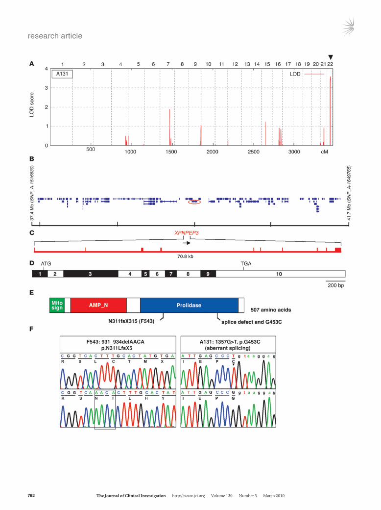
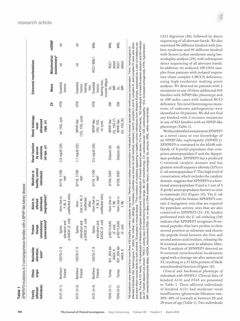
ResultsMutations in XPNPEP3 cause an NPHP-like kidney disease. We per-formed a whole-genome search for linkage in 116 consanguine-ous kindred with NPHP and NPHP-like phenotypes. Linkage analysis in a 5-generation consanguineous family from north-ern Finland (A131), with 3 members affected with an NPHP-like disorder, yielded a significant logarithm of odds (LOD) score of LODmax = 3.6, defining a new locus (NPHP-like 1, NPHPL1) on chromosome 22q13.2 (Figure 1A), within a 4.3-Mb interval flanked by markers SNP_A-1516630 and SNP_A-1649765 (Figure 1B). The critical genetic region overlapped with a homozygous segment detected in a consanguineous kindred of Turkish descent (F543), having 2 members with an NPHP-like phenotype (Supplemental Figure 1; supplemental material available online with this article; doi:10.1172/JCI40076DS1). The NPHPL1 critical genetic interval contained 101 positional candidate genes (Figure 1B).
We performed mutation analysis by sequencing exons in 29 of the candidate genes, prioritizing proteins present in the Ciliary proteome database (http://ciliaproteome.org/; ref. 28). Among them, we iden-tified novel, likely pathogenic variants in only one gene, XPNPEP3 (Figure 1, B–F). Kindred A131 harbored a homozygous splice-site mutation (1357G>T) in an 80% conserved exonic position of the splice donor consensus (Figure 1, B–F). RT-PCR from lymphoblas-toid cells showed that this mutation disrupts correct splicing by activating a cryptic splice site and introducing a frame shift (Sup-plemental Figure 2). The same mutation changed an amino acid residue (G453C) that is conserved throughout evolution, including E. coli (protein ecAPP) (data not shown). In addition, the 2 affected individuals of kindred F543 had a homozygous 4-base deletion in exon 6 (c.931_934 delAACA), resulting in a premature stop codon (p.N311LfsX5) within the predicted prolidase domain, which is the catalytic domain (Figure 1, E and F). The mutation in family F543 segregated from both parents and was absent from 150 people of mixed European descent and 83 Turkish healthy control individuals. Likewise, the mutation of the northern Finnish family A131 segre-gated from both parents; it was absent from 118 Western European healthy control individuals (Table 1) and was present once hetero-zygously in 100 Finnish control individuals, where it occurred on a shared haplotype of more than 2 Mb, suggesting a founder effect.
We then examined 96 kindred with NPHP by direct sequenc-ing of all exons and screened a multiethnic cohort of 376 addi-tional kindred with NPHP using heteroduplex analysis (29) and
Figure 1Positional cloning of the XPNPEP3 gene, as mutated in NPHPL1. (A) Parametric multipoint LOD score profile across the human genome of consanguineous kindred A131. Human chromosomes (numbered on top) are concatenated from pter (left) to qter (right) on the x-axis. Genetic distance is given in cM. Note the presence of a significant max-imum LOD score of 3.6 on human chromosome 22q13.2 (arrowhead), defining a new gene locus (NPHPL1) for an NPHP-like kidney dis-ease. (B) In kindred A131, the NPHPL1 locus, which is homozygous by descent, is delimited by heterozygous markers SNP_A-1516630 and SNP_A-1649765 to a 4.3-Mb interval, which contains 101 posi-tional candidate genes (per the UCSC sequence; http://genome.ucsc.edu/). Mutations were detected in XPNPEP3 (encircled red). (C) The XPNPEP3 gene extends over 70.8 kb and contains 10 exons (verti-cal hatches). (D) Exon structure of human full-length XPNPEP3 cDNA (3,056 bp). Positions of start codon (ATG) at nt +1 and of stop codon (TGA) are indicated. Exon sizes, ranging from 63 bp to 997 bp, are approximated. (E) Positions of the mitochondrial localization signal (Mito sign). The SMART program (http://smart.embl-heidelberg.de) predicts a putative N-terminal aminopeptidase P domain (AMP_N; amino acid 67–213), and prolidase domain (amino acid 253–490), which are drawn in relation to the encoding exon positions in D. (F) Two homozygous mutations of XPNPEP3 detected in families A131 and F543 with NPHPL1 (see Table 1).
research article
794 TheJournalofClinicalInvestigation http://www.jci.org Volume 120 Number 3 March 2010
CELI digestion (30), followed by direct sequencing of all aberrant bands. We also examined 96 different kindred with Jou-bert syndrome and 95 different kindred with Senior-Loken syndrome using het-eroduplex analysis (29), with subsequent direct sequencing of all aberrant bands. In addition, we analyzed 100 DNA sam-ples from patients with isolated respira-tory chain complex I (RCCI) deficiency, using high-resolution melting point analysis. We detected no patients with 2 mutations in any of these additional 643 families with NPHP-like phenotype and in 100 index cases with isolated RCCI deficiency. Ten novel heterozygous muta-tions of unknown pathogenicity were identified in 10 patients. We did not find any kindred with 2 recessive mutations in any of 823 families with an NPHP-like phenotype (Table 1).
We thus identified mutations in XPNPEP3 as a novel cause to our knowledge of an NPHP-like nephropathy (NPHPL1). XPNPEP3 is contained in the M24B sub-family of X-prolyl peptidases that com-prises aminopeptidase P and the dipepti-dase prolidase. XPNPEP3 has a predicted C-terminal catalytic domain and has greatest overall sequence identity (32%) to E. coli aminopeptidase P. This high level of conservation, which includes the catalytic domain, suggests that XPNPEP3 is a func-tional aminopeptidase P and is 1 out of 3 X-prolyl aminopeptidases known to exist in mammals (31) (Figure 1E). The E. coli ortholog and the human XPNPEP3 con-tain 2 manganese ions that are required for peptidase activity, sites that are also conserved in XPNPEP3 (31–33). Studies performed with the E. coli ortholog (34) indicate that XPNPEP3 recognizes N-ter-minal peptides that have proline in their second position as substrate and cleaves the peptide bond between the first and second amino acid residues, releasing the N-terminal amino acid. In addition, Mito-Prot II analysis of XPNPEP3 detected an N-terminal mitochondrial localization signal with a cleavage site after amino acid 53, resulting in a 51-kDa protein of likely mitochondrial function (Figure 1E).
Clinical and biochemical phenotype of individuals with NPHPL1. Clinical data of kindred A131 and F543 are presented in Table 1. Three affected individuals of kindred A131 had moderate renal insufficiency (glomerular filtration rate, 30%–40% of normal) at between 20 and 29 years of age (Table 1). Two individuals
Tab
le 1
XPNP
EP3
mut
atio
ns in
2 c
onsa
ngui
neou
s fa
mili
es w
ith a
NPH
P-lik
e ki
dney
dis
ease
Fam
ily
Ethn
ic
Nucl
eotid
e
Prot
ein
Al
lele
in
Mut
atio
n in
Re
nal f
unct
ion
Re
nal h
isto
logy
; Ex
trare
nal m
anife
stat
ions
(a
ffect
ed
orig
in
alte
ratio
n,
chan
ge
affe
cted
he
alth
y co
ntro
l by
ser
um
rena
l
indi
vidu
al)
ex
onA
(evo
lutio
nary
in
divi
dual
s
indi
vidu
als
crea
tinin
e
ultra
soun
d
cons
erva
tion)
B (s
egre
gatio
n)C
(a
t age
, yr)
(a
ge, y
r)
CV
Neur
o M
itoch
ondr
ial
mtD
NAE
bioc
hem
istr
yD
A131
(II-1
) No
rther
n
1357
G>T,
9
Splic
e
Hom
(het
in
0/11
8, 1
/100
2.
6 m
g/dl
(29)
nd
; ↑EG
, cys
t HT
N Es
sent
ial
nd
nd
Fi
nlan
d
alte
ratio
n an
d
F, M
, 2
HetF
tre
mor
p.G4
53C
(E. c
oli)
unaf
fect
ed s
ibs)
A1
31 (I
I-3)
North
ern
13
57G>
T, 9
Sp
lice
Ho
m
0/11
8, 1
/100
1.
7 m
g/dl
(22)
NP
HP-li
ke
HTN
Esse
ntia
l nd
nd
Finl
and
al
tera
tion
and
(h
et in
F, M
, 2
HetF
(1
5); ↑
EG, c
ysts
trem
or,
p.
G453
C (E
. col
i) un
affe
cted
he
arin
g
sibs
)
lo
ssA1
31 (I
I-4)
North
ern
13
57G>
T, 9
Sp
lice
Ho
m (h
et in
0/
118,
1/1
00
2.5
mg/
dl (2
0)
NPHP
-like
HT
N Es
sent
ial
RCC1
WNL
G W
NL
Fi
nlan
d
alte
ratio
n an
d
F, M
, 2
HetF
(1
6); ↑
EG,
tre
mor
,
G4
53C
(E. c
oli)
unaf
fect
ed s
ibs)
no
cys
ts
he
arin
g lo
ss,
mus
cle
fatig
ue
F543
(II-1
) Tu
rkey
93
1_93
4 de
l p.
N311
LfsX
5
Hom
(het
in
0/15
0, 0
/83F
Dial
ysis
(9)
NPHP
-like
CM
Sz
, RC
C1
WNL
AA
CA, 6
(E
. col
i) F,
M)
(3);
↑EG
(7)
M
R de
fect
F543
(II-2
) Tu
rkey
93
1_93
4 de
l p.
N311
LfsX
5
Hom
(het
in
0/15
0, 0
/83F
Dial
ysis
(8)
NPHP
-like
CM
Sz
, RC
C1
WNL
AA
CA, 6
(E
. col
i) F,
M)
(5);
↑EG
(6)
M
R de
fect
AA
ll m
utat
ions
wer
e ab
sent
from
mor
e th
an 1
50 h
ealth
y co
ntro
l sub
ject
s (s
ee te
xt).
BA
min
o ac
id r
esid
ue c
onse
rvat
ion
is in
dica
ted
for
low
est s
peci
es w
ith c
ontin
uous
con
serv
atio
n. C
Hom
, hom
ozyg
ous
in
affe
cted
indi
vidu
al; H
et, h
eter
ozyg
ous;
F, f
athe
r; M
, mot
her;
sib
s, s
iblin
gs. D
Com
plex
I ac
tiviti
es a
re s
how
n fo
r bo
th k
indr
ed; c
ompl
ex I
and
III, c
ompl
ex II
and
III,
com
plex
III,
and
com
plex
IV w
ere
also
don
e an
d w
ere
with
in n
orm
al li
mits
for
both
kin
dred
. EE
valu
ated
in m
uscl
e bi
opsy
. FT
he n
umbe
rs o
f mut
atio
ns a
ppea
ring
in h
ealth
y co
ntro
l ind
ivid
uals
from
Wes
tern
Eur
ope,
exp
ress
ed a
s a
ratio
of m
utat
ions
per
gr
oup.
For
the
A13
1 fa
mily
, 118
indi
vidu
als
of W
este
rn E
urop
ean
desc
ent a
nd 1
00 in
divi
dual
s of
Fin
nish
des
cent
are
sho
wn.
For
the
F54
3 fa
mily
, 150
indi
vidu
als
of m
ixed
Eur
opea
n de
scen
t and
83
indi
vidu
-al
s of
Tur
kish
des
cent
are
sho
wn.
GR
CC
1 ac
tivity
was
210
nm
ol/m
in/m
g (n
orm
al r
ange
, 85–
337
nmol
/min
/mg)
. CV
, car
diov
ascu
lar;
CM
, car
diom
yopa
thy;
↑E
G, i
ncre
ased
ech
ogen
icity
; HT
N, h
yper
tens
ion;
M
R, m
enta
l/dev
elop
men
tal d
elay
; mtD
NA
, mito
chon
dria
l DN
A; n
d, n
o da
ta o
r D
NA
ava
ilabl
e; N
euro
, neu
rolo
gica
l; S
z, s
eizu
re; W
NL,
with
in n
orm
al li
mits
.
research article
TheJournalofClinicalInvestigation http://www.jci.org Volume 120 Number 3 March 2010 795
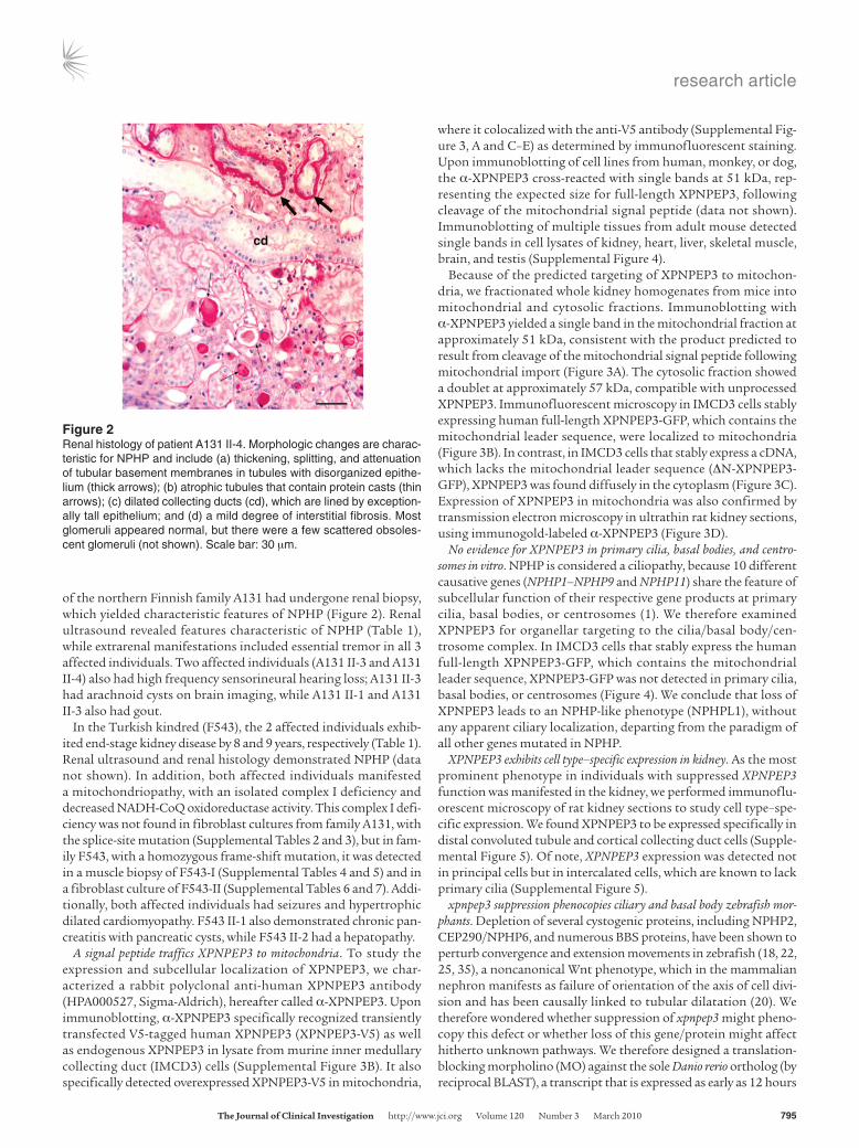
of the northern Finnish family A131 had undergone renal biopsy, which yielded characteristic features of NPHP (Figure 2). Renal ultrasound revealed features characteristic of NPHP (Table 1), while extrarenal manifestations included essential tremor in all 3 affected individuals. Two affected individuals (A131 II-3 and A131 II-4) also had high frequency sensorineural hearing loss; A131 II-3 had arachnoid cysts on brain imaging, while A131 II-1 and A131 II-3 also had gout.
In the Turkish kindred (F543), the 2 affected individuals exhib-ited end-stage kidney disease by 8 and 9 years, respectively (Table 1). Renal ultrasound and renal histology demonstrated NPHP (data not shown). In addition, both affected individuals manifested a mitochondriopathy, with an isolated complex I deficiency and decreased NADH-CoQ oxidoreductase activity. This complex I defi-ciency was not found in fibroblast cultures from family A131, with the splice-site mutation (Supplemental Tables 2 and 3), but in fam-ily F543, with a homozygous frame-shift mutation, it was detected in a muscle biopsy of F543-I (Supplemental Tables 4 and 5) and in a fibroblast culture of F543-II (Supplemental Tables 6 and 7). Addi-tionally, both affected individuals had seizures and hypertrophic dilated cardiomyopathy. F543 II-1 also demonstrated chronic pan-creatitis with pancreatic cysts, while F543 II-2 had a hepatopathy.
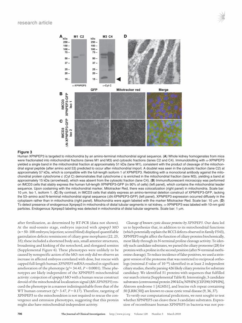
A signal peptide traffics XPNPEP3 to mitochondria. To study the expression and subcellular localization of XPNPEP3, we char-acterized a rabbit polyclonal anti-human XPNPEP3 antibody (HPA000527, Sigma-Aldrich), hereafter called α-XPNPEP3. Upon immunoblotting, α-XPNPEP3 specifically recognized transiently transfected V5-tagged human XPNPEP3 (XPNPEP3-V5) as well as endogenous XPNPEP3 in lysate from murine inner medullary collecting duct (IMCD3) cells (Supplemental Figure 3B). It also specifically detected overexpressed XPNPEP3-V5 in mitochondria,
where it colocalized with the anti-V5 antibody (Supplemental Fig-ure 3, A and C–E) as determined by immunofluorescent staining. Upon immunoblotting of cell lines from human, monkey, or dog, the α-XPNPEP3 cross-reacted with single bands at 51 kDa, rep-resenting the expected size for full-length XPNPEP3, following cleavage of the mitochondrial signal peptide (data not shown). Immunoblotting of multiple tissues from adult mouse detected single bands in cell lysates of kidney, heart, liver, skeletal muscle, brain, and testis (Supplemental Figure 4).
Because of the predicted targeting of XPNPEP3 to mitochon-dria, we fractionated whole kidney homogenates from mice into mitochondrial and cytosolic fractions. Immunoblotting with α-XPNPEP3 yielded a single band in the mitochondrial fraction at approximately 51 kDa, consistent with the product predicted to result from cleavage of the mitochondrial signal peptide following mitochondrial import (Figure 3A). The cytosolic fraction showed a doublet at approximately 57 kDa, compatible with unprocessed XPNPEP3. Immunofluorescent microscopy in IMCD3 cells stably expressing human full-length XPNPEP3-GFP, which contains the mitochondrial leader sequence, were localized to mitochondria (Figure 3B). In contrast, in IMCD3 cells that stably express a cDNA, which lacks the mitochondrial leader sequence (ΔN-XPNPEP3-GFP), XPNPEP3 was found diffusely in the cytoplasm (Figure 3C). Expression of XPNPEP3 in mitochondria was also confirmed by transmission electron microscopy in ultrathin rat kidney sections, using immunogold-labeled α-XPNPEP3 (Figure 3D).
No evidence for XPNPEP3 in primary cilia, basal bodies, and centro-somes in vitro. NPHP is considered a ciliopathy, because 10 different causative genes (NPHP1–NPHP9 and NPHP11) share the feature of subcellular function of their respective gene products at primary cilia, basal bodies, or centrosomes (1). We therefore examined XPNPEP3 for organellar targeting to the cilia/basal body/cen-trosome complex. In IMCD3 cells that stably express the human full-length XPNPEP3-GFP, which contains the mitochondrial leader sequence, XPNPEP3-GFP was not detected in primary cilia, basal bodies, or centrosomes (Figure 4). We conclude that loss of XPNPEP3 leads to an NPHP-like phenotype (NPHPL1), without any apparent ciliary localization, departing from the paradigm of all other genes mutated in NPHP.
XPNPEP3 exhibits cell type–specific expression in kidney. As the most prominent phenotype in individuals with suppressed XPNPEP3 function was manifested in the kidney, we performed immunoflu-orescent microscopy of rat kidney sections to study cell type–spe-cific expression. We found XPNPEP3 to be expressed specifically in distal convoluted tubule and cortical collecting duct cells (Supple-mental Figure 5). Of note, XPNPEP3 expression was detected not in principal cells but in intercalated cells, which are known to lack primary cilia (Supplemental Figure 5).
xpnpep3 suppression phenocopies ciliary and basal body zebrafish mor-phants. Depletion of several cystogenic proteins, including NPHP2, CEP290/NPHP6, and numerous BBS proteins, have been shown to perturb convergence and extension movements in zebrafish (18, 22, 25, 35), a noncanonical Wnt phenotype, which in the mammalian nephron manifests as failure of orientation of the axis of cell divi-sion and has been causally linked to tubular dilatation (20). We therefore wondered whether suppression of xpnpep3 might pheno-copy this defect or whether loss of this gene/protein might affect hitherto unknown pathways. We therefore designed a translation-blocking morpholino (MO) against the sole Danio rerio ortholog (by reciprocal BLAST), a transcript that is expressed as early as 12 hours
Figure 2Renal histology of patient A131 II-4. Morphologic changes are charac-teristic for NPHP and include (a) thickening, splitting, and attenuation of tubular basement membranes in tubules with disorganized epithe-lium (thick arrows); (b) atrophic tubules that contain protein casts (thin arrows); (c) dilated collecting ducts (cd), which are lined by exception-ally tall epithelium; and (d) a mild degree of interstitial fibrosis. Most glomeruli appeared normal, but there were a few scattered obsoles-cent glomeruli (not shown). Scale bar: 30 μm.
research article
796 TheJournalofClinicalInvestigation http://www.jci.org Volume 120 Number 3 March 2010
after fertilization, as determined by RT-PCR (data not shown). At the mid-somite stage, embryos injected with xpnpep3 MO (n = 50–100 embryos/injection; scored blind) displayed quantifiable gastrulation defects reminiscent of ciliary gene morphants (22, 25, 35); these included a shortened body axis, small anterior structures, broadening and kinking of the notochord, and elongated somites (Supplemental Figure 6). These phenotypes were unlikely to be caused by nonspecific action of the MO: not only did we observe an increase in affected embryos correlated with dose, but rescue with capped full-length human XPNPEP3 mRNA resulted in significant amelioration of the phenotype (χ2= 34.45, P < 0.0001). These phe-notypes are likely independent of the XPNPEP3 mitochondrial activity: coinjection of xpnpep3 MO with a human rescue construct devoid of the mitochondrial localization signal (ΔN-XPNPEP3) res-cued the phenotype in a manner indistinguishable from that of the WT human construct (χ2= 3.47, P = 0.17). Therefore, targeting of XPNPEP3 to the mitochondrion is not required to rescue the con-vergence and extension phenotypes, suggesting that this protein might also have mitochondrial-independent activity.
Cleavage of known cystic disease proteins by XPNPEP3. Our data led us to hypothesize that, in addition to its mitochondrial functions (which potentially explain the RCCI defects observed in family F543), XPNPEP3 might affect the function of ciliary proteins biochemically, most likely through its N-terminal proline cleavage activity. To iden-tify such candidate substrates, we parsed the ciliary proteome (28) for proteins with a proline in the second position (after N-terminal methi-onine cleavage). To reduce incidence of false-positives, we used a strin-gent version of the proteome that was restricted to reciprocal ortho-logs (minimal E value of 10–30) identified in at least 2 independent ciliary studies, thereby parsing 426 likely ciliary proteins for substrate candidacy. We identified 51 proteins with sequences that fulfilled our search criteria (Supplemental Table 8). Interestingly, 3 candidate substrates (centrosomal protein 290 kDa/NPHP6 [CEP290/NPHP6], Alstrom syndrome 1 [ALMS1], and leucine rich repeat containing 50 [LRRC50]) are known to cause cystic renal disease (9, 36, 37).
To verify our computational predictions, we next sought to test whether XPNPEP3 can cleave these 3 candidate substrates. Expres-sion of recombinant human XPNPEP3 in bacteria was not pos-
Figure 3Human XPNPEP3 is targeted to mitochondria by an amino-terminal mitochondrial signal sequence. (A) Whole kidney homogenates from mice were fractionated into mitochondrial fractions (lanes M1 and M3) and cytosolic fractions (lanes C2 and C4). Immunoblotting with α-XPNPEP3 yielded a single band in the mitochondrial fraction at approximately 51 kDa (lane M1), consistent with the product of cleavage of the mitochon-drial signal peptide (after amino acid 53) predicted to occur after mitochondrial import. A doublet was seen in the cytosolic fraction (lane C2) at approximately 57 kDa, which is compatible with the full-length isoform 1 of XPNPEP3. Reblotting with a monoclonal antibody against the mito-chondrial protein cytochrome c (Cyt C) demonstrates that cytochrome c is enriched in the mitochondrial fraction (lane M3), yielding a band at approximately 15 kDa (arrowhead), which was absent from the cytosolic fraction (lane C4). (B) Immunofluorescent microscopy was performed on IMCD3 cells that stably express the human full-length XPNPEP3-GFP (in 90% of cells) (left panel), which contains the mitochondrial leader sequence. Upon costaining with the mitochondrial marker, Mitotracker Red, there was colocalization (right panel) in mitochondria. Scale bar: 10 μm. Iso 1, isoform 1. (C) By contrast, in IMCD3 cells that stably express an amino-terminal deletion construct of XPNPEP3-GFP, lacking the 53–amino acid N-terminal mitochondrial signal sequence (ΔN-XPNPEP3-GFP) (left panel), XPNPEP3 expression occurred diffusely in the cytoplasm rather than in mitochondria (right panel). Mitochondria were again labeled with the marker Mitotracker Red. Scale bar: 10 μm. (D) To detect presence of endogenous Xpnpep3 in mitochondria of distal tubular segments in rat kidney, α-XPNPEP3 was labeled with 10-nm gold particles. Endogenous Xpnpep3 labeling was detected in mitochondria of distal tubular segments. Scale bar: 1 μm.

research article
TheJournalofClinicalInvestigation http://www.jci.org Volume 120 Number 3 March 2010 797
sible due to the protein being insoluble under numerous experi-mental conditions. We therefore focused on the E. coli ortholog of XPNPEP3, ecAPP, which we were able to express and purify to more than 99% homogeneity as detected by Coomassie stain-ing and Western blot (Figure 5A). Three 9–amino acid peptides identical to the N-termini of each of CEP290/NPHP6, ALMS1, and LRRC50 (without the start methionine that is cleaved after translation) were synthesized (Figure 5B) and incubated with purified ecAPP, followed by mass spectrometry. We found that CEP290/NPHP6, ALMS1, and LRRC50 are each cleaved effi-ciently by ecAPP (Figure 5C). By contrast, peptides containing residues other than proline in the second position could not be processed by ecAPP (data not shown). Importantly, the cleavage activity of the enzyme is not promiscuous, since we found that dynein, another ciliary protein that also contains a proline in the second residue but is not involved in cystic kidney diseases, is poorly digested by ecAPP (Figure 5C). These data suggested that XPNPEP3 cleavage of other cystogenic proteins might be relevant to their biological function(s).
Requirement of XPNPEP3 N-terminal cleavage for early gastrulation in zebrafish. To probe the importance of the likely XPNPEP3 cleavage sites in vivo, we asked whether cleavage resistance was deleterious
to protein function. Focusing on LRRC50 (each of CEP290/NPHP6 and ALMS1 encode mRNA more than 7 kb, rendering them difficult to transcribe in vitro at suffi-cient purity), we investigated whether a cleavage-resistant human capped mRNA can rescue the gastrulation pheno-types caused by MO-driven suppression of endogenous lrrc50. Injection of 4.5 ng of a translation-blocking MO induced early gastrulation phenotypes that include all the hallmarks of cilia-related convergent extension defects and which could be rescued by coinjection of 150 pg of WT human capped LRRC50 mRNA (Figure 6). How-ever, coinjection of MO with mRNA encoding an N-ter-minal Pro-Val point mutation, followed by blind scoring of embryos, showed complete failure of rescue (WT vs. Pro-Val, χ2 = 39.20, P < 0.0001; n = 80–110 embryos/injec-tion). This experiment could not exclude the possibility that the observed loss of function could be due to the presence of the Val at this position. However, repetition of this assay with Asp or Arg yielded similar data (Figure 6). Still, the possibility remained that the observed failure to rescue might still be driven by the loss of the proline for reasons other than cleavage. To assess this possibility, we searched the zebrafish genome for other N-terminal ami-nopeptidases and identified an alanyl aminopeptidase (ZFIN ID, ZDB-Gene-030131-1253), which predicts that Ala at the position of the N-terminal proline should ren-der lrrc50 subject to cleavage. Consistent with the notion that it is the cleavage that functionalizes lrrc50, injec-tion of MO with an Ala-encoding human mRNA rescued the phenotypes in a manner indistinguishable from WT (Figure 6; χ2 = 0.26, P < 0.87).
DiscussionUsing a whole-genome homozygosity mapping approach, we hereby identify recessive mutations in XPNPEP3 as a new cause of an NPHP-like disease. The phenotypic severity and organ involvement vary widely between the 2 different families with XPNPEP3 mutations, whereas
the phenotypic spectrum is concordant for affected individuals within the same family (Table 1). Specifically, 3 affected individu-als of family A131 exhibited only a mild form of an NPHP-like kidney disease (Figure 2), with residual kidney function beyond the age of 20 years, at which time virtually all patients with “clas-sic” NPHP would have reached ESRD (1). Extrarenal manifesta-tions were limited to mild neurologic involvement with sensori-neural hearing loss and essential tremor (Table 1). In contrast, the 2 affected siblings of F543 exhibited early-onset renal failure, leading to renal replacement therapy at 8 and 9 years of age, with extrarenal manifestations of mental retardation, seizures, and car-diomyopathy. The observed phenotypic variability might be due to different degrees of loss of function for the 2 different homo-zygous XPNPEP3 alleles. Whereas in family A131, residual correct splicing of the 80% conserved splice consensus may produce some WT splice product and thereby residual function (Supplemental Figure 2), the frameshift mutation of F543 will result in complete truncation of the C-terminal third of XPNPEP3 and likely ren-ders the mutant message substrate for nonsense-mediated decay. This notion is also supported by the finding that there was a mea-surable reduction in RCCI function in muscle biopsy samples of F543-I and fibroblasts of F543-II but not those of family A131.
Figure 4Full-length human XPNPEP3-GFP transiently expressed in IMCD3 cells local-izes to mitochondria and not to the cilium/basal body/centrosome complex in IMCD3 cells. Immunofluorescent microscopy was performed in IMCD3 cells that stably express human full-length XPNPEP3-GFP, which contains the mitochondrial signal sequence, demonstrating expression of XPNPEP3 in mitochondria. XPNPEP3-GFP was not detected in primary cilia (A), basal bod-ies (B), or centrosomes (C), when counterstaining with acetylated α-tubulin (Ac-α-tublin) (A), γ-tubulin (B), or pericentrin (C), respectively (middle and right panels). Scale bar: 10 μm.

research article
798 TheJournalofClinicalInvestigation http://www.jci.org Volume 120 Number 3 March 2010
Alternatively, modifiers in either family might potentiate or pro-tect the effect of the respective primary mutations, as has been suggested for other ciliopathies (23, 38).
NPHP is considered a ciliopathy because 10 different causative genes (NPHP1–NPHP9 and NPHP11) share the feature of subcel-lular expression of their respective gene products at primary cilia, basal bodies, or centrosomes (1). The absence of XPNPEP3 in cilia or basal body from in vitro cell culture could be due to the limitation of antibody affinity, or epitope internalization within cilium, or requirement of further posttranslational procession of XPNPEP3 before localizing to that organelle. It is also possible that trafficking of XPNPEP3 to the cilium or basal body requires some specific physiological stimulus, as shown for Smo (39).
Nevertheless, our data suggest a mitochondria-independent, yet cilia-related function, for XPNPEP3. Suppression of xpnpep3 in zebrafish phenocopies the developmental phenotypes induced by the suppression of numerous ciliary and basal body–encoding genes (22, 23, 25). Importantly, these phenotypes can be rescued by the xpnpep3 isoform devoid of mitochondrial targeting sequence, which in mammalian cells leads to cytoplasmic diffusion of the protein. We propose the ciliary phenotypes unmasked by loss of XPNPEP3 might arise from the loss of XPNPEP3-dependent pro-cessing of ciliary proteins. This idea is reinforced by the observed requirement to cleave the N-terminal proline of 3 cystogenic pro-
teins, most likely executed by XPNPEP3, and by the demonstration that, at least for LRRC50, N-terminal proline cleavage is necessary for correct gastrulation movements. Given the observed localiza-tion of XPNPEP3 in mammalian renal cells, we speculate that such processing might occur outside the cilium, especially since there is mounting evidence for the requirement of ciliary proteins in preciliary functions that include vesicular transport to the api-cal membrane/transition zone (41) and for the assembly of some intraflagellar transport particles in the cytoplasm (42). Although, in the present study CEP290/NPHP6, ALMS1, and LRRC50 (loss of each of which can induce renal cysts), have been identified as likely targets of XPNPEP3, we do not know whether it is the dys-function of these molecules that drives NPHP in the 2 families with XPNPEP3 mutations. It will be important to assay the matu-ration of these 3 proteins in patient cells and to examine the pos-sible anatomical and signaling defects of primary renal cilia in the absence of XPNPEP3.
MethodsStudy subjects. We obtained blood samples, clinical data, and pedigree infor-mation after obtaining informed consent from patients with NPHP and/or their parents. Approval for these studies was obtained from the Univer-sity of Michigan Institutional Review Board. The diagnosis of NPHP, as defined by the Online Mendelian Inheritance in Man database, was based
Figure 5Peptide cleavage by the XPNPEP3 ortholog, ecAPP. (A) Expression and purification of ecAPP. Coomassie staining of purified protein (lane 1) and immunoblot with anti-His tag antibody (lane 2) are shown. (B) Peptide substrates designed for enzymatic assay. Arrows indicate the cleavage site(s). Proline residue recognized by ecAPP is highlighted in red. Molecular weights are listed in Da. BN, bradykinin; N6, CEP290/NPHP6; AL, ALMS1; LR, LRRC50; DN, dynein. (C) Peptide cleavage detected by electrospray ionization–liquid chromatography–mass spectrometry. Mass spectrometry of peptides before and after digestion is shown. M+nH indicates the addition of n protons to the mass after ionization. intens, intensity.

research article
TheJournalofClinicalInvestigation http://www.jci.org Volume 120 Number 3 March 2010 799
on the clinical course and renal ultrasound or renal biopsy that were com-patible with the diagnosis of NPHP, as judged by a pediatric nephrologist. Patients were selected for additional mutation analysis using the criteria that they had an isolated RCCI deficiency and a residual RCCI activity beneath the lowest control value from 5% to 90%, with a median of 51%.
Renal involvement was recorded for 11 out of 100 of those patients. Ten patients had renal tubular acidosis and one had renal insufficiency.
SNP genotyping and sequencing. Whole-genome search for linkage was performed in 116 consanguineous families with NPHP using an Affyme-trix SNP array (50 K). Data were evaluated with nonparametric LOD
Figure 6N-terminal cleavage of LRRC50 is required to rescue lrrc50 morphant phenotypes in zebrafish. (A) Quantitative representation of the effect of lrrc50 MO on gastrulation development and rescue efficiency of different human lrrc50 RNA isoforms. The developmental phenotype of embryos was scored as Class I–II as described previously (22). Normal, indistinguishable from WT; Class I, mildly affected with a shortened body axis, small anterior structures, mild somite defects; Class II, severely affected with a short body axis, poorly defined head and eyes, broadening and kinking of the notochord, broad, thin somites, and tail extension defects. (B) Representative examples of embryos showing the gastrulation defect caused by lrrc50 MO, which could be rescued by WT and proline-to-alanine human LRRC50 but not other mutants. PA, proline to alanine; PV, proline to valine; PD, proline to aspartic acid; PR, proline to arginine.

research article
800 TheJournalofClinicalInvestigation http://www.jci.org Volume 120 Number 3 March 2010
scores across the entire genome to identify regions of homozygosity by descent as reported previously (43). Genomic segments of homozygos-ity were confirmed by high-resolution haplotype analysis within those regions using microsatellite markers. Additional SNPs were typed by direct sequencing. The GeneHunter program was used to calculate multipoint LOD scores, assuming recessive inheritance with complete penetrance, a disease allele frequency of 0.001, and allele frequencies for northern European descent (Affymetrix).
Mutational analysis of candidate genes. Exon PCR of candidate genes was performed using genomic DNA from affected individuals. Exon-flank-ing primers were designed using the University of California, Santa Cruz sequence (http://genome.ucsc.edu/) and Primer3 software (http://ihg2.helmholtz-muenchen.de/ihg/ExonPrimer.html). PCR products were puri-fied (Marligen Biosciences) prior to direct sequencing (Genetic Analyzer 3700, Applied Biosystems). Sequence data were analyzed using the soft-ware Mutation Surveyor (SoftGenetics) and Sequencher (Gene Codes). One hundred ninety healthy control individuals were screened as negative controls for each mutation identified.
High-resolution melting analysis was used for mutation scanning technology to analyze the coding region of XPNPEP3 in 100 patients with isolated RCCI deficiency. XPNPEP3 exons were PCR amplified from 5 ng genomic DNA, with a final denaturation step at 94°C for 1 minute (0.25 units Thermo-Start Taq DNA polymerase [Abgene], 1× LCGreen Plus [BIOKE], 0.25 μM of each primer; Supplemental Tables 1–7). High-resolu-tion melting analysis was performed on a LightScanner instrument (Idaho Technology). In the presence of the saturating double-stranded DNA-bind-ing dye, amplicons were denatured, starting at 77°C, while fluorescence intensities were recorded continuously. Melting curves were analyzed by LightScanner software (Idaho Technology), with normalized, tempera-ture-shifted curves displayed as difference plots (−dF/dT). Detected sam-ples with altered melting curves, compared with the average of multiple WT results, were directly sequenced with the BigDye Cycle sequencing kit (Applied Biosystems) (44).
Antibodies and protein expression studies. The rabbit polyclonal α-XPNPEP3 was from Sigma-Aldrich (HPA000527). Secondary antibodies to rabbit, mouse, and goat IgG were conjugated with either Alexa Fluor 488 or 594 (Molecular Probes). Antibodies for β-actin and acetylated α-tubulin and γ-tubulin were from Sigma-Aldrich; V5 was from Invitrogen; and pericen-trin was from Novus. The aquaporin-2 antibody was a goat polyclonal antibody from Santa Cruz Biotechnology Inc. (sc-9882). The cytochrome C oxidase antibody was a mouse monoclonal antibody from Santa Cruz Bio-technology Inc. (sc-658348).
Cloning. Full-length XPNPEP3 and ΔN-XPNPEP3 (amino acid 79–507) were PCR amplified from pENTR-XPNPEP3 (NM_022098.2, clone IOH5999; Invitrogen), with the termination codon removed and inserted into the pDONR221 vector, using BP Clonase II (Invitrogen), and subse-quently transferred into pEF5/FRT/V5-DEST (Invitrogen) and pG-LAP5 (C-terminal GFP-Lap tag) (45) or pCS2+ vectors, using LR Clonase II (Invitrogen). Human LRRC50 was cloned into the pCS2+ vector; vectors encoding mutated LRRC50, with the third proline residue replaced by alanine, valine, aspartic acid, or arginine, were generated by site-directed mutagenesis. The fidelity of all constructs was confirmed by sequence anal-ysis. MitoProt II was used for prediction of mitochondrial localization sig-nal (MitoProt, http://ihg2.helmholtz-muenchen.de/ihg/mitoprot.html).
Immunoblotting. Immunoblotting was performed as described previ-ously (9). For mitochondrial fractionation studies, whole kidneys from 6-month-old male 129S6 mice were isolated, homogenized, and fraction-ated with differential centrifugation into mitochondrial and cytosolic fractions. Then, 50 μg total protein from each fraction was loaded and separated on a denaturing 4%–12% SDS-PAGE gradient gel and trans-
ferred to PVDF membrane. Total protein was visualized using Ponceau S staining. Membranes were then immunoblotted with α-XPNPEP3 over-night at 4°C at a titer of 1:500.
Immunoelectron microscopy. Kidney cortex and medulla from 4-month-old Sprague-Dawley rats were used for immunocytochemical analysis. Tissues were fixed lightly for 5 minutes at 4°C with 3% formaldehyde and 20 mM ethylacetimidate in HEPES-buffered saline (30 mM HEPES, 151 mM NaCl, 4.7 mM KCl, 1.2 mM MgCl2, 7.8 mM glucose, pH 7.3) at 4°C. These samples were washed, further fixed in 3% formaldehyde containing 0.25% glutaraldehyde in HEPES-buffered saline at 4°C for 45 minutes, then dehydrated in ethanol and embedded in LR White Resin (Polysciences Inc.). Thin sections were blocked with PBS containing 1% w/v BSA and 0.01% v/v Tween 20 (PBT). Grids were then incubated with α-XPNPEP3 (HAP000527, Sigma-Aldrich) at 1:10 and 1:25 dilution in PBT for 12 hours at 4°C. Negative controls included normal rabbit serum and PBT replaced as the primary antibody. After washing, grids were incubated for 1 hour in 10-nm gold-conjugated goat anti-rabbit IgG (Amersham Life Sciences) diluted 1:30 in PBT, rinsed with PBS, and fixed with glutaraldehyde to sta-bilize the gold particles. Samples were stained with uranyl acetate and lead citrate, then examined in a Zeiss CEM902 electron microscope.
Tissue culture. XPNPEP3-V5– and XPNPEP3-LAP–expressing cell lines were generated by cotransfecting IMCD3 Flp-In cells (Invitrogen) with pOG44 and pDEST EF/FRT XPNPEP3-V5 or pG-LAP5-XPNPEP3, accord-ing to the manufacturer’s instructions. Stable cells were generated by selec-tion with hygromycin (800 μg/ml).
Measurements of mitochondrial respiratory complex function were per-formed in muscle biopsy samples from family F543 as described previously (46, 47). In individual A131 II-4, mitochondria were isolated from a muscle biopsy sample using the method of Rasmussen et al. (48), and the respira-tory chain complex activities were measured according to the method of Rustin et al. (49). An Aminco DW-2 Dual-Wavelength Spectrophotometer was used to record the enzyme kinetics.
Immunofluorescence studies in IMCD3 cells. Cells were grown on 24-well glass bottom plates, stained with Mitotracker Red CMTMRos (250 nM) (Invitrogen) for 30 minutes at 37°C in 5% CO2, and fixed with 4% paraformaldehyde. For indirect immunofluorescence studies, fixed cells were permeabilized with 0.1% Triton/1% BSA/PBS, pH 7.4, and stained with primary antibodies for 1 hour at 25°C, followed by secondary antibody immunolabeling. Nuclei were counterstained with Hoechst (Invitrogen). Epifluorescence microscopy images were acquired using a Zeiss AxioObserver.Z1, with a 63× 1.4 numerical aperture oil objective, and a Hamamatsu ORCA-ER CCD camera, with image stacks (0.5-μm steps) deconvolved and pertinent images of the cell z-projected, using AxioVision 3.1 software (Zeiss).
Kidney histology and immunofluorescence. Human kidney biopsies were fixed in Dusbosq-Brasil solution and embedded in paraffin; 2- to 6-μm sections were stained with hematoxylin-eosin, PAS, PAS methenamine silver, Masson’s trichrome, and Congo red for routine diagnostics. Immunofluorescence of rat kidney sections was performed as previously described (50).
Protein purification and mass spectrometry. The E. coli ortholog of XPNPEP3, ecAPP, was cloned into the pET28a vector (Novagen) to express a fusion protein with a His tag at the N terminus. The resulting construct was transformed into E. coli BL21 (DE3) codon plus RIL (Strat-agene). After induction by IPTG, ecAPP was purified with Ni-NTA tech-nology (Qiagen). The 9–amino acid peptides identical to the N-terminals of NPHP6, ALMS1, LRRC50, and dynein without methionine were syn-thesized by Genscript. In a typical enzymatic reaction, 100 μM peptide substrate was incubated with 100 nM purified ecAPP, which was preac-tivated by 20 times excess MnCl2 in 20 mM Tris-HCl, pH7.4, (0.75 mM MnCl2). The reaction was stopped by boiling samples at 100°C for

research article
TheJournalofClinicalInvestigation http://www.jci.org Volume 120 Number 3 March 2010 801
5 minutes, and product was detected by ESI-LC-MS (Agilent Technolo-gies). Candidate cleavage substrates were identified from the Ciliary pro-teome database (http://ciliaproteome.org).
Rescue of gastrulation phenotypes in zebrafish. MOs (Gene Tools) targeting the Danio rerio ortholog of XPNPEP3 or LRRC50 were resuspended to appro-priate concentrations, using sterile deionized water, and injected into WT embryos at the 2-cell stage. To rescue mid-somite morphant phenotypes, we generated capped mRNA for MO coinjections, using linearized pCS2+ XPNPEP3 (full length or ΔN-XPNPEP3) or pCS2+ LRRC50 (WT or mutants) as a template to transcribe mRNA with the SP6 mMessage mMachine kit (Ambion). xpnpep3 rescue studies were carried out with 4 ng translation blocker MO and 50 pg mRNA. For lrrc50 rescue studies, 4.5 ng of lrrc50 translation blocker MO was coinjected with 150 pg of RNA. Gastrulation phenotypes were scored at the 10 somite stage.
Statistics. To evaluate zebrafish knockdown phenotypes, Pearson χ2 test was used; P values of less than 0.05 were considered significant (Figure 6). Putative ciliary targets of XPNPEP3 were ranked using expectant values (Supplemental Table 8).
AcknowledgmentsWe sincerely thank the patients and their families for participa-tion. We acknowledge R.H. Lyons for excellent large-scale sequenc-ing and Jun Zhong for technical help with mass spectrometry analysis. We are grateful to the following colleagues for the con-tribution of helpful discussion regarding other animal models and biological systems: Jan Smeitinck (Nijmegen, Netherlands), Dontscho Kerjaschki (Vienna, Austria), Mark Winey and Chan-dra Kilburn (University of Colorado), Keith Gull (Oxford, United Kingdom), and Michel Leroux (Burnaby, Canada). This research was supported by grants from the NIH (DK1069274, DK1068306, and DK064614 to F. Hildebrandt; R01HD04260, R01DK072301,
and R01DK075972 to N. Katsanis; DK079541 to E.E. Davis), the Macular Vision Research Foundation (to N. Katsanis), and the Foundation for Fighting Blindness (to N. Katsanis) and by a NRSA fellowship F32 DK079541 (to E.E. Davis) from the National Institute of Diabetes, Digestive and Kidney Disorders. F. Hildeb-randt is an Investigator of the Howard Hughes Medical Institute, a Doris Duke Distinguished Clinical Scientist, and a Frederick G. L. Huetwell Professor. H. Khanna was supported by NIH grant EY007961 and by a grant from the Foundation for Fighting Blind-ness. L.M. Quarmby was supported by the Canadian Institutes for Health and Research (MOP-378061). J.F. O’Toole was supported by a grant from the NIH (DK071108). J. Uusimaa was supported by the Finnish Medical Foundation, the Alma and K.A. Snellman Foundation, and the Sigrid Juselius Foundation. This work was also supported by the Bundesministerium für Bildung und Forsc-hung (BMBF) funded German Network for Mitochondrial Disor-ders (mitoNET 01GM0862) and Systems Biology of Metabotypes (SysMBo 0315494A).
Received for publication June 4, 2009, and accepted in revised form January 6, 2010.
Address correspondence to: Friedhelm Hildebrandt, Howard Hughes Medical Institute, Department of Pediatrics, University of Michigan Health System, 8220C MSRB III, 1150 West Medical Center Drive, Ann Arbor, MI 48109-5646. Phone: 734.615.7285; Fax: 734.615.1386; E-mail: [email protected]. Or to: Nicholas Katsanis, Center for Human Disease Modeling, Department of Cell Biology, Duke University Medical Center, Durham, NC 27710. Phone: 919.613.4694; Fax: 919.684.1627; E-mail: [email protected].
1. Hildebrandt F, Attanasio M, Otto E. Nephro-nophthisis: disease mechanisms of a ciliopathy. J Am Soc Nephrol. 2009;20(1):23–35.
2. Hildebrandt F, Zhou W. Nephronophthisis-associated ciliopathies. J Am Soc Nephrol. 2007; 18(6):1855–1871.
3. Hildebrandt F, et al. A novel gene encoding an SH3 domain protein is mutated in nephronophthisis type 1. Nat Genet. 1997;17(2):149–153.
4. Saunier S, et al. A novel gene that encodes a protein with a putative src homology 3 domain is a can-didate gene for familial juvenile nephronophthisis. Hum Mol Genet. 1997;6(13):2317–2323.
5. Otto EA, et al. Mutations in INVS encoding inversin cause nephronophthisis type 2, linking renal cystic disease to the function of primary cilia and left-right axis determination. Nat Genet. 2003;34(4):413–420.
6. Olbrich H, et al. Mutations in a novel gene, NPHP3, cause adolescent nephronophthisis, tapeto-reti-nal degeneration and hepatic fibrosis. Nat Genet. 2003;34(4):455–459.
7. Otto E, et al. A gene mutated in nephronophthisis and retinitis pigmentosa encodes a novel protein, nephroretinin, conserved in evolution. Am J Hum Genet. 2002;71(5):1167–1171.
8. Otto EA, et al. Nephrocystin-5, a ciliary IQ domain protein, is mutated in Senior-Loken syndrome and interacts with RPGR and calmodulin. Nat Genet. 2005;37(3):282–288.
9. Sayer JA, et al. The centrosomal protein neph-rocystin-6 is mutated in Joubert syndrome and activates transcription factor ATF4. Nat Genet. 2006;38(6):674–681.
10. Attanasio M, et al. Loss of GLIS2 causes nephronophthisis in humans and mice by increased apoptosis and fibrosis. Nat Genet.
2007;39(8):1018–1024. 11. Delous M, et al. The ciliary gene RPGRIP1L is
mutated in cerebello-oculo-renal syndrome (Jou-bert syndrome type B) and Meckel syndrome. Nat Genet. 2007;39(7):875–881.
12. Otto EA, Trapp ML, Schultheiss UT, Helou J, Quarmby LM, Hildebrandt F. NEK8 mutations affect ciliary and centrosomal localization and may cause nephronophthisis. J Am Soc Nephrol. 2008;19(3):587–592.
13. Otto EA, et al. Hypomorphic mutations in meck-elin (MKS3/TMEM67) cause nephronophthi-sis with liver fibrosis (NPHP11). J Med Genet. 2009;46(10):663–670.
14. Watnick T, Germino G. From cilia to cyst. Nat Genet. 2003;34(4):355–356.
15. Badano JL, Mitsuma N, Beales PL, Katsanis N. The ciliopathies: an emerging class of human genetic disorders. Annu Rev Genomics Hum Genet. 2006;7:125–148.
16. Nauli SM, et al. Polycystins 1 and 2 mediate mecha-nosensation in the primary cilium of kidney cells. Nat Genet. 2003;33(2):129–137.
17. Fischer E, et al. Defective planar cell polarity in poly-cystic kidney disease. Nat Genet. 2006;38(1):21–23.
18. Simons M, et al. Inversin, the gene product mutat-ed in nephronophthisis type II, functions as a molecular switch between Wnt signaling pathways. Nat Genet. 2005;37(5):537–543.
19. Ma Z, et al. Mutations of HNF-1beta inhibit epithelial morphogenesis through dysregu-lation of SOCS-3. Proc Natl Acad Sci U S A. 2007;104(51):20386–20391.
20. Saburi S, et al. Loss of Fat4 disrupts PCP signaling and oriented cell division and leads to cystic kidney disease. Nat Genet. 2008;40(8):1010–1015.
21. Kishimoto N, Cao Y, Park A, Sun Z. Cystic kidney
gene seahorse regulates cilia-mediated processes and Wnt pathways. Dev Cell. 2008;14(6):954–961.
22. Gerdes JM, et al. Disruption of the basal body compromises proteasomal function and per-turbs intracellular Wnt response. Nat Genet. 2007;39(11):1350–1360.
23. Khanna H, et al. A common allele in RPGRIP1L is a modifier of retinal degeneration in ciliopathies. Nat Genet. 2009;41(6):739–745.
24. den Hollander AI, et al. Mutations in the CEP290 (NPHP6) gene are a frequent cause of Leber congeni-tal amaurosis. Am J Hum Genet. 2006;79(3):556–561.
25. Leitch CC, et al. Hypomorphic mutations in syn-dromic encephalocele genes are associated with Bar-det-Biedl syndrome. Nat Genet. 2008;40(4):443–448.
26. Valente EM, et al. Mutations in CEP290, which encodes a centrosomal protein, cause pleiotropic forms of Joubert syndrome. Nat Genet. 2006;38(6):623–625.
27. Baala L, et al. Pleiotropic effects of CEP290 (NPHP6) mutations extend to Meckel syndrome. Am J Hum Genet. 2007;81(1):170–179.
28. Gherman A, Davis EE, Katsanis N. The ciliary pro-teome database: an integrated community resource for the genetic and functional dissection of cilia. Nat Genet. 2006;38(9):961–962.
29. Hoskins BE, Thorn A, Scambler PJ, Beales PL. Evaluation of multiplex capillary heteroduplex analysis: a rapid and sensitive mutation screening technique. Hum Mutat. 2003;22(2):151–157.
30. Otto EA, et al. Mutation analysis in nephronophthi-sis using a combined approach of homozygosity mapping, CEL I endonuclease cleavage, and direct sequencing. Hum Mutat. 2008;29(3):418–426.
31. Ersahin C, Szpaderska AM, Orawski AT, Sim-mons WH. Aminopeptidase P isozyme expres-sion in human tissues and peripheral blood

research article
802 TheJournalofClinicalInvestigation http://www.jci.org Volume 120 Number 3 March 2010
mononuclear cell fractions. Arch Biochem Biophys. 2005;435(2):303–310.
32. Graham SC, Bond CS, Freeman HC, Guss JM. Structural and functional implications of metal ion selection in aminopeptidase P, a metallopro-tease with a dinuclear metal center. Biochemistry. 2005;44(42):13820–13836.
33. Li X, et al. Structure of human cytosolic X-pro-lyl aminopeptidase: a double Mn(II)-dependent dimeric enzyme with a novel three-domain subunit. J Biol Chem. 2008;283(33):22858–22866.
34. Yoshimoto T, Murayama N, Honda T, Tone H, Tsuru D. Cloning and expression of aminopep-tidase P gene from Escherichia coli HB101 and characterization of expressed enzyme. J Biochem. 1988;104(1):93–97.
35. Ross AJ, et al. Disruption of Bardet-Biedl syndrome ciliary proteins perturbs planar cell polarity in ver-tebrates. Nat Genet. 2005;37(10):1135–1140.
36. Li G, et al. A role for Alstrom syndrome protein, alms1, in kidney ciliogenesis and cellular quiescence. PLoS Genet. 2007;3(1):e8.
37. Sullivan-Brown J, et al. Zebrafish mutations affecting cilia motility share similar cystic phe-
notypes and suggest a mechanism of cyst forma-tion that differs from pkd2 morphants. Dev Biol. 2008;314(2):261–275.
38. Badano JL, et al. Dissection of epistasis in oligogenic Bardet-Biedl syndrome. Nature. 2006;439(7074):326–330.
39. Corbit KC, Aanstad P, Singla V, Norman AR, Stainier DY, Reiter JF. Vertebrate Smooth-ened functions at the primary cilium. Nature. 2005;437(7061):1018–1021.
40. Corbit KC, et al. Kif3a constrains beta-catenin-dependent Wnt signalling through dual cili-ary and non-ciliary mechanisms. Nat Cell Biol. 2008;10(1):70–76.
41. Follit JA, Tuft RA, Fogarty KE, Pazour GJ. The intraflagellar transport protein IFT20 is associated with the Golgi complex and is required for cilia assembly. Mol Biol Cell. 2006;17(9):3781–3792.
42. Omran H, et al. Ktu/PF13 is required for cytoplas-mic pre-assembly of axonemal dyneins. Nature. 2008;456(7222):611–616.
43. Hildebrandt F, et al. A systematic approach to map-ping recessive disease genes in individuals from out-bred populations. PLoS Genet. 2009;5(1):e1000353.
44. Meisinger C, et al. A genome-wide association study identifies three loci associated with mean platelet volume. Am J Hum Genet. 2009;84(1):66–71.
45. Torres JZ, Miller JJ, Jackson PK. High-throughput generation of tagged stable cell lines for proteomic analysis. Proteomics. 2009;9(10):2888–2891.
46. Janssen AJ, et al. Spectrophotometric assay for com-plex I of the respiratory chain in tissue samples and cultured fibroblasts. Clin Chem. 2007;53(4):729–734.
47. Janssen AJ, et al. Measurement of the energy-gen-erating capacity of human muscle mitochondria: diagnostic procedure and application to human pathology. Clin Chem. 2006;52(5):860–871.
48. Rasmussen HN, Andersen AJ, Rasmussen UF. Optimization of preparation of mitochondria from 25–100 mg skeletal muscle. Anal Biochem. 1997;252(1):153–159.
49. Rustin P, et al. Biochemical and molecular investi-gations in respiratory chain deficiencies. Clin Chim Acta. 1994;228(1):35–51.
50. Hinkes B, et al. Positional cloning uncovers muta-tions in PLCE1 responsible for a nephrotic syn-drome variant that may be reversible. Nat Genet. 2006;38(12):1397–1405.