rehabilitative ultrasound imaging
TRANSCRIPT
Dott.ssa Alessia Quercioli, PT
https://it.linkedin.com/in/quercioliaphysiotherapist

1968, Ikai e Fukunaga(Tokyo)
1980,Young, Stokes
(Oxford)1990,
Whittaker(Calgary)
1990, Hides(Queensland)
2006, Symposium, (S. Antonio)

Simposio di S. Antonio, Texas, 2006
Viene coniato l’acronimo RUSI “Rehabilitative Ultrasound Imaging”
Inizia la distinzione tra ecografia in campo medico e in campo riabilitativo
USI
DIAGNOSIS OF PATHOLOGY
RadiologistSonographer
RUSI
MUSCLE MORPHOLOGY AND BEHAVIOR
Physiotherapist
MSK Ultrasound Imaging
Muscles , tendons
ligaments …

“La procedura usata in fisioterapia nella valutazione dellamorfologia del muscolo; nel trattamento, come feedback, chepermette di incrementare la funzione neuro-muscolare; nelleattività di ricerca”
RUSI
VALUTAZIONEMorfologia muscolare
TRATTAMENTOFeedback
- Visivo- Knowdlege of results
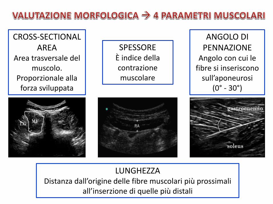
CROSS-SECTIONAL AREA
Area trasversale del muscolo.
Proporzionale alla forza sviluppata
SPESSOREÈ indice della contrazione muscolare
ANGOLO DIPENNAZIONE
Angolo con cui le fibre si inseriscono
sull’aponeurosi (0° - 30°)
LUNGHEZZA Distanza dall’origine delle fibre muscolari più prossimali
all’inserzione di quelle più distali
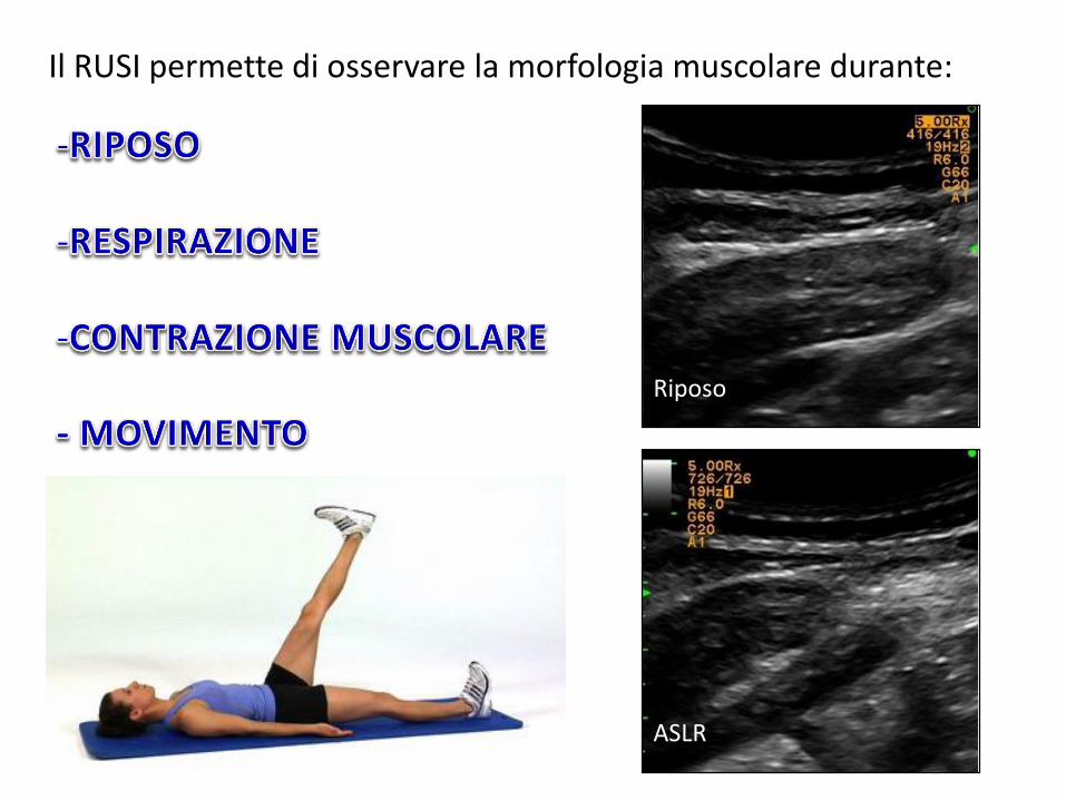
Il RUSI permette di osservare la morfologia muscolare durante:
ASLR
Riposo
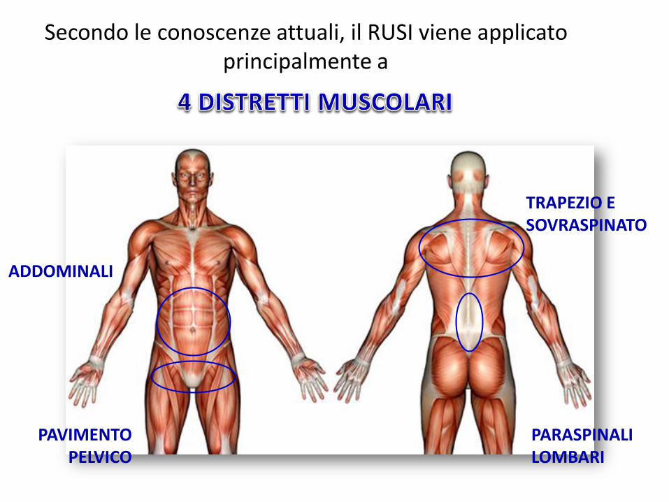
ADDOMINALI
PAVIMENTO PELVICO
TRAPEZIO E SOVRASPINATO
PARASPINALI LOMBARI
Secondo le conoscenze attuali, il RUSI viene applicato principalmente a

Il RUSI presenta elevata validità, riproducibilità edaffidabilità intra-operatore ed inter-operatore nellamisurazione di spessore e CSA di tutti i distretti muscolarisopra citati
Dall’analisi degli studi presenti sulle banche date di Pubmede Pedro pubblicati negli ultimi 10 anni è emerso che:
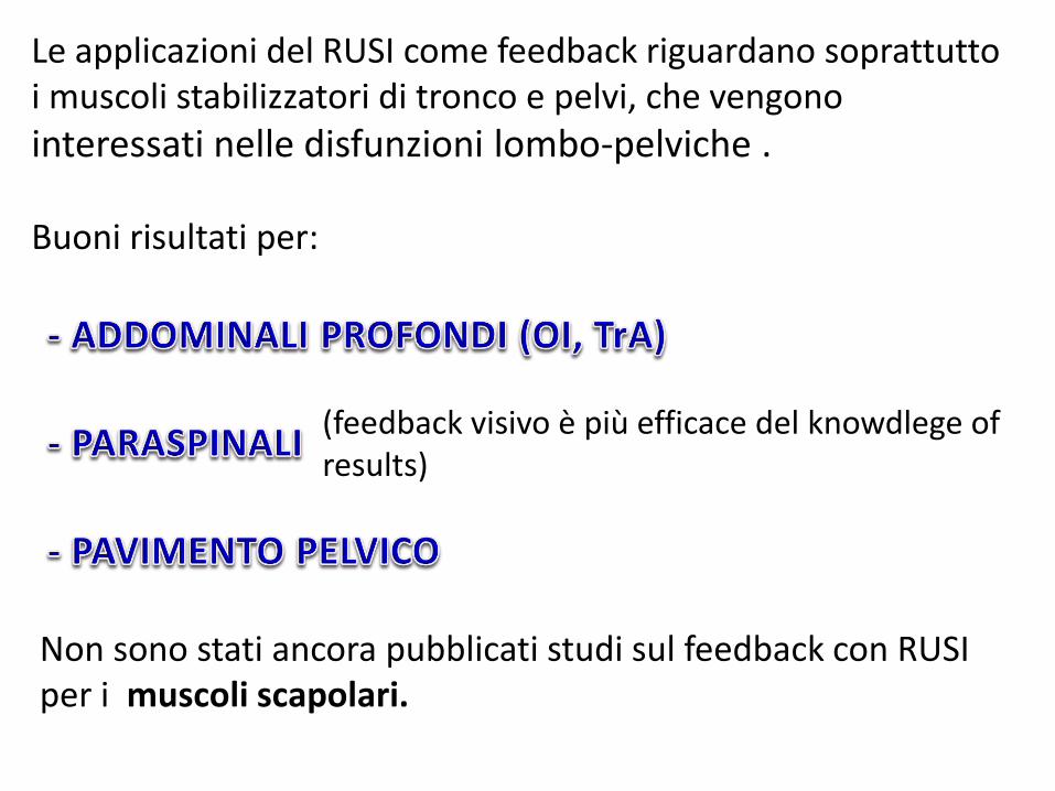
Le applicazioni del RUSI come feedback riguardano soprattutto i muscoli stabilizzatori di tronco e pelvi, che vengono
interessati nelle disfunzioni lombo-pelviche .
Buoni risultati per:
(feedback visivo è più efficace del knowdlege ofresults)
Non sono stati ancora pubblicati studi sul feedback con RUSI per i muscoli scapolari.

Dolore
Disabilità
Eventi futuri di LBP (fino ad un follow-up di 3 anni)
Gli studi dimostrano che il training con feedback è un’ aggiunta importante nella rieducazione neuro-motoria delle disfunzioni lombo-pelviche

Aumenta l’accuratezza della valutazione muscolareMigliora la spiegazione dell’esercizioFornisce un feedback precisoMonitorizza oggettivamente gli outcomes
Aumenta la percezione muscolareMonitorizza oggettivamente i propri progressiMotiva a seguire il trattamento

Bibliografia
Whittaker JL, Teyhen DS, Elliot JM et al. Rehabilitative Ultrasound Imaging: Understanding theTechnology and Its Applications. Journal of Orthopedic & Sports Physical Therapy. 37(8): 434-439. 2007
Teyhen DS. Rehabilitative ultrasound imaging symposium. Journal of Orthopedic & Sports PhysicalTherapy. 36(8): A1-A17. 2006
Teyhen DS, Gill NW, Whittaker JL et al. Rehabilitative ultrasound imaging of the abdominal muscles.Journal of Orthopaedic and Sports Physical Therapy. 37(8): 450-466. 2007
Stokes MW, Hides J, Elliot J et al. Rehabilitative ultrasound imaging of the posterior paraspinalmuscles. Journal of Orthopaedic and Sports Physical Therapy. 37(10): 581-595. 2007
Whittaker JL, Thompson JA, Teyhen DS et al. Rehabilitative ultrasound imaging of pelvic floor musclefunction. Journal of Orthopaedic and Sports Physical Therapy. 37(8): 487-498. 2007
Bentman S, O’Sullivan C and Stokes MW. Thickness of the middle trapezius muscle measured byrehabilitative ultrasound imaging: description of the technique and reliability study. Clinical Physiologyand Functional Imaging. 30(6): 426-431. 2010
O’Sullivan C, Bentman S, Bennetti K et al. Rehabilitative ultrasound imaging of the lower trapeziusmuscle: technkical description and reliability. Journal of Orthopaedic and Sports Physical Therapy.37(10): 620-626. 2007

Schneebeli A, Egloff M, Giampietro A et al. Rehabilitative ultrasound imaging of the sovraspinatusmuscle: intra- and interrater reliability of thickness and cross-sectional area. Journal of Bodywork andMovement Therapies. 18(2): 266-272. 2014
Teyhen DS, Miltenberger CE, Deiters HM et al. The use of ultrasound imaging of the abdominaldrawing-in maneuver in subjects with low-back pain. Journal of Orthopaedic and Sports PhysicalTherapy. 35(6): 346-355. 2005
Worth SGA, Henry SM and Bunn JY. Real-time ultrasound feedback and abdominal hollowing exercisesfor people with low back pain. Journal of Physiotherapy. 35(1). 2007
Henry SM and Westervelt KC. The use of real-time ultrasound feedback in teaching abdominalhollowing exercises to healthy subjects. Journal of Orthopedic & Sports Physical Therapy. 35(6): 338-345. 2005
Van K, Hides JA and Richardson CA. The use of real-time ultrasound imaging for biofeedback of lumbarmultifidus muscle contraction in healthy subjects. Journal of Orthopedic & Sports Physical Therapy.36(12): 920-925. 2006
Painter EE, Ogle MD and Teyhen DS. Lumbopelvic dysfunction and stress urinary incontinence: a casereport applying rehabilitative ultrasound imaging. Journal of Orthopaedic and Sport Physical Therapy.37(8): 499-504. 2007