regulation of plant vascular stem cells by endodermis ... · cambium serves as stem cells for all...
TRANSCRIPT

Journal of Experimental Botany, Vol. 64, No. 17, pp. 5335–5343, 2013doi:10.1093/jxb/ert196 Advance Access publication 23 July, 2013
© The Author 2013. Published by Oxford University Press on behalf of the Society for Experimental Biology. All rights reserved. For permissions, please email: [email protected]
ReseaRch papeR
Regulation of plant vascular stem cells by endodermis-derived EPFL-family peptide hormones and phloem-expressed ERECTA-family receptor kinases
Naoyuki Uchida1,2,* and Masao Tasaka2,*1 WPI-Institute of Transformative Bio-Molecules (WPI-ITbM), Nagoya University, Furo-cho, Chikusa-ku, Nagoya, 464-8602, Japan2 Graduate School of Biological Sciences, Nara Institute of Science and Technology, 8916-5 Takayama, Ikoma 630-0192, Japan
* To whom correspondence should be addressed. E-mail: [email protected] or [email protected]
Received 18 April 2013; Revised 21 May 2013; Accepted 28 May 2013
Abstract
Plant vasculatures are complex tissues consisting of (pro)cambium, phloem, and xylem. The (pro)cambium serves as vascular stem cells that produce all vascular cells. The Arabidopsis ERECTA (ER) receptor kinase is known to regulate the architecture of inflorescence stems. It was recently reported that the er mutation enhances a vascular phenotype induced by a mutation of TDR/PXY, which plays a significant role in procambial proliferation, suggesting that ER participates in vascular development. However, detailed molecular mechanisms of the ER-dependent vascular regu-lation are largely unknown. Here, this work found that ER and its paralogue, ER-LIKE1, were redundantly involved in procambial development of inflorescence stems. Interestingly, their activity in the phloem was sufficient for vascular regulation. Furthermore, two endodermis-derived peptide hormones, EPFL4 and EPFL6, were redundantly involved in such regulation. It has been previously reported that EPFL4 and EPFL6 act as ligands of phloem-expressed ER for stem elongation. Therefore, these findings indicate that cell–cell communication between the endodermis and the phloem plays an important role in procambial development as well as stem elongation. Interestingly, similar EPFL–ER modules control two distinct developmental events by slightly changing their components: the EPFL4/6–ER module for stem elongation and the EPFL4/6–ER/ERL1 module for vascular development.
Key words: Endodermis, EPFL-family peptide hormone, ERECTA-family receptor kinase, ligand, phloem, procambium, vasculature.
Introduction
Various types of stem cells play pivotal roles in well-coordi-nated organogenesis during plant and animal development (Sablowski, 2011; Snippert and Clevers, 2011). They undergo cell division to produce more stem cells that are identical to the parent cells (self-renewal) and give rise to differentiated descendant cells. Plant vasculatures are complex tissues con-sisting of (pro)cambium, phloem, and xylem. During vascu-lar development of Arabidopsis inflorescence stems, the (pro)cambium serves as stem cells for all vascular tissues (Elo et al., 2009) and produces xylem cells on one side and phloem cells on the other side. Because xylem and phloem function
in the conduction of water, minerals, and nutrients through-out the plant body, (pro)cambium should be maintained for a prolonged period to achieve proper vascular organization and support plant growth (Aichinger et al., 2012). Recently, much has been learned about the molecular mechanisms that maintain the (pro)cambium. On the other hand, the new find-ings have revealed further important issues that remain to be elucidated.
TDIF RECEPTOR/PHLOEM INTERCALATED WITH XYLEM (TDR/PXY) is a transmembrane-type recep-tor kinase expressed in (pro)cambium (Fisher and Turner,
5336 | Uchida and Tasaka
2007; Hirakawa et al., 2008). Attenuation of TDR signal-ling induces defects in (pro)cambial development (Fisher and Turner, 2007; Hirakawa et al., 2008). However, although the TDR pathway plays a pivotal role, it has been reported that residual (pro)cambial cells are still maintained even in inflo-rescence stems of tdr mutants (Fisher and Turner, 2007). This implies that another important mechanism maintains (pro)cambial cells independently of the TDR pathway (Fisher and Turner, 2007; Etchells et al., 2012). Recently, it was shown that blocking ethylene signalling enhances the vascular defect of tdr mutants, suggesting that vascular cell division is regu-lated both by both TDR and ethylene signals (Etchells et al., 2012). It has also been reported that the tdr vascular pheno-type is enhanced by a mutation of ERECTA (ER), suggest-ing that TDR signalling and the ER-dependent regulation are integrated for vascular development (Etchells et al., 2013).
ER encodes a transmembrane-type receptor kinase that has been shown to regulate inflorescence architecture (Torii et al., 1996; Bundy et al., 2012). ER activity in phloem tissues plays an important role in such regulation (Uchida et al., 2012). The ER has two paralogous genes, ER-LIKE 1 (ERL1) and ERL2, in the Arabidopsis genome (Shpak et al., 2004). Mutant plants lacking all ER-family members display defects in some developmental aspects of aboveground tissues (Shpak et al., 2004; Pillitteri et al., 2007; Hord et al., 2008) including regu-lation of stomatal density and spacing (Shpak et al., 2005) as well as the shoot apical meristem (SAM) (Uchida et al., 2013). These observations suggest that ER-family members might also redundantly play roles in other aspects of develop-ment. However, information about such redundant roles of ER-family members in vascular development has been largely unknown.
Because the ER family encodes signalling receptors, there should be ligands for each of the related developmental processes. In fact, several ligands have been identified for ER-family receptors (Hara et al., 2007, 2009; Hunt and Gray, 2009; Abrash and Bergmann, 2010; Hunt et al., 2010; Kondo et al., 2010; Sugano et al., 2010; Abrash et al., 2011; Lee et al., 2012; Uchida et al., 2012); EPF1, EPF2, and EPFL9/STOMAGEN for regulation of stomatal development and EPFL4/CHALLAH-LIKE2 and EPFL6/CHALLAH for regulation of inflorescence architecture. All of the identified ligands belong to a superfamily of secreted cysteine-rich pep-tides, the EPFL family (Rychel et al., 2010; Torii, 2012). EPF1 and EPF2 peptides are produced in stomata lineage cells (Hara et al., 2007, 2009; Hunt and Gray, 2009), while EPFL9 peptides are secreted from leaf mesophyll cells (Kondo et al., 2010; Sugano et al., 2010). On the other hand, EPFL4 and EPFL6 peptides are produced in the internal tissues at the vegetative stage (Abrash and Bergmann, 2010; Abrash et al., 2011) and then secreted from endodermal cell layers of inflorescence stems after bolting (Uchida et al., 2012). This observation indicates that the cell–cell communication between the endodermis-derived EPFL4/6 and phloem-expressed ER coordinates proper stem elongation (Uchida et al., 2012). Although the ER-dependent regulation is also involved in vascular development (Etchells et al., 2013), it is unknown in which stem tissue that the ER functions for
vascular regulation. Furthermore, a ligand(s) for ER in this regulation has not been identified yet. Therefore, the spatial aspect is obscure for ER-dependent cell–cell communication in vascular development.
Here, this work found that, among ER-family members, ER and ERL1 redundantly function in phloem tissues of inflores-cence stems for procambial development. It also found that two endodermis-derived peptide ligands, EPFL4 and EPFL6, are redundantly involved in this regulation. These findings indicate that the cell–cell communication between the endo-dermis and the phloem plays an important role in vascular development as well as stem elongation. Interestingly, EPFL–ER modules regulate two distinct developmental events in inflorescence stems by slightly changing their components: the previously reported EPFL4/6–ER module for stem elon-gation (Uchida et al., 2012) and the EPFL4/6–ER/ERL1 module for procambial development.
Materials and methods
Plant materialsThe wild-type accession of Arabidopsis thaliana used in this study was Columbia (Col-0). Materials were previously described as follows: er-105, erl1-2, ERpro:GUS, ERL1pro:GUS, and ERL2pro:GUS (Torii et al., 1996; Shpak et al., 2004, 2005); er-2 (CS3401), epfl4 (Salk_071065), epfl6 (Salk_072522), SUC2pro:ER, IRX3pro:ER, SCRpro:EPFL4, and SCRpro:EPFL6 (Uchida et al., 2012); tdr-1 (SALK_002910), and wox4-1 (GABI_462G01) (Hirakawa et al., 2008, 2010). Plants were grown at 22 °C under continuous light. Vascular bundles at 1 cm from the bottom of inflorescence stems were observed. Five individual plants (5 weeks old) with each geno-type were analysed to check reproducibility.
GUS staining and plastic sectioningThe procedures of GUS staining were previously described (Uchida et al., 2007). To make plastic sections, materials were fixed in FAA and then embedded in Technovit 7100 resin. Sections (4 μm) were stained with 0.02% toluidine blue or 0.04% neutral red before observation.
Results
ER and ERL1 are redundantly involved in procambial development of inflorescence stems
ER was previously shown to be expressed in inflorescence stems to regulate their elongation (Torii et al., 1996; Uchida et al., 2012). ER has two paralogous genes, ERL1 and ERL2, in the Arabidopsis genome (Shpak et al., 2004). Mutant plants lacking all ER-family members display defects in some developmental aspects of aboveground tissues (Shpak et al., 2004, 2005; Pillitteri et al., 2007; Hord et al., 2008; Uchida et al., 2011; Uchida et al., 2012; Uchida et al., 2013), indicating that ER-family members redundantly play roles in plant development. To examine the possibility that ER-family genes redundantly participate in some aspect(s) of stem development other than the previously reported reg-ulation of stem elongation by ER (Uchida et al., 2012), their promoter activities were investigated in stems using

Procambial regulation by EPFL–ER module | 5337
the GUS-reporter genes. As shown in Supplementary Fig. S1A–C (available at JXB online), ER and ERL1, but not ERL2, had strong promoter activities in inflorescence stems, suggesting that ER and ERL1 might redundantly play some roles in inflorescence stems. When cross sections of inflo-rescence stems were compared among wild type, er sin-gle mutant, erl1 single mutant, and er erl1 double-mutant plants, er erl1 plants exhibited a defect in vascular organi-zation (Fig. 1). In wild-type, er single mutant, and erl1 sin-gle mutant plants (Fig. 1A–F), a few layers of procambial cells (brackets), which displayed typical periclinal cell divi-sions along defined division planes, were localized between phloem and xylem tissues. Therefore, phloem and xylem cells (either vessel or fibre cells, both of which produce thick secondary cell walls) did not directly contact each other. On the other hand, er erl1 plants exhibited abnormal organi-zation of the procambium (Fig. 1G, H). The continuity of the layer structure was frequently interrupted in the pro-cambium (brackets in Fig. 1G and arrows in Fig. 1H). In such regions without procambial cells, some phloem cells directly contacted xylem cells (Fig. 1G, H, arrowheads). These results indicate that ER and ERL1 are redundantly involved in procambial development.
Detailed observation of the expression patterns of ER and ERL1 using thin plastic sections of inflorescence stems indicated that both were associated with vascular tissues (Fig. 2A, B). This association between ER/ERL1 and vas-culatures was further supported by analysis of their expres-sion patterns at another developmental stage. As shown in Supplementary Fig. S1D–F, both ER and ERL1, but not ERL2, were expressed in vasculatures (arrows) even at a seedling stage. In contrast, ER, ERL1, and ERL2 were all expressed in shoot apex regions (arrowheads), which is consistent with previous reports showing that all ER-family members are redundantly involved in SAM regulation (Uchida et al., 2011, 2013).
ER activity in the phloem contributes to procambial development
Because ER and ERL1 were redundantly involved in pro-cambial development of inflorescence stems (Fig. 1G, H), the detailed expression patterns of ER and ERL1 were next investigated within vascular bundles in stems. Previously, ER was reported to be expressed in phloem and xylem tissues (Uchida et al., 2012). As shown in Fig. 2A and B, ER and ERL1 displayed very similar and strong promoter activities in phloem and xylem tissues. To determine which activity of ER/ERL1 in phloem or xylem plays an important role in procambial regulation, tissue-specific rescue experiments were carried out by expressing ER in the er erl1 double-mutant background under the control of well-defined tissue-specific promoters: SUC2 for phloem (Imlau et al., 1999; Uchida et al., 2012) and IRX3 for xylem (vessel and fibre cells) (Gardiner et al., 2003; Uchida et al., 2012). To prepare these plants, this work used previously established trans-genic lines in which ER expression had been confirmed from
Fig. 1. Observation of vasculatures in inflorescence stems. Cross-sections of inflorescence stems of wild-type (A, B), er (C, D), erl1 (E, F), and er erl1 (G, H) plants are shown. B, D, F, and H are magnified images of A, C, E, and G, respectively. Pc, procambium; Ph, phloem; Xy, xylem; brackets enclose layer structures of procambium; arrows indicate procambial cells with typical periclinal cell divisions; arrowheads indicate phloem cells directly touching xylem cells. Bars, 40 μm (A, C, E, G) and 10 μm (B, D, F, H).

5338 | Uchida and Tasaka
the corresponding transgenes (Uchida et al., 2012). When the IRX3pro:ER transgene (Uchida et al., 2012) was intro-duced into er erl1 plants, the partial loss of the procambium observed in er erl1 (Fig. 1G, H) was unaffected (Fig. 2C, D). In contrast, SUC2pro:ER (Uchida et al., 2012) rescued the procambial defects (Fig. 2E, F), indicating that ER activity in the phloem is sufficient for procambial development.
ER activity in phloem also participates in the mechanism that maintains residual procambial cells in tdr mutants
TDR is a receptor kinase expressed in the (pro)cambium and attenuations in the TDR signalling induce defects in (pro)cambial development (Fisher and Turner, 2007; Hirakawa et al., 2008). It was recently reported that the tdr phenotypes are enhanced by er mutation, suggesting that TDR signal-ling and the ER-related mechanism cooperate for (pro)cambial development (Etchells et al., 2013). As shown in Supplementary Fig. S2 (Etchells et al., 2013), stems of er tdr double-mutant plants were shorter than those of wild-type, tdr, and er plants. This is a common feature of some reported mutants that show impaired vascular development in stems (Hanzawa et al., 2000; Parker et al., 2003; Pineau et al., 2005). On the other hand, both wild-type and tdr stems grew under the current growth conditions (Supplementary Fig. S2), although tdr plants have been reported to display vascular defects (Fisher and Turner, 2007). When the vascu-latures of tdr stems were examined (Fig. 3A, B), the typical layer structure of the procambium was disrupted but some remaining individual procambial cells were still observed, which displayed disordered division planes (Fig. 3A, B, arrows) (Fisher and Turner, 2007). In er tdr double-mutant plants, vascular bundles were flattened (Fig. 3C, D) com-pared with the triangular-shaped bundles of wild-type, er, and tdr plants (Figs. 1A, C and 3A), which is consistent with a recent report (Etchells et al., 2013). In er tdr vascular bun-dles, phloem cells directly contacted xylem cells without pro-cambial cells between them (Fig. 3C, D, arrowheads). The severe vascular defect in er tdr plants was also indicated by separation of xylem cell clusters (either vessel or fibre cells) within a vascular bundle (Fig. 3C, dotted line). Such discon-tinuous xylem regions were not observed in wild-type, er, or tdr plants (Figs. 1A, C and 3A). The serious disruption of vascular organization induced by simultaneous loss of both ER and TDR activities indicates that ER and TDR pathways are cooperatively involved in vascular development. Thus, it was next examined whether the phloem-specific ER activity also participates in cooperative regulation by ER and TDR in addition to ER/ERL1-mediated regulation (Figs. 1G, H and 2E, F). When IRX3pro:ER or SUC2pro:ER were intro-duced into er tdr double-mutant plants, the severe disruption of vascular organization in er tdr plants (Fig. 3C, D) was unaffected by IRX3pro:ER (Fig. 3E, F). er tdr IRX3pro:ER plants still exhibited flattened vascular bundles without procambial cells between phloem and xylem cells (Fig. 3F, arrowheads) as well as discontinuous xylem regions (Fig. 3E, dotted line). On the other hand, as shown in Fig. 3G and H, SUC2pro:ER restored the severe defects of er tdr vascu-latures to the level of the phenotype of tdr single mutant. Both tdr and er tdr SUC2pro:ER plants showed typical tri-angular-shaped vascular bundles (Fig. 3A and G) and some procambial cells between phloem and xylem cells (Fig. 3A, B, G, H, arrows). Thus, ER activity in the phloem is suffi-cient not only for ER/ERL1-mediated procambial regulation (Fig. 2E, F) but also for the mechanism in which ER and
Fig. 2. Expression patterns of ER and ERL1 and effects of tissue-specific expression of ER on vascular phenotypes. Cross-sections of inflorescence stems of ERpro:GUS (A), ERL1pro:GUS (B), er erl1 IRX3pro:ER (C, D), and er erl1 SUC2pro:ER (E, F) plants are shown. D and F are magnified images of C and E, respectively. For A and B, stems were sectioned after GUS staining. Pc, procambium; Ph, phloem; Xy, xylem; brackets enclose layer structures of procambium; arrows indicate procambial cells with typical periclinal cell divisions; arrowheads indicate phloem cells directly touching xylem cells. Bars, 40 μm (A–C, E) and 10 μm (D, F).

Procambial regulation by EPFL–ER module | 5339
TDR pathways cooperatively act for vascular development (Fig. 3G, H).
Endodermis-derived EPFL4 and EPFL6 peptide ligands for ER act in procambial development
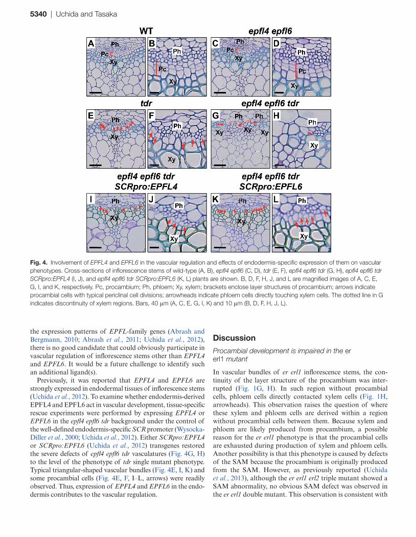
Because ER-family genes encode receptors, the existence of their ligands is highly expected for vascular development. So far, several ligands have been identified for ER-family receptors in some regulation other than vascular develop-ment (Hara et al., 2007, 2009; Hunt and Gray, 2009; Abrash and Bergmann, 2010; Hunt et al., 2010; Kondo et al., 2010; Sugano et al., 2010; Abrash et al., 2011; Uchida et al., 2012) and all of them belong to the EPFL family (Rychel et al., 2010; Torii, 2012). Therefore, to identify ligands involved in vas-cular development of inflorescence stems, this work focused on EPFL-family genes. Among annotated Arabidopsis EPFL genes, previous studies have identified EPFL4 and EPFL6 as being highly expressed in the inflorescence stems (Abrash and Bergmann, 2010; Abrash et al., 2011; Uchida et al., 2012). Each of these ligands physically interacts with an extracellu-lar domain of ER proteins and they are redundantly involved in regulating elongation of inflorescence stems (Abrash et al., 2011; Uchida et al., 2012). In this regulation, recogni-tion of EPFL4 and EPFL6 peptide ligands by ER proteins is required in phloem tissues (Uchida et al., 2012). Because ER activity in the phloem plays a pivotal role in procambial development (Figs. 2 and 3) as well as regulation of inflores-cence elongation (Uchida et al., 2012), it was hypothesized that EPFL4 and EPFL6 may also be involved in procambial development. To examine this hypothesis, vascular bundles in stems of epfl4 epfl6 double-mutant plants were checked. However, unexpectedly, epfl4 epfl6 plants did not show obvi-ous vascular defects (Fig. 4A–D). Then, to further pursue the possibility that EPFL4 and EPFL6 might participate in vascular development, the tdr mutant was made use of as a sensitized background because tdr vascular defects were remarkably enhanced by loss of ER activity (Fig. 3A–D). It was assumed that introduction of epfll4 epfl6 into tdr plants would enhance the tdr vascular phenotype if these ligands played an important role in ER-mediated vascular regulation. Stems of the epfl4 epfl6 tdr triple mutant were shorter than those of tdr and epfl4 epfl6 plants (Supplementary Fig. S3), which was comparable to er tdr stems (Supplementary Fig. S2). This epfl4 epfl6 tdr mutant also exhibited severe defects in vascular organization (Fig. 4G, H), which resembled er tdr vascular defects (Fig. 3C, D). Such defects included flat-tened vascular bundles (Fig. 4G) without procambial cells between phloem and xylem cells (Fig. 4G, H, arrowheads) as well as discontinuous xylem regions (Fig. 4G, dotted line). This observation was in sharp contrast to wild-type, epfl4 epfl6 mutant, and tdr mutant plants, all of which harboured triangular-shaped vascular bundles (Fig. 4A, C, E) and also procambial cells (Fig. 4A–F, brackets or arrows). Thus, it was concluded that EPFL4 and EPFL6 participate in vascu-lar development. One of the possible interpretations for why epfl4 epfl6 plants did not show obvious vascular defects is that other redundant ligands can mask the appearance of vascu-lar defects. However, according to current knowledge about
Fig. 3. Effects of tissue-specific expression of ER on severe vascular phenotypes of er tdr. Cross-sections of inflorescence stems of tdr (A, B), er tdr (C, D), er tdr IRX3pro:ER (E, F), and er tdr SUC2pro:ER (G, H) plants are shown. B, D, F, and H are magnified images of A, C, E, and G, respectively. Pc, procambium; Ph, phloem; Xy, xylem; brackets enclose layer structures of procambium; arrows indicate procambial cells with typical periclinal cell divisions; arrowheads indicate phloem cells directly touching xylem cells. The dotted lines in C and E indicate discontinuity of xylem regions. Bars, 40 μm (A, C, E, G) and 10 μm (B, D, F, H).
5340 | Uchida and Tasaka
the expression patterns of EPFL-family genes (Abrash and Bergmann, 2010; Abrash et al., 2011; Uchida et al., 2012), there is no good candidate that could obviously participate in vascular regulation of inflorescence stems other than EPFL4 and EPFL6. It would be a future challenge to identify such an additional ligand(s).
Previously, it was reported that EPFL4 and EPFL6 are strongly expressed in endodermal tissues of inflorescence stems (Uchida et al., 2012). To examine whether endodermis-derived EPFL4 and EPFL6 act in vascular development, tissue-specific rescue experiments were performed by expressing EPFL4 or EPFL6 in the epfl4 epfl6 tdr background under the control of the well-defined endodermis-specific SCR promoter (Wysocka-Diller et al., 2000; Uchida et al., 2012). Either SCRpro:EPFL4 or SCRpro:EPFL6 (Uchida et al., 2012) transgenes restored the severe defects of epfl4 epfl6 tdr vasculatures (Fig. 4G, H) to the level of the phenotype of tdr single mutant phenotype. Typical triangular-shaped vascular bundles (Fig. 4E, I, K) and some procambial cells (Fig. 4E, F, I–L, arrows) were readily observed. Thus, expression of EPFL4 and EPFL6 in the endo-dermis contributes to the vascular regulation.
Discussion
Procambial development is impaired in the er erl1 mutant
In vascular bundles of er erl1 inflorescence stems, the con-tinuity of the layer structure of the procambium was inter-rupted (Fig. 1G, H). In such region without procambial cells, phloem cells directly contacted xylem cells (Fig. 1H, arrowheads). This observation raises the question of where these xylem and phloem cells are derived within a region without procambial cells between them. Because xylem and phloem are likely produced from procambium, a possible reason for the er erl1 phenotype is that the procambial cells are exhausted during production of xylem and phloem cells. Another possibility is that this phenotype is caused by defects of the SAM because the procambium is originally produced from the SAM. However, as previously reported (Uchida et al., 2013), although the er erl1 erl2 triple mutant showed a SAM abnormality, no obvious SAM defect was observed in the er erl1 double mutant. This observation is consistent with
Fig. 4. Involvement of EPFL4 and EPFL6 in the vascular regulation and effects of endodermis-specific expression of them on vascular phenotypes. Cross-sections of inflorescence stems of wild-type (A, B), epfl4 epfl6 (C, D), tdr (E, F), epfl4 epfl6 tdr (G, H), epfl4 epfl6 tdr SCRpro:EPFL4 (I, J), and epfl4 epfl6 tdr SCRpro:EPFL6 (K, L) plants are shown. B, D, F, H, J, and L are magnified images of A, C, E, G, I, and K, respectively. Pc, procambium; Ph, phloem; Xy, xylem; brackets enclose layer structures of procambium; arrows indicate procambial cells with typical periclinal cell divisions; arrowheads indicate phloem cells directly touching xylem cells. The dotted line in G indicates discontinuity of xylem regions. Bars, 40 μm (A, C, E, G, I, K) and 10 μm (B, D, F, H, J, L).

Procambial regulation by EPFL–ER module | 5341
the fact that ER, ERL1, and ERL2 are all expressed in the SAM (Supplementary Fig. S1D–F, arrowheads) and all of them redundantly participate in the SAM regulation (Uchida et al., 2013). Thus, the procambial phenotype of the er erl1 double mutant is likely due to a defect of procambial main-tenance but not a SAM defect as observed in the er erl1 erl2 triple mutant.
The EPFL–ER signalling module contributes to procambial development
This study demonstrated that, among ER-family receptors, ER and ERL1 redundantly act in procambial development of inflo-rescence stems (Fig. 1). This study also showed that EPFL4 and EPFL6, which can behave as peptide ligands for these recep-tors (Abrash et al., 2011; Uchida et al., 2012), are involved in this mechanism (Fig. 4). Furthermore, ER activity in the phloem (Fig. 2) and expression of EPFL4 and EPFL6 in the endodermis (Fig. 4) are sufficient for this regulation, suggest-ing that EPFL4/EPFL6 peptides secreted from the endodermis are received by ER/ERL1 in the phloem to regulate procam-bial development (Fig. 5A). In this mechanism, an unknown non-cell-autonomous signal(s) from the phloem to procam-bium should be assumed because ER activity in the phloem affects procambial development. It would be an interesting in the future to reveal the nature of such a non-cell-autonomous effect as a downstream event of ER/ERL1 signalling.
Previously, it has been shown that the EPFL4/6–ER module regulates elongation of inflorescence stems (Fig. 5B) (Uchida et al., 2012). On the other hand, the EPFL4/6–ER/ERL1 mod-ule participates in procambial development (Fig. 5A). Thus,
interestingly, the similar EPFL–ER signalling modules regulate the two distinct developmental aspects just by slightly chang-ing their components. Because only three genes of the ER fam-ily regulate multiple and diverse biological events (van Zanten et al., 2009; Torii, 2012), slight differences in components of each module may diversify downstream events governed by the ER family. The current findings may represent an example of an efficient mechanism that utilizes limited genetic resources in the genome for diversification of receptor-based signalling systems. Therefore, this concept might not be limited to the ER family. One receptor or one type of redundant receptors may participate in the regulation of a larger number of distinct bio-logical events through slight changes in components of each signalling module. Recently, it was reported that a MAPK cas-cade functions downstream of the ER signalling to regulate stem elongation (Meng et al., 2012). It would be interesting to examine whether the MAPK cascade also acts downstream of the ER/ERL1 signalling for procambial development.
Possible relationships between the ER pathway and TDR signalling
It has been reported that WOX4, a WUSCHEL-related HOMEOBOX gene, is expressed in the (pro)cambium and is involved in (pro)cambium development (Hirakawa et al., 2010; Ji et al., 2010; Suer et al., 2011) and acts downstream of TDR signalling (Hirakawa et al., 2010). On the other hand, it has been suggested that unknown WOX4-independent mech-anisms mediate a part of the TDR signalling (Hirakawa et al., 2010). Therefore, to further dissect the cooperative regula-tion by ER and TDR, vascular phenotypes were compared
Fig. 5. Illustration of the proposed regulation by cooperative action between ER signalling and TDR signalling. (A) EPFL4/6–ER/ERL1 module for vascular development; (B) EPFL4/6–ER module for stem elongation. Blue indicates the promoter activity of EPFL6 marking endodermal cell layers (Uchida et al., 2012). Green circle indicates the phloem region. Brackets indicate the procambium region. The ligand for TDR is TDIF (tracheary element differentiation inhibitory factor) produced in phloem tissues (Etchells and Turner, 2010; Hirakawa et al., 2008). See text for detailed explanation.

5342 | Uchida and Tasaka
in inflorescence stems between er wox4 and er tdr plants. As shown in Supplementary Fig. S4A, er wox4 plants did not show obvious vascular defects, which was in sharp contrast to er tdr plants exhibiting severe disruption of vascular organi-zation (Supplementary Fig. S4B). This observation is consist-ent with the previous suggestion that the WOX4-independent regulation must contribute to a part of the TDR signalling (Hirakawa et al., 2010). Therefore, it is likely that the WOX4-independent pathway participates in the cooperative regula-tion by ER and TDR (Fig. 5A). It was recently reported that WOX14 participates in procambial development as a down-stream factor of the TDR signalling, which acts redundantly with WOX4 (Etchells et al., 2013). Thus, WOX14 would be a good candidate as a key player in the WOX4-independent pathway in the cooperative regulation by ER and TDR.
Blocking ethylene signalling has been shown to enhance tdr vascular defects (Etchells et al., 2012). Because er muta-tion also enhances the tdr phenotypes (Fig. 3C, D) (Etchells et al., 2013), it may be possible that the ER pathway and the ethylene signalling are integrated in vascular regulation. It would be attractive to hypothesize that ethylene itself may act as the non-cell-autonomous signal that is assumed to act downstream of the ER/ERL1 pathway to regulate procam-bial development (Fig. 5A). Investigating the relationship between the ER pathway and the ethylene signalling might be important to further understand the molecular basis of the ER-dependent regulation for vascular development.
Supplementary material
Supplementary data are available at JXB online.Supplementary Fig. S1. Promoter activities of
ER-family genes.Supplementary Fig. S2. Comparison of 5-week-old plant
statue among wild-type, tdr, er, and er tdr plants.Supplementary Fig. S3. Comparison of 5-week-old plant
statue among tdr, epfl4 epfl6, and epfl4 epfl6 tdr plants.Supplementary Fig. S4. Comparison of vascular pheno-
types in inflorescence stems between er wox4 and er tdr plants.
Acknowledgements
We thank Drs Keiko U Torii and Hiroo Fukuda for providing the ER-family-related materials and tdr-1 seeds, respectively. We also thank Ms Eriko Tanaka for technical assistance. This work was supported by MEXT/JSPS KAKENHI (grant numbers 24570050, 24113513, and 25114511 to NU and 19060007 and 22370019 to MT).
References
Abrash EB, Bergmann DC. 2010. Regional specification of stomatal production by the putative ligand CHALLAH. Development 137, 447–455.
Abrash EB, Davies KA, Bergmann DC. 2011. Generation of signalling specificity in Arabidopsis by spatially restricted buffering of ligand-receptor interactions. The Plant Cell 23, 2864–2879.
Aichinger E, Kornet N, Friedrich T, Laux T. 2012. Plant stem cell niches. Annual Review of Plant Biology 63, 615–636.
Bundy MG, Thompson OA, Sieger MT, Shpak ED. 2012. Patterns of cell division, cell differentiation and cell elongation in epidermis and cortex of Arabidopsis pedicels in the wild type and in erecta. PLoS One 7, e46262.
Elo A, Immanen J, Nieminen K, Helariutta Y. 2009. Stem cell function during plant vascular development. Seminars in Cell and Developmental Biology 20, 1097–1106.
Etchells JP, Provost CM, Mishra L, Turner SR. 2013. WOX4 and WOX14 act downstream of the PXY receptor kinase to regulate plant vascular proliferation independently of any role in vascular organisation. Development 140, 2224–2234.
Etchells JP, Provost CM, Turner SR. 2012. Plant vascular cell division is maintained by an interaction between PXY and ethylene signalling. PLoS Genetics 8, e1002997.
Etchells JP, Turner SR. 2010. The PXY-CLE41 receptor ligand pair defines a multifunctional pathway that controls the rate and orientation of vascular cell division. Development 137, 767–774.
Fisher K, Turner S. 2007. PXY, a receptor-like kinase essential for maintaining polarity during plant vascular-tissue development. Current Biology 17, 1061–1066.
Gardiner JC, Taylor NG, Turner SR. 2003. Control of cellulose synthase complex localization in developing xylem. The Plant Cell 15, 1740–1748.
Hanzawa Y, Takahashi T, Michael AJ, Burtin D, Long D, Pineiro M, Coupland G, Komeda Y. 2000. ACAULIS5, an Arabidopsis gene required for stem elongation, encodes a spermine synthase. EMBO Journal 19, 4248–4256.
Hara K, Kajita R, Torii KU, Bergmann DC, Kakimoto T. 2007. The secretory peptide gene EPF1 enforces the stomatal one-cell-spacing rule. Genes and Development 21, 1720–1725.
Hara K, Yokoo T, Kajita R, Onishi T, Yahata S, Peterson KM, Torii KU, Kakimoto T. 2009. Epidermal cell density is autoregulated via a secretory peptide, EPIDERMAL PATTERNING FACTOR 2 in Arabidopsis leaves. Plant Cell Physiology 50, 1019–1031.
Hirakawa Y, Kondo Y, Fukuda H. 2010. TDIF peptide signalling regulates vascular stem cell proliferation via the WOX4 homeobox gene in Arabidopsis. The Plant Cell 22, 2618–2629.
Hirakawa Y, Shinohara H, Kondo Y, Inoue A, Nakanomyo I, Ogawa M, Sawa S, Ohashi-Ito K, Matsubayashi Y, Fukuda H. 2008. Non-cell-autonomous control of vascular stem cell fate by a CLE peptide/receptor system. Proceedings of the National Academy of Sciences, USA 105, 15208–15213.
Hord CL, Sun YJ, Pillitteri LJ, Torii KU, Wang H, Zhang S, Ma H. 2008. Regulation of Arabidopsis early anther development by the mitogen-activated protein kinases, MPK3 and MPK6, and the ERECTA and related receptor-like kinases. Molecular Plant 1, 645–658.
Hunt L, Bailey KJ, Gray JE. 2010. The signalling peptide EPFL9 is a positive regulator of stomatal development. New Phytologist 186, 609–614.
Hunt L, Gray JE. 2009. The signalling peptide EPF2 controls asymmetric cell divisions during stomatal development. Current Biology 19, 864–869.

Procambial regulation by EPFL–ER module | 5343
Imlau A, Truernit E, Sauer N. 1999. Cell-to-cell and long-distance trafficking of the green fluorescent protein in the phloem and symplastic unloading of the protein into sink tissues. The Plant Cell 11, 309–322.
Ji J, Strable J, Shimizu R, Koenig D, Sinha N, Scanlon MJ. 2010. WOX4 promotes procambial development. Plant Physiology 152, 1346–1356.
Kondo T, Kajita R, Miyazaki A, et al. 2010. Stomatal density is controlled by a mesophyll-derived signalling molecule. Plant Cell Physiology 51, 1–8.
Lee JS, Kuroha T, Hnilova M, Khatayevich D, Kanaoka MM, McAbee JM, Sarikaya M, Tamerler C, Torii KU. 2012. Direct interaction of ligand-receptor pairs specifying stomatal patterning. Genes and Development 26, 126–136.
Meng X, Wang H, He Y, Liu Y, Walker JC, Torii KU, Zhang S. 2012. A MAPK cascade downstream of ERECTA receptor-like protein kinase regulates Arabidopsis inflorescence architecture by promoting localized cell proliferation. The Plant Cell 24, 4948–4960.
Parker G, Schofield R, Sundberg B, Turner S. 2003. Isolation of COV1, a gene involved in the regulation of vascular patterning in the stem of Arabidopsis. Development 130, 2139–2148.
Pillitteri LJ, Bemis SM, Shpak ED, Torii KU. 2007. Haploinsufficiency after successive loss of signalling reveals a role for ERECTA-family genes in Arabidopsis ovule development. Development 134, 3099–3109.
Pineau C, Freydier A, Ranocha P, et al. 2005. hca: an Arabidopsis mutant exhibiting unusual cambial activity and altered vascular patterning. The Plant Journal 44, 271–289.
Rychel AL, Peterson KM, Torii KU. 2010. Plant twitter: ligands under 140 amino acids enforcing stomatal patterning. Journal of Plant Research 123, 275–280.
Sablowski R. 2011. Plant stem cell niches: from signalling to execution. Current Opinion in Plant Biology 14, 4–9.
Shpak ED, Berthiaume CT, Hill EJ, Torii KU. 2004. Synergistic interaction of three ERECTA-family receptor-like kinases controls Arabidopsis organ growth and flower development by promoting cell proliferation. Development 131, 1491–1501.
Shpak ED, McAbee JM, Pillitteri LJ, Torii KU. 2005. Stomatal patterning and differentiation by synergistic interactions of receptor kinases. Science 309, 290–293.
Snippert HJ, Clevers H. 2011. Tracking adult stem cells. EMBO Reports 12, 113–122.
Suer S, Agusti J, Sanchez P, Schwarz M, Greb T. 2011. WOX4 imparts auxin responsiveness to cambium cells in Arabidopsis. The Plant Cell 23, 3247–3259.
Sugano SS, Shimada T, Imai Y, Okawa K, Tamai A, Mori M, Hara-Nishimura I. 2010. Stomagen positively regulates stomatal density in Arabidopsis. Nature 463, 241–244.
Torii KU. 2012. Mix-and-match: ligand-receptor pairs in stomatal development and beyond. Trends in Plant Science 17, 711–719.
Torii KU, Mitsukawa N, Oosumi T, Matsuura Y, Yokoyama R, Whittier RF, Komeda Y. 1996. The Arabidopsis ERECTA gene encodes a putative receptor protein kinase with extracellular leucine-rich repeats. The Plant Cell 8, 735–746.
Uchida N, Igari K, Bogenschutz NL, Torii KU, Tasaka M. 2011. Arabidopsis ERECTA-family receptor kinases mediate morphological alterations stimulated by activation of NB-LRR-type UNI proteins. Plant Cell Physiology 52, 804–814.
Uchida N, Lee JS, Horst RJ, Lai HH, Kajita R, Kakimoto T, Tasaka M, Torii KU. 2012. Regulation of inflorescence architecture by intertissue layer ligand-receptor communication between endodermis and phloem. Proceedings of the National Academy of Sciences, USA 109, 6337–6342.
Uchida N, Shimada M, Tasaka M. 2013. ERECTA-family receptor kinases regulate stem cell homeostasis via buffering its cytokinin responsiveness in the shoot apical meristem. Plant Cell Physiology 54, 343–351.
Uchida N, Townsley B, Chung KH, Sinha N. 2007. Regulation of SHOOT MERISTEMLESS genes via an upstream-conserved noncoding sequence coordinates leaf development. Proceedings of the National Academy of Sciences, USA 104, 15953–15958.
van Zanten M, Snoek LB, Proveniers MC, Peeters AJ. 2009. The many functions of ERECTA. Trends in Plant Science 14, 214–218.
Wysocka-Diller JW, Helariutta Y, Fukaki H, Malamy JE, Benfey PN. 2000. Molecular analysis of SCARECROW function reveals a radial patterning mechanism common to root and shoot. Development 127, 595–603.