regulation of phosphorylation of deoxycytidine and 2’,2
TRANSCRIPT

Biochemical Pharmacology, Vol. 51, pp. 911-918, 1996. Copyright 0 1996 Elsevier Science Inc.
ELSEVIER
ISSN 0006-2952/96/$15.00 + 0.00 SSDI 0006-2952(95)02402+6
Regulation of Phosphorylation of Deoxycytidine
and 2’,2’-Difluorodeoxycytidine (Gemcitabine); Effects of Cytidine 5’0Triphosphate
and Uridine 5’-Triphosphate in Relation to Chemosensitivity for 2’,2’-Difluorodeoxycytidine
Veronique W. T. Ruiz van Haperen, * Gijsbert Veerman, Jan B. Vermorken, Herbert M. Pinedo and Godefridus J. Peters j
DEPARTMENT OF MEDICAL ONCOLOGY, UNIVERSITY HOSPITAL VRIJE UNIVERSITEIT, PO Box 7057, 1007 MB AMSTERDAM, THE NETHERLANDS
ABSTRACT. Deoxycytidine kinase (dCK) and deoxycytidine deaminase (dCDA) are two key enzymes in the activation and inactivation, respectively, of deoxycytidine and its antiviral and anticancer analogues. One purpose of this study was to determine whether or not the deoxycytidine-converting activity of both enzymes would correlate with growth inhibition by 2’,2’-difluorodeoxycytidine (dFdC), a deoxycytidine analogue with established antitumour activity in solid tumours. Another aim of this work was to determine the effects of normal nucleotides on dCK. dCK and dCDA activities were measured with both deoxycytidine and dFdC as substrates in 5 solid tumour cell lines, but no correlation with cellular sensitivity to dFdC was found with either substrate.
The normal dCK activities with deoxycytidine as substrate varied between 0.8 and 13 nmol/hr/lOh cells. The activities determined with dFdC as substrate were remarkably similar in all 5 cell lines (1.1-1.6 nmol/hr/106 cells). dCDA activities varied considerably with both substrates (20-30-fold). Because dFdC markedly affected intracellular concentrations of cytidine 5’-triphosphate (CTP) and uridine 5’-triphosphate (UTP), we studied their effects on deoxycytidine- and dFdC-phosphorylating activities in 3 cell lines (i.e., A2780, WiDr and
C26-10) with a similar dCK activity but major differences in dFdC sensitivity. 1 mM CTP inhibited deoxy- cytidine phosphorylation (at 230 p,M) by 20-30% in A2780 and C26-10 cells, but increased that of WiDr cells by approximately 70%. CTP did not affect dFdC phosphorylation (at 230 p,M) in A2780 cells, but did increase it by 40% in WiDr cells. At 1 and 10 I.LM of deoxycytidine the effects of CTP on dCK activity in A2780, C26-10 and WiDr cells were less pronounced. 1 mM UTP enhanced deoxycytidine phosphorylation at 230 PM in WiDr cells by approximately 40%, whereas dFdC phosphorylation was increased 40% by UTP in C26-10 cells but decreased by 70-80% in WiDr cells. UTP caused a more pronounced increase in dCK activity at 1 and 10 p,M deoxycytidine in C26-10 cells, but provoked a higher inhibition in A2780 and WiDr cells at 10 I.LM. Because of these complex results, dCK kinetics were studied in greater detail. Biphasic kinetics for deoxycytidine were
observed in all 3 cell lines, with K,,, values of 23.2 and 0.4 ~.LM for A2780 cells, 15.9 and 1.5 PM for C26-10 cells, and 27.2 and 0.9 FM for WiDr cells. In all 3 cell lines, adenosine 5’-triphosphate (ATP) was the optimal
phosphate donor, as compared to CTP and UTP. In conclusion, the efficiency of dCK (V,,,/K,,, ratio) seems to correlate with accumulation of dFdCTP, the active metabolite of dFdC, and with cellular sensitivity. UTP and CTP, which are seriously affected in cells exposed to dFdC, display varying effects in these solid tumour cell lines. Both activation and inhibition have been observed; the physiologically low CTP pools and the relatively minor effect on dCK in A2780 cells seem to favour dFdC phosphorylation in these cells, which are the most sensitive. BIOCHEM PHARMACOL 51;7:911-918, 1996.
KEY WORDS. deoxycytidine kinase; deoxycytidine deaminase; gemcitabine; CTP; UTP; regulation; pyrimi-
dine salvage; deoxynucleosides
* Present address: Depr of Clinical Investigatton, Sectton of Cellular & sine 5’striphosphate; CTP, cytidine 5’-triphosphate; dCDA, 2’.deoxycyti- Molecular Pharmacology (Box 71), The University of Texas, M. D. dine deaminase; dCK, 2’-deoxycytidine kinase; dCMP, Z’-deoxycytidine Anderson Cancer Center, 1515 Holcomhe Boulevard, Houston, TX 5’-monophosphate; dCTP, 2’-deoxycytidine 5’-diphosphate; dFdC, 2’,2’- 77030, U.S.A. difluorodeoxycytidine, gemcitabine; dFdCTP, 2’,2’-difluorodeoxycytidine-
+ Corresponding author. 5’-triphosphate; TLC, thin layer chromatography. 8 Abbreviations: ara-C, l-B-D-arabinofuranosylcytosine; ATP, adeno- Received 1 August 1995; accepted

912 V. W. T. Ruiz van Haperen et al.
dCK$ (E.C.2.7.1.74), a py rimidine salvage enzyme, cataly-
ses the phosphorylation of deoxycytidine to its monophosphate dCMP, as well as the phosphorylation of several deoxyribonu- cleoside analogues that play a role in cancer and antiviral treat-
ment [I-3]. One of the physiological roles of this enzyme is deoxycytidine reutilization, although deoxyadenosine and de-
oxyguanosine are also good substrates [l, 31. dCDA (EC 3.5.4.5) catalyses the deamination of cytidine, deoxycytidine, and its ana- logues [4,5]. Both enzymes have been associated with sensitivity
and resistance to 1 -P-D-arabinofuranosylcytosine (am-C) [&8],
the best known deoxycytidine analogue, commonly used in the treatment of leukemia. But, overall, dCK is considered the most important of the two enzymes with respect to the efficacy of
deoxycytidine analogues [3, 691. The regulation of this enzyme is quite complex. In leukemic cells especially, UTP has been
reported to play an important role in the regulation of dCK activity. UTP was found to be the optimal phosphate donor in MOLT-4 and Ehrlich ascites cells [lo, 111, and could also reverse the inhibition of dCK by its most prominent feedback inhibitor
dCTP [12]. Sarup et al. [13] showed that both UTP and CTP could inhibit deoxycytidine phosphorylation in leukemic blasts by 25% and 15-28%, respectively. No other effects of CTP on
dCK activity have been reported until now. Most studies on dCK kinetics have been performed on enzyme extracts obtained from
lymphoid cells or tissues, both normal and malignant [l, 3, l& 131. Only a few studies on dCK activity and regulation have been performed on enzyme from solid tumours and tissues [3, 14, 151.
dFdC (gemcitabine) is a relatively new deoxycytidine
analogue with established clinical activity in solid malignancies, such as ovarian and non-small cell lung cancer [ 16-181. The major mechanisms of action of this compound are exerted by the
phosphorylated metabolites and include incorporation into nucleic acids [19, 201. To gain more insight into the role of both dCK and dCDA in the antitumour activity of dFdC in solid
tumour cell lines, we used dFdC as a substrate in addition to deoxycytidine to evaluate dCK in a panel of five solid tumour cell
lines, characterized previously for their sensitivity to dFdC [21].
One effect of exposing these cell lines to dFdC is a major distur- bance in concentrations of CTP and UTP [21], two nucleotides with effects on dCK activity [lO-131.
Therefore, the effects of these ribonucleotides on dCK
enzyme activity and kinetics with deoxycytidine and dFdC as substrates were studied in three solid tumor cell lines: the
human ovarian carcinoma cell line A2780, the human co- lon carcinoma cell line WiDr, and the murine colon car- cinoma cell line C26-10. These cell lines were selected
because of their different sensitivity to dFdC (despite simi- lar deoxycytidine phosphorylating activity) and the fact that all three were used for an extensive study of dFdCTP accumulation and the accompanying changes in normal
ribonucleotide pools [21].
MATERIALS AND METHODS Materials
dFdC, dFdU, and [5e3H]-dFdC (16.7 Ci/mmol) were kindly provided by Eli Lilly 6r Co., Indianapolis, IN. Deoxycyti-
dine, CTP, and UTP were purchased from Sigma Chemical Co., St. Louis MO. Deoxy-[5-3H]-cytidine (25 Ci/mmol)
was obtained from Amersham International, Buckingham- shire, U.K. All other chemicals were of analytical grade and
commercially available.
Cell Culture
The sources of the human ovarian carcinoma cell lines A2780 and OVCAR-3, the human colon carcinoma cell
line WiDr, the human head and neck squamous cell carci-
noma cell line UM-SSC-14C, and the murine colon carci-
noma C26-10 cells have been described previously [21, 221. Cells were maintained in exponential growth in Dulbecco’s Modification of Eagle’s Medium (Gibco Laboratories,
Grand Island, NY) suppl emented with 5% heat-inactivated fetal calf serum (Gibco), 1 mM L-glutamine (Sigma), and 250 ng/mL gentamicin at 37°C and 5% CO,. Previously we used A2780, C26-10, and WiDr cells for extensive studies
of dFdC metabolism, in which considerable differences in chemosensitivity and metabolism were observed. Given
these results and the similarity of deoxycytidine and dFdC phosphorylation, these cell lines were selected for more
detailed studies. All enzyme assays were performed with
enzyme extracted from cells harvested 2 days after seeding, when cells were in the exponential growth phase.
dCK Assay
The assay for dCK was performed as described previously
[14]. Briefly, for determination of dCK activities in cell lines, a minimum of 25.106 cells were required. The cell pellets were resuspended in cold dCK buffer (0.3 M Tris-
HCl, 50 p,M P-mercaptoethanol, pH 8.0), sonicated and centrifuged. The 10,000 g supernatant was immediately
used in the enzyme assays. One part of the undiluted su- pernatant was taken for determining its protein content using the Biorad Bradford protein assay [23]. To 25 PL
10,000 g supernatant (2-14 pg protein/FL), 25 PL of sub- strate mixture was added. The substrate mixture was pre- pared by mixing 2 volumes of Mg-ATP (50 mM ATP in 2 5
mM MgCl,, pH 7.4) (f’ ma concentration in the reaction 1 mixture: 10 mM ATP), 2 volumes of [5-3H]-deoxycytidine or [5-3H]-dFdC and one volume of dCK buffer. For activity measurements under saturated substrate conditions, the fi-
nal concentration of deoxycytidine (0.04 Ci/mmol) and dFdC (41.8 Ci/mol) was 230 FM. The enzyme activity was measured with and without 0.1 and 1 mM final concentra-
tion CTP or UTP in three of the five solid tumour cell lines: A2780, WiDr, and C26-10 cells. dCK activity was also determined with CTP or UTP as phosphate donors (10 mM final concentration). The reaction mixture was incu- bated for 15-60 min at 37°C. The reaction was terminated by heating at 95°C for 3 min and the subsequent addition of 10 FL 5 mM unlabeled deoxycytidine or dFdC to visu-

Chemosensitivity to Gemcitabine in Relation to Deoxycytidine and Gemcitabine Metabolism; Effects of CTP and UTP 913
alize the spots. The substrate (deoxycytidine or dFdC) was
separated from the product (dCMP or dFdCMP) by thin
layer chromatography on PEI (polyethylene imine) cellu- lose layers, with distilled water as eluent. The spots could be visualised under UV light, marked and cut out. Radioac- tivity was estimated in a liquid scintillation counter, after addition of 9 mL Optima Gold (Packard Instrument B.V., Chemical Operations, Groningen, The Netherlands). En-
zyme activities were expressed as nmol product formed per hour per lo6 cells (nmol/hr/106 cells) and were linear in
time and enzyme concentration. Because dialyzing the sam-
ples or adding tetrahydrouridine to inhibit dCDA did not
change the outcome of the assay, they were, therefore, omitted. Dialysing would remove intracellular nucleotides
and nucleosides that theoretically could interfere with the assay. However, the concentration of UTP is less than 800 pmol/106 cells (21). Cells are suspended in 1 mL and sub-
sequently diluted, resulting in a maximum of 2 FM in the enzyme suspension; in the assay mixture this would be di- luted at least two-fold, resulting in nucleotide concentra-
tions too low to interfere with the assay. Because dCTP concentrations in cells are at least IOO-lOOO-fold lower
than CTP, this possible interference would also be negligi-
ble. The separation procedure used (TLC) enabled us to determine how much label was consumed; this would not
be possible if filter discs similar to those employed by sev- eral other investigators were used. Assays were designed in such a way that deoxycytidine consumption was less than 10%. Due to the higher K, of deoxycytidine for deamina- tion compared to phosphorylation, it would be unlikely
that, under these conditions, deoxycytidine concentrations would be reduced by deamination by more than 10%. ATP measurements showed that no significant degradation oc-
curred during the assay. In other kinase assays, addition of
inhibitors of nucleotide degradation did not affect the en- zyme activities (data not shown).
For the apparent K,,, determinations, the deoxycytidine concentration ranged from 0.4 to 230 p,M. The measured activities were linear in time and corrected for >lO% sub-
strate conversion. K,,, and V,,, were calculated using the statistical application (linear regression analysis) of the
Symphony 2.0 computer program (Lotus Development Corporation).
dCDA Assay
The assay was performed as described previously [14]. Briefly, the reaction was performed in crude cell extracts with a final substrate (deoxycytidine or dFdC) concentra-
tion of 500 PM. Product and substrate were separated by means of HPLC with isocratic elution. Deoxycytidine and deoxyuridine were separated on a LiChrosorb 5-RP-18 col-
umn (Chrompack, Bergen op Zoom, the Netherlands) with 10 mM ammonium dihydrogen phosphate, pH 6.5 as
eluent. dFdC and dFdU were separated using a FBondapack
cl8 column (Waters-Millipore, Etten-Leur, the Nether- lands), with PicB, (Waters) in 15% methanol (final con-
centration heptane sulfonic acid 5 mM), pH 3.1. Peaks were detected and quantitated using their absorption at 254 and 280 nm.
RESULTS dCK and dCDA Measurements with Deoxycytidine and dFdC as Substrates
In Table 1, sensitivity to dFdC and dCK, as well as dCDA
activities, as determined at saturating substrate concentra- tions (230 FM) in five different solid tumour cell lines, are
summarized. The human ovarian carcinoma A2780 cells were most sensitive to dFdC, both at short (4-hr) and long exposure. The two colon carcinoma cell lines, C26-10 (mu- rine) and WiDr (human), were least sensitive to dFdC. The
measured enzyme activities were linear within the incuba- tion time and protein concentration range applied. dCK activities with the natural substrate deoxycytidine varied
considerably, from 1.0 to 12.8 nmol/hr/106 cells. dCK ac- tivities as determined with the deoxycytidine analogue dFdC were remarkably similar between the cell lines, vary-
ing only from 1 .l to 1.6 nmol/hr/106 cells. For dCDA ac- tivities, a considerable variation was observed both with
deoxycytidine (30-fold, from 0.1 to 3 nmol/hr/106 cells) and dFdC (20-fold, from 0.6 to 12 nmol/hr/106 cells). How-
ever, the pattern of activity was different for each substrate, with UMSSC-14C having the highest activity with deox- ycytidine as substrate and OVCAR-3 with dFdC.
Apparent K, and V_ Values
Although C26-10, WiDr, and A2780 cells showed very different sensitivity patterns (Table l), these three cell lines
TABLE 1. Sensitivity to dFdC and deoxycyticlme kinase (with ATP as phosphate donor) and deoxycytidine deaminase activ- ities with different substrates
dCKP dCDAt
Cell line Sensitivity* deoxycytidine dFdC deoxycytidine dFdC
C26-10 238 1.24 I!Z 0.25 1.11 kO.10 2.15 f 0.57 0.92 f 0.18 WiDr 205 1.01 k 0.10 1.18 + 0.04 0.22 f 0.03 1.35 + 0.15 A2780 7 1.01 + 0.09 1.24 f 0.05 0.12 f 0.04 0.63 + 0.15 OVCAR-3 36 12.8 + 1.0 1.21 k 0.13 2.33 L 0.08 11.9f3.6 UM-SSC-14C 34 5.28 If: 0.53 1.61 + 0.04 2.95 f 0.43 4.66 + 0.65
* Sensmvity to dFdC IS expressed as the IC,, in nM after 4-hr exposure followed by a 68-hr drug-free period [Zl].
t Activmes (measured at 230 (LM substrate concentratmn for kmase and 500 FM for the deaminase) are expressed as nmol product formed/hr/106 cells and are means f SEM
of 3-6 separate experiments. Protem content was 0.1-0.2 m&O6 cells.
914 V. W. T. Ruiz van Haperen et al.
TABLE 2. Biochemical characteristics of C26-10, WiDr, and A2780 cells and effect of dFdC on CTP and UTP
C26.10 WiDr A2780
Nucleotide* Effect of dFdC Nucleotide* Effect of dFdC Nucleotide* Effect of dFdC
Nucleotide (pmol/106 cells) (%)-I (pmol/106 cells) (%)-t (pmoY106 cells) (%)P
UTP 789 f 105 161 56OL41 115 630 + 210 111 CTP 458 f 38 153 168 + 17 86 219 i7 65 58 dFdCTPS 31+31 33f 18 lOOf
Values are means + SEM of 3-5 separate experiments [21].
* UTP and CTP concentratmns are those of untreated cells. Values are means k SEM of 3-5 separately harvested cell pellets [21]
+ % of control UTP or CTP concentration after 4-hr exposure to 1 FM dFdC.
$ dFdCTP concenttatmn in pmol/106 cells after a 4-hr treatment with 1 FM dFdC.
were remarkably similar in dCK activity with either sub- strate. Therefore, we selected these three lines to study dCK kinetics to see whether or not differences in apparent K,
and/or V,,, values would explain the differences in dFdC
sensitivity and dFdCTP accumulation. In Table 2, some biochemical characteristics of these cell lines (fully de- scribed previously [21]) are summarized. Data as observed at
a 4-hr exposure to dFdC are presented, because we also used a 4-hr exposure in the growth inhibition experiments. The general pattern was comparable after 24-hr exposure. In
short, UTP concentrations in untreated cells were compa- rable in the three cell lines, but CTP was higher in C26-10 cells. In contrast, the effect of dFdC was different, resulting
in an increase for UTP and CTP in C26-10 but a decrease in CTP in WiDr and A2780 cells. dFdCTP accumulation was higher in A2780 cells compared to the other cell lines.
A similar pattern in dFdCTP accumulation was observed at lower (0.1 FM) and higher (10 p,M) dFdC concentrations.
However, at 0.1 FM it was not always possible to measure dFdCTP accumulation (2 1).
l-
/
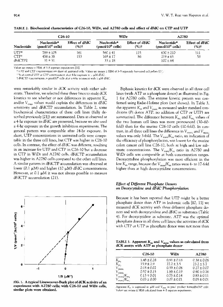
FIG. 1. A typical Lineweaver-Burk plot of dCK activity of an experiment with A2780 cells; with C26-10 and WiDr cells, similar plots were obtained.
Biphasic kinetics for dCK were observed in all three cell lines (with ATP as a phosphate donor) as illustrated in Fig. 1 for A2780 cells. The enzyme kinetic pattern was con-
firmed using Eadie-Hofstee plots (not shown). In Table 3,
the apparent K,,, and V,,, as measured under standard con- ditions (Pi donor ATP, no addition of CTP or UTP) are
summarized. The difference between K,, and Km, values of
the two human cell lines was more pronounced (30-60- fold) than for the murine C26-10 cells (lo-fold). In con- trast, in all three cell lines the difference in V,,,, and V,,,
values was only 3-fold. The V,,,/K,,, ratio, an indication oi the efficiency of phosphorylation, was lowest for the murine colon cancer cell line C26-10, both at high and low sub- strate concentrations. The V,,,/K,,, ratio in A2780 and
WiDr cells was comparable at both concentration ranges. Deoxycytidine phosphorylation was most efficient in the
low K, range, because the V,,,/K,,, ratios were 4- to 17-fold higher than at high deoxycytidine concentrations.
Effect of Different Phosphate Donors
on Deoxycytidine and dFdC Phosphorylation
Because it has been reported that UTP might be a better phosphate donor than ATP in leukemic cells [lo, 111 we measured dCK activity with three different phosphate do-
nors and with deoxycytidine and dFdC as substrates (Table 4). For deoxycytidine as substrate, ATP was the optimal
phosphate donor in all three cell lines; the activities of dCK with CTP or UTP as phosphate donor were not more than
TABLE 3. Apparent K, and V,, values as calculated from dCK assays with ATP as phosphate donor
C26-10 WiDr A2780
K ml 1.48 f 0.28 0.91 f 0.18 0.38 + 0.08 K m2 15.9 f 2.9 27.2 f 3.5 23.2 AZ 5.7 V max, 0.18 + 0.02 0.59 k 0.06 0.27 f 0.07
$::;;;~I 0.57 + 0.15 1.88 + 0.19 0.90 z!z 0.16 0.13 + 0.01 0.75 + 0.14 0.83 + 0.11
Inax rn: 0.03 f 0.00 0.07 + 0.01 0.05 + 0.01
Apparent K,,, is expressed in FM and V,,,, as pmol product formed/hr/lO” cells.
Values are means + SEM calculated from 4-5 separate expenments.

Chemosensitivity to Gemcitabine in Relation to Deoxycytidine and Gemcitabine Metabolism; Effects of CTP and UTP 915
TABLE 4. dCK activities with deoxycytidine and dFdC as substrates, measured with different phosphate donors
Substrate*
deoxycytidine +
dFdC +
Pi donor?
ATP UTP CTP ATP UTP CTP
C26.10 WiDr A2780
1.24$ k 0.25 0.48 + 0.03 (39)§ 0.42 IL 0.03 (34) 1.11 + 0.10
1.01 * 0.03 (94) 0.99 k 0.08 (84)
1.01 + 0.10 0.44 Ik 0.04 (45) 0.33 If: 0.03 (33) 1.18kO.04 0.30 f 0.03 (25) 0.90 + 0.08 (83)
1.01 + 0.09 0.54 f 0.10 (53) 0.57 f 0.13 (56) 1.24 zk 0.05
0.34 k 0.04 (30) 1.12 + 0.06 (86)
* Substrate (deoxycytidine or dFdC) concentratmn was 230 PM.
t Phosphate donor (ATP, UTP, OT CTP) concentration was 10 mM.
i: Activnes are expressed as nmol product formed/hr/lOh cells and are means k SEM of at least 3 separate experiments.
5 Withm parentheses, the relative activq is given as compared wth the enzyme actlvtty with .4TP as phosphate donor, set at lOO%, values are calculated from means of at
least 3 separate expermlents
0
A2780 C26.10
cell line
WiDr
CdR + UTP
TT
A2780 C26-10
cell line
dFdC + CTP
A2780 C26.10
cell line
WiDr
A2780 C26-10 With II I. 6
200 I
t dFdC + UTP
cell me
FIG. 2. The effects of CTP and UTP on deoxycytidine phosphorylation (A) and dFdC phosphorylation (B), both at a con- centration of 230 pM. The open bars represent the relative phosphorylating activity in the presence of 0.1, the hatched bars at 1 mM CTP or UTP compared to that with only ATP in the reaction mixture, which was set at 100%. Values are means +: SEM of 3-5 separate experiments. *CdR, deoxycytidine.

916 V. W. T. Ruiz van Haperen et al.
56% and 62% of the activity with ATP, respectively. ATP
was the optimal phosphate donor for dFdC phosphorylation as well, although with CTP enzyme activities almost similar to those with ATP were observed in all three cell lines, and
in C26-10 cells UTP was as efficient as ATP as a phosphate donor.
Effect of CTP and UTP on dCK Activity with Deoxycytidine or dFdC as Substrates
Figure 2 summarizes the effects of CTP and UTP on dCK activity using ATP as phosphate donor. 0.1 mM CTP had no effect on A2780 cells, decreased dCK activity slightly in
C26-10, and increased it somewhat in WiDr cells. 1 mM CTP decreased dCK activity with deoxycytidine as sub- strate in A2780 and C26-10 cells by 20-30%, but not with
dFdC as substrate. In WiDr cells, however, 1 mM CTP increased deoxycytidine-phosphorylating activity up to 165% and that of dFdC phosphorylation up to 140%. The
effects of UTP were more complex; 0.1 mM UTP increased dCK activity in A2780 and WiDr cells and decreased it in C26-10 cells. Although 1 mM UTP increased enzyme ac-
tivity in WiDr cells, it caused a slight decrease in A2780 cells and did not affect dCK activity in C26-10 cells. UTP,
however, had markedly different effects on dFdC phosphor-
ylation. Although 0.1 mM UTP increased enzyme activity in A2780 and C26-10 cells by 50 and 25%, respectively, activity was inhibited by 40% in WiDr cells. At 1 mM UTP only in C26-10 cells, an increase in activity of 50% was observed, and in WiDr as well as A2780 cells enzyme ac- tivity was inhibited markedly, by 70 and 40%, respectively.
Based on the biphasic enzyme kinetics we also studied the effects of 1 mM CTP and UTP at 2 other deoxycytidine
concentrations: 1 (low) and 10 (intermediate) PM in com- parison with 230 PM (saturating) (Table 5). For CTP, the
inhibitory effect in A2780 cells was comparable at 1, 10,
and 230 FM deoxycytidine and in C26-10 cells at 1 and 230 PM (20-30%). CTP did not affect dCK activity at 10 PM deoxycytidine in C26-10 and WiDr cells. In the latter cell
line, CTP increased enzyme activity at 1 and 230 PM more than 40%. For UTP, a similar inhibiting effect on deoxy-
cytidine phosphorylation was observed at all 3 deoxycyti-
TABLE 5. The relative effects of 1 mM CTP and UTP on dCK activity at different deoxycytidiie concentrations, with ATP as phosphate donor
deoxyc ytidine Addition (l.lM) C26.10 WiDr A2780
CTP + 1 82* f 8 143 f 14 88 f 6 10 105rt3 105 + 12 75 f 23
230 71* 17 165 f 7 78 f 7 UTP + 1 210 + 17 91 f5 88 f 14
10 196f 17 65 f9 54 + 13 230 lOI? 21 142 f 20 86 * 10
* dCK activities measured without CTP or UTP at the given deoxycytidine concen-
trations were set at 100%; values are means k SEM of 34 separate expenments.
dine concentrations in A2780 cells. In contrast, in C26-10
cells, the activity was increased considerably at 1 and 10 I_LM deoxycytidine, and in WiDr cells a marked stimulation was observed only at 230 FM. At 10 FM deoxycytidine, an inhibition of 35% was found.
DISCUSSION
This study shows that, based on enzyme properties, the metabolism of deoxycytidine and dFdC differs markedly in
cell lines of solid tumour origin, even within cell lines
derived from a similar organ. Although, in general, dCDA
activity was higher with dFdC as substrate than with de- oxycytidine, no correlation was evident between sensitivity
and dFdC-deaminating activity, as had already been estab- lished for deoxycytidine deamination and sensitivity to de- oxycytidine analogues [14]. The discrepancies between
dCDA activity measured with deoxycytidine compared to activity with dFdC as substrate, are most probably due to
differences in K,,, and V,,, for the two substrates. In addi-
tion, the substrates may cause a different conformation of the enzyme, resulting in different kinetics and regulation
properties. A remarkable observation was the similarity of dFdC
phosphorylation in all cell lines tested, despite considerable
variation in deoxycytidine phosphorylation rates. Although it can not be excluded that other deoxynucleoside kinases
can use dFdC as a substrate, one can assume that dCK is the major enzyme responsible for dFdC phosphorylation [1, 2, 10, 241. This would mean that dCK in these cells is suffi- ciently high to catalyze dFdC phosphorylation. Thus, the
observed similarity in the cellular capacity for dFdC phos- phorylation cannot account for the differences in sensitiv- ity of the studied cell lines, which varied 1.2 to 3+fold,
depending on exposure time (Table 1) [21]. Differences in nucleoside transport do not seem to be limiting because
dFdCTP accumulation is rather rapid [21] and chemosen- sitivity to other nucleoside analogues, dependent on the same carrier, is similar.
This study also shows that CTP as well as UTP can significantly affect dCK-catalysed phosphorylation of both deoxycytidine and dFdC as measured in relatively crude extracts of disrupted cells. Although this does not neces- sarily reflect the situation in intact cells, it probably gives a better indication of cellular regulation mechanisms than purified enzyme systems. Differences in enzyme regulation may play a role in determining the chemosensitivity of a cell line. We observed biphasic enzyme kinetics for de- oxycytidine phosphorylation in all three cell lines studied with apparent Km, values well below 5 PM. These results, although obtained in cell extracts, are very well in line with those of Bohman and Eriksson [25], who observed similar biphasic kinetics for dCK purified from human leukemic spleen. Most studies on dCK kinetics, however, have been limited to a low deoxycytidine concentration range (~10 p,M) [l, 3, 12, 13, 261, precluding measurement of K,,, val- ues in a higher range; thus, reported apparent K,,, values

Chemosensitivity to Gemcitabine in Relation to Deoxycytidine and Gemcitabine Metabolism; Effects of CTP and UT’P 917
vary from 0.6 to 9 FM, depending on the dCK source and
experimental conditions. The apparent K,,,, values in our
cell lines are in this range. For human leukemia HL-60 cells, only Singhal et al. [27] reported a very high apparent K, of 300 FM. Although Stegmann et al. [28] measured dCK activity in rat leukemia cells at a broad deoxycytidine concentration range (up to 1.5 mM), these authors reported
only one K,,, value of 9.4 p,M deoxycytidine. Considering the V,,,/K,,, ratio, an indicator for phosphorylation effi-
ciency, dCK from the murine colon cancer cell line C26-10
appears to be the least efficient, both at low and higher deoxycytidine concentrations. The ratio is 6-fold lower
than that for A2780 cells. For dFdC and deoxycytidine, an almost similar efficiency was reported for CHO cells [2]. Assuming this also holds for our cell lines, one might ex- tend the differences in V,,,/K,,,, which we found for effi-
ciency of deoxycytidine phosphorylation, to that of dFdC phosphorylation. The pattern in V,,,/K_, ratio correlates
with the accumulation and retention pattern of dFdCTP, as
observed in these cell lines [Zl]. A2780 accumulated the highest dFdCTP levels, closely followed by WiDr (depend- ing on exposure time), both in vitro and in Go. C26-10
cells and colon 26 tumours accumulated the lowest dFdCTP levels and retention was less than 24 hr, in con-
trast to A2780 and WiDr cells and tumours. This pattern
follows the chemosensitivity of the cell lines quite nicely
n41. Most studies on dCK enzyme kinetics are based on dCK
assays performed with ATP as phosphate donor, because ATP is assumed to be the optimal substrate. For dCK from
MOLT-4 cells [lo, 291 and Ehrlich ascites tumour cells [ll], it was shown that UTP was a better phosphate donor than
ATP. In the solid tumour cell lines described in this paper,
ATP was the most efficient phosphate donor for phosphor-
ylation of both deoxycytidine and dFdC. Only in C26-10 cells was UTP as efficient as ATP for dFdC phosphoryla- tion, and in the two human tumour cell lines only 30% of the activity with ATP was measured. CTP was a reasonable phosphate donor for dFdC phosphorylation in all three cell lines.
The effects of CTP and UTP on dCK activity were quite
pronounced, both with deoxycytidine and dFdC as sub- strates. The chosen nucleotide concentrations, 0.1 and 1 mM CTP and UTP, are in the range of normal cellular
concentrations in the three tested cell lines, including the shifts caused by exposure of the cells to dFdC (Table 2) [21]. Considerable differences were observed for the effects
on deoxycytidine and dFdC phosphorylation, possibly re- lated to the mechanism of substrate binding to dCK as proposed by Shewach et al. [29]. This sequential Bi-Bi mechanism may involve different conformational changes for deoxycytidine and dFdC as substrates, leading to differ- ent affinities for CTP and UTP. At lower substrate con- centrations including a more physiological concentration of 1 PM, only for C26-10 cells were considerable differences for the effects of UTP observed, with a 2-fold increase at 1 FM but not at 230 p,M. In 2 other studies, an activation (at
2-10 PM deoxycytidine) by UTP was observed [12,26], and
in another, inhibition was found, albeit at a lower deoxy-
cytidine concentration (0.2 PM) [13]. These effects were all found in cells from a myeloid origin (although different species were used as the source), as well as normal and leukemic cells. Habtayesus et al. [30] reported a different substrate specificity for murine dCK as compared to human dCK. It seems likely that regulation of dCK is very much species-, tissue-, and proliferation-dependent. Extrapola- tion of enzymatic characteristics from one source should be
done with great caution. The implications of these results with respect to the shift
in CTP and UTP pools as a result of exposure to dFdC are not entirely clear. An increase in CTP seems favourable for dFdC phosphorylation. However, probably as a result of CTP-synthetase inhibition, CTP pools decreased initially in all three cell lines, but were restored to levels higher than
control at 24 hr after removal of the drug [21]. The accom- panying increase in UTP seems more favourable for deoxy- cytidine than for dFdC phosphorylation. Because the in-
crease in UTP was lowest in A2780 cells, this may be related to the higher sensitivity of this cell line to dFdC as
compared to C26-10 cells. One possibility to clarify the role of CTP and UTP in dFdC phosphorylation would be the
combination of dFdC with an antimetabolite capable of affecting CTP and UTP pools. It has been suggested that N-phosphon-acetyl-L-aspartate (PALA), a potent inhibitor of pyrimidine de nova synthesis, would increase the effect of ara-C by depletion of CTP and, subsequently, dCTP pools [3 1, 321. Indeed, in several cell lines, PALA can potentiate
the effect of ara-C [31, 331 but this is not true of all cell lines [33]. Under conditions enabling potentiation of ara-C
[33], we could not demonstrate a similar effect for PALA
and dFdC (data not shown) in leukemic cell lines. No data are available on solid tumor cell lines.
Our findings on the effects of UTP and CTP are not in line with those of Shewach et al. [IO]. For dCK purified from MOLT-4 cells, dFdC was phosphorylated most effi- ciently with UTP as phosphate donor, leading these authors to suggest that high UTP concentrations would be favour-
able for dFdC phosphorylation. The discrepancy between these and our findings may be explained by the difference
in dCK source. Thus, an approach representative of the actual physiological situation in the tumour cells seems to give a better prediction as to the regulation of dCK in that
source. In conclusion, the differential effects of CTP and UTP
on deoxycytidine and dFdC phosphorylation seem to potentiate dFdC anabolism more in sensitive A2780 cells compared to the other lines. The efficiency of de- oxycytidine phosphorylation may be an indication of dFdCTP accumulation and, consequently, chemosenstivity to dFdC.
Supported b grant IKA-VU 90-19 from the Dutch Cancer Society.

918 V. W. T. Ruiz van Haperen et al.
References 1. Eriksson S, Kierdaszuk B, Munch-Petersen B, oberg B and
Johansson NG, Comparison of the substrate specificity of hu- man thymidine kinase 1 and 2 and deoxycytidine kinase to- ward antiviral and cytostatic nucleoside analogs. Biochem Bio- phys Res Comm 176: 586-592, 1991.
2. Heinemann V, Hertel LW, Grindey GB and Plunkett W, Comparison of the cellular pharmacokinetics and toxicity of 2’,2’-difluorodeoxycytidine and 1-P-D-arabinofuranosylcyto- sine. Cancer Res 48: 4024-4031, 1988.
3. Ruiz van Haperen VWT and Peters GJ, New targets for py- rimidine antimetabolites for the treatment of solid tumours. 2. Deoxycytidine kinase. Pharm World Sci 16: 104-l 12, 1994.
4. Chabner BA, Johns DG, Coleman CN, Drake JC and Evans WH, Purification and properties of cytidine deaminase from normal and leukemic granulocytes. J Clin Invest 53: 922-931, 1974.
5. Heinemann V, Hertel LW, Grindey GB and Plunkett W, Comparison of the cellular pharmacokinetics and toxicity of 2’,2’-difluorodeoxycytidine and 1-P-D-arabinofuranosylcyto- sine. Cancer Res 48: 4024-4031, 1988.
6. Ho DHW, Distribution of kinase and deaminase of l&D- arabinofuranosylcytosine in tissues of man and mouse. Cancer Res 33: 2816-2820, 1973.
7. Hagenbeek A, Martens ACM and Golly LP, In vivo devel- opment of cytosine arabinoside resistance in the BN acute myelocytic leukemia. Semin Oncol 14: 202-206, 1987.
8. Stueart CD and Burke PJ, Cytidine deaminase and the devel- opment of resistance to cytosine arabinoside. Nature (Neeu Biology) 233: 109, 1971.
9. Grant S, Biochemical modulation of cytosine arabinoside. Pharmacol Ther 48: 29-44, 1990.
10. Shewach DS, Reynolds KK and Hertel LW, Nucleotide spec- ificity of human deoxycytidine kinase. MoI Pharmacol 42: 518-524, 1992.
11. White JC and Hines LH, Role of uridine triphosphate in the phosphorylation of l-P-D-arabinofuranosylcytosine by Ehr- lich ascites tumor cells. Cancer Res 47: 1820-1824, 1987.
12. Durham JP and Ives DH, Deoxycytidine kinase II Purification and general properties of the calf thymus enzyme. J Biol Chem 245: 2276-2284, 1970.
13. Sarup JC, Johnson MA, Verhoeff V and Fridland A, Regula- tion of purine deoxynucleoside phosphorylation by deoxycy- tidine kinase from human leukemic blast cells. Biochem Phar- macol38: 2601-2607, 1989.
14. Ruiz van Haperen VWT, Veerman G, Braakhuis BJM, Ver- morken JB, Boven E, Leyva A and Peters GJ, Deoxycytidine kinase and deoxycytidine deaminase activities in human tu- mour xenografts. Eur J Cancer 29A: 2132-2137, 1993.
15. Spasokoukotskaja T, Arner ESJ, Brosjii 0, Gun&n I’, Julius- son G, Liliemark J and Eriksson S, Expression of deoxycyti- dine kinase and phosphorylation of 2-chlorodeoxyadenosine in human normal and tumor cells and tissues. Eur J Cancer 31A: 202-208, 1995.
16. Lund B, Kristjansen PEG and Hansen H, Clinical and pre- clinical activity of 2’,2’-difluorodeoxycytidine (gemcitabine). Cancer Treatm Rev 19: 45-55, 1993.
17. Kaye SB, Gemcitabine: current status of phase I and phase II trials. J Clin Oncol 12: 1527-1531, 1994.
18. Abratt RI’, Bezwoda WR, Falkson G, Goedhals L, Hacking D and Rugg TA, Efficacy and safety profile of gemcitabine in non-small cell lung cancer: a phase II study. J Clin Oncol 12: 1535-1540, 1994.
19. Huang I’, Chubb S, Hertel LW, Grindey GB and Plunkett W, Action of 2’,2’-difluorodeoxycytidine on DNA synthesis. Cancer Res 51: 6110-6117, 1991.
20. Ruiz van Haperen VWT, Veerman G, Vermorken JB and Peters GJ, 2’,2’,-Difluorodeoxycytidine (gemcitabine) incor- poration into RNA and DNA of tumour cell lines. Biochem PharmacoI46: 762-766, 1993.
21. Ruiz van Haperen VWT, Veerman G, Boven E, Noordhuis I’, Vermorken JB and Peters GJ, Schedule dependence of sensi- tivity to 2’,2’-difluorodeoxycytidine (gemcitabine) in relation to accumulation and retention of its triphosphate in solid tumour cell lines and solid tumours. Biochem Phurmacol 48: 1327-1339, 1994.
22. Peters GJ, Kraal I and Pinedo HM, In vitro and in vivo studies on the combination of brequinar sodium (DUP 785, NSC 368390) with 5-fluorouracil; effects of uridine. Br J Cancer 65: 229-233, 1992.
23. Bradford M, A rapid and sensitive method for the qualifica- tion of microgram quantities of protein using the principle of protein-dye binding. Anulyt Biochem 72: 248-254, 1976.
24. Ruiz van Haperen VWT, Veerman G, Eriksson S, Boven E, Stegmann APA, Hermsen M, Vermorken JB, Pinedo HM and Peters GJ, Development and molecular characterization of a 2’,2’-difluorodeoxycytidine-resistant variant of the human ovarian carcinoma cell line A2780. Cancer Res 54: 4138- 4143, 1994.
25. Bohman C and Eriksson S, Deoxycytidine kinase from human leukemic spleen: preparation and characteristics of homog- enous enzyme. Biochemistry 27: 4258-4265, 1988.
26. Datta NS, Shewach DS, Hurley MC, Mitchell BS and Fox IH, Human T-lymphoblast deoxycytidine kinase: purification and properties. Biochemistry 28: 114-123, 1989.
27. Singhal RL, Yeh YA, Scekeres T and Weber G, Increased de- oxycytidine kinase activity in cancer cells and inhibition by difluorodeoxycytidine. Oncol Res 4: 517-522, 1992.
28. Stegmann APA, Honders MW, Kester MGD, Landegent JE and Willemze R, Role of deoxycytidine kinase in an in vitro model for ara-C and DAC-resistance: substrate-enzyme inter- actions with deoxycytidine, l-P-D-arabinosylcytosine and substrate-enzyme interactions with deoxycytidine, l-p-D-ara- binosylcytosine and 5-aza-2’-deoxycytidine. Leukemia 7: 1005-1011, 1993.
29. Shewach DS, Reynolds KK and Hahn T, Apparent allosteric regulation of deoxycytidine kinase by UTP. Proc Am Assoc Cancer Res 34: 10, 1993.
30. Habteyesus A, Nordenskjald A, Bohman C and Eriksson S, Deoxynucleoside phosphorylating enzymes in monkey and human tissues show great similarities, while mouse deoxycyti- dine kinase has a different substrate specificity. Biochem Phar- macoI42: 1829-1836, 1991.
31. Grant S, Rauscher F III, and Cadman E, Differential effect of N-(phosphonacetyl)-L-aspartate on l-P-D-Arabinofuranosyl- cytosine metabolism and cytotoxicity in human leukemia and normal bone marrow progenitors. Cancer Res 42: 4007-4013, 1982.
32. Plunkett W, Adams T and Keating M, Modulation of Ara- CTP metabolism in leukemia cells during high-dose Ara-C (HD Ara-C) therapy by thymidine and PALA. Proc Am Assoc Clin Oncol 6: 30 (abstract), 1987.
33. Noordhuis P, Kazemier KM, Kaspers GJL and Peters GJ, Modulation of metabolism and cytotoxicity of cytosine ara- binoside with N-(phosphon)-acetyl-L-aspartate in human leukemic blast cells and cell lines. Leukemia Res, in press.